Eur J Clin Chem Clin Biochem 1995; 33:737-742
© 1995 Walter de Gruyter & Co.
Berlin · New York
Automated Fluorimetric Assay Procedure for Glucohydroiases Using a Routine Centrifugal Analyser
Assay of Enzymes of Lysosomal Origin in Plasma, II 1 )
By Giancarlo Goi
1, Emma Giiagnellini
2, Chiara Bairati
1, Marco Besozzi
3, Adriana Lombardo
1, Donatella Boiling, Augiisto Lovagnini* and Guido Tettamanti
l1
Department of Medical Chemistjy and Biochemistry, The Medical School University of Milan, Milan, Italy
2
Laboratory of Clinical Chemistry, S. Paolo Hospital, Milan, Italy
3
Laboratory of Clinical Chemistry, Legnano Hospital, Legnano (Milan), Italy
4
Laboratory of Clinical Chemistry and Microbiology, Bassini Hospital, Cinisello Balsamo (Milan), Italy
(Received February 28/June 23, 1995)
Summary: The manual fluorimetric procedure, considered as a reference method for the determination of N-acetyl-
ß-Z)-glucosaminidase, ß-D-glucuronidase and ß-£>-galactosidase in human plasma, was automated as a routine method, using the IL Monarch centrifugal analyser. Using a liquid standard with a known enzyme content, the automated assay correlated fairly well with the reference manual method (r values very close to 1). Its analytical imprecision was much lower than that of the manual method. The automated assay of N-acetyl-ß-£>-glucosamini- dase, ß-D-glucuronidase and ß-D-galactosidase gave coefficients of variation of 5.7—6.9, 3.6—5.0 and 3.8—4.2%, respectively, detection limits of 4, 2 and 1 mU/1 plasma respectively, and linear responses of up to 73, 8.4 and 0.9 U/l of plasma respectively. Furthermore, the method required only small volumes of undiluted plasma (4—10 ).
This method appears to be reliable, sensitive, simple enough for routine analyses and as cost effective as the most common routine serum enzyme assays.
Introduction
There is evidence that the determination, in body flu- liable, low cost assay procedures, suitable for routine ids, of enzymes of lysosomal origin has potential value analyses. At present, sensitive optimized and reproduc- in the diagnosis of a number of diseases (1—7). For ible fluorimetric methods are available for the determi- instance, the plasma levels of lysosomal enzymes have nation of lysosomal enzymes in plasma (17), including been shown to correlate with the metabolic control of the isozymes of N-acetyl-ß-£>-glucosaminidase (18).
diabetes (8-12), and with the functional conditions of Also, stable reference materials can be prepared for in- kidney and liver (13 — 14), the latter organ being impli- strument calibration and for running of intra- and inter- cated in the clearance of the same enzymes (15). laboratory quality control programmes (19). The only Interestingly, the plasma determination of some lyso- obstacle to the routine application of these methods is somal enzymes has been suggested for monitoring the the fact that they are manual.
functional recovery of the liver after transplantation
(]g\ On these premises and considering the wide availability in clinical laboratories of the IL Monarch centrifugal an- A realistic assessment of the diagnostic potential of ly-
ai
yserequipped with a fluorimetric detector, we decided sosomal enzymes requires the availability of simple, re-
to adapt the manualfluorimetric procedure for lyso-
somal enzyme assays to this automated instrument,
]
) Part I i.e. (19) using the reference calibration material prepared in our
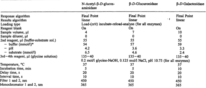
Eur J Clin Chem CHn Biochem 1995; 33 (No 10)Tab. 1 Operational conditions for the automated assay of three enzymes of lysosomal origin using the IL Monarch
N-Acetyl-ß-D-glucos-
aminidase ß-D-Glucuronidase ß-D-Galactosidase
Response algorithm Results algorithm Loading type Reagent blank Sample volume, Sample diluent,
2nd reagent, (buffer-substrate sol.) - buffer (mmol/1)*
- pH— substrate (mmol/1)
3rd-4th reagent, (glycine solution) Temperature, °C
Incubation time, min Delay time, s Interval time, s Filter 1 and 2, nm
Monochromator 1 and 2, nm
Final Point Final Point Final Point linear linear ' ' linear Load-(crit) incubate-reload-analyse (for all enzymes)
On On On 4 7 10
0 0 0
55 55 55 54 57 59 4.2 3.6 3.3 6.5 4.6 2.4 135-40 135-40 135-40 0.2 mol/1 glycine-NaOH, 0.125 mol/1 NaCl, pH 10.75 (for all enzymes)
37 37 37 5 5 10 20 20 20 10 10 10 450 450 450 365 365 365
* citric acid-sodium citrate for N-acetyl-ß-£>-glucosaminidase and acetic acid-sodium acetate for ß-Z)-glucuronidase and ß-Z>-galactosidase
laboratory (19). The three enzymes
2) of lysosomal origin with the highest diagnostic potential, N-acetyl-ß-Z)-glu- cosaminidase, ß-Z)-glucuronidase and ß-D-galactosi- dase, were determined in human plasma. The results showed that all three enzymes can be routinely deter- mined on the IL Monarch centrifugal analyser, simply and reliably, and at relatively low cost.
Materials and Methods
Chemicals and other products
Commercial chemicals were of the highest purity available. The water routinely used was freshly redistilled in a glass apparatus. 4- Methylumbelliferone, purchased from Fluka GmbH (Buchs, Swit- zerland), was recrystallized three times from ethanol; 4-methylum- belliferyl-glycosides, used as substrates for the individual glycohy- drolases, were purchased from Melford (Suffolk, England); ethyl- ene glycol, haemoglobin and bilirubin from Sigma Chem. Co (St.
Louis, Mo, USA); intralipid from Cutter Labs. Inc. (Berkeley, CA 94710).
Blood sampling and preparation of plasma specimens Plasma was prepared from blood treated with sodium citrate (final concentration: 11 mmol/1), which is known not to affect lysosomal enzyme activities (17). Ethylene glycol was added (300 g/1) to the
2) Enzymes:
N-acetyl-ß-D-glucosaminidase:
N-acetyl-ß-D-glucosaminideN-acetylglucosaminohydrolase, JcLx .3..2.1.30
ß-D-galactosidase:
ß-D-galactoside galactohydrolase EC 3.2.1.23 ß-Z)-glucuronidase:
ß-D-glucuronoside glucuronosohydrolase EC 3.2.1.31
plasma samples immediately after collection (19). The samples were then stored at -20 °C until assayed.
Blood was withdrawn from two groups of individuals. The first group provided plasma to be used for studying the analytical char- acteristics of the method (total imprecision, sensitivity) and for evaluating possible interferences. These individuals, who were in- formed of the puropse of the investigation, were healthy adults, both males and females, aged 25-55 years, mostly volunteer blood donors. Some of them were women in the final trimester of preg- nancy. In all cases blood was withdrawn immediately before the control laboratory analyses, and the common blood analytes were found to be within normal ranges. Care was taken to check the absence of antibodies indicative of particular infectious diseases, like AIDS and hepatitis, in all the specimens collected, the plasma specimens were divided into three pools:
pool A, carrying high lysosomal enzyme activities, obtained by pooling plasma from the pregnant women, who are known to have elevated plasma levels of these enzymes (20);
pool B, carrying medium levels of lysosomal enzyme activities, obtained by pooling plasma from normal individuals;
pool C, carrying low levels of lysosomal enzyme activities, ob- tained by dilution 1 + 1 pool B with a sample of the same pool B in which lysosomal enzymes had been inactivated by heating at 56 °C for 5 hours (O pool) (19).
Owing to the extremely low, if any, enzyme activities in the "O pool", the same pool was also utilized to study the sensitivity of the method.
A different plasma pool containing intermediate activity was pre- pared for use as a "standard" for the automated instrument. The enzyme levels of this standard were established by the manual method.
The second group of individuals provided the plasma specimens used to compare the routine automated method with the manual method. This group comprised 60 randomly chosen individuals who entered the Laboratory of Clinical Chemistry and Microbiol- ogy of the Bassini Hospital for scheduled haemato-chemical tests.
Linearity of the automated method
To assess the linearity of the automated method, we studied saples made from various proportions of serum pools with extreme levels of lysosomal enzyme activities. The expected (theoretical) values were calculated from the corresponding values of the original pools. Each determination was performed in triplicate.
Imprecision of the automated method
The analytical imprecision of the automated method was studied using plasma pools A, B and C. Two replicate analyses were per- formed for each enzyme in twenty different analytical series, cov- ering a period of three months, according to the model ofKrouwer (21) and NCCLS recommendations (22).
Fluorimetric assay of lysosomal enzymes on the
"IL Monarch" centrifugal analyser
The following enzymes of lysosomal origin were assayed fluori- metrically: N-acetyl- -Z)-glucosaminidase, -D-glucuronidase, -
£>-galactosidase, using the corresponding 4-methylumbelliferyl glycosides as substrates. The manual assay procedure for these en- zymes, provisionally adopted as the reference method, was as de- scribed (17).
The automated procedure was developed by adapting the assay conditions of the manual method to the random access centrifugal analyser IL Monarch (Instrumentation Laboratory, Lexington, MA, USA). For fluorimetric analysis this instrument uses disposable re- action rotors made of UV-transmitting plastic material. The basic operating conditions were those suggested by the manufacturer.
The plasma specimens were thawed immediately before use, brought to room temperature and employed as such without dilu- tion. The nature, pH and concentration of the buffer, substrate mo- larity and the time of incubation at 37 °C were chosen to give the optimal values for each enzyme activity, and they were found to be the same as those already established for the manual method (17). Details of the conditions used to measure lysosomal enzyme activities on the IL Monarch centrifugal analyser are given in table 1. Regular incubation mixtures containing physiological saline in place of plasma were used as controls (blanks).
All enzyme activities were expressed as units (U, μτηο1β5 hy- drolysed substrate, min""1) per litre.
Interference in the determination of plasma lysosomal enzymes by the routine automated method
For this purpose the plasma was added with increasing amounts of haemolysate (prepared from human erythrocytes), lipids (as Intrali- pid) and bilirubin, following the indication of Click et al. (23). The
Έ
3
D
aωο cοο
10ο ο
20
20 60 80 Ο 2 4 6 8 10 Ο Catalytic concentration (expected values) [U/I]
0.2 0.4 0.6 0.8
Fig. 1 Linearity of the automated method for the determination β-D-galactosidase (c) in human plasma. For details see the experi- of N^acetyl- -D-glucosaminidase (a), -Z)-glucuronidase (b) and mental section.
Tab. 2 Estimation of the imprecision of the automated method for the assay of lysosomal enzymes in plasma.
Enzyme
N-Acetyl- -/>-glucosaminidase
-D-Glucuronidase
-Z^Galactosidase
Plasma pool
AB C AB C AB C
Automated method Enzyme actn (mU/1) Mean 42100 28000 8200 47002400 1100 700300 140
rity Coefficient of variation (%) Within-run
3.93.7 3.6 3.23.0 4.5 3.53.5 3.6
Day to day 4.24.7 5.9 0.52.0 2.2 0.21.1 1.2
Total 5.75.9 6.9 4.13.6 5.0 3.84.1 4.2
Manual method Coefficient of variation (%) Total
5.25.5 9.5 4.24.4 11.9 14.79.3 17,5
Eur J Clin Chem Clin Biochem 1995; 33 (No 10)
haemoglobin content in haemolysate Was quantified by measuring the cyanomethaemoglobin derivative (24).
Statistical analysis
Parametric techniques of analysis were employed. Regression analysis and correlation coefficients were calculated following the Snedecor & Cochran indications (25) for linearity study and with the standard model of Denting (26) for comparison of methods study. The estimation of total imprecision by analysis of variance was performed following the model of Krouwer (21), and NCCLS recommendations (22).
Results
Application of the manual method to a routine centrifu- gal autoanalyser, namely the IL Monarch, presented no particular difficulties, provided the manufacturer's in- structions were rigorously followed. The only discrepant finding was the instrument's response to the liberated fluorophore, 4-methylumbelliferone, which was about three-fold lower with the autoanalyser than with the spectrofluorimeter employed in the manual method. This difference, which remained unchanged after careful cali- bration of the autoanalyser with 4-methylumbelliferone solutions of known concentration, was found to be re- lated to the plastic cuvettes used in the centrifugal ana- lyser. However, when the stable liquid material was used as an analytical internal standard (see later), the method sensitivity remained practically unaffected.
The detection limits, calculated as the minimal enzyme activity detectable above the mean plus three standard deviations measured daily for 15 days in triplicate, were 4.0, 2.0, 1.0 mU/1 for N-acetyl-ß-D-glucosaminidase, ß-
£>-glucuronidase and ß-£>-galactosidase, respectively.
As shown in figure 1, linearity was assured within a relatively wide range of enzyme concentrations (up to 73, 8.4 and 0.9 U/l of plasma for N-acetyl-ß-£)-glucos- aminidase, ß-D-glucuronidase and ß-D-galactosidase, respectively). The correlation coefficients were very high (r = 0.999) for all enzymes and no significant dif- ferences in the internal consistency of the linear regres- sion were detectable.
With the plasma pools of low, medium and high enzyme levels, the automated method showed low and accept- able coefficients of variation: from 5.7 to 6.9 for N-ace- tyl-ß-£)-glucosaminidase, from 3.6 to 5.0 for ß-£>-glu- curonidase and from 3.8 to 4.2 for ß-Z)-galactosidase, the lower values applying to the plasma pool with the highest enzyme activity (tab. 2). Moreover, and as ex- pected, the degree of imprecision decreased with increasing the enzyme concentration in the sample. It is noteworthy that the degree of imprecision was much lower with the automated method than with the manual one (19).
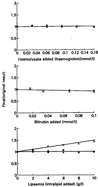
The data in figure 2 on the effects of potential interfer- ents show that haemoglobin did not interfere in the de- termination of these lysosomal enzymes, bilirubin ex- hibited a very modest (-8%) inhibition effect on the enzyme determination, while lipids (as intralipid) caused an increase of ß-£>-galactosidase (40% at 10 g/1), with- out affecting the determination of the other enzymes.
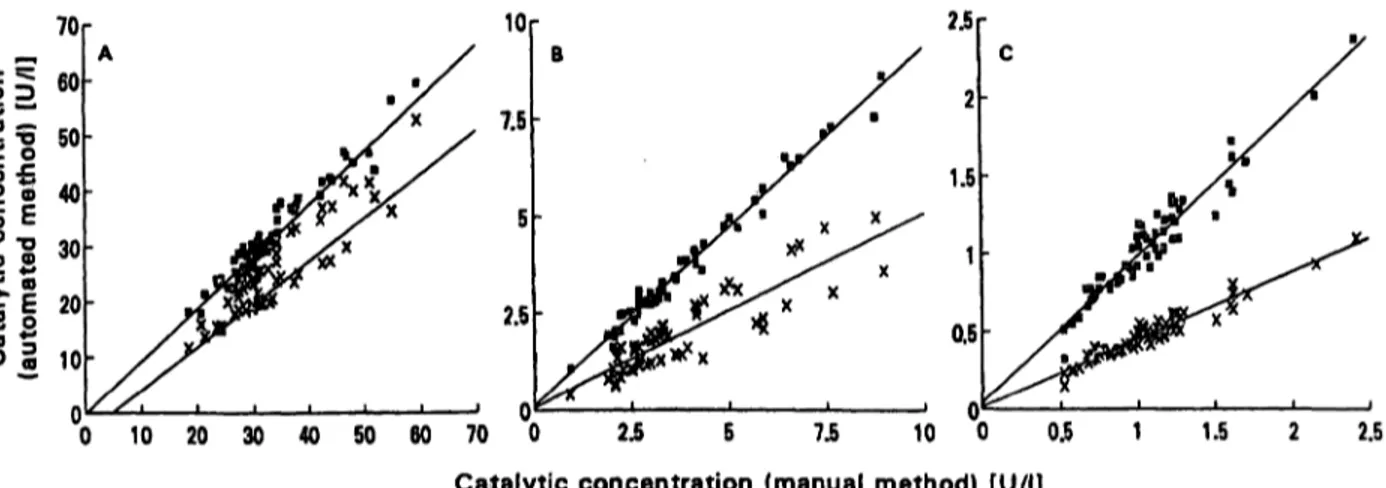
As shown in figure 3 the correlation between the auto^
mated and the manual method, for plasma from 60 in- dividuals, was good for all the considered enzymes. The response of the automated method appeared to be mark- edly lower than that of the manual one, especially for ß- Z)-galactosidase, and the r values, although good, were further from unity for N-äcetyl-ß-Z)-glucosaminidase and ß-£)-glucuronidase. However, the use of the stable liquid material as analytical standard material made the results of the automated method practically identical to those of the manual one for all enzymes, and increased
2 1.5
n
0.5
0 0.02 0.04 0.06 0.08 0.1 0.12 0.14 0.16 Haemolysate added (haemoglobin)[mmol/ll 2r
W
tö
'5>
1.5
0.5
0.02 0.04 0.06 0.08 Bilirubin added [mmol/l]
0.1
2 4 6 8
Lipaemia (intralipid added) [g/l]
10
Fig. 2 . Interference by haemoglobin, bilirubin and intralipid in the determination of plasma N-acetyl-ß-D-glucosaminidase ·, ß^·
D-glucuronidase · and ß-D-gaIactosidase»x by the automated pro- cedure. · »
1.5
0.5
10 20 30 40 50 60 70 0 2.5 5 7.5 10 0 0.5 Catalytic concentration (manual method) [U/l]
1.5 2.5
Fig. 3 Comparison of the automated with the manual method using or not using the enzyme reference material as standard.
Number of subjects: 60.
without the use of the enzyme reference material.
D with the use of the enzyme reference material.
a) N-acetyl-ß-D-glucosaminidase
: r = 0.91, y = -3.8 + 0.86x D: r = 0.97, y = -0.28 + 0.98x b) ß-£>-glucuronidase
x: r = 0.88, y = 0.06 + 0.5x D: r = 0.99, y = 0.087 + 0.94x c) ß-D-galactosidase
x: r = 0.96, y = 0.02 + 0.43x D: r = 0.96, y = 0.02 + 0.98x
the r values to almost unity (0.97, 0.99 and 0.96 for N- acetyl-ß-Z)-glucosaminidase, ß-/5-glucuronidase and ß- D-galactosidase, respectively). Thus, the use of this ma- terial standard is recommended, together with the 4- methylumbelliferone solution-of known concentration for routine instrument calibration.
Discussion
The introduction into the clinical laboratory market of routine autoanalysers suitable for fluorimetric assays, like the IL Monarch centrifugal analyser, prompted us to use this instrument for the optimized fluorimetric as- say of lysosomal enzymes, which are already available as manual methods.
The results showed that the automated procedure pre- sented here, using a stable liquid material as internal standard, is as sensitive as the manual reference method, but more precise than the latter, with coefficients of vari- ation in the range of 3.6-6.9, which are very low for
enzyme assays. The automated method requires small amounts of undiluted plasma (4-10 ), covers a suffi- ciently wide range of enzyme concentration and is as simple and as cost effective as most routine serum en- zyme assays. The availability of this method and of li- quid stable materials for use as reference standards and for instrument calibration will enable large scale screen- ing programmes on selected pathologies, as well as fa- cilitating intra- and inter-laboratory quality control pro- grammes. On this basis, the determination of selected enzymes of lysosomal origin in plasma might gain suffi- cient analytical reliability and diagnostic utility to be in- cluded in current clinical enzymology.
Acknowledgements
This work was supported in part by grants from the "Italian Minis- tery of Education": MURST 40% programme, from Lombardy Re- gion (Ricerca biomedica finalizzata, grant n. 311), and Consiglio Nazionale delle Ricerche (C.N. R.), Progetto strategico per il mez- zogiomo.
References
1. Woolen JV, Turner P. Plasma ß-N-acetyl-D-glucosaminidase and ß-D^glucuronidase in health and disease. Clin Chim Acta 1965; 12:671-83.
2. Niebes P. Determination of enzymes and degradation products of glycosaminoglycans metabolism in the serum of health and varicose subjects. Clin Chim Acta 1972; 42:399-408.
3. Calvp P, Barba JL, Cabezas JA. Serum ß-D-glucuronidase, ß- D-gluGosidase, -L-fueosidase, a^D-galactosidase levels in acute viral hepatitis, pancreatitis, myocardial infarction and breast cancer. Clin Chim Acta 1982; 11:15-9.
4. Regiere A, Carrettero MI, Cabezas JA. Increased serum a-L- fucosidase and ß-D-N-acetylglucosiminidase activities in dia- betic, cirrhotic and gastric cancer patients. Clin Chim Acta
1980; 103:155-8.
5. Hultberg B, Isaksson A. Isoenzyme pattern of serum ß-hexos- aminidase in liver disease, alcohol intoxication and pregnancy.
Enzyme 1983; 30:166-71.
6. Koskinen H, Jarvisolo J, Pitkanen E, Mutanen P, Zitting A.
Serum ß-D-acetylglucosaminidase and ß-glucuronidase activi- ties in silicosis patient and in workers exposed to silica dust.
Br J Dis Chest 1984; 78:217-24.
7. Vigano A, Assael BM, Villa AD, Gagliardi L, Principi N, Ghezzi P, et al. N-acetyl-ß-D-glucosaminidase (NAG) and NAG isoenzymes in children with upper and lower urinary tract infections. Clin Chim Acta 1983; 130:297-304.
8. Belfiore F, Lo Vecchio L, Napoli E, Borzi V. Increased ß-N- acetylglucosaminidase activity in diabetes mellitus. Clin Chem 1974; 20:1229-30.
Eur J Clin Chem Clin Biochem 1995; 33 (No 10)
9. Kohler E, Sheth KJ, Good TA. Plasma acidic glycohydrolases in insulin-dependent diabetes mellitus. Acta Diabetol Lat 1981;
18:243-50.
10. Goi G, Fabi A, Lorenzi R, Lombardo A, Tettamanti G, Burlina AB, et al. Serum enzymes of lysosomal origin as indicators of the metabolic control in diabetes: comparison with glycated hemoglobin and albumin. Acta Diabetol Lat 1986; 23:117-25.
11. Goi G, Lombardo A, Fabi A, Burlina AB, Segalini G, Guagnel- lini E, et al. Serum enzymes of lysosomal origin as indicators of the metabolic control in non-insulin dependent diabetics.
Acta Diabetol Lat 1987; 24:331-40.
12. Burlina AB, Goi G, Fabi A, Lombardo A, Gaburro D, Tetta- manti G. Behaviour of some lysosomal enzyme in the plasma of insulin dependent diabetic patients during artificial pancreas treatment. Clin Biochem 1987; 20:423-7.
13. Yuen CT, Corbett CRR, Kind PRN, Thompson AE, Price RG.
Isoenzymes of urinary N-acetyl-ß-D-glucosaminidase (NAG) in patients with renal transplants. Clin Chim Acta 1987;
164:339-50.
14. Severini G, Aliberti LM, Koch M, Capurso L, Tarquini M.
Clinical evaluation of serum N-acetyl-ß-D-glucosaminidase as a liver function test. Biochem Med Metabol Biol 1990;
44:247-51.
15. Isaksson A, Hultberg B, Sundler R, Äkesson B. Uptake of ß- hexosaminidase by nonparenchymal liver cells and peritoneal macrophages. Enzyme 1983; 30:230—8.
16. Chang CWT, Imai K, Chang YC, Hayashi T, Kohno H, Na- gasue N, Nakamura T. Plasma lysosomal enzymes after liver transplantation in the pig. Enzyme 1991; 45:145-54.
17. Lombardo A, Caimi L, Marchesini S, Goi G, Tettamanti G.
Enzymes of lysosomal origin in human plasma and serum: as- say conditions and parameters influencing the assay. Clin Chim Acta 1980; 108:337-46.
18. Goi G, Bairati C, Roggi C, Maccarini L, Tettamanti G, Meldni C, et al. The lysosomal N-acetyl-ß-D-glucpsaminidase (NAG) isoenzymes in plasma: study of distribution in a general pop- ulation by a simple routine chromatofocusing procedure. Clin Chim Acta 1993; 221:47-57.
19. Goi G, Besozzi M, Bairati C, Guagnellini E, Lombardo A, Tettamenti G. Preparation of a stable liquid material for cali- bration and quality control for lysosomal enzymes in plasma.
Assay of enzymes of lysosomal origin in plasma, I. Eur J Clin Chem Clin Biochem 1992; 30:595-8.
20. Lombardo A, Goi G, Pistolesi E, Rocca E, Agosti S, Fabi A, . et al. Behaviour of several enzymes of lysosomal origin in human plasma during pregnancy. Clin Chim Acta 1984;
143:253-64.
21. Krouwer JS. Observations on comparisons of within-run and day-to-day precision. Clin Chem 1981; 27:202.
22. NCCLS Document EP 10-T Preliminary evaluation of Clinical Chemistry methods 1989; 9.
23. Glick MR, Ryder KW, Jackson S A, Graphical comparison of interferences in clinical chemistry instrumentation. Clin Chem 1986; 32:470-5.
24. van Kämpen EJ, Zijlstra WG. Determination of hemoglobin and its derivatives. Adv Clin Chem 1965; 8:141-87.
25. Snedecor GW, Cochran WG. Statistical methods. Ames: the Yowa State University Press 1967.
26. Deming WE. Statistical adjustment of data. New York: Wiley J and Sons, Publishers. 1943: 184.
Prof. Guido Tettamanti
Dipartimento di Chimica e Biochimica Medica Universita di Milano
Via Saldini, 50 1-20133 Milano Italy