Displacement of the transcription factor B reader domain during transcription initiation
Stefan Dexl
1,†, Robert Reichelt
1,*,†, Katharina Kraatz
1, Sarah Schulz
1, Dina Grohmann
1, Michael Bartlett
2and Michael Thomm
1,*1
Department of Microbiology and Archaea Center, University of Regensburg, 93053 Regensburg, Germany and
2
Department of Biology, Portland State University, Portland, OR 972707-0751, USA
Received March 16, 2018; Revised July 19, 2018; Editorial Decision July 21, 2018; Accepted July 24, 2018
ABSTRACT
Transcription initiation by archaeal RNA polymerase (RNAP) and eukaryotic RNAP II requires the general transcription factor (TF) B/ IIB. Structural analyses of eukaryotic transcription initiation complexes lo- cate the B-reader domain of TFIIB in close proxim- ity to the active site of RNAP II. Here, we present the first crosslinking mapping data that describe the dynamic transitions of an archaeal TFB to provide evidence for structural rearrangements within the transcription complex during transition from initia- tion to early elongation phase of transcription. Us- ing a highly specific UV-inducible crosslinking sys- tem based on the unnatural amino acid para -benzoyl- phenylalanine allowed us to analyze contacts of the Pyrococcus furiosus TFB B-reader domain with site- specific radiolabeled DNA templates in preinitiation and initially transcribing complexes. Crosslink reac- tions at different initiation steps demonstrate inter- actions of TFB with DNA at registers +6 to +14, and reduced contacts at +15, with structural transitions of the B-reader domain detected at register +10. Our data suggest that the B-reader domain of TFB inter- acts with nascent RNA at register +6 and +8 and it is displaced from the transcribed-strand during the transition from +9 to +10, followed by the collapse of the transcription bubble and release of TFB from register +15 onwards.
INTRODUCTION
Transcription is a cyclic process in which information de- rived from DNA is transcribed into RNA molecules by RNA polymerases (RNAPs). Cellular RNAPs need basal transcription factors to carry out promoter-dependent RNA synthesis. The minimal preinitiation complex (PIC) of
the eukaryotic RNAP II system consists of six general tran- scription factors (TFIID, TFIIA, TFIIB, TFIIE, TFIIF and TFIIH) representing a complex and highly regulated transcription machinery (1–4). The archaeal transcription system constitutes a simplified version of that of eukary- otes (5–8). Its preinitiation complex consists of RNAP, the TATA-box binding protein (TBP), transcription factor B (TFB) and E (TFE), which are structurally and functionally related to eukaryotic RNAP II, TBP, TFIIB and TFIIE ␣ (9–16).
Transcription initiation starts with the recognition of the TATA-element by TBP ∼ 25 bp upstream of the transcription start site in the promoter region (17,18).
TFB / TFIIB associates with TBP and DNA to form the TBP / TF(II)B / DNA-complex (19,20), where DNA is found in a bent conformation (21). TFB / TFIIB consists of four domains, the Zn-ribbon, B-reader, B-linker and a C- terminal core domain (Figure 1A) (22). The B-core do- main at the C-terminus of TFB / TFIIB interacts with TBP (20,23,24) as well as with the BRE sequence in the promoter (25,26), to stabilize the complex and to determine the direc- tion of transcription (27). The N-terminal Zn-ribbon do- main is essential for RNAP (II) recruitment to form a PIC, in which the DNA template is still base paired and thus this complex is also referred to as closed complex (CC) (28,29).
After PIC assembly the complex undergoes a structural transition to yield the open complex (OC) characterized by a region of melted DNA around the transcription start site (TSS) (30,31). A recent study showed that promoter-bound TBP and TFB are located closer to the surface of RNAP in the archaeal OC compared to the eukaryotic system. Due to the increase in torsional strain in this structural arrange- ment of TBP and TFB relative to the RNAP and DNA, DNA melting is most likely supported in the archael tran- scription system and occur in an ATP independent manner, without the need for a dedicated translocase/helicase con- taining factor like TFIIH, which is required for DNA melt- ing in the RNAP II system (32–34). OC formation is further supported by the rudder of RNAP (II) and the TFB / TFIIB
*
To whom correspondence should be addressed. Tel: +49 941 943 3161; Fax: +49 941 943 2403; Email: michael.thomm@biologie.uni-regensburg.de Correspondence may also be addressed to Robert Reichelt. Tel: +49 941 943 3150; Fax: +49 941 943 2403; Email: robert.reichelt@ur.de
†
The authors wish it to be known that, in their opinion, the first two authors should be regarded as joint first authors.
C
The Author(s) 2018. Published by Oxford University Press on behalf of Nucleic Acids Research.
This is an Open Access article distributed under the terms of the Creative Commons Attribution Non-Commercial License
(http:
//creativecommons.org
/licenses
/by-nc
/4.0
/), which permits non-commercial re-use, distribution, and reproduction in any medium, provided the original work is properly cited. For commercial re-use, please contact journals.permissions@oup.com
Downloaded from https://academic.oup.com/nar/article-abstract/46/19/10066/5068255 by Universitätsbibliothek user on 28 February 2019
B-linker domain together with the RNAP (II) clamp-coiled coil domain (22,35). In addition, TFE / TFIIE can stimu- late OC formation (29,36–39) and support complex sta- bilization by direct interaction with the non-transcribed DNA strand (NT-strand) and the RNAP (II) clamp do- main, which represents a mobile element of RNAP (II). It exists in two alternative states (closed or open) and through binding of TFE the equilibrium is shifted towards the open state (39). However, this factor is not essential for tran- scription initiation in archaeal in vitro transcription systems (40). The resulting single-stranded part of the DNA repre- sents the initial transcription bubble, which was estimated to be 14 nucleotides in size in the archaeal initiation com- plex (41) and ranges from 6 to 18 nucleotides in eukaryotic systems (36,42,43). Reannealing of the two DNA strands is prevented by interactions of the fork loop 1 and the rudder element of RNAP (II), and the TFB / TFIIB B-linker with the NT-strand (22,32,35).
Cryo-electron microscopy analysis of TFIIB in context of the eukaryotic initiation complexes showed that the linker and the reader domain of TFIIB are disordered in the CC (3,4). Further insights into structural arrangements of TFIIB as well as crosslinking studies between RNA and TFIIB revealed that the B-reader domain is located close to the active center of the polymerase in proximity to the tran- scribed DNA strand (T-strand) after OC formation (42,44–
46). It is suggested that the B-reader loop domain stabi- lizes the T-strand whereas the TSS selection is supported by the B-reader helix domain (4,22). RNAP (II) starts to synthesize and release short RNA fragments during a pro- cess termed abortive initiation (47,48) until a stable DNA- RNA hybrid is formed, which further stabilizes the com- plex (49,50). During this event RNAP (II) pulls downstream DNA into itself, whereas the active site remains stationary, resulting in DNA scrunching (51). It is suggested that the nascent RNA is separated from DNA by charge-dependent interactions with the B-reader loop (45). The RNA further clashes with the B-reader helix because this domain is pre- dicted from structures to be in the path of the advancing 5
end of the RNA (22,44,45). It has been hypothesized that the displacement of the B-reader by the growing RNA chain may trigger transcription bubble collapse, which takes place at position +10 / +11 (41,52) and marks the end of promoter clearance in eukaryotic transcription systems (53). During bubble collapse the upstream edge of the melted DNA re- gion closes suddenly and from that stage on the bubble has a defined size and moves along with RNAP (41,53). TFIIB and RNAP II interactions are attenuated and in the very last step, during the transition from initiation to elongation phase of transcription, the RNA is predicted to clash with the TFIIB Zn-ribbon domain at position +12/+13 (45), causing TFIIB release (54,55). Finally, RNAP (II) enters the elongation phase of the transcription cycle and moves together with the transcription bubble along the DNA until transcription is terminated (41,56).
Several mechanistic aspects of the role of TFB / TFIIB during transcription initiation were deduced from eukary- otic crystal structures and cryo-EM structures. However, so far crystal and cryo-EM structures are not available for all intermediate states that occur during the transition from the initially transcribing complex (ITC, containing a
6-base-pair (bp) DNA–RNA hybrid) to promoter release, and consequently, only limited information on the dynamic behavior of TFB domains during these steps is available.
Thus, in this study we investigated interactions of the TFB B-reader and C-terminal core domains with DNA in the archaeal transcription system of Pyrococcus furiosus. We identified TFB–DNA contacts via UV-inducible crosslink- ing experiments based on the unnatural amino acid para- benzoyl-phenylalanine (Bpa) and analyzed the positioning and topological transitions of TFB domains during tran- scription initiation in the PIC and stalled transcription complexes. The crosslinking mapping data also provide ev- idence for the interaction of TFB with RNA, and demon- strate the displacement of the TFB B-reader domain from the T-strand, which coincides with bubble collapse.
MATERIALS AND METHODS Purification of recombinant proteins
RNAP from P. furiosus cells and recombinant TBP, TFB and TFE were purified as described previously (22,40,57,58).
Mutagenesis, expression and purification of TFB
For TFB mutagenesis, the DNA sequence of tfb (PF1377) with an additional N-terminal histidine tag was cloned into the expression vector pET14b (22). Exchange of amino acid codons at the point of interest with a TAG stop codon was achieved by Phusion® High-Fidelity DNA Polymerase mutagenesis PCR using phosphorylated primers (New Eng- land Biolabs, Ipswich, USA). The linearized fragments were ligated using T4 ligase enzyme and transformed into Es- cherichia coli DH5 ␣ cells (New England Biolabs, Ipswich, USA). The correct nucleotide substitutions of the obtained plasmids were verified by sequencing. The plasmids were transformed into E. coli BL21 (DE3) Star (Thermo Fisher Scientific, Waltham, USA) cells harboring the pEVOL plas- mid (59). This strain contains a plasmid which encodes a modified aminoacyl-tRNA synthetase (aaRS) / suppressor tRNA system under an arabinose-induced promoter, which allows incorporation of the unnatural amino acid into tar- get protein by recognition of the TAG stop codon by the modified tRNA. Protein expression was induced with 0.5 mM IPTG and tRNA expression with 1.3 mM L -arabinose in presence of 0.2 mM p-benzoyl- L -phenylalanine (Bpa;
purchased from Bachem, Bubendorf, Switzerland) (60) at an OD600 = 0.6–0.8 for simultaneous protein expression and incorporation of Bpa. The proteins were purified via Dynabeads
®His-Tag Isolation (Thermo Fisher Scientific, Waltham, USA). The pellet of a 100 ml culture volume was dissolved in 3 ml binding buffer (50 mM phosphate buffer pH 8.0, 300 mM NaCl, 0.01% v/v, Tween 20) and a small pile of lysozyme was added and mixed. The sus- pension was incubated on ice for 45 min, and then the cells were treated with ultra sound for 6 × 1 min (50% intensity, 50% pulse length; Branson Sonifier 250) on ice and subse- quently centrifuged at 4
◦C and 21 000g for 60 min (Hitachi himac CT15RE, VWR). The supernatant was transferred into a new tube and the samples were incubated at 70
◦C for 10 min to denature E. coli proteins. The centrifugation
Downloaded from https://academic.oup.com/nar/article-abstract/46/19/10066/5068255 by Universitätsbibliothek user on 28 February 2019
step at 4
◦C and 21 000g for 60 min was repeated. The su- pernatant was added to 200 l beads and incubated for 30 min at 4
◦C under constant rotation. The magnetic particles were subsequently immobilized by a magnet particle sepa- rator and supernatant was removed. The beads were washed four times with 400 l binding buffer by gentle pipetting for 45 s. Finally, the protein was eluted by adding 100 l elution buffer (50 mM phosphate buffer pH 8.0, 300 mM imidazole, 300 mM NaCl, 0.01% Tween 20) to the beads and incubated at 4
◦C for 30 min under constant rotation.
The beads were immobilized and the eluted protein in the supernatant was stored at -80
◦C in small portions. The con- centrations of purified TFB-Bpa variants were determined by loading distinct volumes of the protein and bovine serum albumin (BSA, purchased from Roche, Mannheim, Ger- many) as reference protein on a denaturing 12% SDS-gel us- ing Tris-Glycine as buffer system. The resulting bands were quantified with ImageJ software (61) and the intensities of the bands corresponding to TFB-Bpa were compared to sig- nal intensities of BSA with known concentrations. Before usage, protein concentrations were adjusted according to a second SDS-PAGE analysis using the TFB WT protein as reference point.
Preparation of the gdh-C DNA-templates
The gdh-C-templates gdh-C6, gdh-C8, gdh-C9, gdh-C10 and gdh-C20 (5
-3
sequence of the gdh-C20 non-transcribed strand: GCCAGGGTTTTCCCAGTCACGACGTTGT AAAACGACGGCCAGTGAA TTCGAGCTCGGTAC CCGGGGATCCGAATTTTAGATTCTTTGAGCCT AATCAAATAAACAAAAGGATTTCCACTCTTGT TTACCGAAAGCTTTATATAGGCTATTGCCCAA AAATGTATCGTTAATGAGGTAATTTGGAGCAT ATGGGGGATCCTCTAGAGTCGACCTGCAGGCA TGCAAGCTTGGCGTAATCATGGTCATAGCTGT TTCCTGTGTGAAATTGTTATCCGCTC; sequences of other cassettes differ in the position of the first cytosine after the TSS at the transcribed strand (Figure 3A) used in this study consist of the strong glutamate dehydrogenase (gdh) promoter of P. furiosus and were described previously (41). Cassettes gdh-C11 to -C15 derived from the stan- dard transcription template gdh-C20 (on pUC19 vector).
The gdh-C20 template was mutated by introduction of a guanidine at the respective site on the transcribed strand via PCR mutagenesis using phosphorylated primers. After ligation with T4 ligase, transformation in E. coli DH5␣, plasmid preparation and sequence validation, the templates for in vitro reactions were amplified via PCR using M13 forward and reverse primer and purified with Promega PCR-purification kit (Promega, Madison, USA).
Electrophoretic mobility shift assays (EMSA)
Assays were carried out using 26 nM RNAP, 56 nM 5
-6-carboxyfluorescein (FAM) -labeled gdh-C20 template DNA, 475 nM TBP and 270 nM TFB in transcription buffer (40 mM HEPES–KOH pH 7.3, 250 mM NaCl, 2.5 mM MgCl
2, 0.1 mM EDTA, 0.1 mM ZnSO
4) supplemented with 1 mM DTT and 0.1 mg / ml BSA and incubated at 70
◦C for 10 min. Subsequently, 0.2 g poly-2
deoxyinosinic-2
- deoxycytidylic acid (poly- dIC) competitor was added to the
sample and incubated for additional 10 min at 70
◦C. Sam- ples were separated by electrophoresis using a Tris-Glycine buffer system and a native 4–10% gradient polyacrylamide gel.
In vitro transcription assays
Transcription initiation assays were performed in tran- scription buffer supplemented with 25 mM DTT and 0.25 mg/ml BSA using 10 nM gdh-C20 template DNA, 10 nM RNAP, 238 nM TBP and 135 nM TFB. Samples were incu- bated with 40 M guanylyl-5
-phosphatidyl-Uracil (GpU) dinucleotide primer and 0.074 MBq [ ␣ -
32P]-UTP (111 TBq/mmol) for 10 min at 70
◦C. 3 nt radiolabeled RNAs were extracted with phenol / chloroform / Isoamyl alcohol (PCI) and separated on a 7 M urea / 28% polyacrylamide gel.
Run-off transcription assays were carried out in tran- scription buffer supplied with 0.25 mg / ml BSA, 25 mM DTT, 440 M ATP, 440 M GTP, 440 M CTP, 2.7 M UTP, 0.049 MBq [ ␣ -
32P]-UTP (111 TBq / mmol) with 10 nM gdh-C20 template DNA, 10 nM RNAP, 238 nM TBP and 135 nM TFB and incubated at 70
◦C for 10 min. The 113 nt radiolabeled run off products were extracted with PCI and separated on a 7 M urea / 8% acrylamide gel. The gel was transferred and fixed to a Whatman chromatography paper.
Both assays were repeated in three individually performed experiments, signals were compared to wild type TFB sig- nals and the averages as well as the standard deviation were calculated.
Transcription complexes were stalled at distinct positions in reactions that contain transcription buffer supplemented with 25 mM DTT and 0.25 mg / ml BSA and 10 nM of the respective gdh-C-cassette, 10 nM RNAP, 238 nM TBP and 135 nM TFB and were preincubated for 5 min at 80
◦C. A nucleotide mix containing 40 M ATP, 40 M GTP, 2.5
M UTP and 0.049 MBq [ ␣ -
32P]-UTP (111 TBq / mmol) together with 2 g heparin were added and incubated for additional 5 min. The 6 nt to 20 nt radiolabeled RNA transcripts were extracted with PCI and separated on a 7 M / urea 28% acrylamide gel.
Potassium permanganate footprinting
Footprinting reactions were performed in 0.5× transcrip- tion buffer supplied with 0.1 g / l BSA, 1 mM DTT and 3 l of radiolabeled and immobilized DNA, 10 nM RNAP, 238 nM TBP and 135 nM TFB and 217 nM TFE in a to- tal volume of 25 l. Samples were incubated at 70
◦C for 10 min followed by addition of 2.5 l of a 250 mM KMnO
4solution and further incubated for 5 min. Reactions were stopped using 1.5 l  -mercaptoethanol and 20 l stop so- lution (100 mM EDTA, 1% w / v SDS) and tubes were sub- sequently placed on ice. Samples were placed on the magnet particle separator and the supernatant was discarded. Beads were resuspended in 18 l H
2O and mixed with 2 l piperi- dine (99%). After incubation at 90
◦C for 30 min the super- natant was transferred into 40 l PCI solution and mixed for 40 s. After centrifugation at 21 000g for 5 min 17 l of the upper phase was transferred into a fresh tube and 30
l H
2O, 5l 3 M NaAc pH 5.3, 1 l glycogen (20 mg/ml) and 125 l ethanol ( > 99.98%) were added. The samples
Downloaded from https://academic.oup.com/nar/article-abstract/46/19/10066/5068255 by Universitätsbibliothek user on 28 February 2019
were mixed and incubated at -80
◦C for 45 minutes, and cen- trifuged at 21 000g for 30 min. The supernatant was re- moved and the pellet was washed with 800 l ethanol (70%), and centrifuged for 10 min at 21 000g. The pellet was dehu- midified using a vacuum concentrator (Concentrator 5301, Eppendorf, Hamburg, Germany). DNA was dissolved in 10
l TE-buffer and mixed with 5 l 3 × loading dye and incu- bated at 95
◦C for 3 min. Samples were separated on a 7 M urea / 6% acrylamide gel.
UV-induced photochemical crosslink reactions
Radioactively labeled DNA templates for crosslinking stud- ies were created with the respective gdh-C-cassettes. Tem- plates for T-strand labeling were amplified with MAT2 re- verse primer (5
-CCAAGCTTGCATGCCTGCAGGTCG- 3
) and biotinylated M13 forward primer, for la- beling the NT-strand gdhShortFOR primer (5
- TAGATTCTTTGAGCCTAATCAAAT-3
) and bi- otinylated M13 reverse primer were used. The 210–231 bp PCR products were purified and 1 pmol DNA was attached to 10 g streptavidin-coated magnetic particles (Thermo Fisher Scientific, Waltham, USA) followed by generating immobilized, single-stranded DNA using alkaline treat- ment (62). In order to radiolabel the DNA templates in a position specific manner (63) 10 pmol of immobilized, single-stranded DNA was hybridized with 5 pmol of a complementary single stranded oligonucleotide, in which the 3
end of the oligonucleotide ends one nucleotide next to the position for labeling. The hybridized DNA was incubated with 2.5 U Klenow Fragment exo- and 2.98 MBq [ ␣ -
32P] dNTP (222 TBq / mmol) for 10 min at 40
◦C in Klenow buffer containing 7.5 g BSA, 30 mM Tris–Cl pH 8.0, 7 mM MgCl
2, 50 mM KCl, 2 mM  -mercaptoethanol and 50% (v / v) glycerol to specifically incorporate the radioactive nucleotide. 2 mM of each dNTP and 4 U Klenow Fragment exo- was added and incubated for 10 min at 40
◦C to complete the strand. Immobilized templates were washed with TE buffer + 0.1% BSA and the DNA was released from the beads either with 20 U EcoRI for T-strand labeling or by heat incubation at 70
◦C for 5 min for NT-strand labeling (64). The supernatant containing the labeled DNA was separated from beads using magnetic particle separator, and the DNA in the supernatant was isolated with PCI, followed by ethanol precipitation. The resulting pellet was dissolved in 10 mM Tris–Cl pH 8.0.
The radioactive concentration was measured with TRI- CARB 2900TR Scintillation Analyzer with QuantaSmart™
software (PerkinElmer, Rodgau-J ¨ugesheim, Germany) by counting activity of defined sample volumes.
UV-induced photochemical crosslink reactions were per- formed in 25 l sample volume with 2 nM of radiolabeled DNA, 13 nM RNAP, 238 nM TBP and 135 nM TFB in transcription buffer with 2 g BSA and 20 mM DTT and, depending on the reaction, with additional 217 nM TFE and / or NTP Mix (40 M ATP, 40 M GTP and 2.7 M UTP). The samples were covered with mineral oil and incu- bated 5 minutes at 80
◦C. 2 g heparin was added as com- petitor and the samples were exposed to UV-light (Philips TUV 15W/G15T8 UV-C) for 20 min at 80
◦C. 15 l of the samples were recovered and digested with 1.65 l DNase
I-mix containing 1 U DNase I, 40 mM Tris–Cl pH 8.0, 7 mM MgCl
2, 100 mM NaCl, 5 mM CaCl
2, 1 mM DTT, 0.1 mg/ml BSA, 1 mM PMSF, 1 g/ml pepstatin, 1 g/ml le- upeptin and 50% (v / v) glycerol for 10 min at 37
◦C. 1 l of a 10% (w/v) SDS solution was added and reactions were in- cubated at 90
◦C for 3 min. Samples were mixed with 1.92
l 12 × Zn / HOAc-mix (0.1 mM ZnSO
4, 12% (v / v) glacial acetic acid) and 1.4 l S1 nuclease-mix containing 20 U S1 nuclease, 20 mM Tris–Cl pH 7.5, 50 mM NaCl, 0.1 mM ZnSO
4, 0.1 mg/ml BSA, 1 mM PMSF, 1 g/ml pepstatin, 1
g / ml leupeptin and 50% (v / v) glycerol and incubated for 10 min at 37
◦C. Reactions were denatured with 6× SDS- Loading Dye (0.35 M Tris–Cl pH 6.8, 30% (v / v) glycerol, 10% (w/v) SDS, 0.6 M DTT, 0.03% (w/v) bromophenol blue) and separated on a 12% SDS-polyacrylamide gel using Tris-Glycine buffer system. The gel was transferred into fix- ation solution (30% (v / v) ethanol, 10% (v / v) glacial acetic acid) and incubated over night. The gel was fixed and trans- ferred to a Whatman chromatography paper.
Analysis of radiolabeled nucleic acids
Gels with radioactive samples were exposed to an Imag- ing Plate for autoradiography. Signals derived from radi- olabeled RNA transcripts or labeled DNA were detected with FUJIFILM FLA 5000 PhosphoImager (Fuji, Japan) and analysed with Aida Image software v4.27 (Raytest, Straubenhardt, Germany).
Single-molecule FRET measurements
Unlabeled transcription factors TBP and TFE from Methanocaldococcus jannaschii were produced as described previously (29,65,66). Subunit Rpo2
of the archaeal RNAP from M. jannaschii were expressed, purified and la- belled as described (32,39,66).
Amber codons were engineered into genes encoding C- terminally hexahistidine-tagged M. jannaschii TFB (po- sitions A124 and G262) using the QuikChange II site- directed mutagenesis kit (Agilent). The recombinant pro- tein was produced in Rosetta(DE3)pLysS (Novagen) cells that additionally carried the arabinose-inducible pEvol- pAzF plasmid an amber-suppressor tRNA (tRNACUA) and an engineered tyrosyl-tRNA synthetase (67). 10 ml cultures were grown in LB medium containing 100 g / ml ampicillin and 25 g/ml tetracycline. Sixteen hours after in- oculation, cells were pelleted and the pellet was resuspended in 50 ml of M9 minimal medium supplemented with 0.4%
glucose, 2 mM MgCl
2, 0.1 M CaCl
2, 3 nM (NH
4)
6Mo
7O
24, 400 nM H
3BO
3, 30 nM CoCl
2, 10 nM CuS0
4, 80 nM MnCl
2, 20 nM ZnSO
4, 2 mg / ml thiamine, 0.4 mg / ml choline chloride, 0.5 mg / ml folic acid, 0.5 mg / ml nicoti- namide, 1 mg/ml myo-inositol, 1 mg/ml pyridoxal, 0.05 mg / ml riboflavin, 1 mg / ml biotin and antibiotics (ampi- cillin and tetracycline). Cells were grown for another 24 h and pelleted (4000 g, 15 min, 4
◦C). The cell pellet was re- suspended in 5 ml of minimal medium and expanded in 500 ml M9 minimal medium (with the supplements as de- scribed above). At an optical density of 0.6–0.8 at 600 nm, 1 mM IPTG was added to induce TFB protein expression.
The cultures were grown for 4–6 h after induction in the
Downloaded from https://academic.oup.com/nar/article-abstract/46/19/10066/5068255 by Universitätsbibliothek user on 28 February 2019
presence of 1 mM of the unnatural amino acid p-azido- L - phenylalanine (Chem-Impex international). The cells were harvested by centrifugation, resuspended in buffer N500 (20 mM Tris-acetate pH 7.5, 10 mM Mg-acetate, 100 M zinc sulfate, 500 mM NaCl, 10% glycerol and 0.5% Triton X-100) and subjected to sonification for cell lysis. Cell de- bris was removed by centrifugation (30 min, 4
◦C, 15 000g).
TFB protein was purified by nickel-affinity chromatogra- phy on a 1 ml Ni-NTA column (GE Healthcare) follow- ing the manufacturer’s protocols. After application of the cell extract supernatant, the column was washed with N500 buffer supplemented with 20 mM Imidazol to remove un- specifically bound proteins. TFB was eluted using a linear gradient from 20 to 300 mM Imidazol in N500 buffer.
MjTFB and MjRNAP subunit Rpo2’ derivatives containing site-specifically incorporated p-azido- L - phenylalanine were labelled by Staudinger ligation using phosphine derivatives of fluorescent probes (68). TFB derivatives were labeled using DyLight550-phosphine (Thermo Fisher) and MjRNAP-subunit Rpo2’ was labeled with DL650-phosphine (Thermo Fisher) as described (32,39,66).
Pre-initiation complexes were assembled by incubating 0.1 M promoter DNA (based on SSVT6 promoter), 10
M TBP, 2 M TFB, 0.67 M RNAP, 5.9 mM DTT, 0.07 mg/ml BSA in 1x TMNE buffer (40mM Tris–HCl, pH 7.3, 250 mM NaCl, 2.5 mM MgCl
2, 0.1 mM EDTA and 5%
glycerol) at 65
◦C for 15 min (39,66). To measure the PIC in presence of the third basal transcription factor TFE, 8
M TFE was added to the PIC and further incubated at 65
◦C for 10 min (39). In order to prevent unspecific bind- ing of the RNAP to DNA, heparin (final concentration of 6 g / ml) was added to the PIC solution followed by an additional incubation step for 15 min at 65
◦C. The PIC complexes were immobilized via the biotin-moiety of the template DNA strand in a measurements chamber coated with Neutravidin-PEG suitable for single-molecule mea- surements using a total internal reflection (TIRF) micro- scope as described in Schulz et al. (39).
Promoter DNA strands used for this study were pur- chased from IBA (G ¨ottingen) (unpaired region of the tran- scription bubble in the DNA heteroduplex is highlighted in red; non-template strand heteroduplex promoter DNA 5
-3
sequence: GATTGATAGAGTAAAGTTTAAATACTTA TAGAGATA GAGTATAGATAGAGGGTTGAGATG ATGGTTAGGGTTGAGATGATGGTTATGGTTCA TCTCGCAGCCACTCCGCA; template strand heterodu- plex promoter DNA 5
-3
sequence: TGCGGAGTGGCTG CGAGATGAACC ATAACCATCATCTCAACCCTAA CCATCATCTCAACCCTCCGCTTATACTCTAT
CTCTATAAGTATTTAAACTTTACTCTATCAATC –
Biotin)
Single-molecule FRET measurements of immobilized pre-initiation complexes were performed as described in Schulz et al. (39) using a TIRF widefield setup. Analysis of the single-molecule data followed the protocol outlined in Schulz et al (2016) (39).
RESULTS AND DISCUSSION
TFB of P. furiosus shows a highly conserved domain orga-
nization and sequence similarity to eukaryotic TFIIB (Fig- ure 1A) (22). It plays a role in RNAP recruitment and con- tributes to promoter opening, but the mechanistic details of how this occurs remained unclear, in part because the precise positioning of its conserved protein domains (e.g.
the B-reader region) in the different steps of initiation is not known. Our aim was to identify TFB B-reader-DNA contacts in context of initially transcribing complexes us- ing UV-inducible crosslinking. Therefore, a set of TFB vari- ants was created by amino acid (AA) substitutions with the unnatural AA Bpa (59,60). This phenylalanine derivate reacts preferentially with unreactive C–H bonds when ex- posed to UV-light at a wavelength of 350–360 nm with a reactive spherical radius of 3.1 ˚ A (69) and has been already successfully applied in both protein-protein and protein–DNA interaction studies (70,71). Site-specifically radiolabeled DNA templates were used to build transcrip- tion complexes containing general TFs and RNAP as in- dicated. After UV-exposure samples were treated with nu- cleases and proteins were separated by SDS-PAGE. If the Bpa-substituted AA of TFB is in close proximity to the radiolabeled position in the DNA template, a short ra- dioactive DNA fragment covalently bound to TFB will re- main after nuclease treatment, which allows detection of the crosslinking signal via SDS-PAGE and autoradiography at the molecular weight of TFB at 37 kDa. In contrast, if the Bpa-substituted AA of TFB is not in close proximity to the DNA, no crosslink occurs and no radioactive signal will be detected. Additionally, if the radiolabeled position in the DNA is too far from the crosslink position, it will be re- moved from TFB by the nuclease treatment and again no crosslink signal will be detectable (Supplementary Figure S1).
Selection of TFB-Bpa variants for UV-inducible crosslinking experiments
In the first step AA positions G41 to A49 of the TFB B- reader helix (E62 to N68 in Saccharomyces cerevisiae TFIIB (ScTFIIB)) and S50 to R57 of the TFB B-reader loop do- main (D69 to R78 in ScTFIIB), which are located closely to the T-strand in the eukaryotic ITC (45) (Figure 1B), were substituted by Bpa. Additionally, position F192 of the TFB B-core domain was included in our study as this TFB Bpa-variant was used to to validate the reliability of the crosslinking system (Micorescu et al., to be submitted). In order to verify that the mutations do not influence the sta- bility and function of TFB, all Bpa containing TFB vari- ants were screened for activity in a set of in vitro assays.
All selected TFB Bpa-variants could be successfully overex- pressed and purified except for TFB P42Bpa (Supplemen- tary Figure S2). In the first step, formation of the PIC was tested in gel shifts assays using fluoresecently-labeled DNA, TBP, RNAP and TFB WT or Bpa-substituted TFB vari- ants (Figure 2A). Addition of TFB WT only or TBP and RNAP only to the promoter DNA did not result in com- plex formation (Figure 2A, lanes 2 and 3). In contrast, ad- dition of TBP and TFB WT led to the formation of a stable ternary complex with DNA (Figure 2A, lanes 4 and 8) and addition of RNAP resulted in a new signal representing the PIC (CC and / or OC) (Figure 2A, lane 5 and 9). PIC forma-
Downloaded from https://academic.oup.com/nar/article-abstract/46/19/10066/5068255 by Universitätsbibliothek user on 28 February 2019
Figure 1.
Structure of TFB
/TFIIB in the initially transcribing complex and schematic organization of TFB
/TFIIB. (A) Schematic representation of the domain organization of TFB. The position of TFB domains like B-reader, B-linker and B-core domain is shown. Color code is as described in panel (B).
Multiple sequence alignment of yeast (Saccharomyces cerevisiae; Sce) and human (Homo sapiens; Hsa) TFIIB and euryarchaeal (Pyrococcus furiosus; Pfu) TFB domains. Residues highlighted in yellow and blue indicate conserved and invariant residues respectively. The alignment was performed with Muscle (79). Bpa substituted amino acids of PfuTFB in the B-reader helix, B-reader loop and B-core domain are underlined. The corresponding PfuTFB variants used for crosslinking experiments are colured in red and the two labeled amino acids in the MjaTFB sequence used for single-molecule FRET experiments are colored in magenta. (B) Crystal structure of the initially transcribing complex including TFIIB
/RNAP II and a 6nt RNA (PDB: 4BBS) (42). TFIIB domain organization is depicted in different colors: N-terminal cyclin fold of the B-core domain (cyan), B-linker (orange) and Zn-ribbon (lime green). The B-reader consists of the helix (green) and loop (blue) and is located in proximity to the template strand (gray). Bpa substituted amino acids are highlighted, the position for PfuTFB is given in bold letters, the corresponding SceTFIIB positions are in brackets, respectively. The active site of RNAP II is indicated by Mg
2+-ions (magenta), RNA (red), and the bridge helix (dark blue).
tion could also be observed using TFB Bpa variants instead of TFB WT (Figure 2A, lanes 6 and 10 to 25). Thus, we also tested the ability of these PICs containing the TFB-Bpa variants to form the first phosphodiester bond as a measure of the capability of RNAP for initial RNA synthesis (Fig- ure 2B). The B-core variant F192Bpa showed transcription activity of two-third in comparison to the TFB WT (Figure 2B, lanes 1 and 2). This AA position was chosen because of its close location to DNA (20), and the incorporated Bpa may influence the binding of TFB F192Bpa to DNA / TBP, which leads to reduced signal intensities in abortive tran- scription assays. Best results ( > 50% of WT level) were ob- tained for Bpa-substitutions in the B-reader helix (Figure 2B, lanes 3–11) at positions A46 and A49 and in the B- reader loop (Figure 2B, lanes 12–20) at positions R52, E53 and S56. Bpa substitutions at other positions led to strongly reduced or no abortive transcription signals. This finding suggests that the substitution of the natural AAs at these
positions with Bpa influences transcription in a way that OC formation, transcription start site selection or other im- portant interactions within the complex are affected and prevent the successful formation of the first phosphodiester bond (72–74).
In addition, run-off transcription assays were performed to analyze the effect of Bpa susbstitutions in TFB on all steps of transcription from initiation to elongation (Figure 2C). Here, similar results were observed as for the initiation assays. Bpa-substitutions in the B-reader helix (Figure 2C, lanes 3–11) at positions A46 and A49 and in the B-reader loop (Figure 2C, lanes 12–20) at positions R52, E53 and S56 only modestly affected TFB function. Interestingly, the B- core position F192Bpa resulted in run-off transcripts with signal intensities of 95% as compared to TFB WT (Figure 2C, lanes 1 and 2), which is in contrast to 67% in the abortive transcription assay. This indicates that the defects during abortive transcription can be compensated so that PICs can
Downloaded from https://academic.oup.com/nar/article-abstract/46/19/10066/5068255 by Universitätsbibliothek user on 28 February 2019
Figure 2.
Functional activity of TFB Bpa-variants. Variants used for crosslinking experiments are colored in red. (A) Electro mobility shift assays of selected TFB-Bpa variants to monitor preinitiation complex formation. 5
-FAM-labeled gdh-C20 template DNA was incubated with TBP, TFB WT or Bpa-substituted TFB variants, and RNAP as indicated and complexes were separated by electrophoresis on a native 4–10% gradient acrylamide gel.
(B) Effects of Bpa substitution on transcription initiation in a first phosphodiester bond formation assay. Addition of one radiolabeled UTP to a 2 nt RNA primer (GpU) by the archaeal RNAP was analyzed on a 7 M urea
/28% polyacrylamide (PA) gel. Intensities of resulting radiolabeled 3 nt RNA products were quantified and both average value (A) and the standard deviation (SD) were calculated from three independent experiments with the TFB WT transcript intensities set to 100%. (C) Run-off transcription assays with TFB-Bpa mutations in comparison to TFB WT. The observed specific 113 nt run-off product is only formed if TBP, TFB and RNAP are present. The resulting radiolabeled transcripts were analyzed on a 7 M urea
/8% polyacrylamide gel. Experiments were performed three times. The intensity of full-length transcription products were quantified and compared to TFB WT to calculate the average value (A) and standard deviation (SD). (D) KMnO
4footprint analysis of the TFB-Bpa variants in the presence of TFE. The gdh-C20 template was used to assemble a complex consisting of TBP, TFB WT or the Bpa variants, TFE and RNAP. After KMnO
4treatment, samples were separated on a 6% sequencing gel. The sequencing ladder is presented on the left ranging from positions –8 to +7 relatively to the TSS and specific patterns of the cleaved radiolabeled non-template-strands are shown for KMnO
4footprint reactions using TFB WT or the Bpa variants. The positions of the T-residues within the initially melted region relatively to the TSS are depicted on the right as described previously (22,41,56).
Downloaded from https://academic.oup.com/nar/article-abstract/46/19/10066/5068255 by Universitätsbibliothek user on 28 February 2019
proceed to complete transcription initiation and enter the elongation phase. Finally, to analyze if the AA substitutions affect the promoter opening event, KMnO
4footprint exper- iments were carried out (Figure 2D). Reactions were per- formed in the presence of TFE as this reflects the cellular conditions and it was demonstrated that TFB-dependent deficiencies in promoter opening can be rescued by the ad- dition of TFE (22,29). The results showed that TFB vari- ants G41Bpa, D48Bpa and R57Bpa are not able to open the initially melted region around the transcription start site providing a rationale for inefficient transcription in the presence of these TFB variants (Figure 2D, lanes 3, 9 and 18). TFB variants F47Bpa, Q51Bpa, R54Bpa and R55Bpa showed strongly reduced opening of the DNA (Figure 2D, lanes 8, 12, 15 and 16), whereas F192Bpa, E43Bpa, R45Bpa W44Bpa, A46Bpa, A49Bpa, S50Bpa and S56Bpa showed signal patterns comparable to TFB WT (Figure 2D, lanes 2, 4, 5, 6, 7, 10, 11 and 16). R52Bpa and E53Bpa showed increased ability to open the DNA (Figure 2D, lanes 13 and 14). These results are in good agreement with published mu- tational studies using TFBs from P. furiosus and other or- ganisms (22,45,75).
Based on this analysis, TFB variants were selected for crosslinking experiments considering on the one hand the transcription activity, and on the other hand the location of the corresponding AAs in the eukaryotic ITC (Figure 1B).
The catalytically fully active TFB R52Bpa variant was cho- sen as it is part of the B-reader loop with the closest distance to the DNA in the crystal structure (Figure 1B). Moreover, this position was already successfully used for adaption of the crosslinking approach to the P. furiosus transcrip- tion system (Micorescu et al., to be submitted) and allowed us in combination with TFB F192Bpa to create and ad- just the experimental setup for crosslinking in stalled tran- scription complexes. Additionally, position A46Bpa was se- lected, which is located in the B-reader helix, and S56Bpa was chosen as an additional position in the B-reader loop, since this AA is located at the tip of the B-reader loop in the ITC model (Figure 1B).
Correct stalling of transcription complexes at registers +6 to +20 using TFB Bpa-variants
Information derived from eukaryotic structures from the PIC and ITC suggest that the B-reader domain of TFIIB is displaced during transcription initiation, when the tran- script reaches a length of 10–11 bases as this domain is in path of the advancing 5
end of the RNA (44,45). Study- ing TFB-DNA interactions on the WT gdh promoter is re- stricted to the PIC and ITC. Thus, variants of this promoter were used, which contain the first cytidine on the NT-strand at increasing distances downstream of the TSS (gdh-C cas- settes) to allow stalling of transcribing complexes from posi- tion +6 to +20 (Figure 3A) (41,40,76). Transcription assays without CTP were performed to validate correct position- ing of the transcription complexes on each template. Pre- dicted RNA sizes were observed for TFB WT using 80
◦C as incubation temperature, indicating correct stalling (Fig- ure 3B, lanes 1–6 and Supplementary Figure S3A). Using TFB-Bpa variants A46Bpa (B-reader helix), R52Bpa and S56Bpa (B-reader loop) and F192Bpa (B-core) yielded cor-
rectly stalled complexes as transcript lengths correspond to the pattern of reactions that included TFB1 WT (Figure 3B, lanes 7–24 and Figure 3C). Corresponding to Figure 2C reduced transcript signals were observed for TFB vari- ants A46Bpa and S56Bpa compared to TFB WT, however the ratios between the transcripts of different length corre- sponds to the pattern using TFB WT. Thus, these variants were used for crosslinking experiments in stalled complexes.
Additionally, we could further demonstrate that the stalled transcription complexes are still competent in transcription after 20 min, since 85% of the complexes can be chased to complete transcription indicated by the run-off signals af- ter addition of non-labeled nucleotides to the reaction (Sup- plementary Figure S3B). Taken together, these results show that our in vitro transcription system using gdh-C cassettes is well suited to analyze topological transitions of TFB do- mains during transcription initiation.
Crosslinking experiments in the PIC
First, the crosslinking behavior of the selected TFB B- reader-Bpa variants was investigated using the gdh-C6 cas- sette with site-specifical radiolabels at position -4 on the T- strand. This position is in close proximity to the B-reader domain in eukaryotic crystal as well as cryo-EM structures (4,45). (Figure 4B). For TFB WT no signals were observed at the expected size of 37 kDa on the SDS gel in different crosslink reactions. Therefore we conclude that TFB with- out Bpa does not crosslink to DNA non-specifically (Figure 4A, lanes 1 to 3 and Supplementary Figure S4). In contrast, TFB-Bpa B-reader variants A46Bpa, R52Bpa, and S56Bpa showed no crosslink signal in the ternary complex (Figure 4A, lanes 4, 7 and 10), but a strong crosslink to position -4 on the T-strand in samples containing RNAP (Figure 4A, lanes 5, 8 and 11). This shows that the B-reader do- main of archael TFB is positioned similarly as observed for eukaryotic TFIIB in the OC. Moreover, using TFE in the reactions, which is assumed to be stably associated with ini- tiating complexes in vivo and which is able to stabilize open complex formation (14,29,40,66), increased the signal inten- sity of these crosslinks (Figure 4A, lanes 6, 9 and 12). Fur- thermore, making use of the TFB1 R57Bpa variant, which is impeded in OC formation (Figure 2D), showed the speci- ficity of crosslinking as for this position, no crosslink sig- nals could be observed (Figure 4A, lanes 13–15). Finally, the TFB B-reader variant R52Bpa did not crosslink to DNA radiolabeled at position -11 on the T-strand and -8 on the NT-strand (Figure 4A, lanes 17 and 19), indicating suffi- cient spatial resolution of the crosslinking system.
Beyond the three TFB B-reader-Bpa variants, we also used a TFB variant substituted with Bpa in the N-terminal cyclin fold of the B-core element at position F192. In the crystal structure of the TBP/TFB-DNA-complex from P.
woeseii contact of TFB-F192 with DNA at the transcribed strand 19 nucleotides upstream of the transcription start site can be found (20,24) (Figure 4D), consistent with the location of the corresponding amino acid I209 of ScTFIIB in the eukaryotic crystal structure (22). A gdh-C6 cassette radiolabeled at position –19 T was designed and crosslink- ing experiments were performed with the B-reader domain variant R52Bpa as negative control and F192Bpa with-
Downloaded from https://academic.oup.com/nar/article-abstract/46/19/10066/5068255 by Universitätsbibliothek user on 28 February 2019
Figure 3.
Correct stalling of transcription complexes using TFB Bpa-variants. (A) Overview of the transcribed strands of the used gdh-C templates used in this study. The radiolabeled positions are highlighted in red. Additionally, the expected transcript length for each gdh-C cassette is given in case CTP is omitted from the transcription assay. (B) Transcription assays without CTP were performed at 80
◦C using different gdh-C cassettes and TFB WT (lanes 1–6), A46Bpa (lanes 7–12), R52Bpa (lanes 13 to 18) and S56Bpa (lanes 19 to 24). The gdh-C cassette used corresponds to the transcript length on the left.
Radiolabeled RNA transcripts of different lengths between 6 and 15 nucleotides were analyzed on a 7 M urea
/28% polyacrylamide gel. All TFB Bpa- variants showed the expected pattern in comparison to the TFB WT, indicating a specific stall position for the transcription complexes. (C) Transcription assays without CTP were performed at 80
◦C using different gdh-C and TFB F192Bpa. The gdh-C cassette used corresponds to the transcript length on the left. Radiolabeled RNA transcripts of the different lengths between 6 and 20 nucleotides were analyzed on a 7 M urea
/28% polyacrylamide gel.
out TBP, without RNAP, with RNAP and with additional TFE (Figure 4B, lanes 2–5). We found that position TFB- F192Bpa crosslinks to DNA at the expected site in both the TBP/TFB/DNA-complex as well as in the OC with and without TFE but not in reactions performed in absence of TBP. TFB R52Bpa did not show a crosslink signal at the respective site (Figure 4B, lane 1), indicating a specific crosslink for F192Bpa. Additionally, TFB F192Bpa did not crosslink using the gdh-C cassette radiolabeled at position -4 T (Figure 4B, lanes 6–8). Therefore, TFB F192Bpa is a suitable variant to demonstrate the absence or presence of TFB at different positions in stalled complexes. In contrast to TFB-A46Bpa, R52Bpa and S56Bpa, the presence of TFE did not increase the signal intensity in experiments using F192Bpa, which indicates that the number of transcription complexes is not increased in presence of TFE. This finding leads to the assumption that the observed increase in the crosslink signal in presence of TFE is a direct effect of TFE possibly due to the increased NT-strand stabilization, which results in a closer location of the T-strand to the B-reader domain of TFB.
As TFE appears to influence the relative position of the TFB linker domain and is generally known to sup- port open complex formation, we aimed to study the ef- fect of TFE in more detail. To this end, we employed the closely related transcription system of Methanocaldococcus jannaschii, for which a single-molecule fluorescence reso- nance energy transfer (smFRET) system is available and has been shown to yield information about conformational transitions of for example the RNAP clamp throughout the transcription cycle (Figure 5) (39,77). Here, we used single-molecule FRET to monitor the distance between po- sitions in TFB and RNA polymerase subunit Rpo2’ in the absence and presence of TFE. First, we investigated open pre-initiation complexes using a TFB variant that carried the fluorescent label at position A124. This residue is lo- cated at the end of the TFB linker domain (Figure 5Bi).
Fluorophores coupled to positions in the B-reader helix or loop resulted in defective PIC formation and transcription and could not be used for smFRET measurements (data not shown). In the absence of TFE, two FRET populations could be detected. The high FRET population (FRET ef- ficiency of 0.86) represents the dominant conformational
Downloaded from https://academic.oup.com/nar/article-abstract/46/19/10066/5068255 by Universitätsbibliothek user on 28 February 2019
Figure 4.
Specific crosslinks of TFB B-reader and TFB core Bpa-variants in the PIC. (A) TFB WT, A46Bpa, R52Bpa, S56Bpa and R57Bpa were used in crosslinking reactions together with a gdh-C6 DNA template radiolabeled at position -4 T (lanes 1–15). The TBP
/TFB
/DNA-complexes for each variant are represented in the first lanes and the open complexes in the second lanes, respectively. TFE was additionally present in the samples as indicated. A radioactive signal was observed on the 12% SDS-PAGE at a size of 37 kDa, which is the result of a covalent bond between TFB and the radiolabeled DNA. Other signals appear and are marked with an asterisk, but they are not factor specific, indicating an unspecific signal. TFB WT and R52Bpa show no crosslinking signal using a ghd-C6 cassette radiolabeled at position -11 on the T-strand (lanes 16 and 17) or at position –8 on the NT-strand (lanes 18 and 19). (B) Postulated eukaryotic yeast open complex model (PDB:3K1F; (22)). TBP is coloured in yellow and TFB domains are depicted in different colors (B-core in cyan; B-reader helix in green, B-reader loop in blue, B-linker in brown) whereas RNAP is excluded. TFB-Bpa positions and radiolabeled sites are shown as red dots. (C) The TFB B-core variant F192Bpa was used in crosslinking reactions together with gdh-C6 labeled at -19 (lanes 2 to 5) or –4 (lanes 6–8) on the T-strand. Specific crosslinks to position -19 T were observed in the ternary complex and the PIC without and with TFE. TFB R52Bpa did not crosslink to position -19 T (lane 1). Unspecific signals are marked with an asterisk. (D) Crystal structure of the TFB–TBP–DNA complex from P. woesei (PDB: 1AIS; (20)). The TFB B-core is shown in cyan), TBP in yellow and the DNA containing a TATA-bix in grey (20). Amino acid F192 and DNA position –19 are indicated by red dots.
state (Figure 5Bii). The FRET efficiency correlates to a dis- tances of 3.8 nm, which is in good agreement with the theo- retical distance of 3.7 nm between TFB A124 and Rpo2’
Q373 in the model of the archaeal open initiation com- plex (32). However, conformational heterogeneity can be detected as a population with lower FRET efficiency (E = 0.71, distance: 4.5 nm) is present. Addition of TFE to the open PIC results in a slight shift in FRET efficiency for the lower FRET population (E = 0.64, distance: 4.7 nm) indicating that the distance between TFB and Rpo2’ in-
creases (Figure 5B). Also, the relative amount of molecules in the two populations shifts towards the low FRET popu- lation. Previously, we found two conformational states for the RNAP clamp in the context of the open PIC. Interest- ingly, TFE shifted the equilibrium towards the open clamp conformation (39). Based on these data and the fact, that the TFB linker, ribbon and reader are lining the inner sur- face of the clamp and protrude into the active site, it seems feasible to assume that TFB moves along with the clamp.
As the clamp adopts a more open state in the open PIC in
Downloaded from https://academic.oup.com/nar/article-abstract/46/19/10066/5068255 by Universitätsbibliothek user on 28 February 2019
Figure 5.
(A) Arrangement of archaeal TFB relative to the RNA polymerase in a model of the archaeal open pre-initiation complex (see (32)). Amino acids positions in TFB (A124, G262) and in Rpo2’ (Q373) used for the incorporation of fluorescent dyes are highlighted as red spheres. Distances between TFB A124 and Rpo2’ Q373 as well as for TFB G262 and Rpo2’ Q373 are shown. The RNAP clamp coiled coil (clamp cc) is highlighted in magenta.
Color code for TFB domains is the same as used in Figure
1. (B) Conformational state of TFB in the open pre-initiation complex upon association ofTFE. Pre-initiation complexes were assembled using transcription initiation factors TBP, a fluorescently labelled TFB variant that carries a donor dye (panel i
/ii: TFBA124AzF
*DL550, panel iii
/iii: TFBG262AzF
*DL550), a labelled RNAP variant that carries an acceptor dye in subunit Rpo2’ (RNAP- Rpo2’Q373AzF
*DL650) and a SSV T6 promoter DNA with a heteroduplex region of 4 bp surrounding the transcription start site (–3 to +1). PICs were immobilized via the biotinylated template strand on a quartz glass slide (grey) for single-molecule TIRF measurements. The histograms (i-iiii) show the smFRET efficiencies determined for the pre-initiation complex in the absence (i,iii) and presence (ii
/iiii) of TFE. Histograms were fitted with a single or double Gaussian and mean FRET efficiencies (E) and the coefficient of determination (R2) are shown with standard errors.
the presence of TFE, the distance between TFB and Rpo2’
also increases if TFB moves along with the clamp. This movement apparently also influences the relative distance of TFB with respect to the template DNA as evident from the crosslinking data. Additionally, we measured the distance between position G262 in the TFB core domain and Rpo2’
Q373. We assumed that the TFB core domain remains rela- tively static when TFE is added to the PIC. In the absence of TFE, we found a single population with a FRET efficiency of 0.58 (Figure 5Biii), which correlates to a distance of ap- proximately 5 nm. This is in good agreement with a greater distance of 4.8 nm between these positions as deduced from the structural model of the PIC. Addition of TFE does not significantly change the FRET distribution even though the FRET efficiency histogram can be fitted better with a dou- ble Gaussian function (Figure 5Biiii). In previous studies, we found two closely spaced populations for this FRET pair (32). However, the TFB core domain does not undergo a significant structural rearrangement upon addition of TFE to the open PIC.
In conclusion, crosslinking experiments under optimized conditions for stalling transcription complexes agreed well with predictions derived from crystal and cryo-EM struc- tures of eukaryotic and archaeal transcription complexes (4,45) and thus provides a reliable system to probe the rela-
tive position of the TFB B-reader domain at different stages of transcription initiation and elongation.
TFB B-reader domain is displaced in stalled complexes at po- sition +10
To analyze structural rearrangements of the TFB B-reader domain during transition from transcription initiation to early elongation, crosslinking experiments were performed with the TFB B-reader mutations A46Bpa, R52Bpa and S56Bpa on stalled transcription complexes. Signal inten- sities of the resulting TFB-DNA contacts in stalled com- plexes were compared to those derived from preinitiation complexes. Altered signal intensities of the crosslinks be- tween OCs and stalled complexes can be interpreted as a change in the distance between the Bpa in the protein and the labeled DNA.
The results of the crosslink reactions with the selected gdh-templates are summarized in Figure 6. At register +6 no change in the signal intensity of the stalled complexes in comparison to the OC was detected for A46Bpa and R52Bpa (Figure 6A and B), whereas S56Bpa showed a slightly reduced signal of 85% (Figure 6C). At register +8 a reduction of the crosslinking signal between the helix po- sition A46Bpa and DNA (72% signal intensity compared to OC) could be monitored, whereas R52Bpa and S56Bpa
Downloaded from https://academic.oup.com/nar/article-abstract/46/19/10066/5068255 by Universitätsbibliothek user on 28 February 2019
Figure 6.
Crosslinking experiments with the TFB B-reader variants A46Bpa, R52Bpa and S56Bpa and -4 T radiolabeled DNA in complexes stalled at +6, +8, +9, +10 and +15. (A) Crosslinking reactions using the B-reader helix variant A46Bpa. (B) Crosslinking reactions using the B-reader loop variant R52Bpa. (C) Crosslinking reactions using the B-reader loop variant S56Bpa. For each figure the TBP
/TFB
/DNA-complex is shown using in the left lane a gdh-C6 template in the absence of RNAP, which serves as control reaction. The second lane represents the open complex, the third lane the stalled complex. For the results with gdh-cassettes –C8 to –C15 the signal originating from open initiation complexes (OC; left lanes) and the signals originating from the respective stalled complexes are shown. Signals marked with an asterisk are due to factor unspecific interactions and are not present in every lane.
Results are shown for three individually experiments and the signal intensity for the stalled complexes were quantified and compared to the respective signal intensity derived from the open complex. The standard deviation (SD) and the average value (A) were calculated and are summarized in a bar diagram.
showed unaltered signal intensities in comparison to reg- ister +6. At register +9 signal intensities for A46Bpa and S56Bpa are only slightly decreased when compared to sig- nals of the OC. However, R52Bpa signal intensitiy started to decrease at this position. The signal intensities of all se- lected TFB positions are markedly reduced at register +10 and remained low at register +15. This finding suggests a translocation of the TFB B-reader domain, which is in agreement with the postulated TFIIB B-reader displace- ment in eukaryotic transcription systems (45,53). We as- sume that the residual signal derived from stalled complexes at register +10 is background, possibly caused by complexes which could not be stalled correctly and are still positioned at the promoter start site. It has been previously described, that during stalling using gdh-C cassettes a minor fraction of the complexes is paused at position +5 and not able to reach register +6 / 7, which represents a major transition state in transcription and is suggested to be rate-limiting (41). Resolving transcripts smaller than 6 nt according to the experiments shown in Figure 3B for TFB R52Bpa re- vealed the presence of an additional transcript in the range of 5 nt for all used gdh-C cassettes (Supplementary Fig- ure S5A), which was also previously demonstrated by Spi-
talny and Thomm (2003) (41). However, it could be shown that these complexes paused at register +5 can be chased by adding a full set of NTPs (41). Thus, crosslinking ex- periments were performed under run-off conditions using R52Bpa and the selected gdh-C templates (Supplementary Figure S5B). This showed that the crosslink signals are com- pletely lost in the run-off crosslinks, which indicates that the residual crosslink signals at register +10 and +15 pre- sumably does not derive from a weak TFB B-reader DNA contact at these positions.
Sainsbury et al (45) postulated a RNA-DNA separation model, which is based on the charge-specific interaction of the B-reader loop and the nascent RNA. In contrast, P. fu- riosus does not comprise a negative charged B-reader loop.
However, in accordance to the model and depending on the structure and homology between TFB and TFIIB, we as- sume a first interaction of RNA with S56 at a length of 6nt. If RNA is extended to 8nt, a clash of the RNA with the TFIIB B-reader helix was predicted (22). We observed a reduced signal intensity of 72% compared to the corre- sponding OC for the helix mutation A46Bpa at this RNA length, indicating a structural change of the B-reader he- lix position likely caused by a clash with the nascent RNA.
Downloaded from https://academic.oup.com/nar/article-abstract/46/19/10066/5068255 by Universitätsbibliothek user on 28 February 2019
The fact that the distance of R52Bpa to DNA seems not to change until position +9 suggests that this region does not interact with RNA directly and showed a steady distance to DNA. This hypothesis is further supported by the fact that R52Bpa always showed the strongest crosslink signals in our experiments compared to other selected TFB vari- ants. This indicates that this position is located closest to DNA.
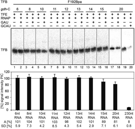
TFB release occurs upon transcription of 15 nt long RNAs To verify that the signal reductions observed for the TFB B- reader domain Bpa variants at register +10 were not caused by an early TFB release event, the TFB Bpa-variant F192 was used in crosslink experiments. This position within TFB is located in the cyclin fold domain far away from the active site, and therefore it should not be affected by structural re- arrangements of the B-reader elements (22). The results of the crosslink experiments with TFB F192 in stalled com- plexes are summarized in Figure 7. Poly-dAdT was addi- tionally used in the reactions in order to trap TFB to prevent re-association of TFB to the promoter site after a possible TFB release event. The experiments showed that the signal intensities of TFB-DNA interactions in stalled complexes at registers +6 to +14 do not change in comparison to the sig- nals derived from the respective PICs, indicating that TFB is not released at these positions. At position +15 and +20 a slight decrease of the signal intensity was observed, whereas crosslinking experiments under run-off conditions showed only background signals.
These data shows that TFB is present in complexes tran- scribed to register +10 and demonstrate further that the loss of interactions of the B-reader domain and DNA men- tioned previously are not due to a TFB release event, but is a result of TFB B-reader translocation. We therefore con- clude that the TFB B-reader domain is displaced at register +10, which causes a bubble collapse at this position consis- tent with predictions derived from eukaryotes (53). Our ob- servations are further in accordance with previous KMnO
4footprint experiments which revealed a reduced size of the open DNA region at registers +10 / +11 in the P. furiosus transcription system (41).
For the human system, TFIIB release was described to take place at an RNA length of 12/13 nt in vitro (55). It was also postulated that a 12 nt RNA clashes with the Zn-ribbon domain of TFIIB in a crystal structure of an initially tran- scribing S. cerevisiae TFIIB / RNAP II complex, since the Zn-ribbon blocks the exit pore of RNAP II (45). Addition- ally, Xie et al. (78) showed that TFB is released in in vitro reactions lacking TFE in the archaeal M. thermoautotroph- icus system when complexes where chased to position +24.
Moreover, in a human in vitro transcription system TFIIB release was shown to take place between +6 and +16 (54).
Therefore, we expected a TFB release event between reg- isters +12 to +14 but in our in vitro system we could not monitor this event at the respective sites. In contrast, we ob- served that the amount of TFB started to decrease at regis- ter +15 whereas the signal is further reduced at register +20, and TFB appears to be completely absent in run-off exper- iments. These findings lead us to the assumption that TFB is destabilized between position +14 and +15 and the con-
tacts between TFB and RNAP, TBP and DNA are further destabilized from these positions onwards. Due to the fact that the signal intensity does not drop from one nucleotide position to the next we further assume that the exact point of release of TFB cannot be pinpointed to a distinct nu- cleotide position in our in vitro transcription system. This may rely on the presence of a too high background signal deriving from complexes, which did not build the PIC, or from complexes, which are still positioned at the promoter start site in the open complex state or abortive transcription state. In addition, it remains unclear if the nascent RNA in stalled complexes has enough kinetic energy to induce the release of TFB from the complex, and if TFB release is an instantaneous event, or if this process happens slowly due to the persisting interactions between TFB and DNA, TBP and RNAP. In addition, the stabilizing effect of TFE and its impact on TFB release is still unclear.
CONCLUSION
In this study, we made use of a highly specific crosslinking method to unravel the conformational changes of the gen- eral transcription factor TFB in archaeal transcription dur- ing the initiation to elongation phase transition. We demon- strated that the TFB B-reader domain is located in close proximity to the active site of the transcription machin- ery as TFB residues of A46Bpa, R52Bpa and S56Bpa can be specifically crosslinked to the template DNA at posi- tion -4 T in the OC. Additionally, TFB-F192Bpa of the B- core element specifically crosslinks to DNA labeled at -19 T in the ternary, as well as in the open initiation complex.
These observations are in agreement with published struc- tural models of eukaryotic transcription complexes (4,3,45), further supporting the conservation of initiation complex architecture between the archaeal and eukaryotic domain of life. For that reason we conclude that the topology of the archaeal TFB is highly similar to that of eukaryotic TFIIB. Our crosslinking system further allowed us to inves- tigate dynamic structural transitions of TFB by analyzing the respective TFB-DNA contacts in stalled complexes that mimic the stepwise transition from the initiation to elonga- tion complex. We could show that transcription complexes with the selected TFB variants can be stalled correctly at the distinct sites on the gdh-C templates. Our experiments provide evidence that nascent RNA interacts with the B- reader loop at a RNA length of six nucleotides, and clashes with the B-reader helix at a length of 8 nucleotides. We fur- ther could demonstrate that the B-reader domain is distant from its DNA site at register +10, resulting in the collapse of the transcription bubble. Using the B-core variant TFB F192Bpa we showed that TFB is present at registers +6 to +14, whereas the amount of TFB in the complexes were re- duced from register +15 onward and is completely absent in crosslinked run-off samples. We assume that after B-reader displacement and transcription bubble collapse at register +10, TFB is destabilized within the complex, which in turn induces TFB release and promoter escape (summarized in Supplementary Figure S6).
Single-molecule FRET experiments furthermore suggest that TFB linker-reader moves along with the RNAP clamp and that the TFE-induced conformational change towards
Downloaded from https://academic.oup.com/nar/article-abstract/46/19/10066/5068255 by Universitätsbibliothek user on 28 February 2019
Figure 7.