Factor B during transcription initiation in Pyrococcus furiosus
Dissertation
zur Erlangung des Doktorgrades der Naturwissenschaften (Dr. rer. nat.) der Fakultät für Biologie und Vorklinische Medizin
der Universität Regensburg
Vorgelegt von
Stefan Albin Dexl
aus
Neumarkt i.d.OPf.
Die Arbeit wurde angeleitet von:
Prof. Dr. Michael Thomm
Unterschrift
Table of contents
Table of contents... i
Introduction ... 1
I. A.Transcription - a crucial step in cellular life... 1
1. Genome organization and promoter-DNA accessibility... 2
2. Promoter architecture and regulation of gene expression... 3
B. Initiation of transcription: Preinitiation complex formation... 6
1. The TATA binding protein... 7
2. The transcription factor B... 9
3. The RNA polymerase ... 14
4. The transcription factor E... 18
5. Additional eukaryotic transcription factors TFIIF and TFIIH... 19
C. From initiation to elongation and termination ... 20
D. The replication protein A of P. furiosus... 22
E. Scientific questioning of this thesis... 23
Materials ... 25
II. A. Chemicals and Reagents... 25
B. Kits... 25
C. Enzymes... 26
D. Strains... 26
E. Services... 26
F. Softwares... 26
G.Plasmids... 27
H. Oligonucleotides... 28
Methods... 31
III. A. DNA preparations... 31
1. DNA templates for in vitro transcription assays and EMSAs... 31
2. 5´end labeled templates for footprint experiments ... 31
3. Mismatch template preparation ... 31
4. gdh-C11 - gdh-C15 template generation using PCR mutagenesis... 32
5. Radio labeled DNA templates for crosslink experiments... 33
B. Protein preparations ... 34
3. Expression and purification of TFB and TFB variants ... 35
4. RNA-polymerase purification ... 36
5. TFE purification... 36
C. Transcription assays ... 37
1. Electro mobility shift assay... 37
2. Abortive transcription assay... 38
3. Run-off transcription assay ... 38
4. Chase experiments and stalled transcription complexes... 38
5. Potassium permanganate footprinting ... 38
6. Crosslinking experiments ... 39
D. FRET measurements and data acquisition ... 39
Results... 41
IV. A. Analysis of the replication protein A during transcription... 41
1. RPA in transcription initiation ... 41
2. RPA in transcription elongation... 42
3. Summary of PfuRPA experiments ... 44
B. DNA bending experiments of P. furiosus TFB using FRET ... 45
C. The role of the TFB B-reader loop in transcription initiation... 48
1. Analysis of TFB Alanine substitutions in transcription assays... 49
2. KMnO
4footprint experiments of TFB B-reader alanine variants ... 51
3. TFE can partially compensate defects in promoter opening ... 53
4. RNA-strand separation at heteroduplex DNA templates... 54
5. Summary of the TFB alanine substitutions... 57
D. TFB-DNA crosslink studies during transcription initiation ... 59
1. Analysis and selection of TFB-Bpa variants ... 60
2. Specificity of UV crosslinking experiments... 66
3. Crosslinking experiments in the preinitiation complex... 68
4. Crosslinking experiments in stalled transcription complexes... 72
5. Summary of the crosslinking experiments... 77
Discussion... 79
V. A. A possible role for RPA during transcription elongation... 79
B. Bending of DNA depends on the presence of TFB in P. furiosus ... 80
C. The charge distribution of the B-reader loop is important for the function of TFB... 81
E. Topology of PfuTFB is almost similar to TFIIB ... 83
F. The TFB B-reader domain is displaced at register +10... 85
G.TFB tends to be released from register +15 onwards... 86
H. Concluding aspects... 86
Abstract... 89
VI. Zusammenfassung ... 90
VII. Appendix ... 91
VIII. A. Abbreviation list ... 91
B. Figure list ... 93
Publication bibliography ... 94
IX. Danksagung... 117
X.
Erklärung ... 118
XI.
Introduction I.
This work should provide more detailed insights into mechanisms and structural functions of the transcription factor B, and to some extent, the possible role of the replication protein A in the process termed transcription. To investigate the interactions of these two factors the in vitro transcription system of the hyperthermophilic organism Pyrococcus furiosus was used.
The strain was isolated at Porto die Levante, Vulcano, Italy, and described by Fiala, G. and Stetter, K.O (Fiala, Stetter 1986). It belongs to the domain Archaea, which was defined by Woese, Kandler and Wheelis by comparison of the ribosomal RNA (Woese et al. 1990).
These studies revealed that basically all living organisms can be referred to one of the three domain of live: Bacteria, Archaea and Eukarya. Pyrococcus furiosus, the “rushing fireball”, grows optimally under anaerobic conditions at 95°C with a doubling time of 37 minutes, and can use different sugars as carbon source (Fiala, Stetter 1986). In 1996, Hethke et al.
established a Pyrococcus cell-free transcription system to enable investigation of transcription processes (Hethke et al. 1996). This artificial system allows one to analyze the functions and mechanisms of different transcription factors, as well as the characterization of distinct subunits of the RNA polymerase using an in vitro reconstitution approach of this enzyme (Fouqueau et al. 2013). In the following years studies of archaeal and eukaryotic organisms showed similarities in the genomic sequences concerning the transcription apparatus, as well as relationships of transcription regulating proteins between bacterial and archaeal organisms (Kyrpides, Ouzounis 1999). Therefore biochemical analysis of the archaeal transcription system can be useful to reveal evolutionary aspects between the three domains, as well as to make statements for eukaryotic systems concerning function and regulatory mechanisms of the transcription machinery.
The following chapters should give a more detailed insight into the process of transcription, the similarities between transcription machineries in the domains of life, and a detailed functional characterization of the transcription factor B. In addition, a short overview on the replication protein A is given at the end of this introduction, which is also characterized in this thesis.
A. Transcription - a crucial step in cellular life
Differentiation, cell division, metabolism as well as communication are major events in the life
of multicellular organisms. In addition, single-cell organisms also need to response to
environmental factors like temperature, nutrients, or toxins for optimal growth. Therefore the
regulation of genetic information is a very important step for cells to perform target-driven
functions and tasks. Experiments of Oswald Avery, Alfred Hershey and Martha Chase, as
well as the discovery of the structure of deoxyribonucleic acid (DNA) by James Watson,
Francis Crick and Rosalind Franklin demonstrated that DNA is the central memory of cellular
information (Avery et al. 1944; HERSHEY, CHASE 1952; WATSON, Crick 1953). The
genetic code is defined as the sequence of the four nucleobases, adenine, cytosine, thymine,
and guanine. The so called “Central Dogma” of molecular biology was proclaimed in the late
1950ths and refined in 1970 (Crick 1958, 1970). Herein it was postulated that information
derived from DNA is transcribed into RNA, which can serve as a template for protein
biosynthesis. The resulting proteins are essential for numerous cellular processes like
metabolism, DNA maintaining and repair, signal pathways for cellular response to various
stimuli, and many more, which defines the phenotype of an organism. Since the last decades
transcriptional RNA processing, were discovered (Shapiro 2009; Koonin 2015). Therefore information does not flow only from DNA to RNA to the protein, moreover, a complete and complex network of information flow exists. RNA, in contrast to DNA, contains a reactive OH- species on the second carbon atom at the ribose, and comprises uracil as nucleobase, the demethylated form of thymine. Nowadays a lot of different RNA molecules are known.
Beside the well-described classes of transfer RNA (tRNA), messenger RNA (mRNA), and ribosomal RNA (rRNA) a new RNA group of non-coding RNAs was revealed. These RNAs are clustered into small non-coding RNAs (snRNAs), like microRNAs (miRNA), small interfering RNAs (siRNA), Piwi-interacting RNAs (piwiRNA), small nucleolar RNAs (snoRNA) and long non-coding RNAs (lncRNAs) (see reviews (Ghildiyal, Zamore 2009; Bratkovic, Rogelj 2014; Fatica, Bozzoni 2014; Bhartiya, Scaria 2016)).
Despite the large number of RNA molecules with numerous different functions the origin is the same for every type of RNA: they have to be transcribed from DNA. This process is termed transcription and is carried out by large multi-subunit DNA-dependent RNA- polymerase (RNAP) enzymes. Eukaryotic organisms possess up to five RNAPs, and archaea and bacteria have only one enzyme to synthesize RNA, whereas the subunits are homolog to eukaryotic RNAP II (Werner, Grohmann 2011). The eukaryotic RNA-polymerases I - III have specific functions. The RNAP I transcribe only rRNA (Engel et al. 2013), the RNAP II synthesizes mRNA and some small non-coding RNAs (Kornberg 2007), whereas the RNAP III transcribe the 5S rRNA, tRNAs and small non-coding RNAs (Arimbasseri, Maraia 2016). The nuclear RNAP IV and RNAP V are only present in plant species and some algae, they contain 10 or more subunits which are more or less related to subunits of other RNAPs, and are important for small interfering RNA-mediated gene silencing (Landick 2009). To synthesize RNA, the RNAPs have to be recruited to the DNA by interaction with specific transcription factors. These general factors need access to specific sequence motifs, and therefore DNA has to be remodeled first.
1. Genome organization and promoter-DNA accessibility
Transcription is a precisely organized process which enables targeted gene expression, and is regulated by numerous cellular processes. To transcribe a gene specifically transcription factors need access to target DNA sequences. The genetic material, which can comprise millions of base pairs, is structurally organized and condensed by proteins to facilitate compression of the DNA into a single cell.
DNA of eukaryotes is packaged and organized in the nucleus as chromatin, a conglomeration of nucleosomes. A nucleosome consists of a histone protein bound to 145- 147bp DNA (Luger et al. 1997). DNA is wrapped around the histones and cannot be the target of transcription factors due to a steric hindrance. Therefore the chromatin structure has to be remodeled in a way that the histones were relocated to expose free DNA. This process is executed in eukaryotic organisms by a large number of proteins which belong to one of four ATP-dependent chromatin remodeling complex families, whereas the histones can also be modified e.g. by acetylation, methylation, phosphorylation or ubiquitination (Witkowski, Foulkes 2015).
Bacteria lack histones or histone-like proteins, and their DNA is packaged as a nucleoid in
the cell, whereas the DNA is bound to and organized by nucleoid-associated proteins (NAPs)
(Dorman 2014). The most abundant chromatin proteins in bacteria are members of the HU
(histone-like protein from E.coli strain U93) protein family, and the related protein HTa can
also be found in some archaeal species which lack histone-like proteins (Dorman 2009;
Archaeal organisms show different DNA packaging strategies of their nucleoid. The genomic DNA of thermophilic organisms is positively supercoiled as a result of the reverse gyrase enzyme (Brochier-Armanet, Forterre 2007). This enzyme is thought to be exclusive for hyperthermophilic organisms and therefore this DNA conformation is preferred possibly due to an adaption to hot environments (Forterre et al. 1996). In addition, the DNA is further stabilized by DNA-binding proteins. A highly abundant chromatin protein distributed in the archaeal domain is alba (acetylation lowers binding affinity), or proteins of this family, respectively (Laurens et al. 2012). Alba can be modified by acetylation and deacetylation (Wardleworth et al. 2002), whereas in vitro experiments revealed that it can condense, bridge and loop DNA, but its in vivo dynamics remains unclear (Jelinska et al. 2005; Laurens et al.
2012). In addition to alba, members of the phylum Euryarchaeota possess mainly histone proteins to organize the DNA (Reeve 2003). These proteins are homologous to the eukaryotic H3 and H4 histone subunits and form dimers in solution and tetramers when bound to DNA (Reeve et al. 2004), but lack the typical N- and C-terminal extensions for modifications (Cheung et al. 2000). In contrast, Crenarchaeota lack eukaryotic-like structures, but have own small basic DNA-binding proteins like Cren7, which are highly conserved and exclusive within this phylum, or the related Sul7 proteins (Guo et al. 2008).
These chromatin proteins show high similarity to bacterial NAPs (Driessen, Dame 2011).
Indeed, genes for eukaryotic-like proteins were also found in some organisms of the Crenarchaeota (Cubonova et al. 2005).
Less is known about the interplay between DNA organizing proteins and transcription factors, which enable recruitment of the RNA polymerase to the promoter site of a gene for RNA synthesis. However, it was shown that if promoter regions are occupied by DNA-binding proteins, the transcription is blocked due to the prevention of factor binding or inhibition of DNA separation (Soares et al. 1998; Xie, Reeve 2004a; Wilkinson et al. 2010). For example, transcription is inhibited in the M. jannaschii in vitro system when nucleosome formation at the promoter site occurs (Wilkinson et al. 2010). Similar effects were observed in M.
thermoautotrophicus, as binding of HMta2 downstream of the transcription start site (TSS) forms a filament that extends to the upstream part of the +1 site, and prevents transcription factor binding (Xie, Reeve 2004a). Interestingly, the same protein does not block the RNA polymerase in the elongation phase, but it lowers the transcription rate (Xie, Reeve 2004a).
Global scale analysis revealed that archaeal histones in general are not present at core promoters of archaeal genes and it was shown that the region directly upstream of the TSS is not occupied by histone proteins (Nalabothula et al. 2013). It was pointed out by Peeters et al. that it is more likely in the genome that sequences direct the positioning of nucleosomes to enable binding of transcription factors rather than the transcription factors block the binding of histones in resulting chromatin-free regions (Peeters et al. 2015).
Taken together, it is still enigmatic how transcription is interlinked to genomic organization in archaeal organisms, because the mechanisms of global gene regulation, as well as the goal- driven deposition of chromatin proteins to make DNA accessible for transcription remains to be determined. However, if DNA becomes accessible for transcription factors, numerous proteins, which regulate transcription by repression or activation, interact with the promoter site of the gene.
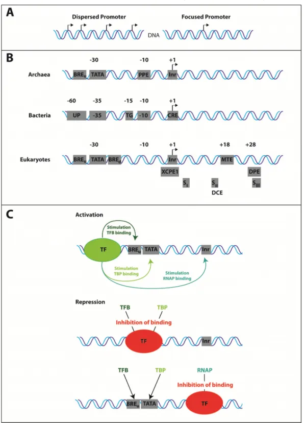
2. Promoter architecture and regulation of gene expression
Basically two types of promoters are known: core promoters, also known as the single peak
or focused promoters, and dispersed or broad peak promoters (Juven-Gershon et al. 2008;
start site (TSS) (Butler, Kadonaga 2002; Müller et al. 2007). The broad peak promoters have several start sites distributed over >100 nucleotides and are typically found in CpG islands in vertebrates (Carninci et al. 2006). Both promoter types have specific elements, which serve as interaction platforms for transcription factors. Dispersed promoters lack the TATA-box, downstream promoter element (DPE) and the motif ten element (MTE), which are typical components of core promoters (Juven-Gershon et al. 2008). Furthermore, genes regulated by core promoters are usually issue-specific (Müller et al. 2007), whereas genes regulated by dispersed promoters are mostly ubiquitously expressed (Carninci et al. 2006).
Core promoters often contain the so called TATA-box, also known as Goldberg-Hogness sequence (Sassone-Corsi et al. 1981) (Figure 1B). It is an AT-rich element with the consensus sequence TATAWAAR, whereas the upstream T is most commonly located at -31 or -30 relative to the transcription start site (TTS) +1 (Hausner et al. 1991; Ponjavic et al.
2006; Carninci et al. 2006). This widely used and ancient element is the most conserved promoter motif in archaea and eukaryotes, and is recognized by the general transcription factor TATA binding protein (TBP) (Thomm, Wich 1988; Hausner et al. 1996). Despite the high abundance only 10% of human RNAP II promoters contain a TATA-box (Bajic et al.
2006). A second motif adjacent to the TATA-box is the transcription factor B recognition element (BRE) which is bound by the transcription factor B (TFB) upstream (BRE
u) and/or downstream (BRE
d) the TATA box (Deng, Roberts 2005; Lagrange et al. 1998). The location of the BRE relative to the TATA and the transcription start site defines the transcription direction (Bell et al. 1999). The BRE and the TATA box are strictly required for core promoter dependent transcription, whereas a third element, the Initiator region (Inr) is not (Gehring et al. 2016). This regulatory element encompasses the TSS +1. Sequence alignments of thousands of mammalian transcription start sites showed that the consensus sequence can be restricted to YR, whereas R is the +1 site (Juven-Gershon et al. 2008) and is often an adenine (Butler, Kadonaga 2002). Inr is recognized by the transcription factor IID (TFIID) in eukaryotes and some transcriptional activators in archaea and comprises a high AT content similar to the TATA box (Gehring et al. 2016). This region is often termed the initially melted region (IMR), and can extend up to 12 base pairs upstream the +1 site (Bell et al. 1998), and is an important determinant for the strength of the stimulatory effect of the transcription factor E (TFE) (Blombach et al. 2015). In addition, a proximal promoter element (PPE) exists in archaeal organisms, which is located approximately 10 base pairs upstream of the transcription start site and can increase transcription output through interaction with general transcription factors (GTFs) (Peng et al. 2009). In contrast, in eukaryotic organisms a downstream core promoter element (DPE) can be found 28 to 33 base pairs downstream the TSS, which is important for basal transcription and interacts with the TATA associated factors (TAF) 6 and 9 of the RNAP I system, and TAFII60 and TAFII40 of TFIID of the RNAP II system (Burke, Kadonaga 1996). Promoters containing DPE usually lacks a TATA-box (Müller et al. 2007). Another sequence in eukaryotes was found by computational and biochemical studies and is called the motif ten element (MTE) (Lim et al. 2004). It is located +18 to +27 downstream of the TSS, and, like DPE, functions with the Inr in a cooperative spacer-dependent manner (Lim et al. 2004). Interestingly, optimization of the core promoter elements TATA-box, DPE, MTE, Inr and BRE
d/BRE
uleads to the strongest known in vitro promoter (Juven-Gershon et al. 2006). A much more specific promoter region is the so called downstream core element (DCE), which was found in the beta-globin promoter (Lewis et al.
2000) and also characterized in the adeno virus major late promoter (Lee et al. 2005). It
approximately 1% of human core promoters which are TATA-less, and are called X core promoter element 1 (XCPE1). This element is located from -8 to +2 and interacts only with sequence specific activators like NRF1, NF-1 and Sp1 (Tokusumi et al. 2007).
Figure 1: Promoter architecture and regulation of gene expression. A) Dispersed and focused (core) promoters differ in the number of their transcription start sites. B) General core promoter elements of archaea, bacteria and eukaryotes. C) Mechanism of activation and repression of transcription.
Transcription factors (TF) bind to sequence motifs upstream the BRE/TATA to activate transcription,
whereas binding of TF to elements downstream the BRE/TATA inhibit binding of GTFs and RNAP.
Typical archaeal promoters contain a TATA-box, the BRE and Inr motif. In contrast, Bacteria differ in their promoter architecture in comparison to eukaryotic and archaeal promoters but comprise also sequences important for the interaction with σ-factors and the RNA polymerase. The important sites for interaction with σ-factors are the -35 (TTGACA) and the - 10 (TATAAT) region, whereas the AT-rich UP region and the start site containing core recognition element (CRE) both interact with the polymerase (Decker, Hinton 2013). An overview on the common promoter architecture of bacteria, archaea and eukaryotes is shown in figure 1B. The distinct motifs shown here are all cis-acting regulatory elements (Butler, Kadonaga 2002), and the presence of distinct motifs and their combinations are one possibility to regulate gene expression (Colgan, Manley 1995). These elements serve as platforms for a variety of transcription factors.
In addition to these combinations gene expression can also be regulated by activators, repressors, enhancers and mediators, which recognize additional specific sequence motifs in proximity to the promoter (Figure 1C). One of the best studied transcriptional regulator in archaea is the Leucine-responsive regulatory protein (Lrp), which possess a typical bacterial helix-turn-helix DNA binding motif, and has a dual role as activator and repressor of transcription (Peeters, Charlier 2010). Members of the Lrp family regulate almost 10% of all genes and are mostly involved in amino acid and central metabolisms in bacteria (Cho et al.
2008). In Pyrococcus furiosus, it was shown that the Lrp-like protein LrpA binds closely downstream the TATA box, forming a TBP/TFB/LrpA complex, which in turn blocks the binding of the RNA polymerase due to steric hindrance (Dahlke, Thomm 2002). In contrast, the putative transcription factor 2 (Ptr2) of Methanococcus jannaschii activates transcription through binding to an upstream element and stimulates recruitment of TBP (Ouhammouch et al. 2003). A further global regulator of transcription with a dual role is the transcriptional regulator of mal B operon like factor 1 (TrmBL1), which recognizes the Thermococcales Glycolytic Motif (TGM) located upstream or downstream of the TATA box to regulate genes involved in sugar metabolism (Gindner et al. 2014). It was shown in ChIP-Seq experiments that TrmBL1 binds to TGMs located downstream of the TATA to repress genes involved in gluconeogenesis, and simultaneously binds to TGMs located upstream of the TATA to switch on genes involved in sugar metabolism under glycolytic growth conditions, whereas TrmBL1 does not bind TGMs under gluconeogenic growth conditions (Reichelt et al. 2016).
The interplay between transcription factors and regulators in combination with distinct promoter elements defines the transcriptional activity and the level of gene expression. The presence of basal factors at the promoter in turn recruits RNAP to initiate RNA-synthesis.
Therefore, the gene expression level of a single cell, as a response mechanism to environmental signals, depends on many different factors.
B. Initiation of transcription: Preinitiation complex formation
The core promoter-dependent transcription process can be divided into three distinct phases.
In the first stage general transcription factors specifically interact with sequence motifs of the
promoter and bind to DNA until the RNA polymerase is recruited to form a preinitiation
complex (PIC). This complex is formed in a stepwise manner as it was shown with native gel
electrophoresis experiments (Buratowski et al. 1989) and later with cryo-EM analysis (He et
al. 2013). RNAP II preinitiation complexes of eukaryotic organisms consist of in minimum six
transcription factors TFIID, TFIIA, TFIIB, TFIIE, TFIIF and TFIIH, whereas archaeal
organisms require basically the three eukaryote-related factors, TBP, TFB and TFE (Bell,
Jackson 2001; Carlo et al. 2010) (Figure 2). The archaeal transcription machinery therefore
2011; Decker, Hinton 2013). In contrast, bacterial complexes contain RNAP and σ (Feng et al. 2016). After complex assembly several structural rearrangements have to take place to convert the initiation complex into an initially transcribing complex. These transitions are shown in chapter I. C (From initiation to elongation and termination). Then RNA synthesis takes place in the elongation phase until transcription is terminated. The proteins which form a preinitiation complex at the core promoter of the three domains are shown in the following chapters.
Figure 2: Comparison of archaeal and eukaryotic Pol II preinitiation complexes. Archaeal PIC consists of TBP (red; PDB: 5FZ5), TFB (green; PDB: 3K1F), RNA polymerase (grey; PDB: 4QIW), and TFE (pale green; PDB: 5FZ5) and bent DNA (PDB: 5FZ5), whereas eukaryotic Pol II PIC consist of the related TBP (red; PDB: 5FZ5), TFIIB (green; PDB: 3K1F), TFIIEα/β (pale green and pale blue; PDB:
5FZ5), and the eukaryote-specific TFIIAα/β (purple; PDB: 5FZ5) and TFIIFα/β (blue; PDB: 5FZ5) and bent DNA (PDB: 5FZ5). Complete Pol II PIC structure was modified from PDB: 5FZ5 (Plaschka et al.
2016). For the archaeal PIC T. kodakarensis RNAP from structure 4QIW (Jun et al. 2014) was fitted to the complex based on exact overlay of conserved residues in PyMol. TFB/TFIIB was taken from structure 3K1F (Kostrewa et al. 2009) due to absent domain structures in 5ZF5. TFIIH is missing in the 5ZF5 structure because of insufficient resolution of the cryo-EM structure.
1. The TATA binding protein
The first factor which interacts with the TATA element of a core promoter via an induced-fit mechanism is the TATA binding protein (TBP) (Chasman et al. 1993; Kim et al. 1993a;
Burley 1996). This protein was formerly referred as the aTFB protein in archaeal organisms,
but because of analogous functions to eukaryotic TBP and the similar structure it was re-
termed TBP in archaea (Hausner et al. 1996). This saddle-shaped protein comprises a
tandem repeat consisting of two conserved domains which are likely the product of ancient
1996). Each of the two domains consists of a five-stranded anti-parallel β-sheet and two α- helices on the opposite site (Kim et al. 1993a; Kim et al. 1993b). Four β-strands of each domain bind to DNA, whereas two α-helices of each domain together with parts of the two β- strands form the convex opposite site and serve as an interface for proteins which are involved in transcription initiation (Akhtar, Veenstra 2011). It was shown that TBP can be exchanged between organisms, e.g. TBP of P. furiosus with Methanococcus TBP, and Methanococcus TBP with human and yeast TBP (Wettach et al. 1995; Hethke et al. 1996).
Bacteria lack the TATA-binding protein and transcription is basically initiated using sigma factors, but it was shown that elements of the conserved TBP are part of the RNase HIII and a DNA glycosylase, likely due to a fusion processes of a TBP core domain and these proteins (Brindefalk et al. 2013). From this point of view, Brindefalk et al. showed that sequences of TBP domains can be found in numerous proteins, indicating that a TBP precursor was present in the last universal common ancestor (LUCA) and evolved either by fusion processes with other proteins or to itself and functions were adapted, or TBP domains originated from DNA-glycosylases and TBP becomes a general transcription factor later (Brindefalk et al. 2013). It is also interesting to note that single TBP-domain sequences were identified e.g. in Halobacteria and in the Pyrococcus furiosus genome, which encodes a monopartite TBP of unidentified function in addition to the regular TBP sequence (Brindefalk et al. 2013).
Genomes of higher eukaryotes encode TBP, TBP-related factors (TRF) and TBP-like factors (TLF), which are involved in development and differentiation, in particular gametogenesis and early embryonic development (Akhtar, Veenstra 2011). For RNAP II transcription TBP together with up to 14 TBP associated factors (TAFs) form the eukaryotic TFIID multi-subunit complex (Matangkasombut et al. 2004). The core of TFIID is formed by a subset consisting of TAF4 - TAF6, TAF 8 - TAF10 and TAF12, but no TBP (Leurent et al. 2004). Therefore, different TFIID variations are present in different tissues and cell types to promote targeted gene expression (Demeny et al. 2007), and TBP is further not the universal initiation factor in metazoans like it is in yeast (Akhtar, Veenstra 2011). Recent single molecule analysis on PIC assembly in eukaryotic Pol II transcription showed that TBP alone indeed binds to the promoter, but the specificity of the interaction between TBP and DNA is strongly increased if TBP is part of the TFIID complex (Zhang et al. 2016). In addition, TBP is not only involved in the initiation of RNAP II promoters, it also has its role in RNAP I and RNAP III initiation. TBP together with the selectivity factor SL1 and five RNAP I-specific TAFs are required to initiate RNAP I transcription, whereas TBP and two RNAP III-specific TAFs (BrtI and BdpI) assemble together with TFIIIB to initiate RNAP III transcription (Drygin et al. 2010; Hoffmann et al. 2016).
After TBP bind to the TATA element, the DNA is highly bent in approximately 90° angle due
to a transition of DNA into a unique partially unwounded right-handed double helix by a kink
(Kim et al. 1993a; Nikolov et al. 1995; Juo et al. 1996). The kink is caused by two
phenylalanine residues (Phe284 and Phe301 in human TBP) which contact DNA in the minor
groove between the first two base pairs of the TATA-box. The second kink is located at the
7
thand 8
thbase of the TATA-box also by insertion of two phenylalanine residues (Phe193
and Phe210 in human TBP), and DNA is restored back to its usual B-conformation. Binding
of TBP and bending of DNA occur simultaneously (Masters et al. 2003) and DNA bending
was shown to be a prerequisite for transcriptional activation (Gietl et al. 2014). Recent
studies using single molecule analysis showed that DNA in archaeal organisms is bent to
the general transcription factor B (TFB) is required to stabilize the bent state (Gietl et al.
2014). For Saccharomyces cerevisiae it was shown that DNA bending follows a three-step binding mechanism, as two different complexes were identified with different bending angles, whereas addition of the transcription factor IIB (TFIIB) leads to a fully bent state of the DNA (Gietl et al. 2014).
Beside the stabilization effect of TFIIB, the RNA polymerase II specific auxiliary factor TFIIA also stabilizes the TBP-DNA interaction in eukaryotic transcription initiation (Kang et al.
1995). This factor consists of two conserved domains, a 12-stranded β-barrel which binds to the upstream DNA of the TATA-box and the TBP saddle, and the other domain consists of a four-helix bundle, forming a boot-shaped heterodimer (Tan et al. 1996; Geiger et al. 1996).
TFIIA is not able to bind DNA alone, but together with TBP or TFIID, binding to DNA is very efficient (Zhang et al. 2016). TFIIA is not strictly required for transcription initiation, but can stimulate basal and activated transcription (Imbalzano et al. 1994).
2. The transcription factor B
The next factor associating to DNA and TBP is the general transcription factor B (TFB), or TFIIB for RNAP II transcription, respectively. Recent studies demonstrated that the eukaryotic TFIIB requires TFIID and TFIIA to bind transiently to the promoter, and addition of RNAP II-TFIIF fully stabilizes the association of TFIIB to the preinitiation complex (Zhang et al. 2016). TFB/TFIIB is a single polypeptide consisting of a carboxyl-terminal B-core domain, an amino-terminal Zn-ribbon domain, and a region in between, which were later termed the linker and the reader domain (Ha et al. 1991; Malik et al. 1991; Pinto et al. 1992; Kostrewa et al. 2009) (Figure 3 A). Magnetic resonance spectroscopy analysis of the human TFIIB C- terminal B-core domain (TFIIBc), revealed that this domain consist of two direct repeats which have similar α-helical structures, whereas each repeat contains five alpha-helices A1 to E1 of repeat one, and A2 to E2 of the more hydrophobic repeat 2 (Bagby et al. 1995). First crystal structures of TFIIB/TBP/DNA-complexes indicated that the B-core contacts TBP as well as DNA at the major groove immediately upstream, and at the minor groove downstream the TATA-box (Nikolov et al. 1995). TFB binds to DNA at the BRE via a helix- turn-helix motif formed by helices D and E, whereas the TFIIB-DNA contacts were also verified by DNase I footprinting (Malik et al. 1993), hydroxyl radical footprinting (Lee, Hahn 1995), fluorescence anisotropy measurements and photochemical crosslinking (Lagrange et al. 1998). Mutational analysis of amino acids of the yeast B-core domain further demonstrated these interactions, and revealed that the basic amino acids K190, K201, and K205 play a major role in the interaction with DNA, as these mutants do not form a TBP/TFIIB/DNA complex in yeast in in vitro gel shift experiments, and showed impairments in growth in vivo (Bangur et al. 1997). Amino acid exchange of the conserved amino acids G153 and R154 (Buratowski, Zhou 1993), as well as amino acid substitutions of G247 and R248 within the second repeat in human TFIIB showed a decreased ability to form TBP/TFIIB/DNA complexes (Bagby et al. 1995). In addition to the mutational analysis, sequence alignments further revealed structural similarities to cyclin A, which is a cell cycle regulating protein (Bagby et al. 1995). From this point of view it was hypothesized that cyclins may have evolved from more fundamental transcription processes in earlier life (Bagby et al. 1995).
Structural analysis of the N-terminus of the archaeal TFB of Pyrococcus furiosus showed that
this domain forms a Zn-ribbon fold (Zhu et al. 1996). This domain of TFIIB is required for the
interaction with the RNA polymerase II associated protein (RAP) 30/74, the small subunit of
Figure 3: Structure, domain organization and multiple sequence alignments of the transcription factor
IIB. A) TFB/TFIIB consists of a C-and N-terminal cyclin fold (cyan), a B-linker region (brown), a B-
reader domain consisting of the loop (blue) and the helix (green), and a Zn-ribbon (pale green) with a
bound Zn
2+ion (red) (modified from PDB: 3K1F). The domain organization of PfuTFB is given below
from N to the C terminal end, and the same color code is used as in the structure. Amino acids are
shown for the respective domain from N36 (reader helix) to S102 (linker helix). B) Sequence
alignments of the highly conserved TFB/TFIIB B-reader domain. PfuTFB was blasted against
RNA polymerase recruitment was shown to be carried out by the Zn-ribbon domain, using amino acid mutagenesis approach in yeast (Pardee et al. 1998). Further site-specific photo crosslinking experiments revealed a specific contact of the TFIIB Zn-ribbon with the surface of the RNAP II dock domain, overlapping the RNA exit point (Chen, Hahn 2003), whereas this location of the ribbon was later confirmed in a crystal structure (Kostrewa et al. 2009).
Therefore the Zn-ribbon is essential for RNAP II/TFIIF recruitment. Because of the fact, that TFIIB stabilizes the TBP/DNA complex by direct interactions with TBP and DNA, and the observation, that TFIIB plays a role in RNAP II/TFIIF recruitment, it was proposed that this factor has only a role in bridging between TBP/DNA and RNAP II (Buratowski et al. 1989;
Orphanides et al. 1996; Hampsey 1998). Therefore the domains between the N- and C- terminal domains where thought to be just a flexible hinge region. Interestingly, first mutational analysis of the region adjacent to the N-terminal Zn-ribbon revealed that this domain provides key features for the initiation process (Bangur et al. 1997). It was also shown that this region of TFIIB is the most highly conserved region amongst known TFB proteins (Na, Hampsey 1993) (Figure 3 B), and amino acids 52-140 of yeast TFIIB can be functionally replaced by the corresponding region of human TFIIB (Shaw et al. 1996).
The TFB B-reader helix domain is important for transcription start site selection. Mutational analysis of this domain in yeast, especially amino acid R64, results in shifts of the transcription start site in vitro and in vivo, a cold-sensitive phenotype and diminished growth rates (Pardee et al. 1998; Bangur et al. 1997; Pinto et al. 1992). Amino acid E62 of yeast TFIIB showed the same effects, but interestingly, the corresponding amino acid E51 of human TFIIB is not affected by substitutions (Cho, Buratowski 1999). Therefore it was assumed that the transcription start site selection also depends on the distance between Inr and TATA, because in yeast promoters the spacing between Inr and TATA differ in comparison to human promoters (Cho, Buratowski 1999). Additional analysis of TFIIB B- reader mutations and different Inr sequences made clear that TSS selection is B-reader helix and RNA polymerase dependent (Li et al. 1994; Faitar et al. 2001). Moreover, it was postulated that the TSS selection is carried out by scanning of the RNA polymerase to search for the correct nucleotide to start RNA synthesis (Giardina, Lis 1993). First models of crystal structures containing TFIIB and RNAP II suggest that the B-reader domain might contact one strand of the DNA, indicating a supporting role for TSS selection (Bushnell et al.
2004). In addition, mutational analysis of the upstream region immediately next to the Inr site, especially position -8 eight nucleotides upstream the TSS, in combination with TFIIB B- reader helix mutations also showed altered patterns in the TSS selection (Kuehner, Brow 2006). In a later published model of a yeast TFIIB/RNAP II crystal structure a contact of TFIIB B-reader helix and DNA eight nucleotides upstream the TSS was proposed, which confirmed previous results and strengthen the DNA scanning hypothesis (Kostrewa et al.
2009).
One of the first biochemical analyses of the B-linker domain of the archaeal Pyrococcus furiosus TFB revealed that mutations or deletion of this domain indeed form a preinitiation complex, but transcriptional activity is completely lost, and promoter DNA is not melted anymore, indicating that this domain of TFB plays a key role in promoter opening (Kostrewa et al. 2009).
The last domain to mention is the TFIIB B-reader loop domain. Because of the close
proximity of the B-reader to the active site of the RNAP in crystal structures containing yeast
RNAP II, TFIIB and DNA it was hypothesized that the B-reader loop stabilizes the
Beside the above-mentioned functions of the transcription factor II B, it was also shown that this factor can be the target for several transcription regulation factors to activate or repress transcription. It was shown in affinity chromatography experiments that members of the Jun activator protein family can directly interact with the B-core domain (Franklin et al. 1995). The receptor for the thyroxine hormone in chicken (cTR3) was shown to bind efficiently TFIIB in in vitro binding studies (Hadzic et al. 1995), and in a yeast two-hybrid protein interaction assay, a specific protein-protein interaction between TFIIB and the vitamin D receptor was shown (MacDonald et al. 1995). In addition, Krüppel, a segmentation protein in Drosophila, also interacts with TFIIB when bound to DNA, and activates transcription (Sauer et al. 1995).
Another example of specific gene regulation is the cAMP-induced transcription of cAMP- controlled genes. Here, the cAMP responsive element binding protein (CREB) can independently and specifically interact with TFIIB in co-immunoprecipitation assays (Xing et al. 1995). These few examples show that, beside the crucial function in basal transcription initiation, TFIIB can also be the target for transcriptional regulators.
Orthologues of TFIIB exist in the transcription system of RNA polymerase I and RNA polymerase III. For RNAP I a TFIIB-like protein was not observed, but with structural predictions based on computational analysis of specific domains of TFIIB, a factor was identified, which comprise the cyclin-folds of the B-core domain, the Zn-ribbon domain and a hinge region similar to the B-reader and B-liker domain (Naidu et al. 2011). This protein is a TBP-associated factor 1B (TAF1B) in human, and is a subunit of the transcription factor SL1.
It was shown that TAF1B interacts with the RNAP I recruitment factor hRRN3, which converts the inactive Pol I to an initiation-competent enzyme (Engel et al. 2016), and therefore plays a role in the recruitment of initiation-competent RNA polymerase I to the rDNA promoter (Miller et al. 2001). TAF1B lacks the highly conserved B-reader and B-linker region, and the Zn-ribbon domain plays a role in post-recruitment of the RNAP I in humans (Naidu et al. 2011). It is also interesting to note that the yeast counterpart of TAF1B, Rrn7, has little homology to TAF1B, suggesting a co-evolution of the two factors with species- specific elements of RNAP I (Naidu et al. 2011).
Transcriptional activity and recruitment of RNAP III requires the transcription factor IIIB. This factor is placed on TATA-less promoters of the Pol III system by TFIIIC, or TFIIIB can autonomously interact with the few TATA-boxes present in the RNAP III system (Dieci et al.
2000). Once TFIIIB is bound to the promoter it repetitively recruits the 17 subunit RNAP III (Kassavetis et al. 1990). TFIIIB consists of three subunits, a TATA binding protein, a TFIIB- related factor 1 (Brf1) and the RNAP III specific B double prime 1 (Bdp1), whereas human RNAP III contains two homologous Brf proteins, hsBrf1 and hsBrf2 (Willis 2002). The N- terminus of Brf1 comprises the Zn-ribbon structure of the corresponding TFIIB, but this domain is not essential for the recruitment of RNAP III (Kassavetis, Geiduschek 2006). It was shown that Brf1 and Bdp1 mutations failed to open the promoter and therefore the N-terminal Brf1 domain likely stabilizes the transcribed strand after DNA melting and is essential for TFIIIB activity (Kassavetis et al. 2001).
Bacteria basically lack TFB and their transcription is initiated by σ-factors. Different types of σ-factors evolved in bacterial organisms to regulate targeted gene expression. These proteins can be classified into two major groups. The housekeeping factors or σ
70, are necessary for transcription of genes important for cell growth, and can be further classified into group 1-4, whereas the members of these groups differ in absence or presence of four distinct σ-domains (σR1.1, σR1.2-2.4, σR3.0-3.2 and σR4.1-4.2 (Lonetto et al. 1992). The
54