• Keine Ergebnisse gefunden

The Dictyostelium discoideum RACK1 orthologue has roles in growth and development Inaugural-Dissertation zur

N/A
N/A
Protected

Academic year: 2021

Aktie "The Dictyostelium discoideum RACK1 orthologue has roles in growth and development Inaugural-Dissertation zur"

Copied!
103
0
0

Wird geladen.... (Jetzt Volltext ansehen)

Volltext

(1)

1

The Dictyostelium discoideum RACK1 orthologue has roles in growth and development

Inaugural-Dissertation zur

Erlangung des Doktorgrades

der Mathematisch-Naturwissenschaftlichen Fakultät der Universität zu Köln

vorgelegt von

Napoleon Nosa Omosigho aus Benin City, Nigeria

Köln, 2014

(2)

2 Vorsitzender: Prof. Dr. H. Arndt

1. Berichterstatter: Prof. Dr. A. A. Noegel

2. Berichterstatter: Prof. Dr. M. Hülskamp

Tag der mündlichen Prüfung: 24 Juni 2014

(3)

3 This research work was carried out from July 2011 to May 2014 at the Centre for Biochemistry, Institute of Biochemistry I, Medical Faculty, University of Cologne, Cologne, Germany, under the supervision of Prof. Dr. Angelika A. Noegel.

Die vorliegende Arbeit wurde in der Zeit von Juli 2011 bis May 2014 unter der Anleitung von

Prof. Dr. Angelika A. Noegel im Institut für Biochemie I der Medizinischen Fakultät der

Universität zu Köln angefertigt.

(4)

4 Acknowledgements

I would first like to thank Prof. Dr. Angelika Noegel for the opportunity given to me to carry out this research work in the Institute of Biochemistry I. Also, her words of advice and encouragement throughout the period of the research were of immense help.

My sincere gratitude also goes to Dr. Tanja Riyahi, who is the pioneer of this project, for incorporating me into the D. discoideum field of studies. Her competence was second to none with respect to problems solving whenever and wherever they arose.

My gratitude also goes to Dr. Annette Müller-Taubenberger (LMU München), Dr. G. Gerisch and Dr. M. Plomann for their support in the provision of reagents during this study.

I also thank the Center for Molecular Medicine Cologne for assistance in protein and DNA sequencing.

Furthermore, my appreciation also goes to all members of Lab. 14 and of the Institute of Biochemistry I which include Rosi (Frau Blau who is retired but never tired), Sonja, Karthik (Herr Swaminathanski, now in UK), Vivek, Salil, Ping Li, Atul (Bling Bling) and Ilknur. I cannot forget Mr. Berthold Gassen for all the great antibodies he provided during the course of this study. Mr. Rolf Müller and Maria Stumpf are not left out for their technical support in cloning experiments. Members of the other laboratories in the Biochemistry Institute too numerous to mention who were in one way or the other of assistance to me during the research are also appreciated.

I want to thank members of my family especially my elder brother, Omosigho Julius and Ursula Freyer, who engineered my trip to Germany from Nigeria for this course. Others are Mr & Mrs Tony Omorogbe, Kennedy, Harrison, Mercy Igbinovia (Opladen), and my parents.

My heart and very warm thanks goes to my wife, Nancy Osarugue Osaghae, for her endless encouragement and assistance. My children, Adonai and Harmony, who were borne during this study, also added new life to this project. I would not forget to thank friends, Mr Sammy (Leverkusen), and Mr and Mrs Oliver and Glory Hoegenkamp (Duisburg), who are very much, like family members to me.

Above all, I give God almighty all the glory for his grace, goodness and mercies upon my life

and that of my family. May He be praised forever!

(5)

5

Table of contents

Abbreviations ……….1

1 Introduction ………2

1.1 The WD-repeat family of proteins ………..2

1.2 Receptor for activated C kinase 1 (RACK1) - a scaffold protein: structure and physiological functions ………....7

1.2.1 Structure of RACK1 ………...8

1.2.2 Physiological functions of RACK1 ………...10

1.2.3 Post-translational Modification of RACK1 ...………..13

1.3 G protein-linked signalling in Dictyostelium discoideum ...………13

1.4 D. discoideum G protein β subunit-null mutants ………....14

1.5 Aim of this study ………15

2 Materials and Methods ………..16

2.1 Kits ………..16

2.2 Enzymes, antibodies and antibiotics ………16

2.2.1 Enzymes for molecular biology ……….16

2.2.2 Antibodies ………...17

2.2.2.1 Primary antibodies ………...17

2.2.2.2 Secondary antibodies ………...……17

2.2.3 Antibiotics ………...17

2.3 Media and Buffers ……….18

2.3.1 Buffers and Solutions ………...18

2.3.2 Bacteria medium and agar plates ………...19

2.3.3 Yeast medium ………...20

2.3.4 Media and buffers for Dictyostelium cultures ………...22

(6)

6

2.3.5 Bacteria, D. discoideum, and yeast strains …..………...23

2.3.6 Oligonucleotides ………...23

2.4 Methods ……….26

2.4.1 Growth, development and transfection ……….26

2.4.2 Cloning of RACK1 cDNA and expression of recombinant proteins …..26

2.4.3 Phosphoinositide binding assay ………...27

2.4.4 Lipid vesicle preparation and sedimentation assay ………..28

2.4.5 Yeast Two-Hybrid Interaction ………....…....………....29

2.4.6 Pull down and immunoprecipitation assays ………..29

2.4.7 In vitro cross-link assay ………...30

2.4.8 Test for presence of phosphotyrosine in DdRACK1 ………30

2.4.9 Immunofluorescence analysis and life cell imaging ………..30

2.5 Cell migration studies ……….31

2.5.1 Miscellaneous methods ………31

3 Results ………...…………32

3.1 Characterization of DdRACK1 ………....………....32

3.2 Subcellular localization of RFP-/GFP-DdRACK1 ……….36

3.3 Subcellular distribution of DdRACK1, GFP-DdRACK1 and DdRACK1mut, and the developmental expression pattern of DdRACK1 ..40

3.4 DdRACK1 oligomerization potential ………42

3.5 Post-translational modification of DdRACK1 ………...45

3.6 Lipid interactions ……….………46

3.7 DdRACK1 interacts with G proteins ……….49

3.8 Growth and development of D. discoideum strains ………..56

3.9 Development is altered in DdRACK1 overexpressor strains ………..61

4 Discussion ………...66

(7)

7

5 Summary ………71

6 References ………..73

7 Erklärung ………...92

8 Lebenslauf ………..93

(8)

8 Abbreviations

Dd Dictyostelium discoideum DNA Deoxyribonucleic acid

EDTA Ethylenediaminetetraacetic acid GFP Green fluorescent protein GpbB G protein beta subunit B GPCR G-protein-coupled receptor GST Glutathion-S-Transferase IPTG Isopropyl-thio-galactoside kDa Kilo Dalton

min Minute mM Millimolar M Molar

PAGE Polyacrylamide Gel electrophoresis PCR Polymerase chain reaction

PIPs Phosphoinositides

PMSF Phenylmethylsulfonyl fluoride

RACK1 Receptor for activated C Kinase 1

(9)

9 RFP Red fluorescent protein

RpkA Receptor Phosphatidylinositol Kinase A SDS Sodium dodecyl sulphate

TAE Tris-Acetate-EDTA-Buffer

Tris Tris(hydroxymethyl)aminomethane TRITC Tetramethylrhodamine isothiocyanate v/v volume per volume

YFP Yellow fluorescent protein

X-Gal 5-bromo-4-chloro-3-indolyl-β-D-Galactopyranoside

(10)

10

1 Introduction

1.1 The WD-repeat family of proteins

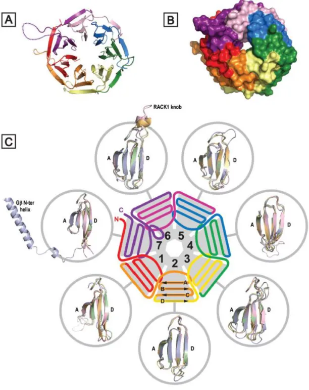
The tryptophan, aspartic acid-repeat (WD-repeat) containing proteins are an ancient conservative family of proteins found in prokaryotes and all eukaryotes (Li and Roberts, 2001). They are involved in almost every signalling pathway and are associated with many genetic diseases. Until now over 100 WD-repeat proteins have been assigned with an approved name and designation in the human nomenclature database (Adams et al., 2011).

WD-repeats themselves are sequences of typically 40-60 amino acids in length ending at the

C-terminus with a signature WD dipeptide or its equivalent (Adams et al., 2011). The motifs

were first identified as repeating segments of homologous sequence within the primary

structure of the transducin Gβ subunit and CDC4 (Fong et al., 1986). The WD-repeats are

also characterised by the presence of a GH dipeptide which is usually in the N-terminus,

though neither the GH nor the WD dipeptide is absolutely conserved. Moreover, WD-repeat

proteins may contain N-terminal and C-terminal regions of variable length (Chen et al.,

2004). Several other characteristic amino acids contribute to the repeat, most notably an

aspartic acid located 6 residues before the WD dipeptide, but it is the collective critical mass

of such features rather than the absolute conservation of any individual amino acid that

establishes the identity of a sequence as a WD-repeat ( Xu and Min, 2011). Given the variable

number of residues at the N-terminal end of these units, sequence databases tend to map the

repeats of WD-proteins between GH and WD dipeptides for convenience (Adams et al.,

2011). The basic criterion for inclusion of a protein into this family is the presence of at least

four of these repeat sequences to generate a WD-domain. These domains adopt a β-propeller

structure, where the propeller fold is characterised by blades that are arranged radially around

a central axis (Figure 1) (Neer et al., 1994; Neer, 1995; Paoli, 2001; Sondek et al., 1996; Wall

et al., 1995).

(11)

11

Figure 1 Structural detail for RACK1 proteins. (A) Crystal structure of RACK1A from

A. thaliana (PDB: 3DM0), illustrating the seven-bladed β-propeller structure. (B) As (A)

but with surface rendition. (C) Schematic representation of RACK1 structure and

organisation of WD-repeats; peripheral circles show superimposition of individual

propeller blades for Gβ1 (blue) (PDB: 1TBG) and for the structurally defined RACK1

(12)

12 orthologues. Gβ is distinguished in blade 7 by a helical N-terminal extension that engages tightly bound Gγ (not shown) in coiled coil interactions (Adams et al., 2011).

The conserved propeller structure is maintained by well-defined hydrogen bonding networks and intra-chain hydrophobic interactions, although different WD-repeat proteins appear to adopt distinct folding orders for their constituent propeller blades (Sondek et al., 1996;

Garcia-Higuera et al., 1998). In principle, s single β-propeller subunit may comprise four to eight blades (Smith, 2008; Paoli, 2001), although at present only 7-bladed or 8-bladed WD- repeat propellers have been characterised by X-ray diffraction studies-the majority being 7- bladed structures consistent with the proposal that this is the optimal β-propeller fold (Murzin, 1992). Proteins are known with more than eight WD-repeats, but these assume tertiary structures with multiple propeller subunits (Adams et al., 2011).

WD-repeat proteins share a common role in scaffolding protein complexes, often with multiple and competing partners, thereby serving as hubs for spatiotemporal orchestration of signalling events across diverse pathways. Enzymatic activities have not been reported thus far for WD-repeat proteins, but their functions can be regulated by post-translational modifications (Adams et al., 2011). The lack of a direct catalytic enzymatic function for the protein family contrasts with other β-propeller-forming proteins, many of which exhibit enzymatic activity (Paoli, 2001).

To date the most extensively studied WD-repeat protein is the G-protein β subunit (Gβ)

(Neer, 1995; Sondek and Siderovski, 2001), which exists in a complex with the γ subunit

(Gγ). Gβγ reversibly complexes with the GDP-bound Gα subunit to form a Gαβγ heterotrimer

that associates with G-protein-coupled receptors (GPCRs) for transduction of extracellular

signals. Agonist-bound GPCRs act as guanine nucleotide exchange factors (GEFs), promoting

the release of the Gα subunit from the Gβγ heterodimer, resulting in the activation of various

(13)

13 signalling cascades (Schwindinger and Robishaw, 2001; Tuteja, 2009). The mode of Gβ interaction with its binding partners has become increasingly clear with the emergence of crystal structures of Gβ in various complexes with Gα/Gγ, (Sondek et al., 1996; Wall et al., 1995; Lambright et al., 1996; Nishimura et al., 2010), phosducin (Gaudet et al., 1996; Gaudet et al., 1999; Loew et al., 1998), GPCR Receptor Kinase 2 (GRK2) (Ladowski et al., 2003;

Tesma et al., 2010; Tesma et al., 2005), and Regulator of G-protein Signalling 9 (RGS9) (Cheever et al., 2008; Slep et al., 2001). Evidence is accumulating for the homodimeric/heterodimeric interactions of the WD-repeat containing proteins including the Receptor for activated C kinase 1 (RACK1) (Thornton et al., 2004; Liu et al., 2007; Chen et al., 2004; Chen et al., 2005). Dimerization events are frequently employed to transduce signals from the cell surface to the nucleus (Klemm et al., 1998). Moreover, some WD-repeat proteins contains some other domains in addition to the WD repeat sequences that increase the number of binding partners, scaffolding properties and overall function of the protein (Adams et al., 2011). For example, the β-transducing repeat-containing protein 1 (β-TrCP1) is a ubiquitin ligase with both WD-repeat and F-Box domains as well as a RING domain. β- TrCP1 is required for the degradation of regulatory proteins such as Snail and p53 (Vinas- Castells et al., 2010; Xia et al., 2009), and the WD-repeats increase its binding cohort and regulate its sub-cellular location, allowing the protein to also have a role in transcription and in regulating circadian rhythm (Seo et al., 2009; Kimbrel and Kung, 2009; Ohsaki et al., 2008).

As the knowledge of the WD-repeat family of proteins increases, the members are being

regularly shown to be involved in most signalling pathways. It is therefore not surprising to

note that WD-repeat proteins play critical roles in several human diseases. It is also interesting

(14)

14 to know that changes in WD-repeat homeostasis can have a dramatic effect on protein complex assembly and on key signalling pathways.

1.2 Receptor for activated C kinase 1 (RACK1) - a scaffold protein: structure and physiological functions

Scaffold proteins are uniquely poised to integrate signals from multiple pathways. Such proteins generate considerable functional diversity by mediating concomitant and/or promiscuous interactions with a vast array of protein partners (Ullah et al., 2008). RACK1 is a highly conserved intracellular adaptor protein with significant homology to Gβ (Adams et al., 2011). RACK1 was originally cloned from a chicken liver cDNA library and human B- lymphoblastoid cell line (B-LCL) (Guillemot et al., 1989). The protein was then cloned from a rat brain cDNA library which was screened for gene products that bind purified rat brain PKC in the presence of its activators (phosphatidyserine, diacylglycerol and calcium) (Ron et al., 1994). Given the association of RACK1 with the active conformation of PKCβII, the protein was named Receptor for Activated C Kinase 1 (RACK1) (Ron et al., 1994; 1995; Ron and Mochly-Rosen, 1995; Stebbins and Mochly-Rosen, 2001).

It is now very well established that RACK1 interacts with a large number of proteins either

directly or as a part of a complex. As a scaffold protein, RACK1 integrates inputs from

distinct signalling pathways and is crucial for fundamental cellular activities such as cell

proliferation, transcription and protein synthesis, as well as various neuronal functions

(Adams et al., 2011). RACK1’s scaffolding properties are mediated by the presence of seven

WD-repeats (McCahill et al., 2002; Sklan et al., 2006) that present multiple protein-binding

sites and facilitate interaction with specialized protein docking modules, including SH2

domains (Src and Fyn) (Chang et al., 2002; Yaka et al., 2002), pleckstrin homology (PH)

(15)

15 domains (dynamin and p120GAP) (Koehler and Moran, 2001; Rodriguez et al., 1999) and C2 domains (Ron et al., 1994; Stebbins and Mochly-Rosen, 2001).

RACK1 functions as a homodimer enabling the expansion of its binding partners (Chen et al., 2005; Thornton et al., 2004; Yaka et al., 2002; Dell et al., 2002; Liu and Semenza, 2007). The formation of the RACK1 heterodimer has been shown to enable efficient cross-talk between signal transduction pathways mediated by GPCRs and by ligand-gated ion channels, specifically between the cAMP/PKA pathway and the N-methyl D-aspartate receptor (NMDAR) (Thornton et al., 2004). RACK1 binding partners have been identified at various cellular locations. One very important function of RACK1 is to shuttle some of its interaction partners to specific sites within the cell. Global control of gene transcription, translation, and ribosome assembly are also emerging as important cellular functions of RACK1 (Ceci et al., 2003; Nilsson et al., 2004). RACK1 orthologs have also been discovered in lower eukaryotes, such as Chlamydomonas and yeast (Schloss, 1990), and are highly conserved in plants and other organisms (van Nocker and Ludwig, 2003), which do not express canonical protein kinase C enzymes (Herold et al., 2002).

1.2.1 Structure of RACK1

Studies probing the evolution of RACK1 suggest that the protein is strongly conserved through evolution (Wang et al., 2003). Sequence alignments of RACK1 species from diverse organisms reveal a significant sequence identity. Gβ was the first WD repeat protein to be characterised by X-ray crystallography. Numerous crystal structures have since emerged for WD-repeat proteins (Sondek et al., 1996; Wall et al., 1995; Lambright et al., 1996;

Stirnimann et al., 2010). These structural studies confirmed that RACK1 adopts a seven-

bladed β-propeller structure consistent with the predictions of earlier homology modelling

studies based on Gβ and other WD-repeat proteins (McCahill et al., 2002; Stirnimann et al.,

(16)

16 2010; Coyle et al., 2009; Rabl et al., 2011; Ullah et al., 2008; Ruiz et al., 2012). The A.

thaliana protein was the first of RACK1 orthologues to be structurally defined. In contrast to metazoans, where RACK1 is expressed as a single gene, A. thaliana possesses three genes.

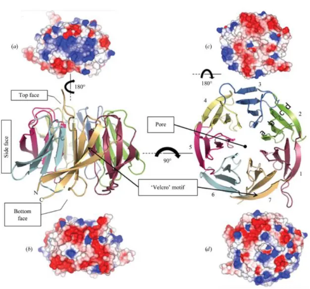
The structure of human RACK1 is shown in Figure 2.

Figure 2 Views of the hRACK1 crystal structure. Central panel: cartoon representations

of hRack1 viewed from the side (left) and from the top (right). The locations of the

protein pore and the ‘velcro’ motif are indicated. Each β-sheet or blade is numbered

sequentially from the N-terminus of the protein and their β-strands are labelled a, b, c,

(17)

17 and d starting from the inside of the propeller close to the pore. The electrostatic surface of hRack1 is shown from four different angles. Side surfaces, bottom and top views are displayed in (a) (blades 1–3), (b) (blades 5–7), (c) and (d), respectively (Ruiz et al., 2012).

Each β-sheet (1-7) shows the same basic architecture comprised of antiparallel strands labelled a, b, c and d from the inner side of the propeller to the outer side. Adjacent blades are connected by a loop bridging from strand d on one blade to strand a on the next. These loops are exposed on the top face of the propeller blade, as are the β-turns linking strands b and c in each blade. The WD-repeats of RACK1, as with all proteins that adopt this fold, overlap two adjacent propeller blades to provide an interlocking architecture. Each repeat encompasses the d-strand of one blade and strands a, b, and c of the next, terminating in the signature WD dipeptide at the end of strand c such that the aspartic acid (or equivalent residue) is exposed on the propeller’s lower face (Adams et al., 2011).

The available crystal structures for Gβ and other WD-repeat proteins have established the structural basis for scaffold interactions with a range of partner proteins (Xu and Min, 2011).

Unfortunately, structural information relating specifically to the interactions of RACK1 with its numerous protein partners currently remains very limited (Adams et al., 2011).

1.2.2 Physiological functions of RACK1

There are emerging critical roles for RACK1 in development. RACK1 has been implicated in

the membrane localization of van gogh-like 2 (Vangl2) in zebrafish. The Vangl2-interacting

region of RACK1 has been shown to exert a dominant-negative effect on Vangl2 localization

and gastrulation (Li et al., 2011). The interaction between RACK1 and tyrosine-protein

kinase-like 7 (PTK7) has also been shown to be a requirement for neural tube closure in

Xenopus (Wehner et al., 2011). In Arabidopsis, where three RACK1 homologues are present,

(18)

18 the RACK1 gene products are essential regulators of plant development (Chen et al., 2006;

Guo and Chen, 2008). RACK1 homologs in Drosophila (Kadrmas et al., 2007), Aspergillus nidulans (Hoffmann et al., 2000), Schizosaccharomyces pombe (McLeod et al., 2000) and Trypanosoma brucei (Rothberg et al., 2006) have similarly been shown to be central to various stages of the developmental process.

The scaffolding of signalling proteins by RACK1 at receptors is particularly important in dynamic processes such as cell migration, cell adhesion and cell spreading (Hermanto et al., 2002; Kiely et al., 2002; 2005; 2009). All of these processes require the highly regulated converging of transient signalling between receptors. For instance, RACK1 was first discovered to be a mediator of cell spreading by establishing contact with the extracellular matrix and growth factor receptors at adhesion sites (Hermanto et al., 2002). As a scaffold protein RACK1 plays very important roles during cell migration. Cell migration is a fundamental process required for embryonic development, wound healing and immune responses, and the components of cell migration are functionally conserved in evolution (Adams et al., 2011). Cell migration is a dynamic process involving multiple steps:

cytoskeletal reorganisation to form leading edge protrusions, turnover of focal adhesions, generation of mechanic forces, retraction of the cell tail, and detachment from the surrounding extracellular matrix (Gandin et al., 2013). Evidence is mounting of RACK1 involvement in various stages of this process. RACK1 is essential for cell migration, and the protein binds to many components of the cell migration machinery including kinases, phosphatases and the cytoplasmic domains of cell surface receptors (McCahill et al., 2002; Sklan et al., 2006).

RACK1 also functions in direction sensing of migrating cells. A complex of RACK1, focal

adhesion kinase (FAK), and the cAMP-degrading phosphodiesterase PDE4D5 is recruited to

nascent adhesions, where it modulates cell polarity (Serrels et al., 2010).

(19)

19 Some of the importance of RACK1 in disease pathology results from its ability to modulate the innate immune response and activation of interferon (IFN) signalling (Gandin et al., 2013). Many pathogens, especially viruses, evade the host immune response by interfering with IFN signalling. RACK1 interacts with IFNα/β receptor and recruits signal transducer and activator of transcription 1 (STAT1) into this complex, which is required for IFN signalling (Usacheva et al., 2001). RACK1 has been suggested to be a prognostic marker in breast cancer, and its elevated expression is associated with poor clinical outcome (Al-Reefy and Mokbel, 2010; Cao et al., 2010). Furthermore, RACK1 promotes proliferation, migration and metastasis of breast cancer cells in vitro and in vivo through activation of the RhoA/Rho kinase pathway (Cao et al., 2010; 2011). RACK1 is also implicated as a key player in ovarian cancer (Williams et al., 2004), prostate cancer (Hellberg et al., 2002), and in cancers caused by pathogens such as human papillomavirus (HPV16) (Boner and Morgan, 2002) and Helicobacter pyroli (Hennig et al., 2001).

As a primary RACK1-binding partner (Ron et al., 1994; 1995), PKCβII has been identified as

an important factor linking ribosomal RACK1 and translational control in mammals (Gandin

et al., 2013). RACK1 recruits active PKCβII onto ribosomes, where it phosphorylates

eukaryotic initiation factor 6 (eIF6) (Sharma et al., 2013). This confirms the importance of

RACK1-PCKβII-eIF6 complex in the regulation of translation. In addition to a positive role

for ribosomal RACK1 in mRNA translation, a negative role for RACK1 in gene expression at

the posttranscriptional level has been reported in yeast (Gandin et al., 2013). Depletion of

RACK1 results in increased ribosomal activity in cell-free in vitro translation assays and a

concomitant increase in protein levels in vivo (Gerbasi et al., 2004).

(20)

20 1.2.3 Post-translational Modification of RACK1

Apart from phosphorylation which appears as an important process that modulates binding of RACK1 to other proteins, not much is known about other possible post-translational modifications. There are lots of tyrosine residues in the RACK1 sequence. Phosphorylation of RACK1 by c-Abl mediates the interaction with FAK (Kiely et al., 2009), while phosphorylation/dephosphorylation of RACK1 also regulates its mutually exclusive association with PP2A and β1 Integrin (Kiely et al., 2008; 2006). Specifically, Src phosphorylates RACK1 on Y-228 and 246 (Chang et al., 2002).

1.3 G protein-linked signalling in Dictyostelium discoideum

In D. discoideum, G protein-linked signal transduction events, in particular, are essential for

chemotaxis, cell aggregation, morphogenesis, gene expression, and pattern formation

(Devreotes, 1994). A major breakthrough in the identification of key proteins that are

involved in eukaryotic chemotaxis was the discovery that cAMP receptors in D. discoideum

are members of GPCRs (Klein et al., 1988; Saxe et al., 1988). Members of the large family of

GPCRs detect various extracellular stimuli, including hormones, neurotransmitters, odorants,

light, and chemoattractants (Jin et al., 2008; Gilmann, 1987). Activation of GPCRs induces

events which lead to dissociation of heterotrimeric G proteins into Gα and Gβγ subunits,

which in turn transduce cascade of signals to intracellular signalling components that

culminate cell responses. Of the 12 Gα subunits present in D. discoideum, some have been

characterised. The Gα2 subunit is required for cellular responses to the extracellular cAMP

signal that directs the aggregation process during development (Kumagai et al., 1989). The

Gα3 subunit is also required for aggregation but only in absence of exogenous cAMP,

suggesting that Gα3 subunit is not directly involved with responses to extracellular cAMP

(Brandon and Podgorski, 1997; Brandon et al., 1997). Finally, the Gα4, Gα5 and Gα8

(21)

21 subunits play important roles in the development of spores and the anterior prestalk cells, respectively, consistent with the increased expression of these subunits upon aggregate formation (Hadwiger and Firtel, 1992; Hadwiger and Srinivasan, 1999; Hadwiger et al., 1991;

1994; Wu and Janetopoulos, 2013).

1.4 D. discoideum G protein β subunit-null mutants

In chemotaxis, amoeboid motile cells, like leukocytes and Dictyostelium cells, respond directionally to chemical gradients; in phagocytosis, they bind and engulf foreign organisms or apoptotic cells (Devreotes and Zigmond, 1988; Rabinovitch, 1995). Chemotaxis and phagocytosis seem to be closely related, suggesting that the underlying signal transduction events and cytoskeletal responses have evolved in parallel (Metchnikoff, 1968). In the simple eukaryote D. discoideum and in amoeboid cells of the immune system of animals, chemotactic and phagocytic stimuli elicit a remarkably similar spectrum of behavioural events and biochemical reactions (Devreotes and Zigmond, 1988; Greenberg, 1995). Foremost among these is the polymerization of actin into filaments that support the extension of pseudopods and the formation of phagocytic cups (McRobbie and Newell, 1993; Greenberg, 1995; Zigmond, 1996). Chemotaxis and phagocytosis involve both G protein-coupled and tyrosine kinase-linked signal transduction pathways (Parent and Devreotes, 1996, Murphy, 1996). With regard to phagocytosis, bound particles activate protein tyrosine kinases, such as syk, leading to actin polymerization and rearrangement, possibly through involvement of the small G protein Rho (Greenberg et al., 1994, 1996; Indik et al., 1995; Hackam et al., 1997).

Heterotrimeric G proteins have been involved in chemotactic activation of macrophages,

which leads to phagocytosis (Thelen and Wirthmueller, 1994), and in phagosome-endosome

fusion (Desjardins et al., 1994)

(22)

22 D. discoideum amoebae contain a single G protein β subunit; its deletion creates cells that lack functional G proteins (Lilly et al., 1993; Wu et al., 1995). These mutants are severely defective in chemotaxis, aggregation, and development. When plated on bacterial lawns, they form smooth plaques consisting of monolayers of undifferentiated cells. These plaques are much smaller than those of wild type (Wu et al., 1995). This slow growth reflects a severe defect in phagocytosis, which is primarily due to a failure in organizing the actin meshwork into a phagocytic cup (Peracino et al., 1998).

1.5 Aim of this study

The aim of this study was to characterise the D. discoideum RACK1 (DdRACK1) protein which we initially identified as a binding partner of Receptor phosphatidylinositol kinase A (RpkA), an unusual GPCR which functions in phagocytosis and antibacterial defense in D.

discoideum (Riyahi et al., 2011). DdRACK1 was characterised based on the following features:

1. Its structure and homology to other RACK1 species 2. Subcellular localisation and its dimerization properties 3. Its ability to interact with G proteins

The final part of this study was focused on the analyses of the effect of D. discoideum

RACK1 (DdRACK1) overexpression during growth and development.

(23)

23

2 Materials and Methods

Standard molecular biology techniques were performed as described in Sambrook et al., 1989.

Light and heavy instruments used were properties of the Department. Standard laboratory materials and reagents were obtained from local suppliers.

2.1 Kits

NucleoSpin Extract 2 in 1 Macherey-Nagel Quick Change Site-Directed Mutagenesis kit Stratagene pGEM-Teasy Vector kit Promega Pure Yield Plasmid System Promega

VIVASPIN 500 sartoriusstedim biotech 2.2 Enzymes, antibodies and antibiotics

2.2.1 Enzymes for molecular biology:

Taq-polymerase Promega

T4 DNA ligase Boehringer Thrombin GE Healthcare Pfu DNA polymerase Promega Alkaline phosphatase Roche

Restriction endonuclease New England Biolabs

Ribonuclease A Sigma

(24)

24 2.2.2 Antibodies

2.2.2.1 Primary antibodies

Mouse monoclonal anti- α-actinin mAb 47-16-8 (Schleicher et al., 1984) Mouse monoclonal anti-csA mAb 33-294 (Berthold et al., 1985) Mouse monoclonal anti-cap32 mAb 188-19-95 (Haus et al., 1993) Mouse monoclonal mAb 5E7 (Fendly et al., 1990) Mouse monoclonal anti-GFP mAb K3-184-2 (Noegel et al., 2004) Monoclonal antibody anti-mRFPmars mAb K73-875-7 (Fischer et al., 2004;

Omosigho et al., submitted) Rabbit polyclonal anti-GST (Xiong et al., 2008) Rabbit polyclonal anti-RACK1 (This study)

2.2.2.2 Secondary antibodies

Goat anti-mouse IgG, peroxidase conjugated Sigma Goat anti-rabbit IgG, peroxidase conjugated Sigma 2.2.3 Antibiotics

Ampicillin Gruenenthal

Blasticidin S Biomedicals

Dihydrostreptomycinsulphate Sigma

(25)

25 Tetracyclin Sigma

Gentamicin Sigma G418 Sigma 2.3 Media and Buffers

All solutions and media used in the course of these experiments were prepared with deionized water from a pure water source in the laboratory. All other buffers and solutions which were not mentioned here are seen in the methods.

2.3.1 Buffers and Solutions

GST-fusion protein elution buffer, pH 7.2 50 mM Tris/HCl, pH 7.2

100 mM NaCl

10 mM reduced glutathione 0.2 % Tween-20

10 mM DTT

10 x NCP-buffer, pH 8.0 12.1 g Tris

87.0 g NaCl 5.0 ml Tween 20 2.0 g NaCl

Made up to 1 litre with deionised water

(26)

26 50 x Tris/Acetate buffer (TAE), pH 8.0

242 g Tris

57.1 ml 16.6 M Glacial acetic acid 100 ml 0.5 M EDTA

Made up to 1 litre with deionized water TBS lysis buffer, pH 7.2

50 mM Tris/HCl, pH 7.2 100 mM NaCl

TE-buffer, pH 8.0

10 mM Tris/HCl, pH 8.0 1 mM EDTA

TBS-T buffer, pH 7.2 50 mM Tris/HCl, pH 7.2 100 mM NaCl

0.2 % Tween-20

2.3.2 Bacteria medium and agar plates LB-Medium (Sambroock et al., 1989) 10 g/L Bacto-Tryptone

5 g/L Yeast Extract 5 g/L NaCl

pH was adjusted to 7.0 with 5 M NaOH and made up to 1 litre with deionised water and then

autoclaved.

(27)

27 Ampicillin-LB-agar Plates

For the preparation of LB-agar plates, 0.9% (w/v) agar was added to LB-medium. After autoclaving and cooling to 55

o

C, 100 mg/l ampicillin was added. Stroage was at 4

o

C.

To prepare plates with ampicillin/IPTG/X-Gal, 10 µl of 1M IPTG and 50 µl of 20 mg/ml X- Gal was spread over the surface of LB-Amp-Plates and allowed to absorb for at least 30 minutes at 37

o

C prior to use.

2.3.3 Yeast medium YEPD-Medium 20 g Difco-Peptone

10 g Yeast Extract Made up to 1 litre with deionized water and then autoclaved YEPD-Agarplates

20 g Difco-Peptone 10 g Yeast Extract 18 g Agar-Agar

Made up to 1 litre and then autoclaved 100x L-Adenine solution

200 mg of L-Adenine dissolved in 100 ml H

2

O. Dissolution was aided with addition of drops

of HCl and then sterile filtered.

100x L-Tyrosine solution

300 mg of L-Tyrosine dissolved in 100 ml H

2

O. Dissolution was aided with addition of drops of NaOH and then sterile filtered.

100x L-Histidine solution

200 mg of L-Histidine dissolved in 100 ml H

2

O and then sterile filtered.

(28)

28 100x L-Leucine solution

200 mg of L-Leucine dissolved in 100 ml H

2

O and then sterile filtered.

100x L-Tryptophan solution

200 mg of L-Tryptophan dissolved in 100 ml H

2

O and then sterile filtered.

100x L-Uracil solution

200 mg of L-Uracil dissolved in 100 ml H

2

O. Dissolution was aided by slightly warming solution before it was sterile filtered.

50x Drop-out Solution 1,500 mg Isoleucine 7,500 mg Valine 1,000 mg Arginine 1,500 mg Lysine 1000 mg Methionine 2,500 mg Phenylalanine 10,000 mg Threonine

Filled up to 1 litre and then sterile filtered 20x Drop-out Solution

20 ml 50x Drop-out solution 10 ml 100x L-Uracil 10 ml 100x L-Tyrosine 10 ml 100x L-Adenine

1 M 3-Amino-1, 2, 4-triazol solution (3AT)

8.4 g 3-Amino-1, 2, 4-triazol dissolved in 100 ml deionised H

2

O and then sterile filtered.

(29)

29 Composition of the Yeast Selection Plates and Selection Medium

SD/-Leu/-Trp SD/-Leu/-Trp/

-His/+3AT Yeast Nitrogen Base (g) 6.7 6.7

Agar-Agar (g) 20 20

Water (ml) 770 745

20% Glucose solution (ml) 100 100 20x Drop-out solution (ml) 50 50 100 x L-Histidine (ml) 10

100 x L-Leucine (ml) - 100 x L-Tryptophan (ml) -

1M 3AT Solution (ml) 25

Yeast selection media were prepared but without the addition of the 20 g Agar to the preparations. To test for protein interactions, transformants on the SD /-Leu /-Trp plates were transferred to the SD /-Leu /-Trp /-His /+3AT plates. After 6-8 days, the colonies which grew were used to perform β-galactosidase activity staining.

2.3.4 Media and buffers for Dictyostelium cultures AX2-Medium, pH 6.7 (Claviez et al., 1982)

7.15 g Yeast extract 14.3 g Peptone 18.0 g Maltose 0.486 g KH

2

PO

4

0.616 g Na

2

HPO2 x H

2

O

Made up with H

2

O to 1 Liter

(30)

30 Soerensen-phosphate buffer, pH 6.0 (Malchow et al., 1972)

2 mM Na

2

HPO

4

14.6 mM KH

2

PO

4

Phosphate-Agar plates, pH 6.0

9 g Agar made up with Soerensen-phosphate buffer pH 6.0 to 1 Litre Water agar plates

1% Agar in Water

2.3.5 Bacteria, D. discoideum, and yeast strains Bacteria Cell Stock

E.coli (XL1 blue) (Bullock et al., 1987) E.coli (BL21) (Studier and Moffat, 1986)

D. discoideum

AX2-214, also known as AX2 is a derivative of NC4 wild isolate (Raper, 1935) which can grow axenically.

Yeast strain

Saccharomyces cerevisiae Y190 (His3 and lacZ reporter genes) (Johnston et al., 1991; Harper et al., 1993).

2.3.6 Oligonucleotides

Oligonucleotides used for PCR (Polymerase Chain Reaction) were purchased from Sigma-

Genosys in Steinheim.

(31)

31 RACK1

RACK1_fw GGATCCATGGAACAACAAAAAGCACCAC RACK1_rev ATCGATTTATTGGGATGATGATTTGTAAAC

Gβ subunit

Gβ_fw ATCGCGGATCCATGTCATCAGATATTTCAGAAAAAATT Gβ_rev ATCGCCTGCAGTTAAGCCCAAATCTTGAGGAGAGAATC Gγ subunit

Gγ_fw GCGGGATCCACCATGGAAATGTCCGAATCACAATTAAAAAAAGTT Gγ_rev ATCGCGGATCCTTATAACACAGAACATCCATTTCCTTT

Gα subunits

Gα1_fw ATCGCGGATCCATGGGTAATATTTGTGGTAAACCA Gα1_rev ATCGCCTCGAGTTAAAGAATCATACCAGCTTCACCCAA Gα2_fw ATCGCGAATTCATGGGTATTTGTGCATCATCAATG

Gα2_rev ATCGCAGATCTTTAGGATCCAGAATATAAACCAGCTTTCATAACACA Gα3_fw ATCGCCTCGAGATGGATTTCAATCCAGTACCACCA

Gα3_rev ATCGCAGATCTTTAACAATAAAATTCTAAAGTTTTTGAAATTAT

Gα4_fw ATCGCGAATTCATGAGATTCAAGTGTTTTGGATCA

Gα4_rev ATCGCCTCGAGTTAGGATCCGAAGTGTTCTAAAGCTTGAGATAA Gα5_fw ATCGCGAATTCATGGGTTGTATATTAACAATTGAAGCA

Gα5_rev ATCGCAGATCTTTAGGATCCATAATTTATGATTGTATTAAAGATATTTTT Gα6_fw ATCGCGAATTCATGGCATTTTTATGTAAATCAAATGAT

Gα6_rev ATCGCAGATCTTTAGGATCCAACAAGATCCATAACATGACCAAC Gα7_fw ATCGCGAATTCATGAGTAGCACTACAACAAATACA

Gα7_rev ATCGCAGATCTTTAGGATCCGATACCTCCTTCCTCCATAGTTTG

(32)

32 Gα8_fw ATCGCGGATCCATGGGTTGCTATCAATCACGTGTT

Gα8_rev ATCGCCTCGAGTTAAGAATTAATTTTGGCGGTTGCACC Gα9_fw ATCGCGAATTCATGGGTTGTAATTCAAGTAGTGAA

Gα9_rev ATCGCAGATCTTTAGGATCCATAGTGAAGTTTTAAGATAGATTGAAT Gα10_fw ATCGCGAATTCATGTCATTTTTATGCTCAGAAAATTCA

Gα10_rev ATCGCAGATCTTTAGGATCCATTTTTTACAGAATTATATGTTATGTT Gα11_fw ATCGCGAATTCATGGGAAGCCAATTTAGTGTTTTA

Gα11_rev ATCGCAGATCTTTAGGATCCAATTGTATCTTCTAATATTTTTTTAAC Gα12_rev ATCGCGGATCCATGTGTACAAGAAATAAAAAAGATATT

Gα12_rev ATCGCCTCGAGTTATAAAATTTCTGCAACATTCATTAATAA

Primers for Site-Directed Mutagenesis

mut_fw CCAGAAGTCAAAGAACAAGCTTTCGACTCAGAGGAAGAGGAA GAATCAAAACCAAAGCACC

mut_rev GGTGCTTTTGGTTTTGATTCTTCCTCTTCCTCTGAGTCGAAAGCT

TGTTCTTTGACTTCTGG

(33)

33 2.4 Methods

2.4.1 Growth, development and transfection

Cells were either grown on a lawn of K. aerogenes on SM agar plates, on a lawn of E. coli B12 on NA-agar or cultivated in shaking suspension (160 rpm) or in a submerged culture at 21-23°C in axenic medium (Harloff et al., 1989). Development was initiated by plating 5 x 10

7

cells which were washed twice with Soerensen phosphate buffer (17 mM Na

+

/K

+

phosphate, pH 6.0) on phosphate agar plates and monitored. Development was also followed for cells starved in Soerensen phosphate buffer in shaken suspension (1 x 10

7

cells/ml; 160 rpm at 22ºC) or in petri dishes. Mutants were maintained in the presence of appropriate antibiotics (2-4 µg/ml G418) (Roche Applied Science) or (3-5 µg/ml Blasticidin) (MP Biomedicals Inc., Eschwege, Germany). The following strains have been used; AX2-214 (wild type) (Noegel et al., 1985), AX2 expressing GFP-, YFP- or RFP-tagged fusion proteins, Gβ null mutants LW6 (Wu et al., 1995; Peracino et al., 1998) and LW6 expressing GFP- DdRACK1. The corresponding plasmids were introduced by electroporation using a Biorad electroporator Gene Pulser Xcell (Biorad, München, Germany) according to the protocol supplied.

2.4.2 Cloning of RACK1 cDNA and expression of recombinant proteins

For expression of recombinant D. discoideum RACK1 and Gα subunits as glutathione S

transferase (GST) fusion proteins in E. coli, full-length cDNAs were respectively cloned into

pGEX-4T-1 vector (GE Healthcare Life Sciences). E. coli strain XL1 Blue was used for

expression of the GST fusion proteins. Induction of protein expression was with 0.25 mM

isopropyl -D-thio-galactoside (IPTG) when an OD

600

of 0.8 was reached. Cells were further

cultured at 30ºC for 3 hours. They were harvested, lysed in 50 mM Tris/HCl, pH 7.4, 100 mM

NaCl, supplemented with Protease inhibitors (0.5 mM PMSF, 1mM Benzamidine and

(34)

34 Complete (Roche) and 1mM DTT with an EmulsiFlex cell homogenizer (Avestin Europe GmbH, Mannheim, Germany). Lysates were separated into soluble and insoluble fractions by centrifugation at 18,000g. The fusion proteins from the soluble fraction were purified using GST-Sepharose beads (GE Healthcare).

For cleavage of proteins from GST-Sepharose beads, the GST fusion proteins were washed 5 times with cleavage buffer (20 mM Tris/HCl, pH 7.4, 150 mM NaCl and 0.2 % Sarcosyl).

Beads were then re-suspended in cleavage buffer and 3-10 U thrombin/mg fusion protein were added to the beads and incubated with little agitation at room temperature overnight. As RACK1 was released from the beads together with some GST, we next performed an anion exchange chromatography step in order to separate the proteins. For this the protein solution was dialyzed against 20 mM Tris/HCl, pH 8.0, and 1 mM EDTA overnight before loading onto a DE-52 Sephadex column which had been calibrated with 50 mM Tris/HCl, pH 8.0, 1 mM EDTA. The protein was eluted with 1 M NaCl and the eluate dialyzed and analyzed by SDS-PAGE.

For expression in AX2 the cDNA was cloned into pBsr-N2-GFP vector and expressed as GFP-RACK1 (GFP N-terminal) under control of the actin 15 promoter and also into mRFPmars plasmid for RFP-RACK1 (RFP N-terminal) (Blau-Wasser et al., 2009; Fischer et al., 2004). A PCR-mediated site-directed mutagenesis (QuikChange Site-Directed Mutagenesis Kit, Stratagene) was used to generate mutations in the GST-RACK1 and GFP- RACK1 plasmids. The mutations were confirmed by sequencing.

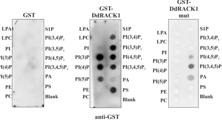
2.4.3 Phosphoinositide binding assay

PIP-strips supplied by Echelon Biosciences, Inc. (Salt Lake City, Utah, USA) were used to

perform phosphoinositide binding according to the supplied protocol. Briefly, GST and GST-

fusion proteins were eluted from the glutathione agarose beads with elution buffer (20 mM

(35)

35 reduced glutathione, 50 mM Tris/HCl, pH 7.4, 100 mM NaCl, 0.2% Tween-20, and 100 mM DTT).

The membranes were blocked with 0.1% ovalbumin (Sigma # A-5253) in TBS for one hour at room temperature. After discarding the blocking solution membranes were incubated with 1 mg/ml GST-fusion proteins in TBS-T (50 mM Tris/HCl, pH 7.4, 100 mM NaCl, 0.2 % Tween-20) at room temperature for one hour. The protein solution was then discarded and the membranes were washed with TBS-T three times 10 minutes each. Bound protein was detected by western blot analysis with GST polyclonal antibodies as primary and anti-rabbit IgG-peroxidase (Sigma # A-6154) as secondary antibody followed by enhanced chemiluminescence.

2.4.4 Lipid vesicle preparation and sedimentation assay

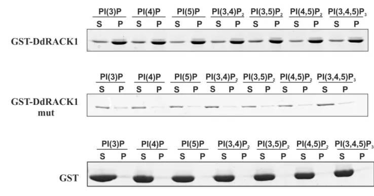
Phosphatidylserine (PS), phosphatidylcholine (PC), phosphatidylethanolamine (PE), PI(3)P, PI(4)P, PI(5)P, PI(3,4)P2, PI(3,5)P2, PI(4,5)P2, and PI(3,4,5)P3 were obtained from Sigma and disolved in chloroform. Liposome binding experiments were performed with a modified published liposome binding assay protocol (Blume et al., 2007). Lipid mixtures containing 65% PC, 20% PE, 5% PS and 10% individual phosphoinositides were produced by mixing appropriate lipid solutions in chloroform/methanol. Slow flow nitrogen gas was used for the production of a film on the glass and vacuum desiccation for 30 min for solvent removal.

Sterile-filtered sucrose binding buffer (20 mM HEPES, pH 7.4, 100 mM KCl, 1 mM EDTA,

0.1 M sucrose) was added to a final lipid concentration of 1 mg/ml and incubated at 37ºC for

2 h. Lipids were then sonicated in a waterbath-sonicator for 10 sec. To test liposome binding,

a 100 μl reaction mixture of freshly prepared liposomes and 5 μg of purified protein were

incubated for 15 min at room temperature and centrifuged at 100,000 x g (42,000 rpm) at 4ºC

for 25 min in a Beckman table top ultracentrifuge Optima TLX (TLA 45 rotor). The

supernatant was saved, and the pellet was resuspended in 100 μl of sucrose binding buffer.

(36)

36 Both fractions were then analyzed by SDS-PAGE followed by Coomassie Blue staining.

ImageJ was used for quantification.

2.4.5 Yeast Two-Hybrid Interaction

For the yeast two-hybrid screen, the full-length cDNAs of D. discoideum G protein β-, γ-, α1-, α2-, α4-, α5-, α6-, α7-, α8-, α9-, α10-, α11- and α12-subunits were cloned in frame into the yeast pAS2-1 vector (Clontech), respectively, resulting in fusion to the GAL4-DNA-BD (BD, binding domain). Full-length cDNA of DdRACK1 was cloned into the yeast pACT2 vector (Clontech) resulting in a fusion to the GAL4-DNA-AD (AD, activation domain). Yeast Y190 strain which has His3 and lacZ reporter genes was used for this assay.

Candidate colonies expressing interacting proteins were screened by plating on SD/-Leu/- Trp/-His/+3AT plates after which membrane colonies-lift β-galactosidase activity assay was performed according to the MATCHMAKER Y2H system manual. Briefly, colonies on SD/- Leu/-Trp/-His/+3AT selection plates were transferred to a Nitrocellulose membrane (Protran BA 85) by placing the membrane over colonies on selection plates for 20 min. The filter was carefully lifted off the agar plates and transferred (with colonies facing up) to a pool of liquid nitrogen for 10 sec. The frozen filter was then allowed to thaw at RT and placed on a Whatman filter paper presoaked in freshly prepared X-Gal solution (60 mM Na

2

HPO

4

, 40 mM NaH

2

PO

4

, 10 mM KCl, 1 mM MgSO

4,

pH 7.0, 50 mM β-mercaptoethanol, X-Gal (1 mg/ml final concentration)) and incubated at 30

o

C and checked between 1 to 6 h for the appearance of blue colonies.

2.4.6 Pull down and immunoprecipitation assays

For pull down and immunoprecipitation experiments D. discoideum cells were lysed in 50

mM (10 mM for immunoprecipitation assay) Tris/HCl, pH 7.4, 150 mM NaCl, 0.5% NP40,

supplemented with protease inhibitor cocktail (Sigma), 0.5 mM PMSF, 0.5 mM EDTA, and 1

mM Benzamidine by passing them through a 25G syringe (10-20 strokes) and incubated with

(37)

37 agitation (1000 rpm) for 15 min at 4ºC (to ensure complete cell lysis) followed by a centrifugation step at 16,000 rpm for 10 min. The supernatants were either incubated with GST and GST-fusion proteins respectively or with GFP-trap beads (ChromoTek, Martinsried, Germany). After incubation for 3 h while GST beads were washed three times with wash buffer (50 mM Tris/HCl, pH 7.4, 150 mM NaCl, protease inhibitor cocktail, 0.5 mM PMSF, 0.5 mM EDTA, 1 mM Benzamidine), GFP-trap beads were washed with a different wash buffer (10 mM Tris/HCl pH 7.4, 150 mM NaCl, protease inhibitor cocktail, 0.5 mM PMSF, 0.5 mM EDTA, 1 mM Benzamidine). The beads were resuspended in SDS sample buffer, incubated at 95ºC for 5 min and the proteins separated by SDS-PAGE and analyzed by western blot. The Gβ and Gγ subunits used in this study were previously cloned into GFP (GFP N-terminal) and YFP (YFP C-terminal) vectors, respectively (Jin et al., 2000; Zhang et al., 2001).

2.4.7 In vitro cross-link assay

Purified DdRACK1 was used for a multimerization experiment as was previously described (Xiong et al., 2008). Briefly, 5-10 µg/100 µl of RACK1 in 1 x PBS, pH 7.4, was incubated at room temperature in the presence of 0.001 % (v/v) glutaraldehyde for various time points.

The reaction was stopped by addition of glycine to a final concentration of 0.1 M after 5, 10 and 20 min, respectively. Samples were analyzed by SDS-PAGE and western blot.

2.4.8 Test for presence of phosphotyrosine in DdRACK1

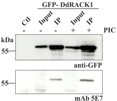
Samples from immunoprecipitation experiments from GFP-DdRACK1 bound to GFP-trap beads in the presence or absence of phosphatase inhibitors were analyzed by western blots and probed with anti-phosphotyrosine monoclonal antibody (5E7) (Fendly et al., 1990).

2.4.9 Immunofluorescence analysis and life cell imaging

Immunofluorescence study was performed as previously described (Blau-Wasser et al., 2009).

Briefly, cells were transferred onto coverslips in Petri dishes and fixed by ice-cold methanol

(38)

38 (5 min, 20 °C). Cells were treated twice for 15 min (room temperature) with blocking solution (1x PBS containing 0.5% (wt/vol) BSA and 0.1% (vol/vol) fish gelatin). The appropriate antibodies were diluted in the blocking solution and applied on the cells for 1 h at room temperature; the excess of antibodies was removed by washing with the blocking solution before the 1 h incubation with the corresponding secondary antibodies. For live cell study, cells were placed in 35 mm Petri dishes (ibidi GmbH-Martinsried, Germany) and allowed to adhere to the surface. Analysis of fixed and live cells was done by laser scanning confocal microscopy using a Leica TCS SPS microscope.

2.5 Cell migration studies

This analysis was done as previously described (Blau-Wasser et al., 2009; Müller et al., 2013). Briefly, cells were plated after ~ 6 h of starvation in a chamber (ibidi GmbH- Martinsried, Germany) and migration towards aggregation centers were followed. Images were recorded at intervals of 6 s using a Leica DM-IL inverse microscope (Deerfield, IL; 40x objective) and a conventional CCD video camera and analyzed using Dynamic Image Analysis Software (DIAS, Soll Technologies, Iowa City, IA).

2.5.1 Miscellaneous methods

Cell fractionation of AX2 cells was done as described (Müller et al., 2013). For generation of rabbit polyclonal antibodies against DdRACK1, the GST-part of GST-DdRACK1 was removed by thrombin cleavage and DdRACK1 was used to immunize rabbits (Pineda, Berlin, Germany). The antibodies specifically recognized the bacterially produced recombinant protein, the RFP- and GFP-tagged fusion proteins as well as the endogenous protein in western blots of whole cell lysates; they were used in immunoprecipitation experiments whereas they were not suitable for immunofluorescence studies.

Protein sequences of RACK1 proteins from H. sapiens (P63244), D. melanogaster (O18640),

A. thaliana (O24456), S. cerevisiae (P38011), and D. discoideum (P46800) were retrieved

(39)

39 from Uniprot protein database and aligned using ClustalW program with Blosum 62 matrix.

The aligned sequences were processed through EsPript for representation. The structural coordinates of S. cerevisiae RACK1 (Asc1p) was obtained from protein databank (PBD:

3FRX) (Figure 4) and used as a template for modelling D. discoideum RACK1. MODELLER

v9 was used to generate DdRACK1 model. Structures in Figures 3B and C were generated

with the aid of the molecular visualization software PyMOL.

(40)

40

3 Results

3.1 Characterization of DdRACK1

gpbB (DDB_G0275045) is located on chromosome 2 of the D. discoideum genome and has 2 exons. The open reading frame encompasses 1136 bp which encodes a protein of 329 amino acids migrating as a 36 kDa protein on SDS polyacrylamide gels. Blast results showed that GpbB is highly related to the RACK1 family of proteins and the alignment of RACK1 sequences from diverse organisms such as H. sapiens, D. melanogaster, A. thaliana, D.

discoideum and S. cerevisiae revealed significant sequence identity. The greatest difference is observed between propeller blades 6 and 7 where an extended loop of mainly basic amino acids is present in the D. discoideum protein and the A. thaliana RACK1 (Figure 3).

Gβ was the first WD-repeat protein to be characterized by X-ray crystallography (Wall et al.,

1995). Since then various other crystal structures have been reported for WD-repeat proteins

(Lambright et al., 1996; Sondek et al., 1996) which include the recently determined structures

for several RACK1 proteins, RACK1A from A. thaliana, Asc1p from S. cerevisiae, RACK1

from T. thermophila and RACK1 from human (Coyle et al., 2009; Rabl et al., 2011; Ruiz et

al., 2012; Ullah et al., 2008; Yatime et al., 2011). These structural studies confirmed the

seven-bladed β-propeller structure. In the RACK1 structure each propeller blade consists of a

four-stranded antiparallel β-sheet, where strand A lines the central canal of the protein, and

strand D is present on the outer circumference. Adjacent blades are connected by a loop

bridging from strand D on one blade to strand A on the next. These loops are exposed on the

top face of the propeller blade as are the β-turns linking strands B and C in each blade. The

loops connecting strand A to B and strand C to D in each blade are located on the reverse,

slightly larger face of the propeller (Adams et al., 2011).

(41)

41

Figure 3 Sequence alignment of RACK1 orthologues and their UniProt accession numbers from H. sapiens (P63244), D. melanogaster (O18640), A. thaliana (O24456), D.

discoideum (P46800) and S. cerevisiae (P38011). The WD40 repeats and β-propeller blade positions are written above the sequences. Alignment was done with the ClustalW program and processed through ESPript for representation. All conserved residues are shown in red and similar residues in yellow

Most notably, the D-A loop between blades 6 and 7 in the RACK1 species is 8 to 19 residues

longer than the cognate region of Gβ

1

and forms a knob-like projection from the upper face of

(42)

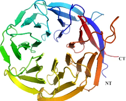
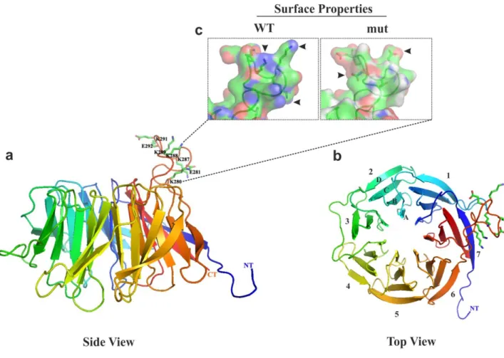
42 the propeller (discovered in the crystal structure of A. thaliana RACK1A) (Adams et al., 2011). This sequence is quite unusual in the D. discoideum protein as it is rich in lysine residues. These general features described for RACK1 proteins are also present in DdRACK1 when we modelled the DdRACK1 sequence to the crystal structure of S. cerevisiae RACK1 (Asc1p) which reveals a comparable structure (Figure 4, 5).

Figure 4 Ribbon diagram of S. cerevisiae RACK1 (Asc1p) (PDB: 3FRX) which was used as template for DdRACK1 model. Shown are the seven β-propeller blades. Coordinates were retrieved from protein data bank (PDB) (www.rcsb.org) and modelled with the aid of MODELLER 9 program and visualized by the software PyMOL.

The DdRACK1 protein harbors in the knob-like projection in the D-A loop between blades 6

and 7 of sequence a polybasic stretch with four lysine residues. These residues were changed

(43)

43 to glutamic acid and the surface properties of this region viewed alongside the wild type protein during the modelling of DdRACK1.

Figure 5 Ribbon diagram of DdRACK1 modelled with the aid of MODELLER9 program, with yeast RACK1, which was retrieved from protein data bank (PDB) (www.rcsb.org), as template. Structures were finally generated using molecular visualization software PyMOL. In (c) the surface charge of the extension after mutation of the basic residues into glutamic acid (see 3.6) is shown.

3.2 Subcellular localization of RFP-/GFP-DdRACK1

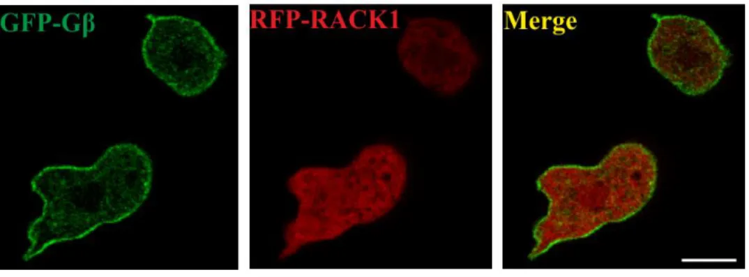
When we expressed RFP-DdRACK1 in AX2 cells expressing the G protein beta-subunit as

GFP-fusion protein for labelling the plasma membrane, we found that RFP-DdRACK1 is

present throughout the cytosol (Figure 6).

(44)

44

Figure 6 Subcellular localization of DdRACK1. To determine the localization of DdRACK1, AX2 wild type cells co-expressing GFP-Gβ and RFP-DdRACK1 were used to perform confocal live cell microscopy. Localization of RFP-DdRACK1 is mainly in the cytosol and GFP-Gβ is distinctly at the plasma membrane. Scale bar, 5 µm.

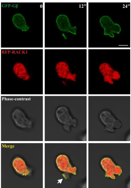
In live cell confocal imaging analysis, RFP-DdRACK1 co-localized with GFP-Gβ at the front

of newly formed membrane protrusions (Figure 7, arrow).

(45)

45

Figure 7 DdRACK1 co-localisation with Gβ. Representative images of a series show co- localization of RFP-DdRACK1 with GFP-Gβ at membrane protrusions (arrow) formed after 12 sec of imaging, which disappeared after 24 sec. Scale bar, 5 µm.

Close inspection of AX2 cells expressing GFP-DdRACK1 revealed that newly formed

protrusions initially were devoid of RACK1 but were later filled with GFP-DdRACK1 after

several seconds (Figure 8, arrow).

(46)

46

Figure 8 Confocal live cell images of GFP-DdRACK1/AX2 cells. Like RFP-DdRACK1 in Figure 7 GFP-DdRACK1 filled membrane protrusions after 50 sec of live imaging which disappeared after 120 sec. Scale bar, 5 µm.

We also generated a mutant protein in which the four lysine residues in the extended loop of

RACK1 (see Figure 5) were exchanged with glutamic acid residues. Analysis of GFP-

DdRACK1mut cellular distribution showed similar localization like GFP-DdRACK1 (Figure

9).

(47)

47 Figure 9 Subcellular localization of DdRACK1mut. Imaging of GFP- DdRACK1mut/AX2 cells showed localization of GFP-DdRACK1mut at membrane protrusions after 70 sec and 90 sec (arrows) respectively. Scale bar, 5 µm.

3.3 Subcellular distribution of DdRACK1, GFP-DdRACK1 and DdRACK1mut, and the developmental expression pattern of DdRACK1

Cell fractionation experiments were performed to further study the subcellular distribution of

endogenous DdRACK1 as well as its GFP-fusion constructs. While reasonable amounts of

endogenous DdRACK1 associated with the pellet fractions, only faint bands of both GFP-

DdRACK1 and GFP-RACK1mut were seen in the pellet fractions (Figure 10). These low

GFP-fusion protein amounts in the pellet fractions may be explained by their moderately low

level of overexpression with respect to endogenous DdRACK1. α-Actinin which served as

cytosolic marker was exclusively present in the cytosolic fraction. A membrane association of

RACK1 is not surprising as it has been repeatedly found in phagosomal preparations from

mouse and Drosophila, and GpbB has been found in phagosomal preparations from D.

(48)

48 discoideum (Boulais et al., 2010; Gotthardt et al., 2006; Rogers and Foster, 2007; Urwyler et al., 2009).

Figure 10 Subcellular fractionation of AX2 and AX2 expressing GFP-DdRACK1 and

GFP-RACK1mut after lysis by passing through Nucleopore filters. Protein aliquots separated by SDS PAGE were used to perform western blot analysis. WL, whole cell lysate; L, supernatant from cell lysate (400 x g); S1, P1 (10,000 x g); S2, P2 (100,000 x g).

S, supernatant; P, pellet. DdRACK1 was detected with polyclonal anti-DdRACK1 antibodies. Antibodies against the cytosolic α-actinin were used as control.

A developmental analysis showed the presence of DdRACK1 protein in nearly unaltered

levels during all stages of Dictyostelium development (Figure 11).

(49)

49 Figure 11 DdRACK expression levels during development. Western blot analysis was performed with AX2 wild type cell samples collected during starvation in shaking suspension at the indicated time points. DdRACK1 was detected with polyclonal anti- DdRACK1 antibodies. For loading control the blot was probed with mAb 188-19-95 detecting cap32.

3.4 DdRACK1 oligomerization potential

It has been reported that RACK1 can dimerize in vivo and this dimerization is required for specific processes including the regulation of the N-methyl-D-aspartate (NMDA) receptor by the Fyn kinase in the brain (Thornton et al., 2004). Here, we tested the capability of DdRACK1 to oligomerize using recombinant DdRACK1 full length protein that had been cleaved from the GST part. In the presence of the cross-linking reagent glutaraldehyde (0.001%), DdRACK1 formed dimers and even higher oligomers with increasing time of incubation as detected by western blots using polyclonal DdRACK1 specific antibodies.

Interestingly, the native non-crosslinked DdRACK1 sample also contained some amount of

dimers and oligomers (Figure 12). This indicates that the dimerization characteristic exhibited

by RACK1 proteins also holds true for DdRACK1.

(50)

50

Figure 12 DdRACK1 forms homodimers and oligomers. 5-10 µg/100 µl of recombinant

DdRACK1 were incubated with 0.001% of glutaraldehyde and samples taken at the indicated time points of 5, 10 and 20 min. In the absence of glutaraldehyde, the monomer (36 kDa, mono) and low amounts of dimer (72 kDa), and trimer (108 kDa) were detected. Proteins were detected with polyclonal anti-DdRACK1 antibodies. The band below the monomer is due to intramolecular crosslinks.

Similarly, DdRACK1mut in which we had exchanged the basic amino acid residues in the

loop between blades 6 and 7 (see below, 3.6) also displayed wild type DdRACK1

oligomerization capability (Figure 13).

Abbildung

Figure 1 Structural detail for RACK1 proteins. (A) Crystal structure of RACK1A from  A
Figure 2 Views of the hRACK1 crystal structure. Central panel: cartoon representations  of  hRack1  viewed  from  the  side  (left)  and  from  the  top  (right)
Figure 4 Ribbon diagram of S. cerevisiae RACK1 (Asc1p) (PDB: 3FRX) which was used  as template for DdRACK1 model
Figure  5  Ribbon  diagram  of  DdRACK1  modelled  with  the  aid  of  MODELLER9  program,  with  yeast  RACK1,  which  was  retrieved  from  protein  data  bank  (PDB)  (www.rcsb.org),  as  template
+7

Referenzen

ÄHNLICHE DOKUMENTE

Moreover, although CNG channels are nonselective cation channels in vitro, the currents recorded from these channels are mainly carried by Ca 2+ in vivo (Ohyama et al.,.. Since no

das Hefe 3-Hybrid System in Frage, mit dessen Hilfe Protein-RNA Interaktionen identifiziert werden können (SenGupta et al., 1996). Außerdem ist die Isolierung der Fen1-

Interestingly, we also showed that despite similar binding affinities of DnmA to different RNA substrates, only tRNA Asp(GUC-1) , tRNA Glu(UUC-5) and tRNA Glu(CUA-5)

A minor degree of overexpression was also observed in knock-outs of the gene dicer A (drnA) and the heterochromatin protein gene hcpA (Kaller et al. In contrast,

In addition to the already known chemotaxis defect, we show here that coronin deletion mutants fail to upregulate genes involved in early development and, as a consequence,

This is not surprising, because microtubule plus end- binding proteins such as DdCP224 and DdEB1 have turned out not only to regulate microtubule dynamics but also to

dis- coideum amebae in this chamber with respect to the effects on chemotaxis of varying the midpoint gradient concentra- tion, the gradient steepness, and temporal

Da die mögliche dPMCA in ihrer C­terminalen Kationen ATPAse keine Calmodulinbinde­ und