INAUGURAL-DISSERTATION
zur
Erlangung des Doktorgrades
der Mathematisch-Naturwissenschaftlichen Fakultät der Universität zu Köln
vorgelegt von
Yogikala Vasudeva Prabhu
aus Mumbai, Indien
Köln, 2005
Referees/Berichterstatter: Prof. Dr. Angelika A. Noegel Prof. Dr. Martin Hülskamp Date of oral examination: 09.02.2005
Tag der mündlichen Prüfung
The present research work was carried out under the supervision of Prof. Dr. Angelika A. Noegel, in the Institute of Biochemistry I, Medical Faculty, University of Cologne, Cologne, Germany.
From April 2003 to November 2005.
Diese Arbeit wurde von April 2003 bis November 2005 am Biochemischen Institut I der Medizinischen Fakultät der Universität zu Köln unter der Leitung von Prof. Dr. Angelika A.
Noegel durchgeführt.
I take this opportunity to let several IMPORTANT people know much how they mean to me.
PS: This is the section I enjoyed writing the most☺
--- First and foremost, I convey my sincere heartfelt gratitude to Prof. Dr. Noegel for letting me work under her esteemed supervision. I would really like her know how much I owe and respect her for providing valuable guidance, creative suggestions and constructive criticisms (and a very polite way of doing that). Her immense support, guidance, encouragement, optimism and promptness are highly acknowledged. It was a privilege to work under her superb guidance.
I would then like to acknowledge Francisco and Ludwig who were ever ready to offer help, constructive ideas and guidance and patiently tolerate all my questions☺. A special thanks to Francisco again for rendering me help with most of the Dictyostelium work and microscopy (I was otherwise allergic towards them) and being friendly. Never to forget Akis (with his wonderful stories) and Julia, who were always so friendly and never let my motivation and enthusiasm down. I want to deeply forward my thanks to all of you for being so wonderful.
A special thanks to Christophe (my collaborator, UCSD, La Jolla) for helping me with some experiments, constructive suggestions (tolerating all my questions) and all his friendly e-mails. I would like to also thank Prof. Dr. Siu for providing few antibodies. I would forward my thanks to the Dicty Stock center for providing few important constructs.
I would then like to sincerely acknowledge Rosi(lein), (I like to annoy her sometimes as she looks more cute when angry☺) for being so kind and friendly and for all the help she offered anytime I needed. I would also like to thank Rolf for helping me in the project by giving away few constructs. Thanks to Maria and Berthold for being really friendly and cooperative. I would never forget to acknowledge the people from the cellar, Bärbel, Brigitte and Sonja without their help, smooth work would be impossible.
Next to all my dear friends who made my stay in the lab extremely wonderful. So here we go…..a sincere heartfelt gratitude to the darling Sorrrrrrrrrrrrrrraya who was so kind, friendly and helpful. Anne (ken)- my lost twin sister---even though we look so different☺. I want to thank you for being genuinely friendly and nice. I would like forward my heartfelt thanks to all my lab members: Mary, Hua, Vivek, Subh, Deen, and ex members from the lab: Nandu, Henning, Maki, Sunil and Hameeda to maintain a friendly working environment.
I would also convey my special thanks to Dhamu and Sabu for being such lovely company. I would never forget those days.
My heartfelt thanks to Carola, Jessica, Georgia, Hafi, Charles, Christhoph, Marion, Kathrin, Hyder, Ria, Wenshu and all others for being so friendly and who made my stay so pleasant.
I would like to forward my sincere thanks to Jianbo to help me patiently and friendly with Microarray work. I would forward my thanks to Vasily for his friendly and practical suggestions. I would then like to forward my thanks to my graduate school friends Magda, Julia, Maurijn and all the people from koeln-friends group for giving me nice company and letting me out of work stress sometimes. I would miss those cooking weekends☺ and the parties with you guys.
Gudrun for her prompt help anytime needed. A very sincere heartfelt thanks to Brigitte (Secretary, Graduate School) for being so kind and offer help patiently anytime I needed.
I would like to forward my thanks to all the people who directly or indirectly helped me towards the completion of the project.
I am indebted to my dear lifetime friends Girish, Annie, Brinda, Sav, Sumi and Alka whithout whom I would not consider myself complete.
My acknowledgments would be highly incomplete if I don’t mention two most important people in my life.
Samidha madam who is a friend, philosopher and guide. Thank you for showing me a postive outlook towards life. And Prof. Dr. Phale, I have no words to let you know how much I owe you. I convey my special and sincere gratitude for offering me support, guidance, help anytime I needed and being so tolerant, friendly, supportive and encouraging no matter what. And I would never forget to mention that wherever I am today is because of you.
My heartfelt sincere gratitude to Kumar, whose presence made my Koeln days memorable. I would deeply like to acknowledge your friendship, and for being standing by my side. I would also like to sincerely thank you for giving me a practical outlook towards life and work.
A special note of thanks to all of you: my sweetest sister Shashi, my loving brothers: Kiran, Kittu, Girish, Manoj, Puru, and Raj. Also to all the kids, Mohit, Yash, Vijay, Riya, Vansh. I can’t complete this section without thanking my sweet jaanu, Gaju.
Last but not the least, I would like to thank, Amma for being the best mother. I deeply acknowledge her patience, tolerance, support, encouragement, practical advices and sacrifices. Thanks amma for everything you have done for me. I would also like to thank Pappa (my late father- I miss you a lot).
Thanks my dear papa for everything. I love u both.
Finally I owe my sincere thanks to the Graduate School in Genetics and Functional Genomics,Unversity of Köln for offering me the fellowship for my PhD studies.
Yogikala Prabhu
7th December 2005
Cologne, Germany
Dedicated to my Parents
1.Introduction
1.1 GPCRs (G-protein coupled receptors) 1
1.2 GABA and its receptor systems 2
1.2.1 GABAB dimerization 4
1.2.2 GABAB structure (schematic representation) and signalling 5 1.3 Dictyostelium as a model organism 6
1.3.1 Dictyostelium developmental checkpoints 7
1.3.2 Late developmental events 8
1.3.3 Terminal differentiation 9
1.3.4 Sporulation 10
1.4 GABAB receptor-like proteins in D. discoideum 11
1.5 Aim of the work 13
2. Materials and Methods 2.1 Kits 14
2.2 Enzymes, antibodies, substrates, radioactive probes and antibiotics 14 2.3 Media and Buffers 15
2.3.1 Media and buffers for Dictyostelium culture 15
2.3.2 Media for E. coli culture 16
2.3.3 Media and buffers for Yeast two-Hybrid studies 16
2.3.4 Buffers and other solutions 17 2.3.5 Biological materials 17
2.3.6 Plasmids 18
2.3.7 Oligonucleotides 18
2.4 Methods 19
2.4.1 Total RNA and cDNA preparation 19 2.4.1.1 Isolation of total RNA from Dictyostelium cells 19
2.4.1.2 Generation of cDNA 19 2.4.2 DNA sequencing 19
2.4.3 Quantitative PCR 19
2.4.5 Semi-quantitative PCR (RT-PCR) 20 2.5 Construction of vectors 20 2.5.1 Generation of knockout mutants and screening of transformants 20
2.5.1.1 Gene replacement vector for GrlA 20 2.5.1.2 Gene disruption vector for GrlJ 20 2.5.1.3 Screening of transformants 20 2.5.2 Vectors for Yeast two-hybrid interactions 21 2.5.3 GrlA-GFP fusion vector 21
2.6. Biochemical methods 21 2.6.1 Western blotting (Kyhse-Andersen, 1984) 21 2.6.2. Yeast two hybrid methods 22
2.6.2.1 Yeast transformation 22 2.6.2.2 X-gal colony-lift filter assay 22 2.7 Dictyostelium cell culture methods 23
2.7.1 Measuring cell size (Rivero et al., 1996) 23
2.7.2 Growth rate measurement 23
2.7.3.1 Aggregation in shaking suspension 23 2.7.3.2 Development on phosphate-agar or water agar plates 24
2.7.4 Cytokinesis 24
2.7.5 Phototaxis experiments 24
2.7.6 Viability of osmotically shocked cells 24
2.7.7 Cell adhesion assay 25
2.7.8 Neutral red staining 25
2.7.9 LacZ reporter gene expression 25
2.7.10 DIF induced gene expression by shaking suspension assay 25
2.7.11 Spore germination assay 26
2.7.12 Sporogenous assay 26
2.7.13 Video imaging and chemotaxis assay 27 2.7.14 Indirect immunofluorescence of Dictyostelium cells 27
2.7.14.1 Preparation of Dictyostelium cells 27
2.7.14.2 Methanol fixation 27
2.7.14.2 Immunolabeling 28
2.7.14.3 Mounting 28
2.8 Microarray Analysis 28
3. Results
3.1 GABAB receptor-like proteins in Dictyostelium 30 3.1.1 Family of GABAB receptor-like genes in Dictyostelium 30 3.1.2 Domain architecture of GrlA and GrlJ 31 3.1.3 Comparison of GrlA and GrlJ with GABAB receptors from higher eukaryotes 31 3.1.4 Multiple alignment of the TMDs of GrlA and GrlJ and GABAB receptors 32
from higher eukaryotes
3.2. Expression and localization of GrlA and GrlJ 34 3.2.1 Expression profile of the grlA and grlJ 34
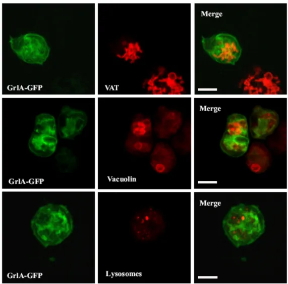
3.2.2 Subcellular localization of GrlA 35
3.3. Dimerization profile of family 3 of Dictyostelium GPCRs 37 3.4. Generation of grlA- and grlJ- mutants and their characterization 39 3.4.1.1 grlA and grlJ gene disruption by homologous recombination 39 3.4.1.2 Recombination event confirmed with PCR and Southern blot analysis and 41 RT-PCR
3.4.2 Characterization of grlA- and grlJ –cells (Mutant Analysis) 42
3.4.2.1 Growth 42
3.4.2.2 Cytokinesis in grlA- and grlJ - 44 3.4.2.3 Measurement of cell size in the mutants 44 3.4.2.4 Resistance to osmotic shock in the grlA− and grlJ− cells 45 3.4.2.5 grlA− and grlJ− sense folic acid as a chemoattractant 46 3.4.2.5 GrlA and GrlJ are not involved in phototaxis 47 3.4.3 Development of grlA−and grlJ− 48
3.4.3.1 Aggregation on solid surface 48
3.4.3.2 Cell motility in grlA- 49 3.4.3.3 Unaltered expression of aggregation-specific genes in grlA−and grlJ− 51 3.4.3.4 Role of GrlA and GrlJ in post aggregation differentiation 52 3.4.3.4.1 Development on phosphate agar plates 52 3.4.3.4.2 Gene expression profile during post aggregation differentiation 54
3.4.4.1 Cell adhesion in grlA and grlJ 55
3.4.4.2 Cell-cell adhesion 56
3.4.4.3 Localization of contact sites A in the mutants 58
3.4.5 Dictyostelium morphogenesis 58
3.4.5.1 Slug analysis (neutral red staining) 58
3.4.5.2 Slug migration 59
3.4.5.2.1 grlJ −slugs leave behind traces during migration 59
3.4.5.2.2 Breaking slugs monitored in grlJ- 60
3.4.5.2 DIF and cAMP induction of stalk and spore specific genes 62 3.4.5.4 Localization of prestalk and prespore cells in the mutants 64
3.4.5.4.1 Patterning of pspA in grlA- 64
3.4.5.4.2 Localization of prestalk cells in grlA− and grlJ−cells 65 3.4.5.5 Sporulation in grlA− and grlJ−cells 66 3.4.5.5.1 Defective spore morphology of the grlJ−strain 66 3.4.5.5.2 Spore germination in grlA−and grlJ−strains 67
3.4.5.5.3 grlA− does not produce SDF-2 68
3.4.5.5.4 grlA− shows a defect in the SDF-2 production involving TagC and DhkA 69
3.4.6 Transcription profile of grlA− 70
3.4.6.1 Experimental design 70
3.4.6.2 Comparison of the genes differentially regulated in grlA− cells 71 3.4.6.2.1 Transcriptional profile of differentially regulated genes in grlA− 72 3.4.6.3 GOAT (Gene Ontology Annotation Tool) 74 4. Discussion
4.1 GABAB receptors-like family of proteins in Dictyostelium 78 4.2 GrlA and GrlJ, expression, localization and dimerization 79 4.3 GrlA and GrlJ, two GPCRs involved in late developmental stages 80 4.3.1 Charactersistics of grlA- and grlJ- 80 4.3.2 grlA- and grlJ - exhibit post aggregation development defects 81
4.3.3 Cell adhesion 82
4.3.4 Probable role of GrlA in morphogenesis 83 4.3.4.1 GrlA is involved in sporulation events 84 4.3.4.2 grlA- has a defect in the production of SDF-2 84 4.3.4.3 Defect in SDF-2 production is mediated by reduced levels of tagC in grlA- 85
4.3.4.4 grlA- displays low levels of the SDF-2 receptor, dhkA 85 4.3.4.5 Transcriptional profile of genes in grlA- 85 4.3.4.6 Hypothetical model depicting the role of GrlA in controlling sporulation
events 86
4.3.5 GrlJ in morphogenesis 88
4.4 Concluding remarks 90
5. Bibliography 91
6. Summary (in English/German) 7. Erklärung
8. Curriculum Vitae (in English/German)
bp base pair(s)
β-ME beta-mercaptoethanol BSA bovine serum albumin Bsr blasticidin resistance cassette cAMP cyclic adenosine monophosphate cDNA complementary DNA
DEPC diethylpyrocarbonate DMSO dimethylsulphoxide DNA deoxyribonucleic acid
dNTP deoxyribonucleotide triphosphate ECL enzymatic chemiluminescence EDTA ethylenediaminetetraacetic acid
EGTA ethyleneglycol-bis (2-amino-ethylene) N,N,N,N-tetraacetic acid EST expressed sequence tag
G418 geneticin
GABA γ-amino butyric acid GFP green fluorescent protein
GPCR G-protein coupled receptors
Grl GABAB or Glutamate like receptors
IgG immunoglobulin G
kb kilo base pairs
MOPS Morpholinopropanesulphonic acid
Mw molecular weight OD optical density ORF open reading frame
PAGE polyacrylamide gel electrophoresis PCR polymerase chain reaction
PKA Protein kinase A RNA ribonucleic acid RNase ribonuclease
rpm rotations per minute
RT-PCR reverse transcript polymerase chain reaction SDF Spore Differentiation Factor
SDS sodium dodecyl sulphate SRF Serum Response Factor v/v volume by volume
vol. volume
X-gal 5-bromo-4-chloro-3-indoyl-D-galactopyranoside
1. INTRODUCTION
1.1 GPCRs (G-protein coupled receptors)
Organisms must readily respond to essential signals in their environment.
Multicellular organisms greatly depend on the capacity of cells to communicate with each other and their environment. It has been recognised that membrane bound receptors devised to recognise sensory messages from the environment (light, phermones, gustative molecules) and intercellular messages such as neurotransmitters, hormones, growth and developmental factors are very similar and derive from common ancestral origins (Bockaert, 2001). They can be classified on the basis of their structure and function into different categories: 1) Channel receptors, 2) Tyrosine kinase receptors, 3) Guanylate cyclase receptor, 4) Serine/threonine kinase receptors, 5) Cytokine receptors, and 6) GPCRs.
The seven-transmembrane spanning G protein-coupled receptors (GPCRs) represent a major group of cell-surface detectors and constitute 3.5 % of the genome in vertebrates. Of the 1000 genes thought to encode GPCRs in humans, about 300-400 mediate effects by endogeneous ligands, with the remainder being sensory receptors. They are characterized structurally by an amino-terminal extracellular domain, a carboxy-terminal intracellular domain and seven hydrophobic transmembrane regions (TMDs). Generally, agonist binding at the GPCR pocket leads to a conformational change in the 7TMDs which in turn allows its association with G-proteins. The activated G-proteins initiate signalling cascade(s) within the cell eliciting a physiological response (Prinster et al., 2005). GPCRs are involved in recognition and transduction of a wide variety of signals as diverse as light, Ca2+, odorants, nucleotides, amino acid residues, peptides as well as proteins (Figure 1).
Figure 1. GPCRs as an illustration of Jacob’s idea ‘evolution is molecular tinkering’. (Bockaert and Pin, 1999) GPCRs have a central common core made of seven transmembrane helices (TM-I to -
VII) connected by three intracellular (i1, i2, i3) and three extracellular (e1, e2, e3) loops. The diversity of messages which activate these receptors is an illustration of their evolutionary success.
Different GPCRs differ in their length, ligands bound or sensed at their N termini, function of their intracellular and extracellular loops, C-terminal interactions and the respective G-proteins coupled. In short, a high degree of molecular tinkering renders them the capability to carry out multitude of functions (Bockaert and Pin, 1999). GPCRs are the oldest known signal transducers present in plants, animals, fungi and protozoa. They carry out signalling largely via intracellular second messenger systems such as 3’, 5’ cyclic AMP (cAMP); 3’, 5’ cyclic GMP (cGMP); 1, 2-diacyl glycerol (DAG); and inositol 1, 4, 5- triphosphate (IP3) and also use Ca2+ and various inositol phospholipids embedded underneath in the membranes.
G-proteins are heterotrimeric molecules composed of α, β and γ subunits. In their active form the α subunit is GTP bound and modulates the activity of downstream effectors in most of the species, whereas in their inactive GDP bound form they are in a complex with one Gβ and one Gγ subunit. There are 20 known G-proteins in mammals each comprising an unique Gα combined with one of the 5 known Gβ and one of the 12 Gγ subunits providing an extraordinary ability to perform a wide range of signalling. These heterotrimeric proteins are also present in many other experimental models including Drosophila melanogaster, D.
discoideum, Xenopus laevis, C. elegans and mouse. The role of G-proteins in development was first reported in D. discoideum (Pupillo et al., 1988). Presently there are 14 Gα, two Gβ and one Gγ known in this organism. However, other than coupling to G-proteins downstream, many GPCRs have also been reported to bind to many other molecules modulating the activity of the receptors, for instance arrestins or PDZ domain containing proteins to name a few (Malbon, 2005).
1.2 GABA and its receptor systems
GABA (gamma-amino-butyric acid) is the most widely distributed inhibitory amino acid derived neurotransmitter in the vertebrate centralnervous system. There are two distinct types of target receptors for GABA present on peripheral nerve terminals each of which mediates synaptic inhibition: the ionotropic GABA types A and C (GABA A/C); and the metabotropic GABA type B (GABAB) receptors. Upon activation of two molecules of GABA, the pentameric GABAA receptor (ion channel) induces a rapid inhibition allowing inward influx of Cl– and a subsequent hyperpolarization of the postsynaptic membrane, whereas activation of GABAB produces a slow, prolonged inhibition and hence is
metabotropic (Blein et al., 2000; Pagano et al., 2001). GABAB receptors, which were first pharmacologically distinguished by Hill and Bowery (1981) act presynaptically and postsynaptically based on their anatomical location and physiological functions. These receptors couple through heterotrimeric G proteins (GI and Go) modulating adenylyl cyclase activity and cause inwardly rectifying K+ channels to open and voltage-dependent Ca2+
channels to close (Bettler et al., 1998; Blein et al., 2000) as illustrated in Figure 2.
Physiologically,GABAB receptors have been implicated in synaptic inhibition, hippocampal long-term potentiation, short-wave sleep, muscle relaxation, andantinociception, which make them attractive therapeutic targets (Kaupmann et al., 1998; Bowery and Enna, 2000). The dysfunction in the GABA system leads to several neuropsychiatric disorders including anxiety, depression, schizophrenia, loss of memory, loss of sleep, epilepsy (Cyran et al., 2005). These receptors have also been implicated in several inherited neurological disorders.
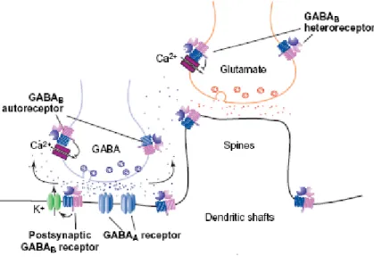
Figure 2. Schematic representation of the GABAB receptor heterodimer and its localization in the brain(Cyran et al., 2005). In the hippocampus, GABAB receptors are located presynaptically, postsynaptically and on extrasynaptic membranes. Presynaptic GABAB autoreceptors on GABA releasing terminals inhibit the release of GABA, whereas GABAB heteroreceptors inhibit the release of several other neurotransmitters (e.g. glutamate) and bioactive peptides. Postsynaptic GABAB
receptors activate K+ channels and induce slow inhibitory postsynaptic potentials, the fast component of which is mediated through GABAA receptors.
GABAB receptors belong to the family 3of GPCRs (G-protein coupled receptors) and as such share homology with metabotropic glutamate (mGlu), Ca2+-sensing (CaS), pheromone, taste and vomeronasal receptors. These proteins share35% sequence identity. In contrast to the mGlu and CaS receptors, which form functional homodimers, the GABAB
receptor is a heteromer constituted ofboth GABABR1 and GABABR2 proteins (Kaupmann et al., 1998).
1.2.1 GABAB dimerization
Although the concept of GPCR heterodimerization was proposed in the early 1980s, the first widely accepted evidence came from GABAB receptors. The first GABAB receptor cDNA was cloned (Kaupmann et al., 1997) and termed as GABABR1. However it was largely non- functional in most cell types when expressed alone, due to its inability to get trafficked to the cell surface (Couve et al., 1998). A year later six independent groups cloned another receptor, termed as GABABR2 (Jones et al., 1998; Kaupmann et al., 1998; Kuner et al., 1999; Martin et al., 1999), which did not bind to GABA when expressed alone. Indeed, thecoexpression of both subunits is required for a fully functionalGABAB receptor (Jones et al., 1998; Kaupmann et al., 1998). Both receptors consist of an extracellular domain (ECD) and aTMD composed of seven transmembrane α-helices, loops and an intracellular C-terminus. The GABABR1 subunit binds all known GABAB ligandsand is responsible for the ligand recognitionby the heteromeric GABAB receptor, whereas the GABABR2 subunit is necessary for the correct trafficking of GABABR1 to the cell surface (Couve et al., 1998; White et al., 1998) by masking a signal responsible for the retention of GABABR1 in the endoplasmic reticulum (ER) (Pagano et al., 2001). While the GABABR1 ECD plays a criticalrole in ligand binding, however, GABABR2 also contributes to agonist binding affinity i.e. in short, both the proteins are essential for efficient ligand binding. The ECD of these proteins shares the same protein fold as bacterial periplasmic binding proteins. Ligand binding into this Venus flytrap like module causes activation of the TMD, which in turn carries out the downstream signalling. It has been shown that the GABABR2 TMD contains enough molecular determinants for coupling to G-proteins, because G-protein activation can be detected with a GABAB receptor combination containing GABABR2 TMDs only (Blein et al., 2000; Pagano et al., 2001;
Grünewald et al., 2002). However, the exact role of each subunit in G-protein recognition and activation by the heterodimeric GABABreceptor is still unknown. The carboxyl-termini of the two receptor subunits were shown to physically interact with each other in yeast two-hybrid and fusion protein pull down assays (White et al., 1998; Kuner et al., 1999) but it was not essential for dimerization (Margeta-Mitrovic et al., 1999; Calver et al., 2001; Pagano et al., 2001). Infact, the N-termini and the transmembrane domain of both the receptor subunits are shown to possess enough determinants required for dimerization (Liu et al., 2004).
Nevertheless, the involvement of C-termini coupling for targeting of the receptors and their function in holding the dimers strongly together through coiled coil interactions cannot be ruled out.
1.2.2 GABAB structure (schematic representation) and signalling
Upon ligand binding, the amino-terminal domain undergoes a hinge-bending motion, as in LIVBP (Leucine Isoleucine Valine Binding Pocket), which is described as a "venus fly trap" mechanism. Receptor activation produces adenylyl cyclase inhibition through the involvement of the α subunit of Gαi/o type G-proteins (Blein et al., 2000). Postsynaptic receptors cause inwardly rectifying K+ channels to open allowing K+ to move down its electrochemical gradient. This effect on K+ channels is mediated by G protein β/γ subunits. It has been proposed that activation of presynaptic receptors results in the closing of voltage- dependent Ca2+ channels (Figure 3).
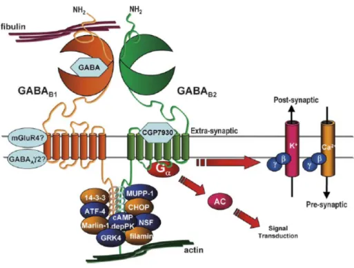
Figure 3. Schematic representation of the components of the putative GABAB receptor
‘‘signalosome.’’ (Couve et al., 2004). The figure illustrates several known interactions and signaling carried out by the functional GABAB receptors but their positions shown in the figure are only schematic.
The functional GABABR1/GABABR2 subunits were also reported to associate with a number of proteins both directly and indirectly, giving rise to a signaling complex capable of producing a diverse array of physiological and pharmacological effects. GABAB agonists such as GABA and baclofen binding to the "Venus Flytrap" structure of GABABR1 were known for a long time but interestingly, an allosteric modulator CGP7930 discovered recently appears to bind to the heptahelical domain of GABABR2. Non-canonical GABAB mediated
signaling involves some proteins that directly interact with the coiled-coil domain of either one or both the receptors, for instance, ATFx and CREB2 (cAMP responsive element binding protein-2, ATF4), which are both members of the ATF/CREB family of transcription factors.
Other proteins, such as GRK4 and cAMP-dependent protein kinase, are thought to be associated indirectly to the signaling complex as illustrated in Figure 3. (White et al., 2000;
Couve et al., 2004).
1.3 Dictyostelium as a model organism
Dictyostelium discoideum is a soil-living amoeba and feeds on bacteria in decaying vegetation. It reproduces by binary fission, as do all amoebae. They enjoy their unicellular fate as long as the food is plentiful and enter a multicellular phase on environmental pressure such as starvation. Growing amoebae chemotax toward folic acid and other nutrients, whereas starved cells undergo a developmental process that transforms them into cAMP sensing machines (Manahan et al., 2004). When synchronously starved, cells begin to spontaneously emit pulses of cyclic AMP (cAMP) - a compound used by these cells to facilitate cell-cell communication - to which other D. discoideum cells are attracted. As cells move towards the source of cAMP, they emit their own pulse of cAMP, thus amplifying and propagating the signal, and enabling large numbers of cells to congregate from a wide area. The cells begin to associate, forming streams of migrating cells, which merge in an aggregate consisting of up to 100,000 cells. The streams eventually come together to form a mound which develops a tip coordinating further development resulting in the formation of a slug that migrates to a favourable environment for culmination into the fruiting body.
D. discoideum, is an excellent organism for the study of the molecular mechanisms of cell motility, signal transduction, cell-type differentiation and developmental processes.
Dictyostelium has a 34 Mb genome spread over six chromosomes encoding approximately 12,500 proteins (Eichinger et al., 2005). A significant number of genes show higher similarities to the genes of vertebrates than to those of other fully sequenced eukaryotes. This results further strengthened the view that the evolutionary position of D. discoideum is located prior to the branching of metazoa and fungi but after the divergence of the plant kingdom, placing it close to the base of metazoan evolution. (Glöckner et al., 2002). The most studied receptors in this amoeba are the four G-protein coupled cAMP receptors, which are induced during starvation. 48 additional GPCRs have been discovered during genome analysis.
1.3.1 Dictyostelium developmental checkpoints
Developmental transitions occur at two points; one is from growth to development mediated by cAMP signalling wherein many aggregation specific proteins are upregulated within a few hours and the cells gradually develop polarized cell morphology. Oscillatory pulses of cAMP with a period of 6 min propagate as waves through cell monolayers binding to the high affinity cAMP receptors (cAR1) which through their βγ subunits activate the downstream components PI3 kinase to form PIP3 and cortical myosin in turn leading to the formation of polarized cells and directional movement towards the chemoattractant (chemotaxis). cAMP also activates CRAC and adenylyl cyclase through Gα2, which in turn synthesizes more cAMP and elicits an autocrine response (Manahan et al., 2004; Strmecki et al., 2005).
Aggregation results in the formation of a multicellular organism, known as a mound. A second transition occurs at the mound stage during which the precursors of the mature spore and stalk cells - prestalk and prespore cells differentiate and sort, forming a tipped aggregate or tipped mound. As development proceeds, the tip extends and an anterior-posterior axis
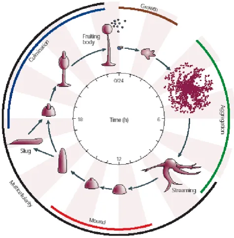
Figure 4. Dictyostelium discoideum morphogenesis (Chisholm and Firtel, 2004).
forms, which is maintained through the slug and early culminant stages. Culmination and the formation of the fruiting body completes morphogenesis. During this stage, the precursor
populations differentiate, producing a sorus on top of a highly vacuolated mass of stalk cells.
The entire process from starvation of vegetative cells to the formation of a mature fruiting body takes 24 hours as depicted in the schematic representation of the life cycle of D.
discoideum (Figure 4)(Chisholm and Firtel, 2004).
1.3.2 Late developmental events
A mound of developing Dictyostelium cells establishes asymmetry and generates the correct proportions of different cell types. Cells expressing the earliest available markers specific for prespore or prestalk cell fate appear at random locations in the mound and these cells then sort by a process which is likely to involve both differential adhesion and different rates of chemotaxis such that by the slug stage the distinct populations are found in separate locations (Williams et al., 1989; Esch and Firtel, 1991; Fosnaugh and Loomis, 1993; Ozaki et al., 1993; Yoder and Blumberg, 1994). Once the aggregates have formed, owing to high extracellular cAMP concentration there is a developmental switch leading to adaptation or downregulation of aggregation specific genes and activation of post aggregation genes. The high and saturating concentration of cAMP results in the induction of the transcription factor GBF which plays as a central role in controlling events pertaining to induction of post aggregation genes and cell-type specific gene expression. This immediately leads to tyrosine phosphorylation of STATa (signal transducer and activator of transcription) that in turn binds to promoter elements so as to activate the ecmA gene and repress ecmB (extracellular matrix proteins A and B respectively). The prespore specific genes are induced by cAMP and repressed by DIF-1. cAMP and DIF together are two essential morphogens required to establish a proper prestalk and prespore population to form the multicellular organism. cAMP receptors are involved in a complex signalling cascade in the post aggregation stages when all the four cARs are active, functional and exhibit distinct spatial expression pattern and different affinities for cAMP which helps in sorting, patterning and morphogenesis into a highly proportional organism (Johnson et al., 1993; Saxe et al., 1993, 1996; Louis et al., 1994). The extracellular cAMP has opposing effects depending on the expression levels and activation of alternate cAR receptor expression in different cell types. The cAMP mediated effect on the prespore genes and pstB (prestalk B) is dependent on a serine/threonine kinase GSK3 controlled by high affinity car3 receptor (glycogen synthase kinase), which helps to keep the ecmB repressed until the right time (Schilde et al., 2004). At mid culmination, the low affinity car4 inhibits and turns on the ecmB expression. The intracellular cAMP also regulates events to control the transition from slug to mature fruiting body and terminal differentiation via the intracellular phosphodiesterase RegA and the adenylyl cyclase ACR
which is required to control late developmental events. PKA (protein kinase A) is essentially involved in all the events governing the cell-type specification and morphogenesis (Loomis, 1998).
1.3.3 Terminal differentiation
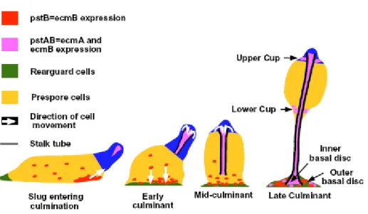
The final morphogenesis to form the mature fruiting body requires spatial reorganization of cell populations within the slug. Slug migration is arrested by different environmental factors including low humidity, reduction of local ammonium ion concentrations followed by movement of the posterior of the slug under the tip, forming a Mexican hat like structure wherein the rearguard cells enriched with ecmB form the base and the prestalk cells present at the anterior of the slug form the tip. Fruiting body formation is initiated when apically localized prestalk cells invaginate into the prespore mass, differentiate into stalk cells and form the stalk tube (Figure 5). The first cells that enter are the pstAB cells followed by the pstA cells, which then induce ecmB and are committed to stalk cell differentiation. A stalk tube progressivley elongates through the prespore mass, raising it off the substratum (Jermyn et al.,1996). Simultaneously, the ALCs (anterior like cells) migrate to form part of the basal disc and upper and lower cups. As the stalk tube is formed, prespore cells enter the terminal differentiation pathway and generates spores. Terminal differentiation is greatly controlled by PKA (Aubry and Firtel, 1999).
Figure 5. Diagramatic representation of the movements of ecmA and ecmB-expressing cells during culmination. (Jermyn et al., 1996)
1.3.4 Sporulation
Stalk cell differentiation and sporulation are highly interconnected. During culmination, PKA activity is highly dependent on intracellular cAMP levels (that controls the dissociation of the catalytic subunit PKA-C from the regulatory PKA-R subunit), which is produced by the late adenlyl cyclase ACR and degraded by the phosphodiesterase RegA. The two component system histidine kinase receptor DhkC and the phosphodonor RdeA activates RegA but is inhibited by another hybrid histidine kinase receptor, DhkA, which inhibits this phosphorelay via RdeA and thereby activates PKA and induces sporulation (and later by DhkB to avoid premature germination) (Loomis et al., 1998). This inhibition is mediated by a peptide, SDF-2 (spore differentiation factor) which is released by the proteolytic cleavage/processing of AcbA (Acyl binding protein) which synthesized in the prespore cells.This proteolytic cleavage is mediated by the dual function transporter/protease TagC present on the surface of the prestalk cells to release SDF-2 which then acts on prespore cells to trigger encapsulation and acts as a feed back loop in the prestalk cells to control its production (Anjard et al., 1998, Anjard and Loomis, 2005) represented in Figure 6.
Figure 6. Proposed signaling pathway leading to sporulation ( Anjard and Loomis, 2005)
The MADS-Box transcription factor SrfA (serum response factor) has been largely implicated in the events regulating the spore maturation. It is known to act downstream of PKA and induces the transcription of a distinct set of genes all of which are known to function in the process of spore coat formation and its maturation (Escalante et al., 2001, 2003;
Escalante and Sastre, 2002).
1.4 GABAB receptor-like proteins in D. discoideum
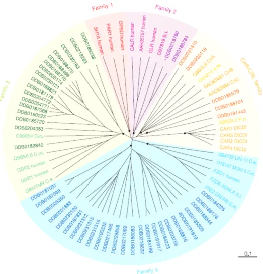
The genome sequence of chromosome 2 (Glöckner et al., 2002) gave a clue for the presence of four genes related to the metabotropic glutamate/GABAB subfamily. The complete sequence (Eichinger et al., 2005) revealed the existence of 17 genes encoding receptors belonging to Family 3 of GPCRs and all of them resembled GABAB or metabotropic glutamate like- receptors. They were recently named by Dale Hereld (Dictyostelium Genomics, 2005) and officially accepted to be represented as Grl (A-R) (GABAB or metabotropic glutamate - receptors like) proteins. The presence of these receptors was indeed a very interesting observation as only the cAMP receptor gene family known to be present in D. discoideum. The presence of the animal specific (as it was thought so far) frizzled like-receptors (25 genes), the secretin family (2 genes) and 17 members representing the Family 3 of GPCRs was rather surprising (Figure 7) (Insall 2005; Williams et al., 2005).
Figure 7. Phylogenetic tree of the G-protein-coupled receptor Family in Dictyostelium discoideum. (Eichinger et al., 2005). A CLUSTALX alignment of the sequences encompassing the seven transmembrane domains of all Dictyostelium GPCRs, and selected GPCRs from other organisms, was used to create an unrooted dendrogram with the TreeView program. A.th., A. thaliana;
B.t., Bos taurus; CAR/CRL, cAMP receptor/cAMP receptorlike; C.e., C. elegans; DICDI, D.
discoideum; D.m., D. melanogaster; G.c., Geodia cydonium; P.p., Polysphondylium pallidum; X.l., Xenopus laevis.
We carried out a series of blast searches with Family 3 proteins and most of them displayed a close resemblance to GABAB. The identity was around 25 percent to the mammalian counterparts but the Grls always clustered well amongst each other. The transmembrane domains of most of the Dictyostelium GABAB receptor-like proteins possess a identity and there is a high degree of conservation observed among GABAB homologsfrom different species with regard to their TMDs. Several amino acids in the intracellular loops 2 were conserved whereas the intracellular loop 3 of Dictyostelium Grls were found very similar to mammalian GABAB receptors. A further search revealed the resemblance of GrlJ, GrlF and GrlE to the receptor 1 subtypes whereas all others were slightly more closer to the sub-type 2.
This observation is in contrast to what is found in mammalian GABAB receptor sub-types as they have many of GABABR1 and fewer of GABABR2 subtypes which are a result of alternative splicing. However the differences between these subtypes are only very slight and their grouping into subtypes is rather weak. Also, a few of these receptors possessed a coiled- coil domains at their C-termini but the N-terminal ligand binding domain was comparatively shorter in most of the receptors belonging to this family. Few of them also possessed a Bmp (Bacterial/ Basic membrane protein) domain at their N - termini which resembled the periplasmic binding proteins found in bacteria.
1.5 Aim of the work
GABAB receptors are widely studied amongst higher eukaryotes and mammals including rat, mouse, Drosophila and humans. The analysis of the Dictyostelium genome revealed the presence of 17 different genes encoding members of the metabotropic glutamate and/or GABAB receptor family which was surprising as this family of receptors was so far considered animal-specific. This opens up a further complexity of signaling in these amoeabe in addition to the role of the well investigated cAMP receptors. It was therefore interesting to investigate the role played by these receptors D.discoideum. Hence, we attempt to address the following questions:
• the function carried out by these receptors in Dictyostelium,
• their expression pattern and localization,
• their probable role in development, signaling pathways used by these receptors, and
• finally the dimerizing pair(s) amongst the receptor pool if any.
We have selected GrlA and GrlJ, two of the first four GABAB homologs identified in Dictyostelium, for the present study.
2. MATERIALS AND METHODS
Details regarding following things are not included in this section: In short,
• Standard molecular biology techniques were carried out as per (Sambrook et al., 1989). A Laboratory Manual. Cold Spring Harbor Laboratory Press, NY, Vols. 1-3 (1989).
• Fine chemicals were obtained from Sigma as otherwise indicated.
• Standard laboratory reagents and materials were obtained from local suppliers.
• Instruments used were from the Departmental facility.
2.1 Kits
Nucleobond AX 100 and 500 Macherey Nagel
NucleoSpin Extract 2 in 1 Macherey-Nagel
NucleoSpin Plus Macherey-Nagel
Original TA Cloning Invitrogen
Qiagen Midi- and Maxi-prep Qiagen
Stratagene PrimeIt II Stratagene
PGEMTeasy Cloning kit Promega
Qiagen RNAeasy Mini kit Qiagen
High pure PCR Template preparation kit Roche
RT-PCR Roche
RT-PCR for Microarray Stratagene
2.2 Enzymes, antibodies, substrates, radioactive probes and antibiotics
Enzymes for molecular biology:
Calf Intestinal Alkaline Phosphatase (CIP) Boehringer
Klenow fragment Boehringer
Lysozyme Sigma
Proteinase K Sigma
Restriction endonucleases Amersham,Life technologies, New England Biolabs
M-MLV reverse transcriptase Promega
Ribonuclease H (RNase H) Boehringer
Ribonuclease A (RNase A) Sigma
T4 DNA ligase Boehringer
Taq-polymerase Life technologies/Boehringer
Antibodies:
Primary antibodies:
Polyclonal Goat anti-GST antibody Pharmacia Mouse monoclonal anti-GFP mAb K3-184-2 Noegel et al., 2004
Mouse monoclonal anti-actin mAb Act-1 Simpson et al., 1984 Mouse monoclonal anti-CAP mAb 231-18-8 Gottwald et al., 1996 Mouse monoclonal anti-csA mAb 33-294 Berthold et al., 1985 Mouse monoclonal anti-vacuolin mAb 221-1-1 Rauchenberger et al., 1997
Mouse monoclonal anti-comitin mAb 190-340-2 Weiner et al., 1993 Mouse monoclonal anti-interaptin mAb 260-60-10 Rivero et al., 1998 Mouse monoclonal anti-PDI mAb 221-135-1 Monnat et al., 1997 Mouse monoclonal anti-lysosmal enzyme mAb 221-342-5 Neuhaus et al, 1998 Mouse monoclonal anti-annexin mAb 185-447-3 Döring et al, 1995 LagC polyclonal antibodies Wang et al., 2000 Dd-CAD1 polyclonal antibodies Wong et al., 1996
Secondary antibodies:
Goat anti-mouse IgG, peroxidase conjugated Sigma Goat anti-rabbit IgG, peroxidase conjugated Sigma Sheep anti-mouse IgG, Cy3 conjugated Sigma
Goat anti-rabbit Alexa 568 conjugated Molecular probes
Antibiotics:
Ampicillin Gruenenthal
Blasticidin S ICN Biomedicals
Dihydrostreptomycinsulphate Sigma
Kanamycin Sigma, Biochrom
Tetracyclin Sigma
Radiolabelled nucleotide:
α-32P-deoxyadenosine triphosphate, (10 mCi/ml) Amersham
2.3 Media and Buffers
All media and buffers were prepared with deionised water, filtered through an ion-exchange unit (Membra Pure). The media and buffers were sterilized by autoclaving at 120ºC and antibiotics were added to the media after cooling to approx. 50ºC. For making agar plates, a semi-automatic plate-pouring machine (Technomat) was used.
2.3.1 Media and buffers for Dictyostelium culture Ax2-medium, pH 6.7 (Claviez et al., 1982):
7.15 g yeast extract, 14.3 g peptone (proteose), 18.0 g maltose, 0.486 g KH2PO4, 0.616 g Na2HPO4.2H2O add H2O to make 1 liter
Phosphate agar plates, pH 6.0:
9 g agar ,add Soerensen phosphate buffer, pH 6.0 to make 1 liter Salt solution (Bonner et al., 1947):
10 mM NaCl,10 mM KCl, 2.7 mM CaCl2
Starvation buffer, pH 6.5 (Shaulsky et al., 1998):
10 mM MES, pH 6.5, 10 mM NaCl,10 mM KCl,1 mM CaCl2,1 mM MgSO4
SM agar plates, pH 6.5 (Sussman, 1951):
9 g agar, 10 g peptone, 10 g glucose, 1 g yeast extract, 1 g MgSO4.7H2O, 2.2 g KH2PO4
1 g K2HPO4, add H2O to make 1 liter
Soerensen phosphate buffer, pH 6.0 (Malchow et al., 1972):
2 mM Na2HPO4, 14.6 mM KH2PO4
2.3.2 Media for E. coli culture
LB medium, pH 7.4 (Sambrook et al., 1989) SOC medium, pH 7.0 (Sambrook et al., 1989)
2.3.3 Media and buffers for Yeast two-Hybrid studies YEPD-Medium:
20 g/l Difco Peptone, 10 g/l Yeast extract YEPD-Agar plates:
20 g/l Difco Peptone ,10 g/l Yeast extract ,18 g/l Agar agar 100 x Adenine solution:
200 mg (1,1 mmol) Adenine in 100 ml water dissolve with addition of little amounts of HCl and filter sterilized.
100 x Histidine solution:
200 mg (1 mmol) Histidine in 100 ml Water and filter sterilized.
100 x Tryptophan solution:
200 mg (1 mmol) Tryptophan in 100 ml Water filter sterilized.
100 x Tyrosine solution:
300 mg (1,7 mmol) Tyrosine in 100 ml dissolve with addition of NaOH solution and filter sterilized.
100 x Leucine solution:
1000 mg (7, 6 mmol) Leucin in 100 ml Water and filter sterilized.
100 x Uracil solution:
200 mg (1, 8 mmol) Uracil in 100 ml Water dissolve by warming in water bath and filter sterilized.
1M 3-Amino-1,2,4-triazol (3AT) solution:
8.4g 3AT in 100 ml water andn filter sterilize 10 x Amino acid solutions:
300 mg (2, 3 mmol) Isoleucine 1500 mg (1, 1 mmol) Valine 200 mg (0, 9 mmol) Arginine 300 mg (1, 6 mmol) Lysine 200 mg (1, 34 mmol) Methionine 500 mg (3 mmol) Phenylalanine 2000 mg (16, 8 mmol) Threonine,
adjust final volume to 1l and filter sterilize.
The composition of the selection media and agar plates is indicated in the following table.
Agar agar and Yeast extract without nitrogen base was dissolved in water and autoclaved. The glucose solution was prepared in water and filter sterilised. The remaining stock solutions were added after cooling to 55 °C.
2.3.4 Buffers and other solutions
Buffers and solutions not listed below are described in the methods section.
10x MOPS (pH 7.0/ pH 8.0):
41.9 g MOPS, 7 ml 3 M sodium acetate , 20 ml 0.5 M EDTA, add H2O to make 1 liter 10x NCP-Puffer (pH 8.0):
12.1 g Tris/HCl, pH 8.0, 87.0 g NaCl, 5.0 ml Tween 20, 2.0 g sodium azide add H2O to make 1 liter
1x PBS (pH 7.4):
8.0 g NaCl, 0.2 g KH2PO4,1.15 g Na2HPO4, 0.2 g KCl dissolve in 900 ml deionised H2 adjust to pH 7.4 add H2O to make 1 liter, autoclave
1.2 M Phosphate buffer (pH 6.8):
1.2 M Na2HPO4, pH 9.1 was mixed with 1.2 M NaH2PO4, pH 4.02 in the ratio of 2:1.
20x SSC (pH 7.0):
3 M NaCl, 0.3 M sodium citrate TE buffer (pH 8.0):
10 mM Tris/HCl, pH 8.0, 1 mM EDTA Tris-phenol:
1 kg phenol was melted at 60ºC in a water-bath and equilibrated with 1 vol. of 1 M Tris/HCl, pH 8.0. The equilibrated phenol was aliquoted in 50 ml Falcon tubes and stored at –20ºC.
10x TAE buffer (pH 8.3):
27.22 g Tris, 13.6 g sodium acetate , 3.72 g EDTA add H2O to make 1 liter 2.3.5 Biological materials
Bacterial strains:
E. coli BL21 (DE) Studier and Moffat,1986
E. coli DH5α Hanahan, 1983
E. coli XL1 blue Bullock et al., 1987
Klebsiella aeorgenes Williams and Newell, 1976
Dictyostelium discoideum strain:
Ax2-214, an axenically growing derivative of wild strain, NC-4 (Raper et al., 1935) commonly referred to as Ax2.
Yeast strain:
Y190 Flick et al., 1990, Harper et al., 1993 2.3.6 Plasmids
pGEM-Teasy Promega
pGEX-4T1 Pharmacia Biotech
pGADT7 Clontech
pGBKT7 Clontech
p1ABsr8 Gräf et. al., 2000
pDNeo II Witke et al., 1987
pBSBsr unpublished
EcmB-Gal Jermyn and Williams, 1991 pspA(D19)-Gal Dingermann et al., 1989 2.3.7 Oligonucleotides
GrlA-CTFp 5’GGAATTCAATCAAACATTTGCAACAAATACAA 3’
GrlA-CTRp 5’CGTCGACTTATACATCATCTGGATCGATTTCAGATTG 3’
GrlJ-CTFp 5’ CGAATTCGAAACTTTTGCAACTAGTAC 3’
GrlJ-CTRp 5’CGGATCCTTAAACATTATTGGAATCAATTTCTGT 3’
Primers for screening knock out
GrlA-5' probeFp 5' CCTAAGAATAAATGCATTGGAGGTTC 3' GrlA-5' probe Rp 5' ACTATTGAACAATCCATTATTG3’
GrlA-3'ProbeFp 5' GAAGCAAGAGTAAAGGCAGAATCAATA 3’
GrlA-3'ProbeRp 5' GTTAAACCAGGAACCAGTGAATGTTGTC 3’
5'intron Fp 5' CTGTAATTGTAGAATTTATACCTG 3' Bsr actin15promoter Rp 5’GATGGGATTAATTAATTTGTAATC 3’
Primers for RT-PCR
Car2 Fp 5' GTTGGTGTTGGATTGGTGATA 3’
Car2Rp 5' CGATTGCAAATACCCAACAAA 3’
Act8 Fp 5' TTAAATCCAAAGGCCAACAGAG 3’
Act8Rp 5' TACCTGGGAACATAGTTGTACC 3’
TagC Fp 5' GTTCACTATATACCACATG 3’
TagCRp 5' CCGAAAACTACCTTTGGTGAT 3’
DhkAFp 5' ATTGAAAATGGAGACATTACCC 3’
DhkARp 5' TAAACTTTGAACCTTGACCGAC 3’
Yeast two- hybrid primers:
GrlA-CT-Fp 5’GGAATTCAATCAAACATTTGCAACAAATACAA 3’
GrlA-CT-Rp 5’ CGTCGACTTATACATCATCTGGATCGATTTCAGATTG 3’
GrlB-CT-Fp 5’ GGAATTCTTAAATCAAACATTCGCTTCAAGTTC 3’
GrlB-CT-Rp 5’ CGTCGACTTAAAGGTTATTAGAATCAATTTC 3’
GrlL-CT-Fp 5’ CGAATTCTTCTGGAAAATTTATAAACCAGTT G 3’
GrlL-CT-Rp 5’ CGTCGACTTAATTATCATTTTGTGCAGCAGC 3’
GrlH-CT-Fp 5’ CGAATTCCCAAAATTCTGGAGAGTATTTAGA 3’
GrlH-CT-Rp 5’ GGTCGACTTAATTATTATTTTCTGAATCATTGAC 3’
GrlJ-CT-Fp 5’ CGAATTCGAAACTTTTGCAACTAGTAC 3’
GrlJ-CT-Rp 5’ CGGATCCTTAAACATTATTGGAATCAATTTCTGT 3’
GrlF-CT-Fp 5’ CGAATTCTTCTTTAAAATGATAACAGTTGGTTTAGAAC 3’
GrlF-CT-Rp 5’CGGATCCTTAAACATCATTCGAATCAATTTCAG 3’
2.4 Methods
2.4.1 Total RNA and cDNA preparation
2.4.1.1 Isolation of total RNA from Dictyostelium cells
Total RNA was extracted from either Ax2 or grlA- or grlJ- from different developmental stages of the Dictyostelium life cycle or different assay conditions using the Qiagen RNeasy Mini kit. The manufacturer's protocol for the isolation of RNA from the cytoplasm of animal cells was used for preparation. The RNA samples were used directly for northern blot analysis or after reverse transcription for RT-PCR, Real-Time PCR and Microarray analysis.
2.4.1.2 Generation of cDNA
cDNA was generated using the M-MLV reverse transcriptase, RNAse H minus (Roche) according to the manufacturers protocol. Usually 1-5µg of the respective total RNA was used for each RT reaction.
2.4.2 DNA sequencing
Sequencing of the PCR-amplified product or plasmid DNA was performed at the sequencing facility of the Centre for Molecular Medicine, University of Cologne, Cologne by modified dideoxy nucleotide termination method using a ‘Perkin Elmer ABI prism 377’ DNA sequencer.
2.4.3 Quantitative PCR
Total RNA was extracted at different time points of Dictyostelium development and cDNA was prepared as described as under (section 2.4.1.2). Primers were selected such that the expected product size was between 250-500bp. Prior to use in real time experiments the quality of the cDNA and the primers were tested by PCR. Real Time PCR was carried out with the Quantited TMSYBR® green PCR kit (Qiagen) according to the manufacturer's protocol. For each sample gene specific primers (10 pmole) and 1 µl of cDNA was used. As a quantification standard defined concentrations (10ng, 1ng, 100pg, 10pg and 1pg) of GrlA-C- terminal and GrlJ-C-terminal gene sequences in pGEMTeasy were used. Actin specific primers were used as positive control and to ensure comparable concentrations of cDNA in samples of wild type and mutant cells.
2.4.5 Semi-quantitative PCR (RT-PCR)
cDNA generated as described in section 2.4.1.2 and specific primers were used to carry out PCR and is mentioned in the text as and when.
2.5 Construction of vectors
2.5.1 Generation of knockout mutants and screening of transformants 2.5.1.1 Gene replacement vector for GrlA
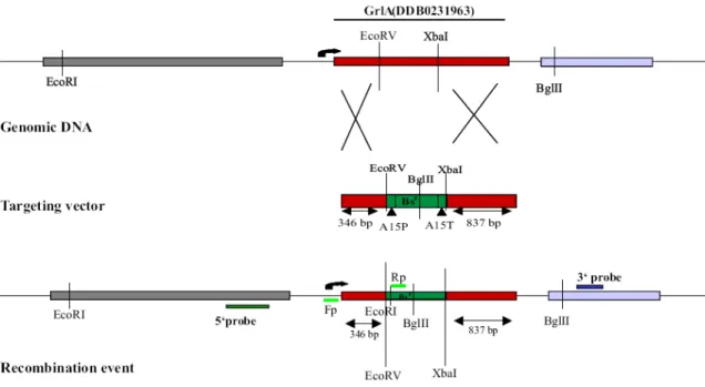
The Blasticidin resistance gene with the act15 promoter and act15 terminator sequences were retrieved using EcoRV and XbaI from the pBs-Bsr vector and inserted into the GrlA gene such that 1.3 kb of the GrlA was replaced by the 1.4 kb Bsr gene. GrlA sequences carrying the Blasticidin resistance cassette were purified and transformed into the wild type Ax2 cells by electroporation.
2.5.1.2 Gene disruption vector for GrlJ
The neomycin resistance cassette (2.2 kb) was obtained from pDNeoII vector by EcoRV digestion and cloned into the GrlJ gene that was present in pGEMTeasy vector. The targeting vector was transformed into the wild type Ax2 cells by electroporation.
2.5.1.3 Screening of transformants
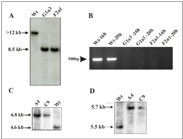
A PCR based approach was used for screening the grlA- cells followed by Southern blotting whereas the grlJ- was screened using Southern blotting. The respective transformants obtained were selected with 3 µg/ml of Blasticidin or 4 µg/ml G418 for Neomycin resistance.
Single colonies were obtained by spreader dilution of the whole pool of transformants onto SM agar plates overlaid with Klebsiella areogenes. The single transformants were then grown with the respective selection medium in a 96-well plate and eventually transferred to a 24- well plate and a 6-well plate. The cells were used to isolate genomic DNA using the kit (High pure PCR Template preparation kit) for PCR based screening whereas amoebae spread on K.
aerogenes were used to isolate genomic DNA for Southern blotting. However the final confirmation of the recombination event for individual single colonies for both the mutants was done using Southern blot analysis.
2.5.2 Vectors for Yeast two-hybrid interactions Heterodimerization:
C-termini immediately after the transmembrane domains (as obtained from SMART) of the following genes, grlA, grlB, grlH, grlL each were cloned in frame with the binding domain (BD) of Gal4 in pGBKT7 plasmid using EcoRI and SalI respectively. The C-termini of grlJ and grlF each were cloned in frame with the activating domain (AD) of Gal4 in pGADT7 with BamHI and EcoRI respectively. Each of the (AD) contructs were cotransformed in the yeast strain Y190 with each BD fusion constructs separately and grown on SD-Trp-Leu medium plates. Negative controls were carried out with cotransformation of BD fusion constructs with the AD alone and AD fusion constructs with the BD alone. Similarly, a positive control was also performed along with by cotransforming pCL1 and pGBKT7, which are known interactors The growth of transformed yeast cells streaked on SD-Trp-Leu-His (+3AT, 60mM) were monitored over a period of 4 to 5 days.
Homodimerization:
C-termini of the respective genes, grlA, grlB, grlH, grlL each were further cloned in fusion with the AD of Gal4 in pGADT7 with EcoRI and BamHI, respectively. grlA, grlB, grlH, grlL fused with BD and AD each were cotransformed in yeast strain Y190 and selected in the similar way ( see above).
2.5.3 GrlA-GFP fusion vector
The full-length GrlA was amplified as a HindIII - BamHI fragment and cloned in p1ABsr8 vector and transformed into Ax2 cells. The transformants were selected using blasticidin. The resultant cells were fixed and observed for the localization of the protein and alternatively stained with the respective antibodies for colocalization studies.
2.6. Biochemical methods
2.6.1 Western blotting (Kyhse-Andersen, 1984)
Protein samples prepared at the respective time points of Dictyostelium development and separated by SDS-PAGE gels (8%, 10% or 15% respectively) were blotted on to nitrocellulose membranes. They were blocked with 5 % milk in 1x NCP and probed with different dilutions of the respective primary antibodies and POD-conjugated secondary antibodies. They were then detected by ECL (Enzyme chemiluminescence) reactions.
2.6.2.Yeast two-hybrid methods
2.6.2.1 Yeast transformation Stock solutions
50% PEG 3350 prepared in water
100% DMSO : 0.1 M Tris-HCl (pH 7.5), 10 mM EDTA 10X LiAc: 1M LiAc, pH 7.5
50 ml of YPD or SD medium are inoculated overnight with several colonies of Y190 yeast strain and incubated overnight at 30°C for 16 to 18 hours with shaking at 250 rpm to stationary phase. An overnight culture (30 ml) is transferred 300 ml of YPD or SD and incubated at 30°C for 3 hours until the OD600 becomes 0.4 to 0.6. The cells are centrifuged and resuspended in TE buffer or water and centrifuged again. The pellet is resusupended again in 1X TE/1X LiAc (the resuspended cells are called competent cells). 0.1 µg of plasmid DNA and 0.1 mg of herring testes carrier DNA are added to a 1.5 ml tube and mixed. 0.1 ml of competent cells is added. 0.6 ml of sterile PEG/LiAc solution is added and vortexed and incubation is at 30°C overnight. 70 µl of DMSO is added and mixed gently and the cells heat shocked at 42°C for 30 minutes. The cells are centifuged for 10 seconds and the supernatant discarded. The pellet is resuspended in 100 µl of water and plated on SD-Trp-Leu plates and the plates incubated at 30°C for 3 to 4 days. The transformed cells were further streaked onto SD-Trp-Leu-His with 3-AT concentrations varying from 25 to 60 mM and the growth monitored over a period of one week.
2.6.2.2 X-gal colony-lift filter assay
Z buffer: Na2HPO4 7 H2O: 16.1 g/l, NaH2PO4 H2O: 5.50 g/l, KCl: 0.75 g/l and MgSO4 7H2O:
0.246 g/l (pH 7.0)
X-gal stock solution: 20 mg/ml in DMF, β-mercaptoethanol
Z buffer / X-gal solution: Add 0.27 ml X-gal stock solution and 1.67 ml β-mercaptoethanol to 100 ml of Z- buffer.
Yeast colonies grown on SD-Trp-Leu plates are carefully transferred onto a nitrocellulose membrane by placing it on the yeast plate. The membrane containing the yeast colonies is soaked in liquid nitrogen and then placed on a petri plate containing a filter paper, which is already soaked in Z buffer/X-gal solution. The plate is then kept at 30°C in the dark until there is a development of blue colour.
2.7 Dictyostelium cell culture methods 2.7.1 Measuring cell size (Rivero et al., 1996)
Ax2 and grlA- and grlJ- cells were washed twice with Soerensen phosphate buffer and resuspended at a density of 1x10⁷ cells/ ml with the same buffer supplemented with 20 mM EDTA and shaken at 160 rpm at 21oC until the cells were rounded. The spherical cells were allowed to settle on a cover slip for 15 min and were then photographed using an inverted microscope (1X70, Olympus) equipped with a 40X objective and a charge coupled device (CCD) camera (CVM10, Progressive Scan, Japan). The diameters of the cells were measured from the prints. The values were processed using Microsoft Excel program.
2.7.2 Growth rate measurement Growth in liquid nutrient medium
Log phase wild type and mutant cells were inoculated in equal volume of medium at a density of 2x10⁵ cells/ml and grown at 21ºC with shaking at 160 rpm. Cells were counted at different time points using the Neubauer chamber. The experiments were carried out twice in
triplicates.
Growth on SM agar plates
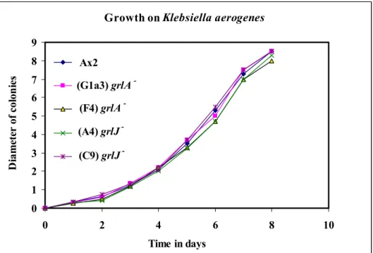
Ax2 and the respective mutant cells were plated onto SM agar plates overlaid with Klebsiella aerogenes and incubated at 21ºC for 3 to 4 days until Dictyostelium plaques appeared on the bacterial lawns. Cells were taken from the clearing zones of single plaques with sterile toothpicks and spotted onto fresh plates, and the diameter of growth was measured. The experiments were done in triplicates.
2.7.3 Aggregation analysis and development on phosphate-buffered agar plates or water agar plates
2.7.3.1 Aggregation in shaking suspension:
Vegetative cells grown axenically to a density of 3x10⁶ cells/ml were harvested and washed twice in ice-cold Soerensen buffer. The cells were again reconstituted at a density of 5x10⁷ cells/ml in Soerensen buffer and plated as monolayers on a 6 well plates (2x10⁷ cells/ml/well). At different time points during aggregation images were captured using an Olympus IX70 inverse microscope.
2.7.3.2 Development on phosphate-agar or water agar plates:
Cells at a density of 2-3x10⁶ cells/ml were washed twice with equal volumes of Soerensen phosphate buffer. 5x10⁷ cells were then resuspended in 1 ml Soerensen phosphate buffer and evenly distributed onto a phosphate-buffered agar plate (90 mm) or water agar plate (90 mm).
The plates were allowed to air dry and any excess liquid was carefully aspirated without disturbing the cell layer. The plates were then incubated at 21°C. Different stages of development were observed and the images were captured using a Stereomicroscope at the indicated time points.
2.7.4 Cytokinesis
Axenically growing cells were prepared as described in Materials and methods, 2.7.3.1 and harvested immediately after washing, fixed with methanol (Materials and methods, 2.7.14.1) and stained with DAPI, a dye that binds to DNA. Bright field images and fluorescent images were captured in parallel from different fields and the number of nuclei per cell was scored.
2.7.5 Phototaxis experiments (Wallraff and Wallraff, 1997)
Cells were harvested by centrifugation at 2000 rpm for 2 minutes and washed twice in water before placing 106 cells each on 1% water agar plates. The plates were incubated in a petridish storage containers (thermocol box with walls painted completely black from within) containing a vertical 3 mm wide perforation along the length of the container and were incubated in constant subdued light for 48 hours at 22°C. The slime trails left behind the migrating slugs were blotted onto nitrocellulose membranes and stained with 0.1% amido black in 25% isopropanol and 10% acetic acid (Staining solution) for 10 minutes, destained in 25% isopropanol and 10% acetic acid and washed with water and air dried.
2.7.6 Viability of osmotically shocked cells (Rivero et al., 1996)
Axenically grown cells (Ax2 and grlA- and grlJ-) were harvested and washed twice each with Soerensen phosphate buffer and kept shaking (160 rpm) at 21oC at a density of 3x10⁷ cells/ml.
After one hour 0.4 M sorbitol was added and shaking continued for 2 hours. The cells were then diluted such that 100 µl corresponding to 200 cells were plated onto a SM agar plate along with K. aerogenes and incubated at 21oC. The colonies appearing after 3-4 days were counted and calculated for percentage of surviving cells.