AUS DEM LEHRSTUHL
FÜR KLINISCHE CHEMIE, LABORATORIUMSMEDIZIN UND TRANSFUSIONSMEDIZIN
DIREKTOR: PROF. DR. MED. GERD SCHMITZ DER FAKULTÄT FÜR MEDIZIN
DER UNIVERSITÄT REGENSBURG
PLATELET-DERIVED EXTRACELLULAR VESICLES IN PLATELETPHERESIS CONCENTRATES
AS A QUALITY CONTROL APPROACH
Dissertation
zur Erlangung des Doktorgrades der Medizin
der
Fakultät für Medizin der Universität Regensburg
vorgelegt von Anne Black (geb. Dzikus)
2013
Dekan: Prof. Dr. Dr. Torsten E. Reichert
1. Berichterstatter: Prof. Dr. Gerd Schmitz
2. Berichterstatter: Prof. Dr. Ernst Holler Tag der mündlichen Prüfung: 16.09.2014
Table of contents
Index of figures ... ix
Index of tables ... xii
List of abbreviations and acronyms ... xiv
I Introduction ... 1
1.1 Platelet-derived extracellular vesicles in platelet storage lesion and platelet senescence 1 1.2 Plateletpheresis concentrate quality 7 1.3 Extracellular vesicles in general 10 1.3.1. Definition ... 10
1.3.2. Function of extracellular vesicles ... 14
1.3.3. Microvesicle formation ... 17
1.3.4. Formation of exosomes ... 20
1.4 Platelet-derived extracellular vesicles 21 1.4.1. Background and clinical potential ... 21
1.4.2. Distribution of PL-EVs ... 25
1.4.3. Characteristics of PL-EVs ... 26
1.4.4. Specific features of PL-EV formation ... 27
1.4.5. Clearance of PL-EVs ... 28
1.4.6. Detection techniques ... 29
II Aims of the study ... 36
III Materials ... 38
3.1 Technical equipment 38 3.2 Consumables 39 3.3 Reagents 40 3.4 Analytical Software 42 3.5 Preparation of solutions 43 3.5.1 TRAP-6 ... 43
3.5.2 Tyrode buffer ... 43
3.6 Plateletpheresis concentrates 43
3.7 Red blood cell units 44
3.8 Blood samples of healthy donors 44
IV Methods ... 45
4.1 Background 45 4.2 Flow chart of plateletpheresis and sampling for quality control 46 4.3 Sampling of platelet concentrates 47 4.4 Sample preparation of platelet concentrates for measurement of platelet-derived and red blood cell-derived extracellular vesicles 50 4.4.1 Dilution of PC samples ... 50
4.4.2 Platelet- and EV-containing plasma preparation ... 50
4.4.3 Preparative isolation of EVs from plateletpheresis concentrates ... 50
4.5 Sample preparation of red blood cell units for measurement of platelet- derived and red blood cell-derived extracellular vesicles 50 4.6 Flow cytometry 51 4.6.1 Principles and parameters ... 51
4.6.2 Platelet function test ... 52
4.6.3 Platelet-derived extracellular vesicles ... 54
4.6.3.1 Navios™ ... 54
4.6.3.2 FACS Canto™ II ... 57
4.6.3.3 Apogee ... 59
4.7 Nanoparticle Tracking Analysis 60 4.7.1 Background ... 60
4.7.2 Practical details ... 61
4.8 Aggregometry 61 4.8.1 Impedance aggregometry ... 61
4.8.2 Light transmission aggregometry ... 61
4.9 Total blood count and platelet count analysis 62
4.10 Photometry 62
4.11 Nephelometry 62
4.12 Enzym-Linked Immunosorbent Assay (ELISA) 63
4.13 Chemiluminescence immunoassay (CLIA) 63
4.14 Coagulometry 63
4.15 Chromogenic method 63
4.16 Immunturbidometry 64
4.17 Potentiometric method 64
4.18 Statistical analysis 64
V Results ... 65
5.1 Results of methodological issues 65
5.1.1 Validation of PL-EV measurement with standard flow
cytometry ... 65 5.1.2 Validation of nanoparticle tracking analysis ... 67 5.1.3 Analysis of plasma-vesicle-measurement by different flow
cytometers and nanoparticle tracking system ... 69 5.1.4 PL-EV-boost after TRAP-6 activation ... 72 5.1.5 Rebound phenomenon, dependent on type of sampling and
shear-stress ... 73 5.1.6 Aggregometry ... 76
5.2 Donor and apheresis specific values 77
5.3 Platelet concentrate specific values 84
5.3.1 Characteristics of PCs over time ... 84 5.3.2 Platelet-derived extracellular vesicles and storage ... 85 5.3.3 Comparability of sample types (PC samples and tube samples) ... 87 5.3.4 Effects of irradiation (Differences between non-irradiated
versus irradiated PCs) ... 89 5.3.5 Alteration of PCs during storage ... 94 5.3.6 Linear regression analysis of platelet CD62P expression and
PL-EV levels (sd-FCM) ... 100 5.3.7 Correlation between standard PL-EV analysis (sd-FCM) and the
new systems (hs-FCM and NTA) ... 102
5.3.8 Correlation between platelets CD62P and PL-EV analyses by
hs-FCM and NTA ... 105
5.4 Extracellular vesicle composition in plateletpheresis concentrates and red blood cell units 110 VI Discussion ... 115
VII Summary ... 134
VIII Zusammenfassung ... 135
IX Publications ... 137
X Curriculum vitae ... 138
XI Acknowledgements ... 140
XII Eidesstattliche Erklärung ... 141
XIII References ... 142
Index of figures
Figure I-1: Summary of changes related to platelet storage lesion (PSL) and formation of
platelet-derived extracellular vesicles (PL-EVs) ... 2
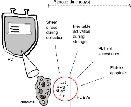
Figure I-2: Potential mechanisms behind the presence of platelet-derived extracellular vesicles (PL-EVs) in plateletpheresis concentrates (PCs) ... 4
Figure I-3: Scheme of eukaryotic cell forming microvesicles, exosomes and apoptotic bodies . 12 Figure I-4: Summary of the main functions of extracellular vesicles (EVs) ... 14
Figure I-5: The formation of microvesicles from the cell membrane ... 18
Figure II-1: Investigation of quality in plateletpheresis concentrates in vitro involves the analysis of separate concentrate components, i.e. plasma, platelets and platelet-derived extracellular vesicles (PL-EVs) ... 37
Figure IV-1: Workflow diagram for manufacturing and sampling of platelet concentrates (PC) on several days after apheresis ... 46
Figure IV-2: Flowchart of sample count (n=) from plateletpheresis concentrates ... 46
Figure IV-3: Disconnection of the tube of plateletpheresis set after donation ... 48
Figure IV-4: Flexible tube filled with plateletpheresis product after disconnection from the apheresis set from a double donation ... 48
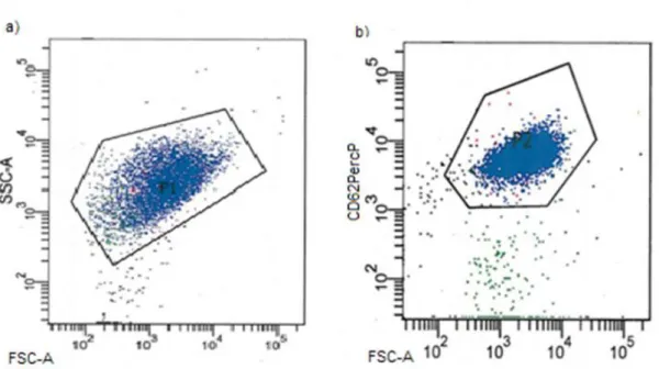
Figure IV-5: Gating strategy by flow cytometry with Canto™ II ... 53
Figure IV-6: Absolute values of platelet events and Mean Fluorescence Intensity by analytical FACSDiva Software ... 54
Figure IV-7: Changes of externalization of CD62P after activation with TRAP-6 by flow cytometry ... 54
Figure IV-8: Calibration of instrument settings for EV-analysis with Navios™ ... 55
Figure IV-9: Gating strategy for EV analysis by flow cytometry with Navios™ ... 56
Figure IV-10: Scatter plots of events for platelets and platelet-derived EVs by flow cytometry with Navios™ ... 56
Figure IV-11: Calibration of the instrument settings for EV-analysis with Canto™ II ... 57
Figure IV-12: Gating strategies for EV-analysis by flow cytometry with Canto™ II ... 58
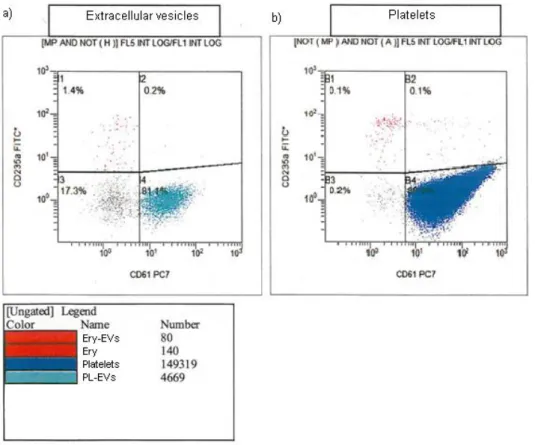
Figure IV-13: Platelet-derived EV analysis by flow cytometry with Canto™ II ... 58
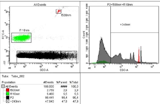
Figure IV-14: Histogram of calibration of the instrument settings of Apogee A-50 Micro ... 59
Figure V-1: Validation of PL-EV measurement by standard flow cytometry ... 66
Figure V-2: Linearity of the method for nanoparticle tracking analysis (NTA) with beads ... 67
Figure V-3: Linearity and accuracy of plasma vesicle size analysis (A) and recovery and accuracy of plasma vesicle concentration (B) by NTA ... 68
Figure V-4: Cumulative graph represents proof of size recovery for NTA with monodispers bead solutions ... 68
Figure V-5: Accuracy of size measurements with mixed up polystyrene bead solutions ... 69
Figure V-6: Line chart of plasma vesicle size of plateletpheresis concentrates ... 71
Figure V-7: Inter-assay coefficients of variability ... 71
Figure V-8: Release of PL-EVs in response to TRAP-6 in fresh PCs on day 0 ... 72
Figure V-9: Rebound effect of platelet vesiculation and platelet function over time ... 74
Figure V-10: Differences in rebound phenomenon depending on shear stress ... 75
Figure V-11: Effects of PL-EVs on platelet aggregation ... 76
Figure V-12: Effect of single-needle versus double-needle apheresis system on CD62P ... 81
Figure V-13: Effect of apheresis instruments on PL-EV levels in PCs ... 81
Figure V-14: Box plots for platelet count values of two sample types of plateletpheresis quality control analysis ... 87
Figure V-15: Box plots for CD62P expression on platelets from PCs of two sample types ... 88
Figure V-16: Box plots of the interquartile range (IQR) for PL-EV raw data of different sample types ... 89
Figure V-17: Box plots illustrating the effect of irradiation on platelet count ... 90
Figure V-18: Box plots illustrating the effect of irradiation on PL-EV levels measured by sd-FCM ... 90
Figure V-19: Effect of irradiation on the PL-EV/ PLT ratio in fresh PCs measured by hs-FCM .. 91
Figure V-20: Effect of irradiation on PL-EV and PLT count in PCs measured by hs-FCM ... 92
Figure V-21: Effect of irradiation on the vesicle concentration in PCs measured by NTA ... 93
Figure V-22: Effect of irradiation on size distribution of vesicles in PCs measured by NTA ... 93
Figure V-23: Box plots demonstrating changes of platelet count over time ... 94
Figure V-24: Box plots of different CD62P externalization on platelets of PCs over time (A) and after irradiation (B) ... 95
Figure V-25: Box plots of shedded PL-EVs in PCs over time measured by sd-FCM ... 96
Figure V-26: Box plots of differences in PL-EVs depending on irradiation measured by sd-FCM
... 96
Figure V-27: Scatter plot of the correlation between PL-EVs of fresh versus expired PCs measured by sd-FCM ... 97
Figure V-28: Box plots of PL-EV ratio of irradiated PCs over time measured by hs-FCM... 98
Figure V-29: Box plots of plasma vesicles concentration of irradiated PCs over time measured by NTA ... 98
Figure V-30: Box plots of IPF of fresh and stored PCs measured by automated hematology analyzer ... 99
Figure V-31: Box plots of pH values of PCs over time and the effects of irradiation ... 99
Figure V-32: Scatter plot distribution of PL-EVs in correlation/ linear regression to CD62P externalization ... 101
Figure V-33: Comparison of sd-FCM and hs-FCM PL-EV analysis on day 0 ... 102
Figure V-34: PL-EV analysis by hs-FCM (A) and sd-FCM (B) ... 103
Figure V-35: Evaluation of plasma vesicles from PCs by NTA ... 104
Figure V-36: Correlation between CD62P expression and the PL-EV-ratio by hs-FCM... 106
Figure V-37: Correlation between CD62P (after stimulation with TRAP-6) and PL-EV subpopulations in fresh non-irradiated PCs measured by hs-FCM ... 108
Figure V-38: Correlation between CD62P (after TRAP-6 stimulation) and PL-EV subpopulations in fresh irradiated PCs measured by hs-FCM ... 109
Figure V-39: Correlation between CD62P (after stimulation with TRAP-6) and PL-EV subpopulations in senescent irradiated PCs measured by hs-FCM ... 109
Figure V-40: Composition of cell-derived vesicles in PCs ... 111
Figure V-41: Pie chart of the cell-derived EV percentage in red blood cell units ... 112
Figure V-42: Platelet-derived EVs and erythrocyte-derived EVs from red blood cell units ... 113
Figure V-43: Plasma and cell-derived vesicles from red blood cell units measured by NTA ... 114
Index of tables
Table I-1: Potential mechanisms of PL-EV formation during collection and storage ... 5
Table I-2: In vitro tests of platelet concentrate quality monitoring ... 8
Table I-3: Parameters and criteria of quality control in manufacturing of plateletpheresis concentrates in Germany... 10
Table I-4: Overview of three subgroups of extracellular vesicles ... 13
Table I-5: Platelet-derived vesicles in several diseases ... 22
Table I-6: This overview shows common and variable glycoprotein (GP) receptors or activation markers of platelet-derived and megakaryocyte-derived extracellular vesicles (EVs) ... 27
Table I-7: Pros and cons of detection techniques for measurement of extracellular vesicles .... 30
Table I-8: Designation of PL-EV concentrations from plasma samples of healthy individuals by flow cytometry as reported in the literature... 32
Table I-9: PL-EV concentration of platelet concentrate samples measured by flow cytometry as reported in literature ... 33
Table I-10: Bead and vesicle characteristics for detection by light scatter ... 34
Table IV-1: Summary of count (n=) from plateletpheresis concentrate (PC) samples and tube samples for analysis on day 0 and day 5 (red numbers belong to the irradiated samples) 49 Table IV-2: Preparation of red blood cell units for analysis of extracellular vesicles by ultracentrifugation and density gradient centrifugation ... 51
Table IV-3: Scheme of immunostaining for analyzing PLT function test ... 53
Table V-1: Vesicle quantification of plateletpheresis concentrates (PCs) by NTA ... 70
Table V-2: Characteristics of donors and of the apheresis process ... 77
Table V-3: Blood count parameter of platelet donors... 78
Table V-4: Serum characteristics of donors ... 78
Table V-5: Relevant correlation of donor-specific, clinical or laboratory parameters to PC quality ... 80
Table V-6: Relevant donor specific parameters in correlation to one another ... 83
Table V-7: Characteristic laboratory analysis of PCs over time ... 84
Table V-8: PL-EV and plasma vesicle values in PCs analyzed by flow cytometry and NTA ... 86
Table V-9: PL-EV analysis by sd-FCM versus hs-FCM or NTA ... 105
Table V-10: Correlation between platelet CD62P expression after TRAP-6 determined by hs- FCM and by NTA ... 107 Table VI-1: Comparison of PL-EV concentration from PCs on day 0 or day 1 reported in recent
literature ... 119
List of abbreviations and acronyms
ADP adenosine diphosphate
AFM atomic force microscopy
Apo A-I apolipoprotein A-I Apo B100 apolipoprotein B100 Apo B48 apolipoprotein B48 Apo C-I apolipoprotein C-I Apo E apolipoprotein E
Apo J apolipoprotein J (clusterin) approx. approximately
ATIII antithrombin III
ATP adenosine triphosphate
BMI body mass index
BMP bis-(monoacyl)-glycerophosphate
C3c complement factor C3
cc correlation coefficient CD cluster of differentiation CD40L CD40 ligand, or CD154 CD41 glycoprotein (GP) IIb CD42b glycoprotein (GP) Ib CD61 glycoprotein (GP) IIIa
CD62P p-selectin
CD63 a member of the tetraspanin superfamily of integralmembrane proteins CD95L CD95 ligand, FASL, galectin 9
CHOL cholesterol
CR1 complement receptor 1
CRACM1 calcium release-activated calcium channel protein/ modulator 1
CRP C-reactive protein
CV coefficient of variation C1-INH C1 esterase inhibitor
DC dendritic cells
DIGE differential in-gel electrophoresis
DNA deoxyribonucleic acid Dt diffusion coefficient
e.g. exempli gratia
EGF epidermal growth factor
EGFR epidermal growth factor receptor
EM electron microscopy
ER endoplasmic reticulum
ERK1/2 extracellular signal-regulated kinase 1/2
ESCRT endosomal sorting complex required for transport esRNA exosomal shuttle RNA
et al. et aliae
et seqq. et sequentes EV extracellular vesicle
EXs exosomes
FCM flow cytometry
FITC fluorescein isothiocyanate FSC forward scatter (=FS) GLA γ-carboxyglutamic acid
GLC glucose
GP glycoprotein
GPI glycosyl-phospatidyl-inositol
GTP guanosine triphosphate
GvHD see also TA-GvHD
Gy gray
HBA1c [IFCC] hemoglobin A1c [International Federation of Clinical Chemistry]
HBA1c [NGSP] hemoglobin A1c [National Glycohemoglobin Standardization Program]
HCT hematocrit
HDL-C high density lipoprotein cholesterol
HGB hemoglobin
HPA human platelet antigen HSC hematopoietic stem cell hs-FCM high sensitivity flow cytometry
HSP heat shock protein
i.e. id est
IGF-1 insulin-like growth factor 1
IGFBP3 insulin-like growth factor binding protein 3 ILV intraluminal vesicle
IPF immature platelet fraction
ITP idiopathic thrombocytopenic purpura
KB Boltzmann´s constant
LALS large angle light scatter ≈ SSC
LAMP-1 lysosome-associated membrane glycoprotein-1 LDL-C low-density lipoprotein cholesterol
L-EV leukocyte-derived extracellular vesicle Lp(a) lipoprotein (a)
LPA lysophosphatidic acid
LPS lipopolysaccharide
MFG-E8 milk fat globule - epidermal growth factor - 8 MFI mean fluorescent intensity
MHC major histocompatibility complex
min minutes
miRNA microRNA
MK megakaryocyte
moAb monoclonal antibody
MODS multiple organ dysfunction syndrome
MP microparticle
MPV mean platelet volume
mRNA messenger RNA
MV microvesicle
MVB multivesicular body
MVE multivesicular endosomes
mW milliwatt
NTA nanoparticle tracking analysis
Orai1 see also CRACM1
P2X7 receptor purinergic receptor P2X, ligand-gated ion channel 7 PAI-1 plasminogen activator inhibitor-1
PBS PBS-Dulbecco, w/o Ca2+/Mg2+ Buffer PC platelet(pheresis) concentrate PC7 phycoerythrin cyanin 7 PDGF platelet-derived growth factor PDW platelet distribution width
PE phycoerythrin
PF3 platelet factor 3
PKC protein kinase C
PLT platelet
PL-EV platelet-derived extracellular vesicle PMP platelet microparticles
PNH paroxysmal nocturnal hemoglobinuria
PPI particles per image
PS phosphatidylserine
PSD platelet storage defect
PSGL-1 p-selectin glycoprotein ligand-1 PSL platelet storage lesion
P2X1 purinergic receptor P2X, ligand-gated ion channel 1 P2X7 purinergic receptor P2X, ligand-gated ion channel 7 Rab Ras-related in brain
Rap1 Ras-related protein 1
RBC red blood cell
RBC-EV RBC-derived vesicle
RDW-CV red cell distribution width as coefficient of variation from the mean red cell size
RDW-SD red cell distribution width as standard deviation from the mean red cell size
RES reticuloendothelial system
rh hydrodynamic radius
RI refractive index (ɳ)
RNA ribonucleic acid
ROCK Rho-associated coiled-coil-containing protein kinase RR blood pressure according to Riva-Rocci
SALS small angle light scatter ≈ FSC sd-FCM standard flow cytometry
sec seconds
SERCAs sarco/ endoplasmic reticulum Ca2+-ATPases SFLLRN ser-phen-leu-leu-arg-asn
SNARE soluble N-ethylmaleimide-sensitive-factor attachment receptor SOC store-operated calcium
SOCE store-operated calcium entry
SSC side scatter (=SS)
STIM1 stromal-interacting molecule 1
T temperature
TA-GvHD transfusion associated graft-versus-host disease TCTP translation controlled tumor protein
TF tissue factor
TFPI tissue factor pathway inhibitor TFG-β1 transforming growth factor β 1 TLR4 toll-like receptor 4
TNF-α tumor necrosis factor α
TRAP-6 thrombin receptor activating peptide 6 TRF transferrin receptor
TRIG triglyceride
TSG101 tumor susceptibility gene 101
TXA2 thromboxane A2
UC ultracentrifuge
V volt
VEGF vascular endothelial growth factor VLDL-C very low density lipoprotein cholesterol VSMC vascular smooth muscle cells
vWF von Willebrand factor
vWfr von Willebrand factor receptor
η solvent viscosity
𝜋
Pi°C degree centigrade
I Introduction
1.1 Platelet-derived extracellular vesicles in platelet storage lesion and platelet senescence
Anucleated platelets are small (0.5 - 1 µm), discoid blood cells with a lifespan up to 10 days in the human circulatory system (1, 2). They play an essential role in primary hemostasis and are filled with α-granules, dense granules and lysosomes (3). Intracellular α-granules are surrounded by membranes which contain proteins, chemokines, growth factors and immune mediators. Immune mediators constitute a large group of active molecules which includes, among the others, glycoprotein (GP) IIb-IIIa (integrin αIIbβ3), p-selectin, factor V (FV), factor IX (FIX), protein S, tissue factor (TF), plasminogen, plasminogen activator inhibitor 1 (PAI-1), fibrinogen, von Willebrand factor (vWF), thrombospondin, growth factors (epidermal, insulin-like, vascular endothelium, fibroblast, platelet-derived), complement C3, complement C4, C1 inhibitor, β1H globulin (factor H) and immunoglobulin G (IgG). Dense granules contain Ca2+, Mg2+ and K+, polyphosphate, pyrophosphate, serotonin, histamine and nucleotides (e.g. ADP and ATP). Platelet lysosomes are filled with enzymes degrading proteins (e.g. cathepsin, elastase and collagenase), carbohydrates (e.g. glucosidase, fucosidase and galactosidase) and lipids (e.g. acid phosphatase).
Additionally to granules and cytoskeletal components, platelets also contain other organelles such as mitochondria and the dense tubular system which is analogous to the endoplasmic reticulum connected to the open canalicular system and glycogen stores.
Circulating platelets undergo senescence prior to clearance; the cells are not capable of division, they are metabolically active, though. Ex-vivo resting platelets undergo platelet storage lesion and they are subjected to either activation-mediated death or to storage-induced death.
The storage “phenotype” of platelets, which translates to cell death, may also be responsible for necrotic or apoptotic changes (1), as well as for activation (2).
Platelet storage lesion (PSL) or platelet storage defect (PSD) comprise platelets which lost their typical functional characteristics. This means loss of membrane integrity, p-selectin release, shedding of surface proteins (receptors), diminished mitochondrial membrane potential, increase of the intracellular calcium level with a secondary activation of proteases (caspases), externalization of phosphatidylserine (PS) and secretion of platelet-derived extracellular vesicles (PL-EVs) (2), see Figure I-1. The presence of PSL justifiably corresponds to the reductions of in vivo survival and haemostatic activity after transfusion (4). Changes in PSL platelets are influenced by alterations during collection, processing and storage in platelet concentrates (PCs) (5).
Figure I-1: Summary of changes related to platelet storage lesion (PSL) and formation of platelet-derived extracellular vesicles (PL-EVs)
The figure demonstrates the effects of storage conditions ex vivo (left) and the modulation of platelet morphology, physiology and function (right).
Storage of platelet concentrates is performed under continuous agitation under standard blood banking conditions at 22°C ± 2°C for 5 days, including the day of apheresis. Agitation is carried out in bags with oxygen exchange in order to maintain the aerobic adenosine triphosphate (ATP) formation. Obviously, there are several factors which influence PSL such as temperature, storage duration, form and intensity of agitation, volume of suspending plasma, permeability of the membranes of a storage container and a leukodepletion technique (5). Exemplarily, storage carried out almost at physiological temperatures (37°C) improves the viability of platelets due to a decrease of a rapid ATP turnover and a lower metabolic activity of stored platelets (5, 6).
Additionally, a 16%-release of α-granules within seven days of storage was described and the release was independent from different rotators used. This leads to a loss of functional proteins, essential for the adequate coagulation response after transfusion in vivo (7). To control the effects of these factors during storage of PCs, several techniques were used and incorporated into the quality control process (explained in detail in chapter 1.2, page 7).
PL-EVs were described first in 1967 as a coagulable “platelet dust” (8). They are also connected with the term of platelet factor 3 (PF3) activity and its role in coagulation (9). Platelet vesiculation led back to the observation of a 10-fold rise in the PF3 activity during platelet storage (10). The raise in PF3 activity is accompanied by a stable platelet count. Nearly three decades ago, an active PF3 was regarded as a part of PSL in a process of platelet activation and damage (11). The presence of PL-EVs or platelet-derived microvesicles (PL-MVs) in stored PCs has been well demonstrated. Already in 1986/87, Solberg et al. recognized it and found a correlation between the presence of PL-MVs and PF3 activity (12, 13). Formation of platelet- derived vesicles particularly elevates in PCs exhibiting high pH, associated with LDH release.
In 1988, George et al. showed rotation dependency on PL-MV level over 7 days storage as a shear stress model. Inappropriate shear stress and/ or activation of platelets during storage result in elevated vesicle levels (7). During a study on supplementation of platelets with activation inhibitors that protect platelets from damage, the group of Bode demonstrated in 1991 that solely partial inhibition of vesiculation occurred (40%), resulting in a hypothesis that not only activated platelets shed MVs. However, MVs seemed to appear already at physiological concentrations in matured platelets (14). The group of Bode delineated two populations of microparticles (MPs): one population corresponding to fluorescent beads smaller than 0.5 µm and another one corresponding to fluorescent beads, larger than 0.5 µm.
Nowadays, it is well known that strongly procoagulant PL-MVs or PL-EVs released from membranes of intact platelets are present in PCs but there are many questions in platelet transfusion medicine which still remain unanswered.
Various functions of PL-MVs in PCs can be ascribed to four pathologic conditions (Figure I-2).
The first condition is related to mechanical injury and shear stress during collection and processing. It is not distinguishable to which extent PL-MV components are released from donor platelets in vivo and ”collected” in the suspended donor plasma as compared to the release via the non-physiological shear force of the apheresis systems. Rank et al. isolated annexin V positive microvesicles which reached the concentrates and the group compared the vesicle content of PCs to the MV content in the donor plasma (15). PL-MVs in PCs (93% of all MVs) differed by 39% from the PL-MV amount obtained from donor plasma (1.6 to 1), suggesting that plateletpheresis concentrates contain more PL-MVs than the donor´s pre-donation samples and they seem to be constant in PL-MV enrichment during storage over 5 days. Rank et al.
assumed that the most abundant amount of PL-MVs in PCs results from the collection process and not from the donor plasma. PL-MVs originate to a lesser extent from activated platelets (p- selectin positive PL-MVs in 4.8%) rather than from resting platelets with stable annexin V positive MVs. However, the group of Rank found increased degranulation of p-selectin in PL- MVs dependent on the storage time. Furthermore, the results indicated that CD63 positive platelet-derived exosomes (PL-EXs) are present in isolated MVs. Additionally, exosomes showed a significant increase on the fifth day of storage thus representing a minor fraction (2.6%) in comparison to the dominant larger PL-MVs (15).
Figure I-2: Potential mechanisms behind the presence of platelet-derived extracellular vesicles (PL-EVs) in plateletpheresis concentrates (PCs)
Sloand et al. observed that the application of different preparation techniques (e.g. apheresis versus whole blood-derived PCs) and different anticoagulants (e.g. ACD versus CPDA-1) result in different amounts of PL-MVs (16). These findings support the hypothesis that the abundant amount of PL-MV in PCs is dependent on the conditions of collection and not necessarily on the donor plasma at the time of apheresis. However, at this point it cannot be excluded that there are donors, who have more sensitive platelet membranes and therefore generate more PL-MVs during plateletpheresis.
In addition, activation of stored platelets due to their contact with the plastic surface of a storage container takes place in the vesiculation fraction of PCs. Reduction of the surface-to-volume ratio (S/V) in PCs resulted in a lower LDH release and it significantly correlated to the total MP count, suggesting that the cytoplasmic content of all vesicles is regulated by the vesiculation of platelets (14). With the decrease in the adhesion of the platelets to the surface of the storage container, fewer platelets become activated thus being more prone to form platelet-derived vesicles.
Since 1997, various mechanisms have already been observed which “lead to microvesiculation of platelets during storage” (17), see Table I-1. Seghatchian et al. described available methods for quantification and characterization of PL-MVs. Nowadays, these methods overlap such evaluation approaches as flow cytometric analysis of specific markers (GPIb, GPIIb/IIIa) using monoclonal antibodies (moAbs) or analysis of PS exposure by annexin V labeling. Although, since then flow cytometry has been the favored method, the comparison of standard flow
cytometers with advanced new high-resolution flow cytometers indicates more possibilities in detection of vesicles as far as their lower size and higher sensitivity are concerned.
Table I-1: Potential mechanisms of PL-EV formation during collection and storage
Mechanical disruption and excessive shear stress, e.g. passage through low gauge needles or leukocyte filters, high g force, rigorous agitation
Activation by platelet aggregation agonists, e.g. ADP, thrombin, during collection and storage
Activation and secretion of platelets caused by poor handling and prolonged exposure to low temperatures (4°C) and high pH > 7.6
Lysis of platelets caused by freezing-thawing, lyophilization and rehydration; prolonged exposure to low pH <6.2
Exposure to activated complement components, leukocyte- and platelet-derived cytokines and enzymes, e.g. calpain, histamine
(Table adjusted to (17) )
Senescence and apoptosis of platelets in PCs stored for five days could induce increased vesicle formation. With the purpose of explaining to which extent PL-MVs or PL-EXs originate from senescent or apoptotic platelets, it is essential to better define the “phenotype” of these vesicles compared to the vesicles from activated platelets. However, no evidence has been yet found to prove any principal differences between these vesicle groups. On the other hand, understanding of the process of vesicle formation during senescence and/ or apoptosis in PCs is indispensable and needs reinvestigation with the help of currently available more sophisticated methods. Though, despite of the fact that mechanisms of PSL and senescence may overlap, PSL and the apoptotic program are two distinct processes, even if some authors use the names of PSL and senescence interchangeably (1, 18).
As aforementioned, when inhibitors applied only, the reduction of activation-dependent generation of PL-EVs in stored platelets averages approximately 40%. Nevertheless, significantly lower PL-EV levels within the storage time of 9 days versus control (14) suggest that remaining PL-EVs originate from resting/ senescent platelets. “In vitro experiments demonstrate that platelet apoptosis can be induced by calcium ionophores, other platelet agonists [during] storage at room temperature under blood banking conditions” (5). It is evident that platelets undergo apoptosis at 37°C, accompanied by a loss of platelet viability and a gradual rise in caspase-3 and caspase-9 activities (19). The inhibition of platelet apoptosis by
cell-permeable caspase-inhibitors could improve the viability of platelets but this approach turned out to be ineffective in this study. Under this condition, gelsolin, a caspase-3 substrate affected in apoptosis, was cleaved as caspase-3 activity rose. This process, also observed by Thiele et al., became acknowledged as a cytosolic protein biomarker of apoptosis of platelets subjected to 9-day storage (20). Whereas 97% of cytosolic proteins remained unchanged, gelsolin and septin 2 belong to the group of 3% of proteins which undergo changes as analyzed by differential gel electrophoresis (DIGE) and mass spectrometry.
Thiele et al. also found proteins involved in the early storage lesion, e.g. the focal adhesion signaling integrin αIIbβ3 (21), which plays a role as a proteomic biomarker of platelet quality.
The investigation of changes of cytosolic proteins during storage, especially of the proteins connected to signaling pathways underlying storage lesion development (22), may at least partially correspond to activation, senescence and apoptosis of platelets and the underlying vesiculation.
PS exposure, detectable by annexin V binding and release of p-selectin provide evidence for an effect of apoptosis on platelet viability.
Although platelet mitochondria maintain their transmembrane potential (Δψ(m)) upon storage over 7 days, which does not correlate to the downstream protein markers, selected stressors (i.e. calcium ionophore stimulation) lead to higher degrees of mitochondrial depolarization (23).
The preparation techniques also affect the Δψ(m) of platelets and the percentage of platelets with depolarized Δψ(m) significantly increases in buffy-coat PCs as compared with PCs produced by apheresis (24).
In addition to the observed mitochondrial changes, the group of Albanyan showed growing values, both percentage and absolute, of the immature platelet fraction (IPF) as determined by Sysmex XE-2100 in PCs over 7-day storage (25). Simultaneously, the platelet count remained stable, whereas mean platelet volume (MPV) and platelet distribution width (PDW) exhibited a small increase. The fluorescent dye Thiazole Orange, used for detection of immature platelets with residuals of nucleic acid shows non-specific binding to basic proteins and anionic phospholipids which appear on the platelet surface during senescence. Dense granular or other platelet structures may become also unspecifically dyed because of their nucleotides or messenger RNA (mRNA) which affect the elevation of IPF levels. Thus far, no investigation has confirmed the presence of mRNA or micro RNA (miRNA) in PL-EVs. Yet, their presence is very likely to the expression of miRNA, detected in human peripheral blood vesicles (26-28) and most vesicles in circulating blood originate from platelets (or megakaryocytes, see chapter 1.4.2, page 25). It cannot be excluded that platelet-derived vesicles, which contain mRNA or miRNA, contribute to unspecific staining.
Gamma irradiation of platelet concentrates prevents transfusion-associated graft-versus-host disease (TA-GvHD) and may lead to an increase of vesiculation. Upon application of the required irradiation dose of 25 Gray (Gy), as assessed by Food and Drug Administration (FDA),
no differences were found between irradiated and non-irradiated PCs, including swirling, hypotonic shock response (HSR), the percentage of GPIb-expressing cells and pH during 7 days of storage (29). On the other hand, development of elasticity points to a slower progression after storage in the irradiated group leading to the assumption, that vesiculation increases under the condition of the loss of membrane elasticity. Gamma irradiation applied on the first day of storage causes pronounced proteome changes, responsible for specific catalytic activities and/ or protein/ nucleic acid binding capacity, involved in the platelet storage lesion in PCs (30).
1.2 Plateletpheresis concentrate quality
It is essential to carry out laboratory tests in order to guarantee consistent and uniform quality of plateletpheresis concentrates. However, there is no single laboratory test which would reflect potential hemostatic effects of platelets in vivo and there is no established benchmark which can be used to translate the measured loss of platelet function to an appropriate graduation of PC quality (17). Many applicable tests were validated for identifying PSL of platelets.
Nevertheless, there are established standards for platelet concentrate quality within quality control (QC) programs to detect problems during collection, processing or storage (5) according to the corresponding guidelines. There are currently several protocols for quality control used;
the most popular are as follows: (i) the AABB (American Association of Blood Banks) technical manual (31) for the United States, (ii) the Guide to preparation, use and quality assurance of blood components (32) for Europe and (iii) the German guidelines for production and application of blood and blood components (33).
These guidelines recommend determination of platelet count (number), concentrate volume, supernatant pH and residual leukocytes in clinical practice, although single test criteria deviate, e.g. the required pH in the United States is lower than 6.2, whereas in Germany it varies between 6.4 and 7.8. Routinely applied tests in PCs reflect only a minor part of the platelet changes during storage, also mentioned as the PSL. In addition, all these tests (see Table I-2) only determine the viability and function of platelets in vitro. Many investigators favor the measurement of the percentage recovery and in vivo survival of radiolabeled (generally with
51chromium or with 111indium), autologous platelets as a gold standard (34). This fact should be considered if altered or new storage conditions and/ or platelet substituents are evaluated (5). In healthy individuals, the mean in vivo recovery of fresh platelets from platelet rich plasma (PRP) amounts to 60-70%, whereas, after 5 days of storage, the mean recovery amounts to 45-50%.
Higher recovery rates were observed for apheresis concentrates on the 5th day.
However, an acceptable “standard for approval” by FDA does not exist for platelet recovery and a standardization of practical implementation has not yet been established as compared with the standardized techniques for vesicle measurement (see chapter 1.4.6; page 29).
Table I-2: In vitro tests of platelet concentrate quality monitoring Platelet structure Cellular content (platelet count)
Visual inspection of swirling phenomena Platelet morphology by microscopy
Platelet size distribution by automated counters (PMV, PDW) Measurement of reticulated platelets
Immature platelet fraction (IPF)
Functional tests Platelet aggregation, spontaneous and agonist-directed Hypotonic shock response (HSP)
Extent of shape change
Thrombin-stimulated ATP release
Metabolic status Supernatant pH, pO2, pO2, HCO3
Glucose consumption Lactate production
Platelet activation p-selectin (CD62P) surface expression Soluble p-selectin release into supernatant Platelet factor 4 and β-thromboglobulin
Annexin V binding to estimate the reorganization of phosphatidylserine (PS) exposure
Lactate dehydrogenase (LDH) release into supernatant Platelet microparticle formation
Possibility of transfusion reactions (TR)
Bacteriological growth Cytokines
Activated complement components, i.e. C3a
Otherwise Residual leukocytes
Residual red blood cells (RBC) (Table adjusted to (17, 23 ))
Two in vitro tests should be explained as examples for all tests listed in Table I-2. The simplest evaluations include visual examination of PCs before they are applied to patients. Upon exposing of the storage bag to any light source, a “swirling” phenomenon occurs due to the fact that resting platelets are discoid and refract light within the moved storage bag. If pH of stored PCs drops below the value of 6.2 or platelets underlie activation, the former discoid/ spherical shape of platelets undergoes morphological changes with formation of pseudopodia. Shape change causes the disappearance of the “swirling” phenomenon caused by the fact that shape- changed platelets are not able to refract light (5). The weaker “swirling” phenomenon correlates to a lower platelet increment in vivo which is measurable after transfusion (5, 35). This happens because pH lower than 6.3 translates to diminished in vivo survival after transfusion (5). The subsequent discoid/ spherical conversion and microvesiculation result in decrease of mean platelet volume (MPV) (17). MPV is a common parameter, which is measured by automated hematology blood counters and could be easily implemented in the running procedures of clinical practice to maintain the visualized “swirling” phenomenon, which considerably depends on individuals and status of training.
The analysis of platelet activation during manufacturing by flow cytometry and evaluation of the p-selectin (CD62P) exposure at the platelet surface (36) is another useful measurement of PC- QC and it is applied as the most common measurement of platelet activation in PCs (5). In contrast to the physiological behavior of activated platelets, ex vivo p-selectin expression on the platelet surface upon activation by cold or pH below 6.2 is not reversible (37). In whole blood and platelet-rich-plasma (PRP), p-selectin expression amounted to 2-10% versus 20-30% in PCs (37). The percentage of CD62P positive platelets significantly increased within five days of storage, whereas the mean fluorescent intensity (MFI) values of labeled lysosome-associated membrane proteins 1 and 2 (LAMP-1, LAMP-2) decreased. Simultaneously, the reduction of CD62P positive platelets, LAMP-1 and LAMP-2 on the sixths day of storage was observed (36).
The relative microparticle count and p-selectin expression on platelets surface were also determined and an increased MV number over the storage time in correlation to free and membrane-bound p-selectin was demonstrated. In general, the activation of platelets entails the occurrence of adverse effects during storage, but it is hardly evident that the rate of activation is associated with post-transfusion recovery (5). Several studies reported that p-selectin expression does not affect platelet clearance (38, 39) and may not reflect platelet survival and function in vivo (5).
To highlight the importance of the applied tests for plateletpheresis concentrate quality monitoring, all subsequent parameters, which are beyond standard quality control (see Table I-3) for testing platelet viability at the university hospital of Regensburg, are described later (chapter 4.6; page 51). More parameters are necessary to guarantee platelet integrity in concentrates and to promote the improvement of cell component based quality control at the cellular, biochemical and molecular level.
Table I-3: Parameters and criteria of quality control in manufacturing of plateletpheresis concentrates in Germany
Test parameters Test criteria Time of testing
Volume according to specifications after preparation
Platelet count per unit >2 x10¹¹/ unit after preparation and at the end of expiry Platelet count per mL according to approval after preparation
Residual erythrocytes <1 x10⁶/ unit after preparation
Residual leukocytes <3 x10⁹/ unit after preparation
pH value 6.4 - 7.8 at the end of expiry
Visual valuation undamaged PC container and swirling
at the end of expiry and before application
Sterility sterile at the end of expiry
The same criteria are applicable to irradiated plateletpheresis concentrates with 30 Gy.
Table has been adjusted to “Richtlinien zur Gewinnung von Blut und Blutbestandteilen und zur Anwendung von Blutprodukten (Hämotherapie)“ (33).
1.3 Extracellular vesicles in general
1.3.1. Definition
Under physiological or pathological conditions, all human body fluids, such as plasma (40), serum (41), breast milk (42), amniotic fluid (43) or urine (44) in vivo, as well as media from cultured cells in vitro (45) contain cell-derived vesicles. Cell membrane vesicles, which undergo secretion, constitute spherical or tubular structures surrounded by a phospholipid bilayer.
Vesicular membranes expose receptors that are constructed as transmembrane proteins with cytosolic components, i.e. soluble hydrophilic or hydrophobic molecules. Because the membrane orientation of cell-derived vesicles is the same as that of the donor cells, vesicles can be considered as non-nucleated miniature versions of a cell (46). Both, eukaryotic and prokaryotic cells have the ability to release such vesicles. The characteristics of vesicles are determined by size, density, visibility under the microscope, sedimentation, lipid composition, main protein markers and intracellular membranes or organelles of subcellular origin (46, 47).
Cell-derived vesicles possess different physiochemical features. Therefore, there is currently no consensus about a uniform nomenclature for all cell-derived vesicles. While scientists still debate the nomenclature, the term “extracellular vesicles (EVs)” has been proposed as a general term covering the complete diversity of vesicles. In relation to the current nomenclature, the International Society for Extracellular Vesicles (ISEV) meanders through multiple definitions of vesicles and offers suggestions for scientists and authors. In addition, a general term of EVs used for subgroups should base on logical arguments referring to physiochemical and functional characteristics and should clearly define the methods for isolation and measurement (48).
There are two subgroups of EVs, i.e. exosomes and microvesicles. Apoptotic vesicles form a third, separate class (46, 47). There are more types of vesicles described, such as endosomes, ectosomes, membrane particles or exosome-like vesicles (46). This division is based on different characteristics explained above. The association of the latter four types with platelet- derived vesicles is currently unknown and therefore not discussed in this study. The size of the largest subgroup of vesicles, i.e. apoptotic bodies, released during apoptotic cell death, ranges from 500 nm to 5 µm (46); (see Figure I-3 and Table I-4).
Microvesicles of a size ranging between 100 nm to 1000 nm (46) are released from the plasma membrane during cell stress. The smallest vesicles, also known as exosomes, vary in their size from 50 to 100 nm (46). They are formed from intracellular multivesicular bodies (MVB;
sometimes also used as multivesicular endosomes, MVE) in a process described as the “classic pathway” (47) or produced in a direct pathway (46, 49).
The characteristics of these three subtypes (apoptotic vesicles, microvesicles and exosomes) are summarized in Table I-4. The efficient purification of vesicle subgroups is limited (50) and depends on isolation strategies. As a means to interpret the analytical results, it is necessary to assume a small contamination of vesicles neighbored to the refined vesicle subgroup. Rather, the vesicle subgroups represent a “continuum of vesicle types with overlapping properties [which are almost entirely] present in body fluids” (47). It should be considered that “ex vivo purified vesicles” can be “artificially generated during manual dissociation of tissues” (46) and that exosomes could represent intracellular multivesicular bodies, released from ruptured vesicles and non-secreted exosomes.
Figure I-3: Scheme of eukaryotic cell forming microvesicles, exosomes and apoptotic bodies Cells release small exosomes (EXs) via a classic pathway involving formation of intraluminal vesicles (ILV) within multivesicular endosomes (MVE) and via a direct pathway from the plasma membrane. Larger, secreted microvesicles (MVs) can be formed at the plasma membrane by direct budding. Apoptotic bodies including cellular compartments appear during apoptosis.
Table I-4: Overview of three subgroups of extracellular vesicles
Exosomes Microvesicles Apoptotic vesicles
Size of diameter [nm]
50 - 100 100 - 1000or
20 - 1000
50 - 500or 1000 - 5000
Density in sucrose [g/ ml]
1.13 - 1.19 unknown 1.16 - 1.28
Sedimentation 100,000 g 10,000 g 1,200 g, 10,000 g or
100,000 g
Morphology by EM*
cup-shaped, homogenous
irregular shape, electron-dense
heterogeneous
Cellular origin most cell types most cell types all cell types
Cell origin multivesicular bodies plasma membrane plasma membrane and endoplasmic reticulum during cell death
Composition cholesterol,
sphingomyelin, ceramide, lipid rafts,
glycerophospholipids, phophatidylserine- exposure, Rab proteins, annexin, flotillin, intergins and tetraspanins (CD63, CD9, CD81, CD82), Alix, TSG101, HSP (hsc70, hsc90), mRNA, microRNA
phosphatidylserine- exposure, intergrins, selectins and CD40 ligand; insufficiently known
histone, DNA, organelles
References (27, 46, 47, 51-54) (46, 47, 54, 55) (46, 47, 56-59)
*EM: electron microscopy; (adapted to (46, 49))
1.3.2. Function of extracellular vesicles
The EV subgroups are released by cells during different stages of cell life. Thereby, EVs reflect dissimilar formation processes and are related to different mechanisms of shedding of intracellular and/ or plasma membrane components into the intercellular environment. Released EVs are recognized by acceptor cells. It is obvious, that both, donor and acceptor cells benefit from the transposition of these cell fragments, where donor cells release vesicles, whereas target cells, which are brought into contact with the released vesicles, are acceptors. There is an increasing interest in functions of EVs and the most relevant properties of EVs shall be elucidated.
In general, cell-derived vesicles may act as “multi-purpose” carriers (45), (see Figure I-4) to express the main functions of extracellular vesicles discussed in this work.
Figure I-4: Summary of the main functions of extracellular vesicles (EVs)
As indicated, the process of intercellular signaling via vesicles as potential communicators and, thus, understanding the function of cell-derived vesicles is an important current topic. The transport of receptors or cytokines was characterized and found to be similar to the transfer of the TF receptor and p-selectin glycoprotein ligand-1 (PSGL-1) between leukocyte-derived vesicles and activated platelets. As a result of this transfer, proteolytic activity of the TF-VIIa complex increases after fusion of leukocyte-derived microvesicles and platelets (60).
The transfer of ligands, e.g. CD95L (FASL), located on tumor cell-derived vesicles induced apoptosis of T cells (61) which is a proof for immunosuppressive effects of the intercellular communication.
The mechanism of promotion of humoral immune response via CD40L (CD154) of platelet- derived vesicles followed by B cell activation and production of immunoglobulin G can be considered as sufficiently understood (62). The communication aspect involves an
“orchestrating function” of immune response including antigen presentation and the transfer of cellular components.
A further possibility of the communication between released vesicles and cells is defined through the exchange of genetic information. This was shown in many tumor cell lines, e.g.
transfer of functional mRNA and miRNA in vesicles from glioblastoma cells (63), or mRNA in colorectal cancer cell line-derived vesicles (64) and in physiological cells, e.g. miRNA from T cells (65) or miRNA from dendritic cells (DCs) (66). The exchange of functional miRNA among DCs results in the repression of target mRNA in acceptor cells. This may be an important key mechanism in saving the function of non-coding regulatory miRNA of cellular origin outside the cell, in which it was produced, because, as in the case of vesicles, miRNA is protected from degradation carried out by RNases (67). The ability of vesicles to facilitate the transfer of functional genetic information was pictured in exosomes of mouse and human mast cell lines and primary bone marrow-derived mouse mast cells. Exosomes contain functional mRNA of approximately 1300 genes undetected in the cytoplasm of the donor cells and miRNA called
"exosomal shuttle RNA" (esRNA) (27). Induced expression of target proteins was also observed. Lässer´s work presented detectable levels of RNA in vesicles of several body fluids, e.g. human saliva, plasma or milk (68). These findings indicate a flow of information through RNA transfer.
“Cellular waste management” or “protection against intra- and extracellular stress”(45) using the extracorporeal release of harmful substances in vesicles may be a survival strategy of cells.
External stress, e.g. complement C5b-9 complex incubated with platelets, causes the release of vesicles enriched with this complex to protect the platelets from complement-induced lysis (69).
Reticulocyte-derived transferrin receptor (TFR) or tumor cell-derived shedded vesicles, which contain chemotherapeutics, are active in the process of survival or drug resistance mechanisms (70) and were described as extracellular stress protectors.
Active caspase 3 is the main executioner of apoptosis. Caspase 3 is abundant in vesicles of different cell types and it is assumed that it acts as a protection against internal stress and nemesis of cells (71, 72).
Vesicles also play a role in cell adhesion, vascular integrity and repair which results from their expression of adhesion molecules and from the increasingly significant relation of the surface to cell compartment. “All membrane vesicles of any cellular origin express adhesion molecules on their surface, which could favor their capture by recipient cells” (46).
Platelet-derived vesicles were observed how they bind to fibrinogen-, fibronectin-, and collagen- coated surfaces and minimally to vitronectin and von Willebrand factor (73). In a rabbit model, an increased vesicle binding to injured endothelium in vivo, but not to uninjured surfaces, was observed and elevated binding of platelets to immobilized vesicles was shown. This behavior was interpreted as a mechanism by which vesicles promote thrombus formation.
Another example of exosome-mediated adhesion was described for integrins on B cell-derived exosomes, which interact with extracellular matrix components to mediate adhesion to collagen-I, fibronectin and surface adhesion molecules of tumor necrosis factor (TNF)-α- activated fibroblasts (74). Similar to the mechanism of protection against intracellular stress through caspase 3 activity, an increased release of endothelial-derived vesicles antagonize apoptotic blebs. Tumor susceptibility gene 101 and translation controlled tumor protein (TCTP), which are abundant in vesicles, trigger an extracellular signal-regulated kinase 1/2 (ERK1/2)- dependent antiapoptotic phenotype in vascular smooth muscle cells (VSMCs) (75). In contrast to the antiapoptotic effect, the vesicles from endotoxin-stimulated monocytes trigger caspase 1- dependent apoptosis of vascular smooth muscle cells.
These observations suggest functions beyond the communication between cells and vesicles, and underline the role of vesicles in damage control of vasculature and a potential role of vesicles in coagulation. Section 1.4.1 summarizes historical information on potential coagulatory features under clinical aspects of vesicles.
Two topics referring to coagulation should be discussed. The first topic should concentrate on vesicles as specific or unspecific procoagulants and the second topic should refer to vesicles with anticoagulant activity. In the context of microvesicle-related coagulability, the results of analyses of the material of patients with Castaman syndrome are of interest (76, 77). In four unrelated patients with life-long bleeding tendency, a reduced formation of platelet microvesicles was found. These patients were without von Willebrand factor defect and with no evidence of any other platelet function abnormalities accompanied by normal prothrombin consumption. Castaman syndrome, which is at variance with Scott syndrome (see in 1.3.3), was described as disorder in vesicle formation and the EV generation was closely connected with coagulability under physiological conditions. In addition, the procoagulant activity of EVs results from the molecular composition of the vesicle membrane including presentation of anionic phospholipids, e.g. phosphatidylserine, in the outer monolayer (78). This corresponds to positively charged γ-carboxyglutamic acid (GLA) domains in proteins of the clotting system, such as factors VII, IX, X and II (prothrombin) (79). There is no consensus concerning PS exposure on exosomes and microvesicles. However, PS-positive EVs (51, 80) and PS-negative EVs (54, 81, 82) were reported in context of several diseases.
Tissue factor, as a cofactor of the initiation of the extrinsic coagulation cascade by factor VIIa, is also present as a procoagulant on the surface of EVs. The TF/FVIIa complex, however, is counter-regulated by tissue factor pathway inhibitor (TFPI), suggesting that not all TF/FVIIa
complexes on EVs remain active (or become activated) (79, 83, 84). Similarly to PS exposing EVs, the procoagulant activity of TF bearing EVs remains also controversial. The pro- or anti- coagulant activity of exosomes and, most likely, of microvesicles depend on physiologic or pathologic conditions (85). It was reported that TF exposing exosomes and microparticles from human saliva, promotes a shortened clotting time in human pericardial wound blood (85).
Microvesicles triggered coagulation in a TF-depending pathway. The same working group showed that microvesicles from platelets, erythrocytes and granulocytes in blood samples also support coagulation via a TF-independent way. Antibodies against TF or FVII were ineffective to stop coagulation, suggesting PS-exposure as an initial step of coagulation.
Further, an anticoagulant function was concluded. An inverse correlation of annexin V-positive microparticles and thrombin generation to levels of thrombin-antithrombin-complexes was found. Thrombin in a lower concentration activated protein C and led to an anticoagulant effect of microvesicles (86). PS-positive vesicles present in blood of healthy individuals predominantly originate from platelets, megakaryocytes, and the surface of platelet-derived vesicles showed 50- to 100-fold higher procoagulant activity than that of a single platelet (87). Monocytes, however, are likely to be the major donor cells of TF-positive vesicles (79). Moreover, in several diseases, the procoagulant activity of vesicles, e.g. tumor-derived vesicles exposing TF, was found associated with an increased risk of venous thromboembolism (VTE) (79, 88). An increased incidence of thrombotic events was observed in patients with paroxysmal nocturnal hemoglobinuria (PNH) with ascending concentrations of microparticles (89). In contrast to these procoagulant activities, the results of TF-exposing microparticles in patients with multiple organ dysfunction syndrome (MODS) and sepsis indicate an inverse effect. Lower numbers of vesicles in these patients could explain this effect (82). Altogether, PS- and TF-bearing EVs play an undisputed role in hemostasis and thrombosis, but the contribution of exosomes or microvesicles is still unclear.
1.3.3. Microvesicle formation
Lipid translocases, ions and their plasma membrane channels, as well as such processes as cytoskeletal reorganization, signal transduction, lipid translocase-independent and apoptotic mechanisms are all involved in vesicle formation (78). The role of translocases in vesicle formation and shedding is coupled to the loss of asymmetric distribution of lipids in the outer and inner plasma membrane (90). Exposure of PS towards the outer cell membrane by an ATP- dependent protein “floppase” (91) is an initial event in shedding of membrane vesicles (92). A normal bilayer membrane consists of positively charged polar phospholipids, such as phosphatidylcholine and sphingomyelin in the outer leaflet, and negatively charged anionic phospholipids in the inner leaflet, e.g. aminophospholipids, phophatidylserine (PS) or phosphatidylethanolamine (PE) (93). Aminophospholipid translocases with “flippase” activity direct PS and PE back to the inner leaflet of the cell membrane, whereas lipid scramblase is responsible for a bi-directional transfer of phospholipids and thus maintaining membrane
integrity (91). Scramblase is also the base of a rapid PS exposure towards the cell surface as a part of the physiologic hemostasis in coagulation response (94, 95).
As far as the lipid transport mechanisms are concerned, stimulation-dependent increase in cytosolic Ca2+ provokes a disruption of membrane lipid distribution by activation of scramblases and floppases, whereas flippase activity is inhibited (96). Scott Syndrome is a bleeding disorder characterized by a deficiency in floppase activity, impaired shedding of vesicles and decreased PS expression on vascular cells (97, 98). Sorting of membrane compartments into the vesicle bleb may be regulated by lipid rafts of the cell membrane (99). The activation of the Ca2+ - dependent protease calpain after intracellular influx of calcium ions induces a release of microvesicles from platelets (78) and also promotes proteolysis of the cytoskeleton, which subsequently induces the formation of vesicles (92). The entry of calcium seems to be a common mechanism in platelet activation, while in the resting state of platelets a steady calcium [Ca2+]i is maintained by the endoplasmic reticulum (ER) as a calcium store.
Figure I-5: The formation of microvesicles from the cell membrane
Upon stimulation of resting cells, the cytosolic Ca2+ level increases, the activation of floppases and scramblases and inhibition of flippases result in the externalization of phophatidylserine on the outer leaflet of the cell membrane. Lipid and protein rafts may control the sorting of membrane compartments, which are excluded from the vesicle blebs (figure from (100)).
There are two mechanisms which regulate cytosolic calcium [Ca2+]i in platelets. The first mechanism is based on the calcium release from the cytosol and on the influx of Ca2+ through the cell membrane mediated by store-operated calcium entry (SOCE). The second mechanism involves the ligand-gated ion channel P2X1 (purinergic receptor P2X1) (101). In monocytes, P2X7 receptor (purinergic receptor P2X, ligand-gated ion channel 7) can be activated by extracellular ATP (102).
The calcium entry via SOCE mechanism may act as a modulator of PS exposure and of a refilling of the ER calcium stores (103, 104). To maintain thousand fold lower concentration of [Ca2+]i than in the extracellular space or in the intracellular Ca2+ pools, Ca2+-ATPases translocate Ca2+ across the cell membrane using the energy of ATP hydrolysis. In platelets, different isoforms of sarco/ endoplasmic reticulum Ca2+-ATPases (SERCAs) have been found.
They become activated through the intracellular Ca2+ influx and pump Ca2+ back into the intracellular Ca2+ pools (105). Other key proteins acting as ion channels are involved in the store-operated calcium entry (SOCE). As store-operated calcium (SOC) channels, the stromal interaction molecule 1 (STIM1) and further in the system of tubular denses the Orai1 (also known as CRACM1 = calcium release-activated calcium channel protein/ modulator 1) were detected (106). Both, the STIM1/Orai1 pathway and the thrombin receptor pathway (54) operate in platelets on Ca2+ influx and PS externalization, together with SOCE-independent and cell- type specific Ca2+ signaling, as well as other ions such as K+, Cl-, H+, and Na+ (78).
Destabilization of the actin cytoskeleton in platelets is stimulated by the αIIbβ3 signaling pathway and by calpain activation accompanied by high concentrations of Ca2+ (107).
Caspases, as dominant regulators of the apoptosis cascade, are involved in the reorganization of the cytoskeleton (78). Proteases, caspases and calpain dominate the proteolysis of filamin-1, talin and myosin (108). Recent studies suggest that Rho-associated coiled-coil-containing protein kinase (ROCK) II, activated by caspase-2 in endothelial cells, is linked to the cytoskeletal reorganization, release of microvesicles (109, 110) and may be related to inflammation.
Signal transduction pathways regulate formation and secretion of microvesicles from activated cells. Examples are provided in a comprehensive review of Morel who demonstrated that platelet-derived microvesicle formation is associated with phosphorylation of platelet proteins by calmodulin, myosin light chain kinase, and other calmodulin-regulated effectors. This microvesicle formation process was found to be similar to the pathway of activation-induced protein tyrosine dephosphorylation (78). Results from a study on endothelial cells showed reduced microvesicle formation induced by TNF-α through inhibition of p38 mitogen-activated protein kinase (111). In fact, the release of microvesicles is regulated by stimuli which activate cell specific receptors, e.g. Toll-like receptor 4 (TLR4), the receptor for gram-negative bacterial lipopolysaccharide (LPS) on DCs (112) or on platelets (113), which may influence vesicle formation.
