• Keine Ergebnisse gefunden

AUS DEM LEHRSTUHL FÜR ORTHOPÄDIE DIREKTOR PROF. DR. MED. DR. H.C. J. GRIFKA DER FAKULTÄT FÜR MEDIZIN DER UNIVERSITÄT REGENSBURG

N/A
N/A
Protected

Academic year: 2021

Aktie "AUS DEM LEHRSTUHL FÜR ORTHOPÄDIE DIREKTOR PROF. DR. MED. DR. H.C. J. GRIFKA DER FAKULTÄT FÜR MEDIZIN DER UNIVERSITÄT REGENSBURG"

Copied!
102
0
0

Wird geladen.... (Jetzt Volltext ansehen)

Volltext

(1)

AUS DEM LEHRSTUHL FÜR ORTHOPÄDIE

DIREKTOR PROF. DR. MED. DR. H.C. J. GRIFKA DER FAKULTÄT FÜR MEDIZIN

DER UNIVERSITÄT REGENSBURG

KINEMATIC AND RADIOLOGICAL CHANGES OF THE PATELLA DUE TO THE IMPLANTATION OF A NAVIGATED LIGAMENT-BALANCED TOTAL KNEE

ARTHROPLASTY IN VIVO

Inaugural – Dissertation zur Erlangung des Doktorgrads

der Medizin

der

Fakultät für Medizin der Universität Regensburg

vorgelegt von

Stephanie Diwald

(2)
(3)

AUS DEM LEHRSTUHL FÜR ORTHOPÄDIE

DIREKTOR PROF. DR. MED. DR. H.C. J. GRIFKA DER FAKULTÄT FÜR MEDIZIN

DER UNIVERSITÄT REGENSBURG

KINEMATIC AND RADIOLOGICAL CHANGES OF THE PATELLA DUE TO THE IMPLANTATION OF A NAVIGATED LIGAMENT-BALANCED TOTAL KNEE

ARTHROPLASTY IN VIVO

Inaugural – Dissertation zur Erlangung des Doktorgrads

der Medizin

der

Fakultät für Medizin der Universität Regensburg

vorgelegt von

Stephanie Diwald

(4)

Dekan: Prof. Dr. Dr. Torsten E. Reichert

1. Berichterstatter: Prof. Dr. med. Dr. h.c. Joachim Grifka

2. Berichterstatter: Prof. Dr. med. Martin Fleck

(5)

Die Graphiken table 4 und figure 42 und die zugehörigen Ergebnisse wurden bereits

veröffentlicht unter dem Titel: The influence of component alignment on patellar kinematics in total knee arthroplasty:

An in vivo study using a navigation system

DOI: 10.3109/17453674.2015.1005907

Armin Keshmiri a* , Günther Maderbacher a* , Clemens Baier a* , Ernst Sendtner a* , Jens Schaumburger a* , Florian Zeman b* , Joachim Grifka a* & Hans R Springorum a*

 Received: 17 May 2014

 Accepted: 24 Nov 2014

 Published online: 13 Jan 2015

Copyright: © Nordic Orthopaedic Federation

(6)

Index

1. Abstract ... 6

1.1 Abstract (English) ... 6

1.2 Abstract (German)………..6

2. Introduction ... 9

2.1 Anatomy of the knee joint ... 9

2.2 Osteoarthritis of the knee joint ... 11

2.2.1 Definition ... 11

2.2.2 Therapeutical options ... 12

2.3 The history of total knee arthroplasty... 13

2.4 Surgical techniques in total knee arthroplasty ... 14

2.4.1 The traditional measured resection technique ... 14

2.4.2 CT free navigation ... 15

2.5 Problems of conventional measured resection technique ... 15

2.5.1 Mechanical leg axis ... 15

2.5.2 Tibial and femoral angle of the prosthesis... 16

2.5.3 Tibial slope ... 17

2.5.4 Femoral rotation ... 17

2.5.5 Tibial rotation ... 18

2.6 Patellar tracking ... 19

2.7 Patellar maltracking and postoperative anterior knee pain ... 21

2.8 Important parametres ... 22

2.8.1 Q-angle (Quadriceps angle) ... 22

2.8.2 Modified Insall-Salvati-Index ... 24

2.8.3 Joint line ... 24

(7)

3. Materials and methods ... 28

3.1 Background of the study... 28

3.2 Patients ... 29

3.3 Surgical procedure using patellar navigation ... 30

3.4 Examinations ... 34

3.4.1 Radiological assessment ... 34

3.4.1.1 Leg axis ... 34

3.4.1.2 Q-angle ... 35

3.4.1.3 Modified Insall-Salvati-Index ... 35

3.4.1.4 Patellar shift and tilt ... 36

3.4.1.5 Joint line ... 36

3.4.1.6 Femoral rotation by means of navigation ... 37

3.4.2 Clinical assessment ... 38

3.4.3 Statistical evaluation………...38

3.4.4 Complications……….38

4. Results ... 39

4.1 Radiological assessment ... 39

4.1.1 Leg axis... 39

4.1.2 Q-angle ... 41

4.1.3 Modified Insall-Salvati-Index ... 43

4.1.4 Patellar shift ... 44

4.1.5 Patellar tilt... 46

4.1.6 Joint line ... 48

4.1.7 Femoral rotation by means of navigation ... 50

4.1.8 Overview: Patellar tracking using navigation …..………..…51

4.2 Clinical assessment ... 53

4.2.1 The Knee Society Score (KSS) ... 53

4.2.2 Western Ontario and McMaster Universities Osteoarthritis Index (WOMAC).54 4.2.3 Feller/Patellar Score ... 55

4.2.4 HAKEMP-90 ... 56

4.2.5 Complications……….56

(8)

5. Discussion ... 57

5.1 Radiological assessment ... 58

5.1.1 Effects of the postoperative leg axis on the longevity of the implant ... 58

5.1.2 Radiological errors of evaluation ... 59

5.1.3 Q-angle ... 59

5.1.4 Modified Insall-Salvati-Index ... 60

5.1.5 Patellar shift and tilt ... 60

5.1.6 Patellar resurfacing……….61

5.1.7 Joint line ... 62

5.1.8 Femoral rotation ... 63

5.1.9 Effects of component alignment………...63

5.1.10 Patellar tracking………64

5.2 Clinical assessment ... 65

5.2.1 Knee Society Score ... 65

5.2.2 WOMAC Score ... 66

5.2.3 Feller/Patellar Score ... 66

5.2.4 HAKEMP-90 ... 67

5.2.5 Patients' satisfaction after total knee arthroplasty... 67

5.2.6 Limitations of the study ... 68

5.2.7 Conclusion ... 68

6. Literature ... 69

7. Index of figures and tables ... 74

8. Appendix………..77

9. Acknowledgements ... 96

10. Curriculum vitae ... 97

(9)

1. Abstract

1.1 Abstract (English) Background and purpose:

Due to increased life expectancy the demand of medical care, especially concerning joint arthroplasty, is growing. Hence, the number of Total Knee Arthroplasty (TKA) rose significantly. To ensure the longevity of implants, it is important to bear in mind one of the major complications after TKA, the patellofemoral pain syndrome (PFPS). {Springorum 2012

#4}{Borelli 2011 #74}

The purpose of this study was to examine the changes in patellar tracking after total knee arthroplasty using the ligament-balanced navigated technique and radiological evaluation.

Patients and Methods:

In this prospective study patellar tracking was measured pre- and postoperatively in 40 patients after ligament-balanced TKA using computer navigation. Furthermore, radiological parameters as mechanical leg axis, Q-angle, modified Insall-Salvati-Index, mediolateral shift and tilt of the patella and the joint line were recorded. Clinical results were assessed by 4 different questionnaires (Knee Society Score (KSS), Western Ontario and McMaster Universities Arthritis Index (WOMAC), Feller Score, HAKEMP-90).

Results:

After implantation of the TKA the mechanical axis of the leg, the Q-angle, the modified Insall-Salvati-Index and the deviation of the joint line were within the reference range according to current literature. Because of the implant there were significant changes in mediolateral shift and tilt of the patella in the dynamic measurement by means of navigation.

In the static measurement using X-ray the mediolateral shift had not changed significantly, the patellar tilt, the height of the joint line and the modified Insall-Salvati-Index had changed significantly. The questionnaires KSS, WOMAC and Feller Score showed a significant improvement.

Conclusion:

After TKA there are differences in patellar kinematics compared to the preoperative arthritic

knee, which the orthopaedic surgeon should be aware of. By means of a CT free navigation

system the changes can be shown intraoperatively.

(10)

1.2 Abstract (German) Hintergrund und Zielsetzung:

Aufgrund der gestiegenen Lebenserwartung nehmen auch die Ansprüche an die medizinische Versorgung zu, vor allem in Bezug auf die Gelenkendoprothetik. Folglich stieg die Anzahl an Knie-Totalendoprothesen (TKA) bedeutend. Um eine lange Standzeit der Prothesen zu gewährleisten, ist es wichtig, einer der Hauptkomplikationen nach dem Eingriff vorzubeugen, nämlich dem patellofemoralen Schmerzsyndrom (PFPS). {Springorum 2012 #4}{Borelli 2011 #74}

Ziel dieser Studie war es, die Unterschiede im Lauf der Patella zu untersuchen, nachdem eine Knie-Totalendoprothese mithilfe einer navigierten bandspannungsadaptierten Methode implantiert wurde und unter Zuhilfenahme der Auswertung von Röntgenbildern.

Patienten und Methoden:

In dieser prospektiven Studie wurde der Lauf der Patella prä- und postoperativ bei 40 Patienten gemessen, die eine bandspannungsadaptierte Knieendoprothese mithilfe der Computernavigation erhielten. Zusätzlich wurden die radiologischen Parameter Beinachse, Q-Winkel, modifizierter Insall-Salvati-Index, mediolaterale Verschiebung und Verkippung der Patella und die Abweichung der Gelenklinie miteinbezogen. Die klinischen Ergebnisse wurden mittels 4 verschiedener Fragebögen erfasst (Knee Society Score (KSS), Western Ontario and McMaster Universities Arthritis Index (WOMAC), Feller Score, HAKEMP-90).

Ergebnisse:

Nach der Implantation der Prothese waren die mechanische Beinachse, der Q-Winkel, der

Modifizierte Insall-Salvati-Index und die Abweichung der Gelenklinie innerhalb der

Referenzbereiche in Übereinstimmung mit der gängigen Literatur. Durch die

Prothesenimplantation änderten sich signifikant die mediolaterale Verschiebung und

Verkippung der Patella in der dynamischen Messung mittels Navigation. In der statischen

Messung im Röntgen zeigte sich die mediolaterale Verschiebung nicht signifikant verändert,

die Verkippung der Patella, die Höhe der Gelenklinie und der modifizierte Insall-Salvati-

(11)

Diskussion:

Nach Prothesenimplantation zeigen sich im Gegensatz zum präoperativen arthrotischen Knie Unterschiede in der Kinematik der Patella, worüber sich der Chirurg im Klaren sein sollte.

Mithilfe des CT-freien Navigationssystems kann die Veränderung intraoperativ dargestellt

werden.

(12)

2. Introduction

2.1 Anatomy of the knee joint

The knee is the largest articulation of the body where thigh and lower leg show an opposite movement. It consists of three parts:

- Femoropatellar articulation - Medial tibiofemoral articulation - Lateral tibiofemoral articulation

The fibula forms an independent articulation with the tibia, the tibiofibular articulation.

Hence the following movements are possible:

- Flexion and extension with a simultaneous forward glide during flexion and backward glide during extension.

Furthermore, there is a slight external rotation of about 10°

between the lower leg and the thigh at the end of the extension.

- Internal and external rotation of about 40-50° in flexion with slackened capsule and ligaments.

Stability is necessarily ensured by static and dynamic strengths because of the missing bony guidance:

- The static strength is provided by the capsule, ligaments and the curvature of the femoral condyles.

The menisci have the effect of an

absorber and load balancer which

gives stability against translation

(13)

- The dynamic strengths are defined by the muscles running around the knee joint:

M. quadriceps femoris (extension); M. gastrocnemicus (flexion); M. biceps femoris (flexion and external rotation); M. sartorius, M. gracilis, M. semitendinosus, M.

semimembranosus, and M. popliteus (flexion and internal rotation).

The static and dynamic strengths are equally placed around the central axis of the knee.

The femoropatellar compartment receives strong forces under flexion which might lead to overstrain. Complications in this articulation are one of the most common problems after total knee arthroplasty (TKA). {Krämer 2007 #3}{Niethard 2005 #2}{Kainz 2012 #30}

Figure 2: Femoropatellar articulation {Schulte 2007 #44}

The problem for patellar kinematics is how unstable the patella gets when the knee is slightly flexed. At the beginning of the flexion the patella is only guided by the ligaments, a centralisation of the patella starts at 30° of flexion. When there is a deeper flexion, the patella is guided by the bones towards the trochlea, since it becomes more stable. {Springorum 2011

#19}

‘The proximal tibiofibular joint is a plane type synovial joint’ between the lateral tibial

condyle and the caput fibulae. It is independent from the articulatio genus. Its most important

function is to dissipate torsional stresses which act on the ankle and on the lateral tibial

bending moments. The distal tibiofibular articulation is a syndesmosis at the distal lower leg

and at this point only mentioned for the sake of completeness. {Schulte 2007 #44}{Ogden

1974 #103}

(14)

2.2 Osteoarthritis of the knee joint 2.2.1 Definition

Gonarthrosis is one of the most frequent degenerative diseases within the field of orthopaedics. For about 5% of the elderly are affected, it plays an important sociomedical role.

Gonarthrosis is a progredient degeneration of the knee joint with cartilage destruction (chondromalacia), secondary bone lesions, osteophytes and capsule shrinkage. This leads to minimised and painful movement and osteoarthritis. {Niethard 2005 #2}{Wülker 2005 #31}

Osteoarthritis of the knee joint is divided into primary and secondary causes. Primary osteoarthritis is caused by poor genetic disposition of the articular cartilage tissue. The particular reasons therefore are unknown.

Secondary osteoarthritis can be caused by overload (e.g.

deformations like varus/valgus

malalignment, genu

recurvatum/antecurvatum), traumata, inflammatory joint diseases (e.g. rheumatoid arthritis), metabolic diseases (e.g. gout) or by endocrinary diseases (e.g. hypothyroidism).

{Krämer 2007 #3}{Niethard 2005 #2}

Figure 3: X-ray of varus (a) and valgus (b) gonarthrosis

{Niethard 2005 #2}

(15)

2.2.2 Therapeutical options

There are various options, surgical and nonsurgical ones, depending on the severity of the disease. Both surgical and nonsurgical methods are used singularly as well as combined.

As it is helpful to keep the joints moving, physiotherapy (e.g. swimming, bicycling, balneotherapy) is a proven method. Furthermore it strengthens the muscles. {Niethard 2005

#2}

To reduce pain, it is important to reduce the loading factors, e.g. by losing weight, using a walking cane, or orthopaedic insoles.

Corticosteroids, nonsteroidal antiphlogistic drugs, monolytics or hyaluronic acid derivatives are used to relief pain. This medication abates pain rapidly because of its easy application.

At an advanced stage of gonarthrosis, several surgical options are available depending on the symptoms and the severity of the disease. During open debridements, all mechanical disturbing factors like inflamed synovia, osteophytes and floating/lose joint bodies are removed. In the last decade, minimal invasive arthroscopic techniques have become more and more important and thus have overcome open surgical techniques.

In cases of progressive gonarthrosis knee arthroplasty can be an encouring option.

Endoprosthetic replacements can be performed as partial or as total knee replacement . Both are routine operations nowadays and they provide highly satisfactory results with significant pain decrease and a noticeable increase of function and quality of life in patients.

The satisfaction rate in TKA patients, however, is only between 75-89 %.

Reasons could be postoperative complications such as infections (38%), instabilities (27%), insufficient integration (13%) or others.

Up to 50% - depending on literature - of patients complain about patellofemoral pain (PFP) after the operation, what could be considered as one of the most frequent postoperative complications. {Niethard 2005 #2}{Springorum 2012 #4}{Boyd 1993 #5}{Ip 2004

#6}{Callaghan 2004 #21}{Bonnin 2011 #22}{Bourne 2010 #23}{Kohn 2000 #32}{Wülker

2005 #31}

(16)

2.3 The history of total knee arthroplasty

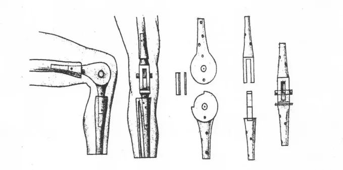
In 1890, Themistokles Gluck, a surgeon from Berlin, published for the very first time his experiences of artificial bones and joints.

The treated patients suffered from osseous tuberculosis. In a first operation, he replaced the complete knee joint. In a second operation, he implanted a hinge implant made of ivory.

Gluck also analysed the fixation using wedges (e.g. of pergament, cork or felt) and an early type of cement (decalcined cattle bone or a mixture of cholophony and gypsum). The problem Gluck was confronted with was increased septic loosening. Although he could remedy the effect of tuberculosis by implanting the prosthesis he could not reduce the infection, which caused the loosening. That dissuaded Gluck and his colleagues from the idea of an endoprosthesis. {Stuhler 1996 #33}{Rabenseifner 1998 #34}

Figure 4: First models of prosthesis of Themistokles Gluck {Rabenseifner 1998 #34}

Only in 1947, the brothers Judet developed a hinge prosthesis made of acrylic after having seen positive results in hip arthroplasty. But acrylic could not resist the high shear forces in the knee permanently.

Ten years later, in 1957, Wallidus used successfully another material called ‘vitallium’ which

(17)

Another ten years later, at the end of the 1960s, the sliding implant lead to the breakthrough.

With the salvage of the ligaments additional stability could be achieved. {Charnley, Love

#36}

In 1970, in New York and Boston the first ‘Duocondylar-Knees’ were implanted, resurfacing the medial and the lateral compartment. 4 years later the ‘Duopatellar Knee’ was developed based on these experiences. It had the advantage of a femoropatellar slide bearing and salvage of the posterior cruciate ligament.

In the 1980s, the ‘press fit condylar (PFC) – knee system’ was tested for the first time and further developed afterwards. It offers a variety of options including fixed or rotating platform, cruciate-retaining or cruciate-substituting implantation technique, or optional patellar resurfacing. The implant was designed to mirror the biomechanics of a natural knee.

The latest innovation in knee arthroplasty are customized surgical cutting guides or customized implants. {Kim YH, Kook, Kim JS #37}{Schunck, Jerosch #38}{Scott, Thornhill

#39} {Orthopedic company celebrates 25 years #40}

2.4 Surgical techniques in total knee arthroplasty 2.4.1 The traditional measured resection technique

Before surgery a preoperative radiological examination, including an a/p view standing under load, an X-ray of the knee joint in two planes and a ‘patella sunrise view’, is essential. With these data it is possible to calculate the correction of the axis and the approximately expected size of the implants using X-ray templates.

After opening the knee joint with a medial parapatellar approach, a stylet is positioned intercondylarly in the femur with intramedullar direction. Depending on the preoperative X- rays the distal cutting block is usually aligned 5° to 7° towards the anatomical axis of the femur.

After that the size of the femoral component is determined. The rotation of the femoral component is marked in orientation of the posterior condylar line, – standardised with 3° of external rotation.

The ‘tibial resection line’ can also be determined using a stylet or using an extramedullar

system. The latter is applied to measure the tibial axis via the malleoli and the tibial

tuberosity. It is also possible to adjust the dorsal descent of the tibial plateau with the aid of a

setscrew at the end of the tibial alignment.

(18)

To measure the particular size of the inlay, either a sensor or a spacer of different height is placed in the rectangular gap where the resection was made. {Jerosch 2000 #42}{Kohn 2000

#32}{Rabenseifner 2013 #43}

2.4.2 CT free navigation

To get the information the CT based method is used basically, that means imaging of the femoral head, the knee joint and the upper ankle joint. But the difference is that the references are located in femur and tibia simultaneously. This allows the operating surgeon to assess the stress ration of the ligament in flexion and extension and to assess the range of motion. This way, it is possible to adapt the rotational adjustment of the femoral component depending on the stress ration of the ligament.

Because of the inherent imperfection of the human being, precision in surgery will always be defective and the introduction of navigation systems in surgery is therefore largely extended.

{Bäthis 2003 #45}

In this study, patella navigation during the operation was used to assess the aforementioned patellar tracking.

2.5 Problems of conventional measured resection technique 2.5.1 Mechanical leg axis

The mechanical axis of the leg is the axis between the centre of the femoral head (caput femoris) and the middle of the talus. The mechanical centre line is called Mikulicz line. In the physiological leg it runs directly through the middle of the knee joint so that the strengths are distributed optimally there.

If there is a deformity of the axis, this line can be located laterally (= genu valgum) or medially (=genu varum). {Wülker 2005 #31}

(19)

To plan a TKA standardised X-ray diagnosis is necessary. Thereby the central beam of the X- ray should be positioned in the middle of the patella and there should be no rotation in the hip joint above.

During the operation the prosthesis should be positioned perpendicularly to the mechanical leg axis. A deviation leads to a significant additional load of the particular compartment where the prosthesis moved to. This leads to higher wear up to less retention. {Bäthis 2004

#46} {Ritter 1994 #55} {Jeffery 1991 #56}

2.5.2 Tibial and femoral angle of the prosthesis

An exact femoral portal of entry and an intramedullary orientation of the stylet are necessary to achieve optimal

implant alignment. It is important to avoid deviations because of a wrong positioned access to the medullary cavity.

A tilting of the stylet can cause a deviation of the axis up to 8°. The

operating surgeon should Figure 6: Exactly positioned stylet also be aware of anatomical {Munzinger 2004 #49}

deformities, e.g. a fractured femur,

that can cause deviation, too. {Jeffery 1991 #56}{Novotny 2001 #47}{Reed 1997 #48}

If the surgeon decided to make an intramedullary alignment, this problem could also occur at

the tibia. An extramedullar stylet can cause errors because it can be unstable or the exact

orientation towards the landmarks can be error-prone. {Sambatakakis 1991 #57}

(20)

2.5.3 Tibial slope

The tibial plateau descends dorsally. Its alignment is adapted depending on the tibial alignment using a traction screw. Thereby, a physiological slope of about 3°-5° should be achieved. If this angle is too large, the femoral component slides dorsally during flexion causing luxation or simply higher contact pressure ending in higher wear of the posterior compartment. {Wasielewski 1994 #58}

Assuming that the tibial plateau descends ventrally, it can cause an anterior tilt which can limit the flexion. Furthermore, if the flexion gap is too narrow ‘posterior wedging’ can be a result. This can also be a reason for uneven movement resulting in early loosening. {Dorr 1986 #53}

2.5.4 Femoral rotation

The femoral rotation has a significant effect regarding localised contact pressure and patellar maltracking. There are two methods to achieve exact alignment: the anterior and the posterior referenced method which offer 0°, 3°, 5°, or 7° of femoral rotation. In both methods the cutting block is adjusted to the anterior femoral cortex (anterior referenced alignment) or the posterior condyles (posterior referenced alignment). The rotation should be adjusted parallel to the epicondylar axis and rectangular to the Whiteside Line.

If there is suspected malrotation in a TKA, a CT scan can deliver objective results. Thus, the surgical ‘epicondylar line’ is usually a means to

measuring. The CT scan is made in the supine position with extended knee. In correct rotation the surgical

‘epicondylar line’ and the posterior ‘condylar line’

should be parallel as shown in figure 7. {Springorum 2012 #4}

(21)

2.5.5 Tibial rotation

The tibial component can be aligned statically or dynamically. The static alignment is defined via the bony landmarks of the tibia. Therefore, the axis of the tibial tray is positioned towards the medial third of the tuberositas tibiae. Another possible bony landmark is the posterior edge of the tibia.

In the dynamic alignment, the operating surgeon moves the knee using an inlay on trial basis

and marks the tibial rotation. This method is only possible after slight soft tissue release

before. {Hofmann 2003 #54} {Laskin 2003 #50} {Romero 2003 #51}

(22)

2.6 Patellar tracking

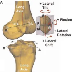
Patella tracking is based on the parameters shift, tilt, flexion and rotation:

Figure 8: Coordinate systems used to quantify the clinical motions of the patella {Nha 2008 #41}

The patellar shift is defined as medial or lateral movement of the centre of the patella along the transepicondylar axis (TEA). During physiological movement the patella shifts slightly medially until the early flexion of 30°, beyond it shifts consistently laterally

up to 90°. At flexion angles more than Figure 9: ML shift of the patella 90° there is only a slight lateral shift. {Nha 2008 #41}

Patellar tilt is defined as the rotation of

the patella around its long axis. In a

physiologic knee the patella tilts

(23)

Patellar flexion is defined as the rotation of the patella around the transepicondylar axis. Patellar flexion increases with knee flexion, but at a lower rate. E.g. at a maximum knee flexion of 135°, the patellar flexion is about 95°.

Figure 11: Patellar flexion as a function of knee flexion

{Nha 2008 #41}

Patellar rotation is defined as the rotation of the patella around it’s a/p axis. The patella rotates laterally with increasing flexion up to 120°. Beyond this angle the rotation reduces.

Figure 12: Patellar rotation with respect to knee flexion

{Nha 2008 #41}

In conclusion, patellar movement is relatively small in relation to the femoral movement

during in vivo weightbearing knee flexion. {Nha 2008 #41}

(24)

2.7 Patellar maltracking and postoperative anterior knee pain

Patellar maltracking means that the patella does not slide properly during knee flexion. So, it does not move smoothly within the trochlear groove of the distal femur.

Hence, it is a purpose to balance the patella during surgery to achieve physiological kinematics. This prevents increased retropatellar pressure postoperatively. Pressure on the femoropatellar articulation is made responsible inter alia for patellofemoral pain (PFP) after TKA. {Rosenstein 2007 #7} .

Altogether, there are various causes for PFP but increasing and localised contact pressure and patellar maltracking are primarily responsible. {Kessler 2008 #8}{Malo 2003 #9}{Catani 2013 #28}

Various causes are discussed:

For example Miller et al., Belvedere et al., Luring et al., Kessler et al., Steinbrück et al.

described rotational malalignment of the tibial and/or femoral component as reason for patellar maltracking.

Berger et al., Kienapfel et al., Farrokhi et al. mentioned femoral internal rotation to change patellofemoral kinematics. Internal rotation of the femoral component is a possible reason for APFP.

Berger et al. detected correlations between combined internal rotation of the femoral and tibial component and the severity of postoperative patellofemoral complications.

Olcott and Scott, Miller et al., and Luring et al. noticed that the rotation of the femoral component according to the transepicondylar line showed better results than the orientation according to Whiteside’s line or 3° external relative to the posterior condyles because the rotation of the femoral component to the transepicondylar line achieves a better restoration of the physiological patellar kinematics.

Only 2 studies, the studies of Anglin et al., and Belvedere et al. measured patellar kinematics before and after TKA in vivo using the accuracy of computer navigation.

To our best knowledge, there exists no data on patellar kinematics intraoperatively so far.

{Anglin 2015 #61}{Belvedere 2007 #62}{Berger 1998 #63}{Farrokhi 2011 #64}{Kessler

(25)

2.8 Important parametres

2.8.1 Q-angle (Quadriceps angle)

The Q-angle provides information about the stability of the TKA.

The anatomical shape of the patella and the anatomical groove of the femoral trochlea are important factors for patellar stability. It should have a ‘V-shape with wide angle and circular base’ to ensure normal patellar tracking.

Therefore Walker et al. analysed the ‘forces between the trochlea and the patella for various Q-angles and flexion angles’ whether the patella tilts or rotates in the femoral trochlea as causes for wear and deformation.

The results revealed ‘that the patella is fully stable and without tilting at all flexion angles for a Q-angle of 14° or less’. Q-angles more than 14° lead to severe misalignment and instability. So, in surgical practice the Q- angles are usually reduced in TKA. {Walker

2001 #15} Figure 13: The three main types of patellae component used in total knees

{Walker 2001 #15}

It is shown up in Fig. 14 that even at full extension of the knee QF (force in quadriceps group) and TF (force in patella tendon) are not collinear which leads to shear stresses that are applied to the patella. Only if the patella is treated as frictionless pulley, which it is not in vivo, then QF = TF. {Walker 2001

#15}{Matthews 1977 #16}

Figure 14: Definition of the Q-angle

{O´Donoghue 1976 #14}

(26)

In Fig. 15, the connection between the flexion angle of the knee and the shear/compression ratio (= S/C ratio) is portrayed as result of the analysis by Walker et al. It can be seen that an increasing Q-angle causes an increasing S/C ratio and in this way a decreasing stability of the patella.

Figure 15: As the Q-angle increases, the S/C ratio increases, and the patella becomes less stable. As the flexion angle increases, the patella becomes more stable {Walker 2001 #15}

In Fig. 16, the concrete figures of instability are described. For Q = 14° there is only a small unstable region up to 10° of flexion; for decreasing Q-angles full stability in all flexion angles can be gathered from this table. {Walker 2001 #15}

Figure 16: The unstable region is

shown up to 20° for a Q-angle of 16° and up to 10° for a Q-angle of 14°. Below 14° there is complete

stability

{Walker 2001 #15}

(27)

2.8.2 Modified Insall-Salvati-Index

The distance between the tibial tuberosity and the most distal patellofemoral point of the articulation is divided through the length of the patellar articular surface. The standard values are in between 0.74 and 1.50.

A numeric value more than 1.50 defines a patella alta. A numeric value less than 0.74 defines a patella baja. There are several reasons for a patella baja. It can be a patella baja because of (scarred) shortening of the ligamentum patellae. Otherwise it can be a pseudo patella baja caused by a proximalisation of the joint line. {Pfitzner 2009 #18}{Springorum 2011

#19}{Kazemi 2011 #20}{Shabshin 2004 #26}

2.8.3 Joint line

It is important and challenging to reproduce the natural joint line in TKA. The functional results of a replaced knee can be significantly compromised if the joint line is far from its natural position. This can cause an increase in patellofemoral forces and leads to subluxation, dislocation or fracture. Apart from the patella the postoperative flexion can also be affected or there can be an increase in varus-valgus laxity.

A review of several clinical studies shows a range of proposed joint line displacement

thresholds from 3-13 mm. At values over these thresholds motion, pain, above all

patellofemoral pain and function of the knee are affected negatively. {Mason 2006 #52}

(28)

2.9 Clinical assessment

For the clinical assessment the following standardised questionnaires were used:

1. The KSS (Knee Society Score) consists of the following parts: the Knee Score and the Function Score. In each score the maximum is 100 points. The Knee Score is based on pain, range of movement, flexion contractures or extension lag and alignment. The Function Score quantifies activities of daily living, such as walking distance, stair climbing and the use of walking aids. Thus, a total score of 200 points indicates full function associated with no pain.

{Liow 2000 #24}

2. The WOMAC Index (Western Ontario and McMaster Universities Osteoarthritis Index) is a generalised scoring system for osteoarthritis. It contains the 3 parts pain, stiffness and functionality. The total score ranges from 0 to 96 points. Here, the lower the score, the better the result. To receive a better comparison pre- and postoperatively, the results are expressed as a percentage. {Ackerman 2009 #10}

3. The PATELLAR SCORE / FELLER SCORE is a scoring system which evaluates especially the patellar function by enquiring about anterior knee pain, the strength of the quadriceps, the ability of getting up from a chair as well as climbing stairs. A total score of 30 points indicates full function and no pain regarding the patella. {Feller 1996 #69}

4. The HAKEMP-90 (Handlungskontrolle nach Erfolg, Misserfolg und prospektiv) was used as personality questionnaire. It evaluates the action control after success and failure. In this study the two sections HOM (Handlungsorientierung nach Misserfolg = self-calming) and HOP (= prospektive Handlungsorientierung = self-motivation) in stressful situations are assessed {Kuhl 1990 #25}. In each section it is possible to achieve 12 points at maximum, so in this study 24 points in total signify full action control.

A third score, the score HOT/LOT is possible to include, but it is influenced by many other variables, so it can be excluded according to the author. {Kuhl 2012 #12}

This third score was not used in this study.

(29)

2.10 Patella navigation

Because of the former discussed errors of the conventional technique the navigation technique got a standard method in daily clinical practice. Although navigation demands longer operation time – depending on literature between 8 and 15 minutes it results in much better alignment of the implants. Controlled ligament-balancing achieves better stability in flexion and extension. {Jerosch 2007 #70}{Bäthis 2003 #45}

Bäthis et al. investigated the difference between CT based and CT free navigation. The CT based version allows a precise preoperative planning but this is associated with additional costs and administrative effort, as well as exposure to radiation. The CT free navigation provides ‘intraoperative visualisation of the leg axis, ligament balancing and joint kinematics’.

Errors in cutting are avoided in both modules similarly. {Bäthis 2003 #45}

(30)

2.11 Objective of the study

Patellar tracking is defined as patellar shift, patellar tilt, patellar flexion and patellar rotation.

Every change of the above mentioned physiological conditions leads to patellar maltracking.

But also extrapatellar parametres like the mechanical axis of the leg, the Q-angle, the modified Insall-Salvati-Index, the femoral rotation, or the joint line have an influence on patellar tracking and are important to be carried out precisely during the operation.

The purpose of this study was to examine the changes in patellar tracking before and after a

ligament-balanced navigated TKA. Patellar kinematics after TKA are not well understood so

far.

(31)

3. Materials and methods

3.1 Background of the study

We included 40 patients (19 women, 21 men) designated for TKA between November 2012 and October 2013.

As mentioned before, anterior knee pain is one of the most common postoperative complications after TKA, and according to various studies, for example of Miller et al.

(2001b), Kienapfel et al. (2003), and Heinert et al. (2011), patellar maltracking is one of the underlying reasons. {Miller 2001 #66}{Kienapfel 2003 #65}{Heinert 2011 #71}

In this study the preoperative patellar kinematics of the osteoarthritic knee were compared with the kinematics after the operation. All patients received a navigated TKA using a ligament-balanced technique.

The following parametres were measured intraoperatively before and after TKA:

- Patellar shift - Patellar tilt - Rotation

- Epicondylar distance

- Axial and sagittal femoral and tibial component alignment and its influence on patellar kinematics using generalised linear models. This concept, the ‘analysis of combined component alignment and its effect on patellar kinematics intraoperatively using patellar navigation’ {Keshmiri 2015 #72} shall prevent the feared patellar maltracking.

Patients were asked to complete the questionnaires preoperatively, 7 and 28 days after the surgical procedure.

Radiological assessment was carried out pre- and 7 days postoperatively.

(32)

3.2 Patients

For this study 46 patients were recruited. All of them had primary osteoarthritis of the knee (Kellgren and Lawrence grade III-IV), were designated for TKA, and received a standard, cemented, cruciate retaining TKA with fixed platform (PFC Sigma; DePuy, Warsaw, IN). The TKA was implanted in ligament-balanced technique using computer navigation, including the record of intraoperative patellar tracking (BrainLAB, Feldkirchen, Germany).

6 patients were excluded according to the criteria of ‘varus/valgus deformity > 15°; sagittal or medio-lateral instability > 5 mm (grade 1+); extension deficiency; insufficient, or missing posterior cruciate ligament; tibial or femoral bone loss; previous patella dislocation; or previous surgical intervention on the relevant knee’. {Keshmiri 2015 #72}

Hence, the final study population includes 40 patients (19 women, 21 men). The parametres age, weight and BMI are displayed in Table 1.

Age (years) Weight (kilogram) BMI

Female (average) 64.6 83 32.01

Male (average) 65.3 93 30.81

Total (average)  range (from –to)

65

51-89 years

88

58-144 kg

31.41

22.72 – 49.83 Table 1: Demographic data of patients

To create equal conditions before and after TKA neither patella replacements nor other surgical patellar interventions were performed.

The patients have been suffering from pain before undergoing surgery for an average period of 53 months (4.4 years). Remarkable is, that men waited almost twice as long to undergo the operation of the procedure.

Time of pain (months)

(33)

3.3 Surgical procedure using patellar navigation

- The operation was started with a midline skin incision.

- Then, a standard medial parapatellar approach was used.

- The capsule was marked at predefined locations with the result that later anatomical reconstruction was ascertained.

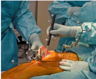
- 2 passive optical reference arrays were attached on the distal medial femur and the proximal medial tibia.

- The centre of the hip was measured by circumduction.

- The landmarks for the femorotibial kinematics were reported by the navigation system in digital form.

Figure 17: Measuring the landmarks {Keshmiri #100}

- Next step was to fix the patellar array onto the anterior side of the patella.

Figure 18: Fixing the navigation array on the patella by using a screw {Keshmiri

#100}

(34)

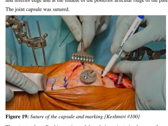
- BrainLab specifies the ‘patella coordinate frame’ as ‘a point at the medial, superior, and inferior edge and at the middle of the posterior articular ridge of the patella’.

- The joint capsule was sutured.

Figure 19: Suture of the capsule and marking {Keshmiri #100}

- Then, ‘natural patellar kinematics and the relative orientation between femur, tibia and patella were recorded between 30° and 90° flexion during passive motion.’

- Navigation calculated during the motion cycle the position of the ‘registered patella coordinate frame relative to the coordinate frame of the femur’. Furthermore, the absolute and relative values for ‘patellar mediolateral shift (medial: +, lateral: -); axial tilt (medial: - lateral: +), and coronal rotation (external: -, internal: +) of the patella’

were gathered. During the motion cycle, the epicondylar distance was recorded. It is described as ‘the line from the previously chosen point at the middle of the posterior articular ride of the patella perpendicular to the transepicondylar line, which is built from the registered femoral epicondyles’. The epicondylar distance is important as it

‘gives information about the anterior-posterior position of the patella throughout the

flexion cycle in relation to the femur.’

(35)

Figure 20: Patella tracking before the implantation {Keshmiri #100}

- The osteophytes were removed at the medial and the lateral compartment.

- The tibial cut was made.

- The double tensiometre was fixed at ‘0° of extension and 90° of flexion with a distraction force of 90 N.’

- In the frontal plane 0° were targeted between the femoral and tibial mechanical axis.

- To gain ligament balancing, the ‘flexion gap was adapted through bony cuts by the navigation software.’

- According to Berger et al. the femoral component rotation was ‘set by ligament balancing and the rotation of the tibial component.’

- After the implantation of the prosthesis the patellar kinematics were carried out with the component placement suggested by the navigation system.

{Keshmiri 2015 #72}{Bäthis 2003 #45}{Berger 1998 #63}

(36)

- In all cases the patellae were left in their natural state, that means no surgical intervention was performed regarding the patella.

- Then, the measurement was made by the surgeon lifting the limbs vertically at the distal femur without touching the tibia. This motion cycle was performed twice.

Thereby, values up to 30° of flexion were removed as these values were irregular because of missing muscle tone and floppy patellae. Furthermore, the ‘definitive femoral component rotation and flexion and the tibial component rotation and slope were recorded intraoperatively’. All data were gathered using patellar tracking software application for TKA (Patellar Tracking; BrainLab AG, Feldkirchen, Germany).

Figure 21: Patella tracking after the implantation with fixed components

(37)

3.4 Examinations

For this study standardised radiological and clinical assessments were used pre- and postoperatively.

3.4.1 Radiological assessment

In this study radiological assessment was carried out pre- and postoperatively with regard to the alignment (varus/valgus angle), the Q-angle, the modified Insall-Salvati-Index, the patellar shift and tilt and the joint line. Therefore long leg imaging (a/p and lateral) and a patella sunrise view were taken.

3.4.1.1 Leg axis

It is defined by the angle between the centre of the hip to the femoral centre of the knee joint and the centre of the tibial knee joint to the centre of the ankle joint. It was measured before and after the operation aiming at an angle of 180° postoperatively to achieve homogeneous distribution of the pressure.

This is important to counteract uneven wear as one of the causes of aseptic loosening. The a/p X-ray image should be taken while the patient is standing and the axial measurement can only be precisely done by taking a long leg radiograph. {Pietsch 2006 #13}

The physiological axis of the leg is defined as

180°. Values between 177° and 183° define Figure 22: Long leg measurement the neutral average value. Angles < -177° {Pietsch 2006 #13}

are defined as genua vara, angles > 183° as genua valga. {Bäthis 2004 #46}

(38)

3.4.1.2 Q-angle

The Q-angle (Quadriceps angle) was assessed by calculating the angle between the direction of the rectus femoris (QF) as resultant force of the quadriceps group and the patella ligament (=TF). The reference value is 8-10° in males and 12-16° in females. {O´Donoghue 1976

#14}

Figure 14: Definition of the Q-Angle {O´Donoghue 1976 #14}

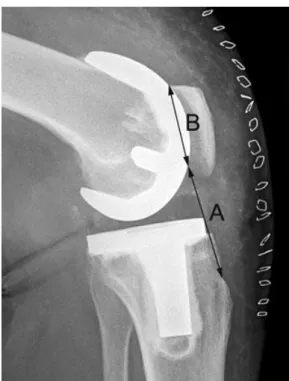
3.4.1.3. Modified Insall-Salvati-Index The knee should be 30° flexed, ideally. Then, the modified Insall-Salvati-Index was calculated by dividing the distance between the tibial tuberosity and the most distal patellofemoral point of the articulation (A) by the length of the patella articular surface (B). The standard of the modified method is the ratio A/B between 0.74 and 1.50. {Shabshin 2004 #26} {Springorum 2011 #19}

Figure 23: Modified Insall-Salvati-Index

{Springorum 2011 #19}

(39)

3.4.1.4 Patellar shift and tilt

Patellar shift and tilt were measured pre-and postoperatively using patella defilée imaging in 60° flexion.

The patellar shift is defined as medial or lateral movement of the centre of the patella along the transepicondylar axis (TEA) (medial shift: +, lateral shift: -).

The patellar tilt is defined as the rotation of the patella about its long axis (medial tilt: +, lateral tilt: -) {Nha 2008 #41}

3.4.1.5 Joint line

In this study the deviation of the joint line was assessed using a long-leg axis imaging under weight-bearing. The joint line is defined as the contact line of the distal femur and the proximal tibia. The height was measured as distance to the fibula head in the tibial shaft axis.

{Maderbacher 2015 #73}

Figure 24: Measurements in long-leg radiographs {Maderbacher 2015 #73}

(40)

Figure 25: Measurements in both knee radiographs {Maderbacher 2015 #73}

3.4.1.6 Femoral rotation by means of navigation

Femoral rotation was assessed during the operation by means of navigation. It was measured

related to the epicondylar line. External rotation is reported as a negative value, internal

rotation as a positive value. {Keshmiri 2015 #72}

(41)

3.4.2 Clinical assessment

The 4 questionnaires: KSS, WOMAC, Feller/Patellar Score and the HAKEMP-90 were carried out preoperatively, and 7 and 28 days postoperatively.

3.4.3 Statistical evaluation

For the statistical evaluation first, the normal distribution of the data was checked using the Kolmogorow-Smirnow-Test. For normally distributed data the paired t-test was further used, for not normally distributed data the Wilcoxon signed rank test was further used (SigmaPlot, Erkrath – Amtsgericht Wuppertal, Germany).

3.4.4 Complications

Postoperative complications were monitored until the 28 th day after surgery.

(42)

neutral 22.5%

valgus 10%

varus 67.5%

4. Results

4.1 Radiological assessment 4.1.1 Leg axis

The mean of the preoperative leg axis was 175.15° (SD 5.67; range 162° - 189°).

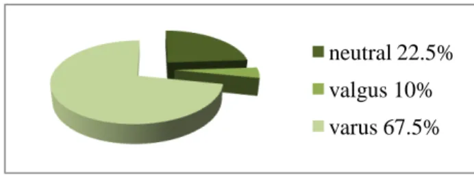
There were 27 (67.5%) patients with genua vara, 9 (22.5%) patients within the neutral average value and 4 (10%) with genua valga.

After the operation the mean of the leg axis was 180.53° (SD 1.4; range 177° - 184°).

There were 39 (97.5%) patients within the neutral average value. 1 (2.5%) patient had a slight genu valgum of 184°, none had a genu varum.

Figure 26: Preoperative deviation of the axis (tolerance range)

3 6 9 12 15

Axis pre-op Gaussian Fit Axis post-op Gaussian Fit

N u m b e r o f p a ti e n ts

Reference range

(43)

5 10 15 20 25 30 35 40

-10 -5 0 5 10 15 20

P a ti e n t n u m b e r

pre

190 185 180 175 170 165 160

Difference in axis of the leg pre

valgus

varus diff

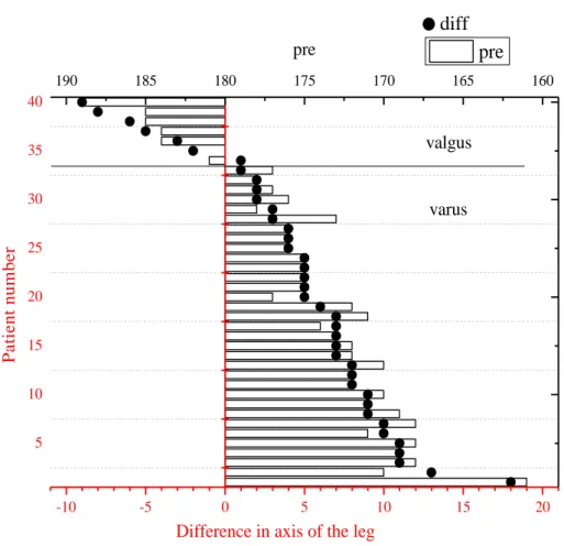
Figure 28: Pre- and postoperative deviation of the axis in each case

Figure 28 shows the deviation of the preoperative leg axis (bar chart) in relation to how many

degrees the axis has been modified (dots) in each case.

(44)

4.1.2 Q-angle Females preoperatively

Preoperatively, the mean Q-angle in females was 14.26° (SD 2.2; range 12° – 18°).

6 7 8 9 10 11 12 13 14 15 16 17 18 19

0 1 2 3 4 5 6

N u m b e r o f p a ti e n ts

Q-Angle in °

pre

Figure 29: Preoperative deviation of the Q-angle (females) Males preoperatively

Preoperatively, the mean Q-angle in males was 10.19° (SD 2.8; range 7° - 17°).

6 7 8 9 10 11 12 13 14 15 16 17 18 19

0 1 2 3 4 5

N u m b e r o f p a ti e n ts

Q-Angle in °

pre

Figure 30: Preoperative deviation of the Q-angle (males) Reference range

Reference range

(45)

Females postoperatively

Postoperatively, the mean Q-angle in females was 12.32° (SD 1.2; range 10° - 15°) .

6 7 8 9 10 11 12 13 14 15 16 17 18 19

0 1 2 3 4 5 6 7 8

N u m b e r o f p a ti e n ts

Q-Angle in °

post

Figure 31: Postoperative deviation of the Q-angle (females)

Males postoperatively

Postoperatively, the mean Q-angle in males was 8.48° (SD 0.9; range 7° - 10°).

6 7 8 9 10 11 12 13 14 15 16 17 18 19

0 1 2 3 4 5 6 7 8

N u m b e r o f p a ti e n ts

Q-Angle in °

post

Figure 32: Postoperative deviation of the Q-angle

Reference range

Reference range

(46)

4.1.3 Modified Insall-Salvati-Index

The preoperative mean modified Insall-Salvati-Index was 1.40 (SD 0.2; range 1.0 - 1.7).

0,9 1,0 1,1 1,2 1,3 1,4 1,5 1,6 1,7

0 2 4 6 8 10

N u m b e r o f p a ti e n ts

Modified Insall-Salvati-Index

pre

Figure 33: Preoperative modified Insall-Salvati-Index (absolute values)

Postoperatively, the modified Insall-Salvati-Index was 1.24 (SD 0.1; range 0.9 - 1.4).

0,9 1,0 1,1 1,2 1,3 1,4 1,5 1,6 1,7

0 2 4 6 8 10 12

N u m b e r o f p a ti e n ts

Modified Insall-Salvati-Index

post

Figure 34: Postoperative modified Insall-Salvati-Index (absolute values) Reference range

Reference range

(47)

4.1.4 Patellar shift

The patellar shift was measured using X-ray.

Preoperatively, the mean patellar shift was -3.3 mm (SD 4; range -14mm – 5mm).

32 (80%) patients had a lateral patellar shift, 8 patients (20%) had medial patellar shift.

Postoperatively, the mean shift was -2.4 mm (SD 3.8; range -10mm – 3 mm).

28 (70%) patients had a lateral shift of the patella, 12 (30%) patients had a medial shift.

The mean difference pre- and postoperatively was -0.9 mm (SD 5 mm; range 11 – 1). The changes were not significant (p=0.26).

-15 -12 -9 -6 -3 0 3 6

0 2 4 6 8

Shift pre-op Gaussian Fit Shift post-op Gaussian Fit

N u m b e r o f p a ti e n ts

Patellar shift [mm]

lateral medial

Figure 35: Pre- and postoperative deviation of the patellar shift (absolute values)

Reference range

(48)

5 10 15 20 25 30 35 40

-10 -5 0 5 10 15

Difference of the patellar shift

P a ti e n t n u m b e r

diff

10 5 0 -5 -10 -15

5 10 15 20 25 30 35 40

pre

preoperative shift

[mm]

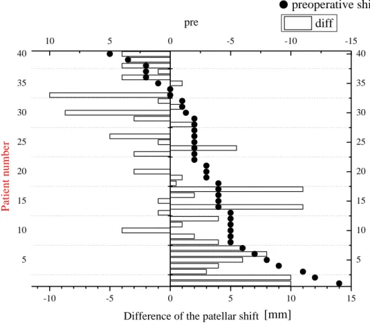
Figure 36: Pre- and postoperative deviation of the patellar shift in each case

Figure 36 shows the deviation of the patellar shift preoperatively (dots) in relation to how

much it has been modified during the operation (bar chart) in each case.

(49)

4.1.5 Patellar tilt

The patellar tilt was measured using X-ray.

Preoperatively, the mean patellar tilt was -4.19° (SD 5.4; range -16° - 14.5°).

In 31 (77.5%) patients the patella tilted laterally, in 9 (22.5%) patients the patella tilted medially.

Postoperatively, the mean tilt was -0.2° (SD 4.3; range -9° - 8°).

In 18 (45%) patients the patella tilted laterally, in 22 (55%) patients the patella tilted medially.

The X-ray evaluation pre- and postoperatively showed a mean difference of 3.99° (SD 4.8°, range -14 – 9). The changes were statistically significant (p<0.001).

-15 -10 -5 0 5 10 15

0 1 2 3 4 5 6 7 8

N u m b e r o f p a ti e n ts

Patellar tilt angle [°]

Tilt pre-op Gaussian Fit Tilt post-op Gaussian Fit

lateral medial

Figure 37: Pre- and postoperative deviation of the patellar tilt (absolute values) (chequered: reference range; striped: extended reference range = patients were found pain-free)

Reference range

(50)

5 10 15 20 25 30 35 40

-15 -10 -5 0 5 10 15

Difference of the patellar tilt

P a ti e n t n u m b e r

diff

15 10 5 0 -5 -10 -15

5 10 15 20 25 30 35 40 pre

preoperative tilt

[°]

Figure 38: Pre- and postoperative deviation of the patellar tilt in each case

Figure 38 shows the deviation of the patellar tilt preoperatively (boxes) in relation to how

much it has been modified during the operation (bar chart) in each case.

(51)

4.1.6 Joint line

Preoperatively, the mean distance was 10.44 mm (SD 5.47; range 0 mm – 23 mm).

Postoperatively, the mean distance was 12.53 mm (SD 3.8; range 5 mm - 21 mm).

The preoperative and postoperative joint line showed a mean difference of 2.1 mm of elevation (±3.5 mm; range -5 to +12 mm). The change of the joint line was significant (p<0.001).

0 3 6 9 12 15 18 21

0 1 2 3 4 5 6

N u m b e r o f p a ti e n ts

Joint line [mm]

Joint line pre-op Gaussian Fit Joint line post-op Gaussian Fit

Figure 39: Pre- and postoperative deviation of the joint line (absolute values)

(52)

5 10 15 20 25 30 35 40

-5 0 5 10

Difference of the joint line

P a ti e n t n u m b e r

diff

-10 -8 -6 -4 -2 0 2 4 6 8 10 12 14 16 18 20 22 24 26

pre

preoperative joint line

[ mm ]

Figure 40: Pre- and postoperative deviation of the joint line in each case

Figure 40 shows the deviation of the joint line (boxes) in relation to how much it has been

modified during the operation (bar chart) in each case.

(53)

4.1.7 Femoral rotation by means of navigation

Postoperatively, the mean deviation of the rotation of the femoral component was -4.68° (SD 6.3; range: -16° - 13.5°).

In 34 (85%) patients the femoral component was implanted in external rotation, in 5 (12.5%) patients in internal rotation. In 1 (2.5%) patient there was nearly no rotation.

-15 -10 -5 0 5 10 15

0 1 2 3 4 5 6

N u m b e r o f p a ti e n ts

Femoral rotation [°]

Number of patients Gaussian Fit

External Internal

Figure 41: Postoperative deviation of the femoral rotation (absolute values)

(54)

4.1.8 Overview: Patellar tracking using navigation

Table 4: Pre- vs. postoperative patellar tracking (lateral shift: +; reduction in epicondylar distance: +; internal rotation: -; lateral tilt: -)

Flexion Mean differ-

ence (SD) 95% CI p-value

Shift 30° 1.6 (4.2) 0.2 to 2.9 0.02

45° 1.4 (4.2) 0.1 to 2.8 0.03 60° 1.3 (4.1) 0.0 to 2.6 0.04 75° 1.1 (4.2) –0.1 to 2.5 0.1 90° 0.6 (3.9) –0.6 to 1.9 0.3 Epicondylar distance 30° 1.2 (4.4) –0.2 to 2.6 0.1 45° 1.7 (3.6) 0.5 to 2.8 < 0.01 60° 1.7 (3.6) 0.5 to 2.8 < 0.01 75° 1.6 (3.4) 0.5 to 2.7 < 0.01 90° 0.9 (3.6) –0.2 to 2.1 0.1 Rotation 30° –0.5 (3.5) –1.6 to 0.6 0.4 45° –0.5 (3.9) –1.8 to 0.7 0.4 60° –1.0 (4.1) –2.3 to 0.2 0.1 75° –0.9 (4.3) –2.3 to 0.4 0.2 90° –1.2 (4.0) –2.5 to 0.0 0.1

Tilt 30° –1.2 (6.4) –3.3 to 0.8 0.2

45° –1.3 (5.6) –3.2 to 0.4 0.1 60° –0.4 (4.9) –2.0 to 1.1 0.6 75° 1.2 (4.7) –0.2 to 2.7 0.1 90° 3.2 (4.4) 1.8 to 4.6 < 0.01

(55)

Postoperatively, there was a statistically significant difference between the pre- and

postoperative shift between 30° and 60° of flexion. Between 45° - 75° of flexion there was a

significant decrease in epicondylar distances during the whole motion cycle. Between 30° and

60° of flexion the patellae tilted more laterally after the operation, beyond (60°-90°) the

patellae tilted statistically significant more medially compared to the tilt before surgery. The

patellae rotated more medially but this was not statistically significant.

(56)

4.2 Clinical assessment

4.2.1 Knee Society Score (KSS)

Preoperatively, the mean was 97 points (SD 20.5; range 61 – 154 points), 7 days postoperatively 97 points as well (SD 29.73; range 54 - 190), and 28 days postoperatively 134 points (SD 19.8; range 85 - 187).

4 0 6 0 8 0 100 120 140 160 180

0 1 2 3 4 5 6 7

pre 7 d ay s

2 8 d a y s

pre 7 days 28 days

N u m b e r o f p a ti e n ts

Av aila ble po int s

Figure 43: Knee Society Score pre- and postoperatively

The Knee Society Score is often given separately in Knee Score (A) and Function Score (B):

pre 7 days post 28 days post

A B A B A B

mean 43.23 53.5 68.98 28.5 78.38 55

SD 14.02 13.07 16.21 21.43 14.16 11.55

(57)

4.2.2 Western Ontario and McMaster Universities Osteoarthritis Index (WOMAC) Preoperatively, the mean was 55.7% (SD 12.2; range 26% - 75%), 7 days postoperatively 33.65% (SD 14.4%; range 17% – 70%) and 28 days postoperatively 28.3% (SD 15.9%; range 0% - 71%).

20 0 60 40

80

0 2

4 6

8

p r e

7 d a y s 2 8 d a y s

pre 7 days 28 days

N u m b er o f p at ie n ts

A v aila

b le p o in ts in %

Figure 44: WOMAC Score pre- and postoperatively

The change was statistically significant (p = < 0.001).

(58)

4.2.3 Feller/Patella Score

Preoperatively, the mean was 18 points (SD 6.16; range 4 – 29 points), postoperatively 22 points (SD 3.75; range 13 – 28 points).

5 10 15 20 25 30

0 2 4 6 8 10 12 14 16

N u m b e r o f p a ti e n ts

Available points pre post

Figure 45: Patellar Score pre- and 7 days postoperatively

The change was statistically significant (p = < 0.001).

(59)

4.2.4 HAKEMP-90

The mean was 18 points (SD 8; range 0 - 24 points).

0 5 10 15 20

0 2 4 6 8 10 12 14 16 18

N u m b e r o f p a ti e n ts

Available points

HAKEMP90

Figure 46: HAKEMP -90 (absolute values)

4.2.5 Complications

There were 2 patients with delayed wound healing on day 7 without revision surgery and 1

patient with deep vein thrombosis at day 28.

(60)

5. Discussion

Due to the increasing number of TKA – in 2009 alone 175.000 primary TKAs were implanted in Germany, the associated revision rate is rising, too. {Borelli 2011 #74}

Patellofemoral problems are known as one of the major reasons for revision surgery. Thus, the patellofemoral joint is a challenge in treatment. The aim, above all, is to reduce the retropatellar pain syndrome. {Springorum 2012 #4} {Lygre 2010 #75}

Correct patellar tracking is only achieved successfully if the patella moves smoothly in the trochlear groove. To receive such a smooth movement it is necessary to know how the patella moves in the natural knee. {Keshmiri 2014 #110}

The objective of this study was to examine the kinematic and radiological changes of the

patella following a ligament-balanced navigated TKA.

Abbildung

Figure 4: First models of prosthesis of Themistokles Gluck {Rabenseifner 1998 #34}
Figure 8: Coordinate systems used to quantify the clinical motions of the patella   {Nha 2008 #41}
Figure  18:  Fixing  the  navigation  array  on  the  patella  by  using  a  screw  {Keshmiri
Figure 19: Suture of the capsule and marking {Keshmiri #100}
+7

Referenzen

ÄHNLICHE DOKUMENTE

Zusammenfassend kann man sagen, dass sich in unserer Studie sowohl deutliche Vorteile für die Patienten nach erfolgreicher Pankreastransplantation im Vergleich zur Einstellung

Die Verschiebung in der Altersstruktur als Folge von niedrigen Geburtenraten und steigender Lebenserwartung ist ein Phänomen, welches die Bundesrepublik Deutschland vor neue

Da sich IL-6 nach einer Endoprothesenimplantation innerhalb von 2 Tagen normalisiert, kann dies ein sensitiver Indikator für eine periprothetische Infektion in der

Auch  in  der  Indikation  der  rheumatoiden  Arthritis  fallen  Unterschiede  in  den  jeweiligen  Fachinformationen  auf.  Zunächst  zeigen  sich 

Diese Kenntnisse über das implizite Gedächtnis legen die Vermutung nahe, dass sich Patienten nach der Implantation einer Knieendoprothese unter Sedierung zwar

In mehreren Studien konnte nachgewiesen werden, dass die katabolischen Effekte auf den Knorpel unter anderem über eine Hemmung der die Chondrogenese fördernden Faktoren TGF-ß

Diese Druckerhöhung wird unter anderem dadurch erklärt, dass es bei den Patienten im Bereich des Hüftkopfes zu einer venösen Stase kommt, da nicht nur die Anzahl

Dabei verwendet man T1 gewichtete Bilder, um Ausdehnung, Gruppierung und Homogenität der Läsion zu beurteilen, T2 gewichtete Darstellungen, um den Knorpel und die