AUS DEM LEHRSTUHL FÜR ORTHOPÄDIE
PROF. DR. MED. DR. H.C. J. GRIFKA DER FAKULTÄT FÜR MEDIZIN DER UNIVERSITÄT REGENSBURG
OSTEONECROSIS OF THE FEMORAL HEAD – A RETROSPECTIVE TRIAL OF JOINT PRESERVING TREATMENT OPTIONS AND A PROSPECT TO FUTURE
CLINICAL POSSIBILITIES
Inaugural – Dissertation zur Erlangung des Doktorgrades
der Medizin der
Fakultät für Medizin der Universität Regensburg
vorgelegt von Korbinian Völkl
2019
AUS DEM LEHRSTUHL FÜR ORTHOPÄDIE
PROF. DR. MED. DR. H.C. J. GRIFKA DER FAKULTÄT FÜR MEDIZIN DER UNIVERSITÄT REGENSBURG
OSTEONECROSIS OF THE FEMORAL HEAD – A RETROSPECTIVE TRIAL OF JOINT PRESERVING TREATMENT OPTIONS AND A PROSPECT TO FUTURE
CLINICAL POSSIBILITIES
Inaugural – Dissertation zur Erlangung des Doktorgrades
der Medizin
der
Fakultät für Medizin der Universität Regensburg
vorgelegt von Korbinian Völkl
2019
Dekan: Prof. Dr. Dr. Torsten E. Reichert
1. Berichterstatter: Prof. Dr. Benjamin Craiovan
2. Berichterstatter: Prof. Dr. Martin Fleck
Tag der mündlichen Prüfung: 23.12.2019
Zusammenfassung in deutscher Sprache
Die Hüftkopfnekrose – eine retrospektive Auswertung gelenkerhaltender Therapiemöglichkeiten und ein Ausblick auf zukünftige klinische Möglichkeiten
Einleitung:
Die hier vorliegende Arbeit zur aseptischen Hüftkopfnekrose des Erwachsenen gliedert sich im Wesentlichen in zwei große Teile. Im ersten Teil wird ein umfangreicher und vollständiger Überblick über die aktuellsten klinischen und wissenschaftlichen Erkenntnisse zur Ätiologie, Diagnostik und Therapie in der gängigen Fachliteratur gegeben.
Im zweiten Teil, welcher sich wiederum in drei Abschnitte, gliedert werden die selbst angestellten klinischen und wissenschaftlichen Untersuchungen, sowie deren Ergebnisse präsentiert.
Die Femurkopfnekrose ist eine Erkrankung, welche vor allem jüngere Menschen, zwischen 30 und 50 Jahren und somit in Mitten ihrer größten Leistungsfähigkeit betrifft.
Dies hat nicht nur für jeden Betroffenen individuell belastende und einschränkende Auswirkungen, sondern ist auch für die Gesellschaft und das Gesundheitssystem eine enorme Belastung [1] [2]. Die aseptische Nekrose des Hüftkopfes beginnt langsam und verläuft unbehandelt progredient bis zur vollständigen arthrotischen Zerstörung des Gelenkes. Die Patienten berichten in vielen Fällen über eine zunehmende diffuse Schmerzhaftigkeit und Bewegungseinschränkung des betroffenen Hüftgelenkes [3]. Die treffende Diagnose wird oft erst spät und somit in bereits fortgeschrittenen Stadien gestellt. Den diagnostischen Goldstandard stellt die Magnetresonanztomographie (MRT) dar. Mittels MRT können bereits die Frühformen der Osteonekrose erkannt werden, wenn das normale Röntgenbild noch völlig unauffällig erscheint [4].
Es haben sich verschiedene Stadien-Klassifikationen der Hüftkopfnekrose entwickelt. Die weitaus geläufigste ist hierbei diejenige der „Association Research Circulation Osseous“
(ARCO). Diese berücksichtigt vor allem radiologische Aspekte. Sie ermöglicht eine Einteilung in die Stadien I bis IV, wobei im potentiell reversiblen Stadium I nur im MRT pathologische Veränderungen sichtbar sind. Im Stadium ARCO II wird auch das normale
Röntgen positiv, mit vermehrter Sklerosierung des Nekroseareals, bei jedoch völlig erhaltener Hüftkopfkontur. Im Stadium III kommt es zu subchondralen Einbrüchen, welche sich im Röntgenbild als eine Entrundung des Hüftkopfes darstellen. Das finale Stadium IV stellt die vollständige Zerstörung des Gelenkpartners dar [5] [6]. Diese Einteilung ist neben dem akademischen Interesse vor allem für die Therapie von höchster Relevanz.
Obwohl viele Risikofaktoren bekannt sind, ist die exakte Ätiologie der Erkrankung noch unklar. Als mögliche Risikofaktoren gelten neben den „Lifestyle-Faktoren“ Rauchen und Alkoholabusus, vor allem eine längere oder höher dosierte Therapie mit Kortikosteroiden. Desweiteren werden physische Überlastung mit Mikrotraumata, metabolische Erkrankungen, wie der Morbus Gaucher und genetische Vorerkrankungen, wie die Sichelzellanämie oder Thrombophilien genannt [7] [8].
Pathomechanismus:
Der Pathomechanismus der Hüftkopfnekrose war lange Zeit unklar, jedoch scheint sich mittlerweile die Theorie eines erhöhten intra-ossären Druckes zu bestätigen [9] [1]. Dabei wird davon ausgegangen, dass im erkrankten Femurkopf der intramedulläre Druck gesteigert ist. Dies führt zu einer Kompression und in Folge dessen zu einer Obstruktion von kleinsten venösen Gefäßen. Dadurch kommt es zu einem gestörten Abfluss des Blutes und somit zu einem erhöhten Blutvolumen im Knochenmark. Dies führt wiederum zu einer Steigerung des Druckes im spongiösen Knochen. Dieser circulus vitiosus bedingt eine Minderversorgung des Knochenmarks mit Sauerstoff und endet letztlich im nekrotischen Untergang des Gewebes [10].
Hintergrund:
Aus der Pathogenese ergeben sich auch die geläufigsten gelenkerhaltenden Therapien.
Diese haben als primäre Grundidee den Versuch der Entlastung des gestauten Kompartiments, da man sich dadurch eine Normalisierung des Blutflusses und folglich eine Regeneration des Gewebes erhofft. Bei erhöhtem intrakompartimentellem Druck erfolgt somit nach Ficat und Arlet eine Core Decompression [9]. Das primäre Ziel aller Therapieversuche ist in frühen Stadien immer die Erhaltung des Hüftgelenkes. Dies sollte wenn möglich dauerhaft sein, jedoch sollte dadurch zumindest Zeit gewonnen werden, bis die Implantation einer Totalendoprothese (TEP) nötig wird. Als mögliche konservative Therapieoption wird vor allem die Entlastung des betroffenen Gelenkes
mittels Gehstützen empfohlen, um in der akuten Phase eine weitere Eskalation zu fortgeschrittenen Stadien zu vermeiden [5]. Ob eine spontane Remission, allein dadurch zustande kommt ist unklar. Auch vielfältige medikamentöse Therapiemöglichkeiten von Statinen, über Bisphosphonate, bis hin zu Rheologika, wie das stabile Prostazyklin- Analogon Ilomedin werden in der klinischen Praxis eingesetzt. Durch das am häufigsten angewandte und am besten erforschte Ilomedin erhofft man sich eine Steigerung des intramedullären Blutflusses, mit einer konsekutiven Regeneration des Knochengewebes.
Ilomedin wird intravenös in einem fünf tägigen Schema mit ansteigender patientenadaptierter Dosis verabreicht [11]. Es konnte gezeigt werden, dass Patienten, welchen das Medikament in den frühen ARCO Stadien I und II verabreicht wurde, eine deutliche Reduktion der Schmerzen, eine Zunahme der Mobilität und sogar eine radiologische Verbesserung zeigten [12]. Ein großer Vorteil dieser Therapie ist die fehlende Invasivität und ihre relativ geringen Nebenwirkungen.
Als operative gelenkerhaltende Therapie ist die sogenannte „Core Decompression“ am weitesten verbreitet und auch am besten untersucht. Hierbei handelt es sich um eine Anbohrung des Hüftkopfes, mit dem Ziel der Druckentlastung [13]. Daneben soll durch das bewusste Verursachen einer intramedullären Blutung ein Regenerationsreiz gesetzt werden. Die Anbohrung erfolgt meistens mittels Kirschnerdrähte, welche unter Bildwandlerkontrolle in das nekrotische Areal eingebracht werden. Klassischerweise wird die Nekrose fächerförmig angebohrt, um einen größeren Anteil des defekten Bereiches zu erfassen [14]. Neben der Anbohrung mit K-Drähten kann dafür auch eine etwa zehn Millimeter starke Hohlfräße verwendet werden [15]. Dies eröffnet die Möglichkeit das nekrotische Material direkt zu entfernen und bei einer autologen Spongiosaplastik durch andernorts gewonnene intakte Spongiosa zu ersetzen [16]. Der Nachteil hierbei ist jedoch der größere kortikale Defekt und die damit verbundene Schwächung und Frakturgefährdung des Schenkelhalses. Dieses Risiko wird jedoch durch eine postoperative Ent- oder Teilbelastung minimiert. Jedoch ist der Eingriff mit einem größeren Aufwand verbunden, da hierfür ein zweiter OP-Situs (Beckenkamm) geschaffen werden muss. Durch die in der gesunden, durchbluteten Spongiosa enthaltenen Osteoprogenitor Zellen erwartet man sich einen zusätzlichen Wachstums- und Heilungsstimulus. In der Literatur werden auch weitere autologe Knochentransplantate, wie gefäßgestielte Fibula-Transplantate beschrieben [17]. Diese konnten sich aber nicht in der breiten Anwendung durchsetzen. Als weitere gelenkerhaltende Therapie sind Umstellungsosteotomien zu nennen. Diese sind jedoch
auf Grund der Komplexizität des Eingriffes und der geringen Patientenakzeptanz eher von untergeordneter Bedeutung und speziellen Ausnahmen vorbehalten. Als letzte Therapiemöglichkeit bei weit fortgeschrittener Nekrose oder bei Versagen der gelenkerhaltenden Optionen bleibt die Hüftgelenkstotalendoprothese (Hüft-TEP) [18].
Diese stellt eine sehr gute Therapieform dar, ist bei den überwiegend jungen Patienten mit Femurkopfnekrose jedoch so lange wie möglich hinaus zu zögern, um perspektivisch häufige Wechsel- und Revisionseingriffe zu vermeiden.
Hauptteil
Im zweiten Abschnitt der hier vorliegenden Arbeit werden die selbst durchgeführten Untersuchungen und Auswertungen dargelegt. Dieser Abschnitt gliedert sich wiederum in drei Fragestellungen. Zum ersten wird ein retrospektiver klinischer Vergleich zwischen der Anbohrung und der Anbohrung mit autologer Spongiosaplastik angestellt.
In der zweiten Fragestellung wird die Zielgenauigkeit der beiden Therapievarianten untersucht.
Im dritten Abschnitt erfolgt die Vorstellung einer bisher so noch nicht beschriebenen Darstellung der intramedullären Perfusion des Hüftkopfes mittels MRT-basierter Perfusionsmessung.
Da in der Literatur viele verschiedene Konzepte zur gelenkerhaltenden Therapie bei Hüftkopfnekrose beschrieben werden, soll im ersten Teil der vorliegenden Arbeit ein Vergleich zwischen der konventionellen Hüftkopfanbohrung und der Anbohrung mit autologer Spongiosaplastik gemacht werden.
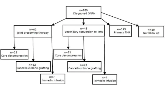
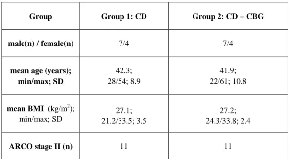
In einem retrospektiven Studiendesign wurden Patienten, welche im orthopädischen Universitätsklinikum Bad Abbach gelenkerhaltend therapiert worden waren, nachuntersucht. Hierfür wurden aus einem Gesamtkollektiv von 289 Hüften mit der Diagnose Hüftkopfnekrose aus dem Zeitraum von 2006 bis 2012 die 62 Fälle mit einer gelenkerhaltenden Therapie ausgewählt. Davon konnten 31 Fälle für die Nachuntersuchung rekrutiert werden und für die folgenden Untersuchungen eingeschlossen werden.
Die Nachuntersuchung setzte sich zusammen aus einer klinischen Untersuchung, mit Ermittlung der Bewegungsfähigkeit (range of motion, ROM), eventuellen Kontrakturen und Feststellung der Schmerzhaftigkeit in Ruhe und unter Belastung mittels der visuellen Analogskala (VAS 10). Ergänzend wurden die Patienten gebeten einige Fragebögen und
Scores zu beantworten. Hierbei kamen der Harris Hip Score (HHS), der Hip disability and Osteoarthritis Outcome Score (HOOS), der EuroQol-5D (EQ-5D) und der Short Form 36 (SF-36) zum Einsatz. Für die radiologische Beurteilung des Hüftgelenkes wurde ein konventionelles Röntgen in Hüftübersicht und in der Lauenstein-Projektion angefertigt. Hierauf wurde bei allen Patienten eine Magnetresonanztomographie (MRT) mit Kontrastmittel durchgeführt. An Hand dieser Daten erfolgte eine Gegenüberstellung von jeweils elf Patienten mit Anbohrung oder Anbohrung mit Spongiosaplastik. Diese relativ kleine Anzahl an Studienteilnehmern in dieser Subgruppe war vor allem durch den Versuch der bestmöglichen Vergleichbarkeit begründet, da durch „matched pairs“ eine möglichst homogene Verteilung der Gruppen erreicht werden sollte. Das „matching“
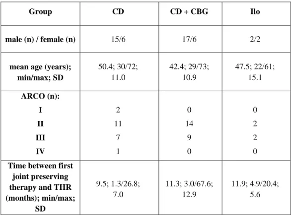
erfolgte nach den Parametern Geschlecht und Alter. Außerdem wurden in jeder Gruppe nur Patienten berücksichtigt, welche von dem jeweils gleichen Operateur behandelt worden waren. Zusätzlich lag bei allen Eingeschlossenen ein initiales ARCO II Stadium vor. Die Nachuntersuchung erfolgte im Mittel etwa vier Jahre nach der Intervention in der Gruppe der „Core Decompression“ und etwa fünf Jahre nach Therapie bei den Patienten die eine autologe Spongiosaplastik erhalten hatten.
Vergleich zwischen Core Decompression und autologer Spongiosaplastik
Die Analyse der Daten erbrachte zwar keine statistisch signifikanten Unterschiede bezüglich der klinischen und radiologischen Ergebnisse zwischen den beiden Gruppen, jedoch generell gute Langzeitergebnisse mit hoher Patientenzufriedenheit. In den klinischen Auswertungen des Bewegungsausmaßes der behandelten Hüfte, als Indikator für die Funktionalität des Gelenkes und der Schmerzhaftigkeit während Belastung, erzielten die Patienten mit einer autologen Spongiosaplastik etwas bessere Ergebnisse.
Dies spiegelte sich auch in den Angaben der Fragebögen wieder. Dort gaben Patienten nach autologer Spongiosaplastik etwas bessere hüftspezifische Werte an. Allerdings war das Abschneiden in der Gruppe der reinen Anbohrung etwas besser in Hinblick auf die Einschätzung der generellen und psychischen Gesundheit. In der radiologischen Beurteilung, also einer erneuten Stadieneinteilung waren im Durchschnitt keine Unterschiede feststellbar. Dies bedeutet, dass beide Therapieformen sowohl im klinischen, als auch im subjektiven und im radiologischen Outcome sehr ähnlich waren.
Dies spricht wiederum für die Gleichwertigkeit beider Therapie Optionen.
Zielgenauigkeit
Der zweite Aspekt dieser Arbeit beschäftigt sich mit einer relativ neuen technischen Möglichkeit, welche zunehmend Einzug in die operative Medizin erhält. Es geht hierbei um die dreidimensionale Rekonstruktion anatomischer oder pathologischer Formationen.
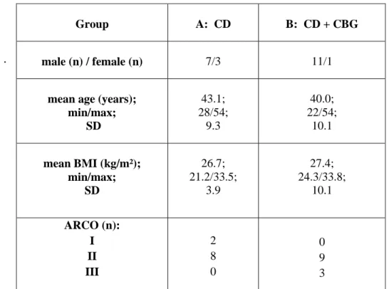
In der vorliegenden Untersuchung wurden bei 22 angebohrten Hüften die Nekrose und der Bohrkanal dreidimensional aus den MRT-Aufnahmen rekonstruiert. Aus der Rekonstruktion erfolgte eine Vermessung der Ausmaße und der Volumina der nekrotischen Areale. Danach wurden die Abweichungen der jeweiligen Bohrkanäle vom Mittelpunkt der Nekrose für jede einzelne Raumachse ausgemessen. Es erfolgte eine Gegenüberstellung von zehn Hüften mit reiner Core Decompression gegen 12 Hüften mit Spongiosaplastik. Aus den gewonnenen Ergebnissen zeigte sich, dass alle nekrotischen Areale von der Anbohrung getroffen wurden. Ein Vergleich der Genauigkeit der Intervention zwischen der klassischen Anbohrung und der Anbohrung mit anschließender Spongiosaplastik ergab keine statistisch signifikanten Unterschiede zwischen den beiden Methoden.
Im Zuge der Volumenbestimmung der Nekrose konnte der bereits in der Literatur beschriebene Zusammenhang zwischen größerem Ausmaß der Nekrose und einer wahrscheinlicheren Progredienz der Erkrankung bestätigt werden.
Intramedulläre Perfusionsmessung
Im dritten Teil dieser Studie wurde das neuartige Verfahren der intramedullären Perfusionsmessung untersucht. Das Grundprinzip dieser Methode beruht auf MRT- Aufnahmen zu bestimmten Zeitintervallen nach Kontrastmittelapplikation. Hierfür wurde den Patienten ein Kontrastmittel injiziert und nach festen Zeiten wurden bestimmte MRT-Sequenzen durchgeführt. Die Idee hinter dieser Methode besagt, dass sich die Kontrastmittelanreicherung im Knochenmark proportional zur Durchblutung verhält. Das heißt, dass sich aus der Signalsteigerung im MRT Rückschlüsse auf die Perfusion ziehen lassen müssten. Dieser Abschnitt der Studie wurde als rein deskriptiver Versuch zur Anwendbarkeit dieser neuartigen und bisher kaum beschriebenen Methode durchgeführt.
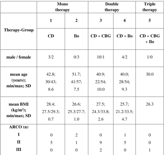
Dies konnte an 26 der behandelten Hüften erfolgen, unabhängig von der Art der Therapie oder dem initialen Stadium. Ergänzend wurden auch die gesunden, beziehungsweise unbehandelten Hüften betrachtet. Es ließ sich erkennen, dass Patienten mit einer Ilomedin-Infusionstherapie erhöhte Perfusionswerte aufwiesen. Die Bedeutung und Zusammenhänge dieser Ergebnisse müssen in weiteren Untersuchungen ebenso dargelegt
werden, wie die Möglichkeit durch die Perfusionsmessung noch früher Hinweise auf eine Störung in der intramedullären Perfusion zu bekommen und damit eine Risikoabschätzung für das Auftreten einer avaskulären Knochennekrose.
3
Index
1.1 Osteonecrosis of the femoral head (ONFH) ... 5
1.2 Epidemiology ... 5
1.3 Etiology and risk factors ... 6
1.3.1 Corticosteroid ... 6
1.3.2 Alcohol ... 7
1.3.3 Nicotine... 8
1.3.4 Physical strain ... 8
1.3.5 Genetic ... 9
1.4 Clinic... 10
1.5 Radiology Imaging ... 10
1.6 Staging ... 12
1.7 Therapy ... 16
1.7.1 Nonoperative therapy ... 16
1.7.1.1 Drugs ... 18
1.7.1.2 Other conservative measures ... 19
1.7.2 Operative therapy ... 20
1.7.2.1 Core decompression ... 22
1.7.2.2 Proximal femoral osteotomy... 24
1.7.2.3 Total hip arthroplasty ... 25
2. Question. ... 26
3. Material and Methods ... 27
3.1 Patients ... 27
3.2 Groups ... 31
3.2.1 Comparison between core decompression and cancellous bone grafting ... 31
3.2.2 Precision measurement of core decompression ... 32
3.2.3 Quantitative measurement of femoral head perfusion ... 34
4
3.3 Clinical protocol and scores ... 36
3.3.1 Clinical comparison between core decompression and cancellous bone grafting ... 36
3.3.2 Three dimensional measurement of drilling precision ... 37
3.3.3 Semiquantitative measurement of the hip perfusion ... 41
3.4 Statistics ... 44
4 Results……… ... 45
4.1 Clinical comparison of core decompression and cancellous bone grafting ... 45
4.2 Three dimensional measurement of drilling precision ... 49
4.3 Semiquantitative measurement of bone marrow perfusion ... 53
5 Discussion…. ... 56
5.1 Comparison of core decompression and cancellous bone grafting ... 56
5.2 Three dimensional measuring of drilling precision ... 63
5.3 Quantitative measuring of femoral head perfusion ... 69
6 Summary ... 73
7 References……. ... 75
8 Lebenslauf………90
5
1.1 Osteonecrosis of the femoral head (ONFH)
Osteonecrosis of the femoral head (ONFH) is a progressive locally limited disease, due to a reduced blood flow in the affected area. The impaired perfusion leads to a gradual destruction of the femoral head and thus to an enormous effect on the patient’s lifestyle [18]. As there are mostly younger people in the third to fifth decade affected, they are forced to change their work and leisure activities [2] [19]. In most cases the necrosis is restricted to the weight bearing area of the femoral head. Normally the disease starts unnoticed without any pain, but as it progresses most patients suffer from an enormous pain during weight bearing and in progressed stages also at rest [20]. Untreated the avascular necrosis leads to secondary arthrosis of the hip and to the necessity of total hip replacement [21] [22]. The natural course of the disease leads within two or three years to a complete joint destruction and in consequence to total hip replacement [23].
Synonyms are avascular or aseptic necrosis of the femoral head (AVN).
It is also possible to distinguish between idiopathic or secondary necrosis. In case of secondary necrosis there can be found causes like trauma, metabolic diseases or medication. For the idiopathic genesis on the other hand there are no evidential trigger factors, although even there can be found certain risk factors [2].
1.2 Epidemiology
In Germany there is a reported prevalence of 5,000 to 7,000 new diseases per year [24].
The diagnosis of avascular necrosis of the femoral head accounts for 5 to 12 percent of total hip replacements [2]. With about 10,000 to 20,000 new cases per year only in the USA it is obvious that ONFH is an important factor in health care systems worldwide [20].
In about 50 percent the patients suffer a bilateral involvement and in even 80 percent of steroid induced necrosis there is a bilateral affection [9].
6
1.3 Etiology and risk factors
There is a wide range of different etiological factors for the development of an ONFH. In general it has to be distinguished between a traumatic and a non-traumatic genesis. In traumatic conditions with fracture or dislocation of the femoral head the reduced blood flow, as a result of structural interruption of the blood vessels is obvious. Nevertheless, there are known several risk factors, like steroid medication, alcohol abuse, smoking or metabolic diseases [2] [25] [10].
1.3.1 Corticosteroid
A) Distribution
One of the major risk factors is the intake of corticosteroids. Studies showed, that about 10 to 30 percent of osteonecrosis can be associated with steroid medication [2] [21] [26].
However, only in 8 to 12 percent of patients treated with high dose corticosteroids for a longer period appeared signs of osteonecrosis. Felson T. et al. came to the conclusion that “The oral dose effect amounts to a 4 to 6 percent increase in the risk of AVN for every 10 mg/day rise in oral steroids during the first 6 months of therapy” [7]. Other reports cite an elevated risk for AVN at a dose of more than 2 g of steroids within a period of about two months [10] [27].
B) Pathogenesis
It is still unclear how corticosteroid therapy leads to osteonecrosis. One explanation could be a raised intraosseus pressure, which is either caused by a swelling of bone marrow fat cells or fat emboli [28]. The increased pressure causes an obstruction of small femoral vessels with a following ischemia and death of osteocytes [29] [30]. A possible model for increased fat cell content in bone marrow could be that, in vitro, dexamethasone has the competence to induce primary marrow stroma cells to differentiate to adipocytes. As bone cells and fat cells share a common precursor and dexamethasone can induce
7 adipogenesis, it results in a mismatch between bone and fat cells. This may lead to increased intraosseus pressure, venous stasis and decreased arterial perfusion [31].
However, the negative effects of steroid intake are not only attributable to the compromised vascularity, but even more to osteoblasts and osteocytes apoptosis [32] [30]
[33]. Within the last years it became obvious that apoptosis of osteocytes and lining cells leads to interruption in the mechanosensory network of the femoral head and thus to a reduced repair mechanism of structural micro damages, followed by collapse of the bone [30]. Glucocorticoids have an effect on all bone cells, even on osteoclasts. They decrease the production of osteoblasts and osteoclasts, increase the apoptosis of osteoblasts and prolong the lifespan of osteoclasts [34]. This process leads to an abnormal bone metabolism and thus to reduced bone mass and decreased stability. The thesis of osteocytes and osteoblast apoptosis as the major pathogenetic factor in steroid induced osteonecrosis is emphasized by the fact that there are not found apoptotic cells in osteonecrosis of other etiology, but only in steroid induced cases [34] [35] [33].
1.3.2 Alcohol
Studies showed that about one third of patients with idiopathic ONFH have a history of regular or excessive alcohol intake [36] [37]. The risk seems to be dose related, but it is, nevertheless, difficult to define a harmful quantity. However, it could be seen that an intake of more than 400 ml ethanol per week increases the risk of osteonecrosis 9.8 fold [2] [25]. Although the exact pathomechanism is still not clear, it seems there is a similarity to steroid induced osteonecrosis [25]. Histological surveys on alcohol induced ONFH showed an increase of fat cells and marrow fat and thus a reduction of osteogenetic cells. In addition to that the increased intraosseus fat content leads to elevated intraosseus pressure and thus to venous stasis, diminished blood circulation and finally to ischemic necrosis [3]. Besides, there are found elevated serum lipid peroxides, which have a pro inflammatory effect and induce degeneration of small blood vessels.
8 This can lead to a reduced bone perfusion, an effect which has also been observed in hepatic steatosis [25] [38].
1.3.3 Nicotine
Various surveys showed that smoking is a potential risk factor for osteonecrosis [22]. It is assumed that there is an increased risk for common smokers and a cumulative effect of a minimum of 20 pack years [10]. Exposition to cigarette smoke does affect every human organ system, so it also leads to intramedullary vasoconstriction and in the following to an ischemic necrosis of the bone [1]. Nicotine abuse can decrease osteogenesis, bone volume and vascular reactivity, which may also explain prolonged healing processes. The reduced blood supply and the decline of bone producing and repairing cells lead to a vulnerable tissue and an increased risk for necrosis of the femoral head [39] [10].
1.3.4 Physical strain
It seems that hard physical strain does not increase the possibility of ONFH. However, it has an impact on the proceeding of a preexisting AVN. This may be the result of physical stress induced micro lesions, which can worsen the vascular supply of the necrotic bone area [40]. Another reason for strict avoidance of physical strain in patients with osteonecrosis is on the one hand a symptomatic improvement of the pain and on the other hand a reduced number of insufficiency bone fractures [41].
9
1.3.5 Genetic
There seem to be various genetic disorders, connected with avascular necrosis in general and ONFH in particular. Especially patients with sickle cell anemia are confronted with an increased risk of getting ONFH [42]. Approximately about half of patients at the age of 35 are affected [43] [2]. These patients suffer from a higher disposition for an activation of the coagulation system, which can lead to occlusion of small blood vessels and thus to ischemic osteonecrosis [44]. There is also a discussion about other coagulation abnormalities, like Factor V Leiden mutation, thrombophilia or hypofibrinolysis. They lead to increased incidents of blood clots or to a reduced ability to dissolve these clots. It has been shown that up to 82% of patients with ONFH (in contrast to 32% in the control group) had at least one coagulation abnormality [10]. Koo, Lee et all. found that certain genetic polymorphisms in the endothelial nitric oxide synthase (eNOS) gene were frequently present in patients with ONFH. This leads to a reduction of vasodilatative effects and an inhibition of platelet aggregation of eNOS and thus to an impaired blood supply in the femoral head [39]. A deprived vascularization, as it can be found in VEGF-gene polymorphisms, may also be a co-factor for osteonecrosis [45]. Liu, Chen et all. succeeded in finding a genetic mutation in three families with autosomal dominant inheritance. They detected a genetic disorder in the type II collagene, the major structural protein in cartilage [46]. It is hard to decide whether all these genetic disorders are risk factors for themselves or they are just acting as co-factors along with other risk factors. This would explain why some people with a certain risk exposition get sick, whereas others stay healthy. However, the knowledge of the genetic risk is important, because it makes it possible to screen people at an early stage. But this is not yet a clinical standard.
Although there are many different risk factors and the exact pathomechanism is still unclear, it seems quite likely that a disturbed blood supply, an increased intraosseous pressure and abnormal cellular mechanisms lead to impaired nourishment of osseous structures and thereby to an aseptic necrosis of the bone [39] [2].
10
1.4 Clinic
As the clinical course of OFNH is very unspecific and often without any symptoms at the beginning, it is difficult to recognize the disease in an early stage, when there would be good chances for conservative treatments. The first clinical sign is mostly a deep pain in the groin, which may derive from the associated bone marrow edema within the femoral head [11] [47]. In this stadium a reduced range of motion, especially internal rotation and abduction, of the affected hip is already possible [9]. A clicking sound during movement and enormous pain might be a sign for a collapsed femoral head and therefore an advanced stage [2].
1.5 Radiological Imaging
As the disease starts in most cases with rather unspecific clinical signs, radiological imaging is very important in early diagnostics. The first step should always be plain radiography, with an anterior-posterior and a frog-leg lateral radiograph [2]. In an early stage of the disease there are no specific signs on the x-ray visible, but it is a good tool to get an overview about the integrity and the constitution of the hip joint [10]. Radiological signs, like cysts, sclerosis, the crescent sign, which derives from subchondral collapsed bone, or complete collapse and destruction of the joint, appear on the plain radiograph months after the onset of pain [47] [2] [5] [4]. As radiography is a tool for advanced stages of ONFH or for the differential diagnosis to arthrotic degenerations of the hip joint, the standard for early diagnosis of ONFH is the magnet resonance imaging (MRI) [48] [49]. With sensitivity and specificity of 99%, MR imaging is the most accurate way for early diagnosis of AVN [2] [5]. A healthy femur shows a high intensity signal, due to the hydrogen rich marrow fat. Invasive processes or displace of the fat, like in necrosis, lead to an altered signal intensity [4]. The MRI of painful hips often shows a bone marrow edema (BME), which appears with low signal intensity in T1-weighted images and high signal intensity in the T2-weighted fat suppressed STIR-MRI (Short-Tau
11 Inversion Recovery-MRI) [47] [5]. It is not definitely clear if the BME can be seen as a direct precursor of ONFH or just of transient osteoporosis [47] [50]. However, in early stages of osteonecrosis there is often a bone marrow edema visible, which can progress to a higher stage of ONFH, with subchondral micro-fracture, or disappear completely after a certain period of time [5]. Other typical signs of ONFH in MR images, like the double line sign or signal intensity changes, hint at the necrotic lesion. A single density line on the T1 pictures demarcates the normal-ischemic-bone interface, whereas a double density line on T2 weighted images represents the hypervascular granulation tissue, which derives from the healthy bone and is a repair mechanism of the necrotic area [2] [51].
Another advantage of MRI is the 3-dimensional overview about the whole joint, which makes it possible to oversee the exact location and extent of the lesion [51].
Bone scan or szintigraphy is a previously used nuclear medicine method in osteonecrosis diagnostic. Nowadays with high specific MR imaging allowing exact diagnosis of ischemic process in the bone, the use of these methods is not state of the art anymore. The sensitivity of szintigraphy with technetium-99 is only about 60 % with a false negative rate of about 25 to 45% [2] [5]. Mostly in the beginning of the disease these techniques are very unspecific, but in the course of the disease they become more sensitive. The typical increased uptake as a “cold in hot” pattern can also be seen in infectious processes like osteomyelitis or coxitis. With a full-body-scan it is possible to detect multiple osteonecrotic lesions [48].
Also not within standard diagnostic procedures is the computed tomography (CT), although it is a good way to detect a cartilage collapse on an early stage. But its high costs and the enormous radiation load make this technique more or less obsolete [2].
Other diagnostic tools, like intraosseous pressure measurement, venography or biopsy, are not used, because they are very invasive and their sensitivity is not better than MRI [2].
12
1.6 Staging
Osteonecrosis almost always progresses in different stages. These can last interindividually very differently and are, until a certain point, at least theoretically totally reversible [6]. As stage is the most important factor for prognosis and for the chosen treatment, an exact and diagnostically conclusive staging is of great importance [2] [10]
[3]. The prognosis is highly related to the extent and location of the necrotic lesion. That is why almost all staging types include this information, beside different clinical or radiological findings [6] [3] [10]. In general, every classification combines clinical signs, like pain or reduced range of motion, and radiological pathologies. The most important radiographic factors are the integrity of the femoral head, whether the bone is pre- or postcollapse. The collapse derives from a mechanical failure of the bone structure. Large lesions can be seen by a contour change of the femoral surface or in advanced course by femoral head depression, known as the “out of round” appearance. The smaller ones are only visible on MRI, by the crescent sign. The larger the collapse, the greater is the risk for necessity of total hip replacement. Another prognostic factor is the size of the lesion and a possible acetabular involvement. Both are crucial for the kind of treatment and the prognosis [10] [9].
Also of great interest is the location of the lesion. There is a difference whether the necrosis is in the weight bearing area or apart from it. In the weight bearing area there is greater mechanical strain, what may lead to early collapse of the damaged tissue. Because of the importance of correct classification there exist many different types of classification.
One of the elder and currently sometimes used in clinical routine is the staging of Ficat and Arlt. They described five different stages, from 0 to IV. Stage 0 is the preclinical and pre-radiological so called “silent hip”, with an increased intramedullar pressure. This diagnosis is only possible if there is a confirmed osteonecrosis of the contralateral hip.
Stage I is only clinical signs, with reduced movement and pain in the groin or the thigh, but no radiographic signs. Stage II presents with persistent or increased pain, limited range of motion and first radiographic signs, like sclerosis, cysts or diffuse osteopenia.
Stage III of Ficat and Arlt contains subchondral fracture, with the crescent sign and a segmental flattening of the femoral head (“out of round” appearance). The patient suffers
13 from continuing strong pain and limited motion. The loss of articular cartilage, arthritic deformation and a diminished range of motion define stage IV [9] [2].
More commonly used are the classifications of Steinberg (table 1) and of the ARCO (association research circulation osseous) (table 2). The Steinberg classification describes seven stages plus three stages of extent and grades of involvement of the femoral head, whereas the five ARCO stages also take the localization into account. Both classifications combine pathopysiological basics and radiologic imaging [6] [5] [52] [47] [48].
Stage Radiograph signs MRI and bone scan Extent or grade of involvement 0 Normal or non diagnostic Normal or non
diagnostic Not visible
I Normal Abnormal, changes of
marrow or bone cells
A: mild <15%
B: moderate 15-30%
C: severe >30%
II
Abnormal, cystic and sclerotic changes
Sclerotic changes, repair mechanism
A: mild <15%
B: moderate 15-30%
C: severe >30%
III
Crescent sign due to subchondral collapse
Subchondral micro fractures, crescent sign
A: mild <15%
B: moderate 15-30%
C: severe >30%
IV
Flattening of articular surface Depression of femoral
head surface
Flattening of articular surface Depression of femoral
head surface
A: mild <15%, < 2 mm B: moderate 15-30%, 2-
4 mm
C: severe >30%, >4 mm
V
Joint narrowing, with or without acetabular
involvement
Joint narrowing, with or without acetabular
involvement
A: mild <15%, < 2 mm B: moderate 15-30%, 2-
4 mm
C: severe >30%, >4 mm
VI Advanced degeneration, osteoarthritic deformation
Advanced degeneration,
osteoartritic deformation
Table 1: Steinberg classification [44]
14
Table 1: ARCO classification [5] [6]
The ARCO classification is the most common one in clinical routine. In addition to the extent of the lesion, which is described in stages I to III as shown in the table above, there can be indicated a localization for the stages II and III: A: medial, B: central and C:
lateral. This may be of importance, because grad C has the worst prognosis of these three.
Although there are five stages (0-IV) in the traditional ARCO classification, there are only four stages (I-IV) in clinical use, because stage 0 is neither clinically nor radiologically visible, but only histologically [5] [6].
Due to numerous different staging systems, without a standard unified classification, and a lack of inter observer reliability, it is very difficult to compare different studies or clinical courses [2] [53].
Stage Radiograph signs MRI and Bone scan Extent or grade of involvement 0
(Initial stage)
Negative Negative Not visible
I
(Early Reversible)
Negative
Bone marrow edema, diffuse subchondral
changes
A: < 15%
B: 15-30%
C: >30%
II
(Early irreversible)
Sclerosis, osteopenia, unspecific
Necrotic lesion, reactive interface “double line
sign”, “cold in hot”
A: < 15%
B: 15-30%
C: >30%
III (transition stage)
Subchondral collaps
“crescent sign”, flattening of femoral head surface, sclerosis
Subchondral fractures
“crescent sign”, “cold in hot”
A: < 15%, < 2mm flattening
B: 15-30%, 2-4mm flattening
C: >30%, > 4mm flattening
IV (final stage)
Complete joint destruction, Arthritic
signs, joint space narrowing
Joint destruction, arthritic changes, loss of articular cartilage, “cold in hot”
15 It is also possible to perform an exact histological staging, as described by Arlet and Durroux in 1973 [9]. But as it is necessary to get tissue by invasive procedure and as the histological findings are not consistent with clinic or radiology, it is not performed anymore.
As another radiological staging parameter the combined necrotic angle of Kerboul has to be mentioned. Although it´s use in clinical routine declined, it is a quite meaningful tool to assess the extent and the prognosis of a necrotic lesion of the femoral head. The combined necrotic angle of Kerboul is determined by adding the necrotic angles in the anterior-posterior and the axial plane of the plain radiograph. First of all the centre of the femoral head has to be defined. Thereafter a sector, which describes the necrotic lesion on the femoral surface, is measured in both projections (see figure 1 below).
The two angles are added. [54] [55]
A Kerboul angle <200 degrees counts as a small lesion with a rather good prognosis.
Angles >200 degrees showed a significant worse outcome after joint preserving therapy [56].
Figure 1: Schematic depiction of the determination of the Kerboul angle [54]
16
1.7 Therapy
As most patients are quite young and naturally the course of untreated osteonecrosis leads to the collapse of the femoral head, joint preserving therapy is necessary. There are many different treatment options; however, none could proof uniformly successful [57].
1.7.1 Nonoperative therapy
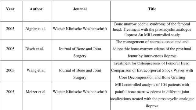
Table 3 below shows a small excerpt of the literature dealing with conservative treatment opportunities for femoral head necrosis. There is a large amount of studies, most of them about pharmaceutical therapies. The most commonly used drug in clinical routine is the stable prostacyclin analogue Iloprost. There are also some reports about other conservative methods, but not that many. A limitation of most works is the small patient numbers and the rather short follow up times. Figure 2 shows a chronological timeline of the mentioned literature.
Year Author Journal Title
2005 Aigner et al. Wiener Klinische Wochenschrift
Bone marrow edema syndrome of the femoral head: Treatment with the prostacyclin analogue
iloprost An MRI-controlled study
2005 Disch et al. Journal of Bone and Joint Surgery
The management of necrosis-associated and idiopathic bone-marrow edema of the proximal
femur by intravenous iloprost
2005 Wang et al. Journal of Bone and Joint Surgery
Treatment for Osteonecrosis of Femoral Head:
Comparison of Extracorporeal Shock Waves with Core Decompression and Bone Grafting
2005 Meizer et al. Wiener Klinische Wochenschrift
MRI-controlled analysis of 104 patients with painful bone marrow edema in different joint localizations treated with the prostacyclin analogue
iloprost
17
2006 Mont et al. Journal of Bone and Joint Surgery
Nontraumatic osteonecrosis of the femoral head:
Ten years later
2011 Jäger et al. International Orthopaedics Efficiency of iloprost treatment for osseous malperfusion
2011 Rajpura et al. Hip International Medical management of osteonecrosis of the hip:
A review 2012 Beckmann et al. Rheumatology International
Infusion, core decompression, or infusion following core decompression in the treatment of
bone edema syndrome and early avascular osteonecrosis of the femoral head 2015 Roth et al. Zeitschrift für Orthopädie und
Unfallchirurgie
S3-Leitlinie. Teil 2: Atraumatische Femurkopfnekrose des Erwachsenen – unbehandelter Verlauf und konservative
Behandlung Table 3: Literature for conservative treatment options
Figure 2: Literature non operative therapies: Iloprost, Extracorporeal Shockwave Therapy (ESWT) and Hyperbaric Oxygen Treatment (HBO)
If the disease is diagnosed in an early stage, it is possible to try conservative methods. It is described in literature that early detected small lesions have a chance to recover spontaneously [10]. Because of that it is often recommended to reduce weight bearing of the affected hip by using a cane or crutches [58]. This may slow down the progression of
18 the disease or lead to a normalization of clinical and radiological findings. It is supposed that the collapse of the joint can be caused by trauma related epiphyseal trabecular impaction or by an insufficiency bone fracture [41]. The weight bearing reduction can be supported by analgesic or anti-inflammatory medication and physical therapy [11].
However, this may be very long lasting or even only delay the collapse of the femoral head. It has been shown that non weight bearing has a clinical success rate of 22.7%, whereas core decompression has a success rate of about 53% [3]. However, the outcome can be improved by the avoidance of risk factors, as discussed before. A lifestyle modification can also help to save the hip joint. This means a reduction of alcohol consumption and a stop of cigarette smoking, as well as a moderate physical training without shock load to the affected hip [8].
1.7.1.1 Drugs
There are several medicaments in use for treatment of osteonecrosis. The most common is the stable prostacyclin analogue Iloprost. It is approved for therapy of critical ischemia secondary to peripheral arteriosclerosis, diabetic angiopathy or pulmonary hypertension [5] [11] [59]. It causes arterial and venous dilatation, reduces capillary permeability and reduces platelet aggregation. Besides the rheological effects on the terminal vascular bed, it has also the ability to reduce the concentration of oxygen radicals and leukotriens [58].
These effects improve microcirculation and so diminish the edema and thereby intraosseous pressure [12]. Administration of Iloprost is only helpful in very early stages, like bone marrow edema and ARCO I or II stages [23] [60]. When administered in these stages, it leads to a fast improvement of clinical symptoms and a significant pain reduction [61] [12]. The drug is given intravenously for five days, with a patient adapted increasing dosage [59]. However, there are several side effects of Iloprost. During infusion it may come to headache, nausea, flush or cardiac effects, like arrhythmia. A therapy with Iloprost is contraindicated during pregnancy, for patients with warfarin or heparin anticoagulation and for patients with peptic ulcers or with cardiological illness [58] [5] [62]. To improve success rate it is also possible to combine the operative core
19 decompression with an Iloprost infusion. Both treatments alone show similar success rates, in combination there are less non responders and a higher success rate [11].
Other medicaments used for therapy of osteonecrosis are statins, which are mainly used in steroid induced ONFH or bisphosphonates, like Alendronate [59] [63].
Bisphosphonates have an antireabsorptive effect, by inhibiting osteoclast activity. Thus it leads to an increased bone density and may prevent subchondral collapse, in order to avoid surgical intervention [64]. Several studies showed that the short term results for Alendronate are mainly in ARCO I or II stages with small lesions quite promising. Pain as well as functionality improved significantly during a therapy with 10 mg Alendronate per day in combination with calcium and vitamin D [64]. In the long term results it could be seen that bisphosphonates given in pre-collapse stages can delay bone collapse up to 4.2 years [23] [59]. Nevertheless, there are some serious side effects, like mucosal irritation of the upper gastro-intestinal-system, osteonecrosis of the jaw bone or atypical femur fractures, which have to be taken into consideration [64].
1.7.1.2 Other conservative measures
There exist some other, less popular nonoperative treatment ideas, like hyperbaric oxygen treatment (HBO) or extracorporeal shockwaves [10]. The hyperbaric oxygen is meant to reverse ischemia, by increasing the oxygen concentration in the tissue [59]. Although there is a pain reduction in the beginning, it has been shown that it cannot prevent subchondral collapse [23].
Extracorporeal shockwave therapy (ESWT) is a controversial discussed treatment option for early ARCO stages or in combination with other joint preserving therapies. It is meant to support neovascularization and regenerative, anti-inflammatory processes [65].
According to current data, ESWT is not able to delay the collapse of the femoral head [23].
20 1.7.2 Operative therapy
In the following tables 4 and 5 and in figure 3 there is given a short chronological overview of the literature about operative treatment of osteonecrosis of the femoral head.
Year Author Journal Title
1955 Koo et al. Journal of Bone and Joint Surgery
Preventing collapse in early osteonecrosis of the femoral head- A randomised clinical trial of core
decompression 1985 Ficat et al. Journal of Bone and Joint
Surgery
Idiopathic Bone Necrosis of the Femoral Head
1994 Adrian et al. Journal of Bone and Joint Surgery
Long term results of core decompression for ischemic osteonecrosis of the femoral head 2005 Aigner et al. Wiener Klinische
Wochenschrift
Bone marrow edema syndrome of the femoral head: Treatment with the prostacyclin analogue
iloprost An MRI-controlled study 2006 Mont et al. Journal of Bone and Joint
Surgery
Nontraumatic osteonecrosis of the femoral head:
Ten years later
2010 Lee et al. Chang Gung Medical Journal Non-traumatic Osteonecrosis of the Femoral Head – From Clinical to Bench 2014 Li et al.
International Journal of Clinical and Experimental Pathology
Comparison of bone marrow mesenchymal stem cells and core decompression in treatment of
osteonecrosis of the femoral head: a meta- analysis
Table 4: Literature Core decompression
21
Year Author Journal Title
2001 Steinberg et al.
Clinical Orthopaedics and Related Research
Core decompression with bone grafting for osteonecrosis of the femoral head
2010 Fang et al. Chinese Journal of reparative and reconstructive Surgery
Treatment of osteonecrosis of femoral head by core decompression combining with autologous
cortical sustaining bone and cancellous bone graft
2013 Kang et al. Yonsei Medical Journal
Clinical results of auto-iliac cancellous bone grafts combined with implantation of autologous
bone marrow cells for osteonecrosis of the femoral head: a minimum 5-year follow-up 2015 Shah et al.
Journal of Clinical Orthopaedics and Trauma
Analysis of outcome of avascular necrosis of femoral head treated by core decompression and
bone grafting Table 5: Literature Cancellous Bone Grafting
Figure 3: Literature operative therapies
22 1.7.2.1 Core decompression
Core decompression is the most common operative hip preserving therapy at the time [57] [66] [58]. The primary aim of this intervention is a reduction of intraosseous pressure, which is meant to be the main pathological factor of osteonecrosis. With a decreased intramedullary pressure, there is a chance for normal blood circulation and so even revascularization is possible. This may lead, at least in theory, to bone remodeling and restitution of the necrosis [11] [5] [67]. The reduced hypertension in the bone explains why most patients have a significant pain reduction after the operation [68] [69]
[70]. Nevertheless, there are many different success rates described in literature. When Ficat first brought up this method, he found an overall clinical success rate of 89.5%. He also showed that outcome is very much dependent on the stage of the necrosis, as he came up with success rates of 93.9% for stage I and 82.5% for stage II. The radiographic results, however, were not that positive. Here could have been shown an improvement rate of 78.9% [9]. Later studies show a wide range of success rates. It is settled somewhere between 63.5% and 84%, but only in early stages of the disease [66] [13] [3]
[10]. In most cases the clinical symptoms of patients can be improved, although radiographic progression of the necrosis is visible. Only in very early stages the disease is reversible. But the earlier core decompression is performed, the better are the long term results for the patients. Size and location of the affected area are also important risk factors for the outcome of core decompression [66] [13] [67].
The procedure of core decompression is quite easy. The patient is in general or spinal anesthesia and the location of the necrotic area is detected with C-arm imaging. Then surgery is performed by a lateral approach. A guide pin is drilled from the proximal lateral femur, about 4 cm below the trochanteric ridge, into the lesion. When aiming is confirmed by C-arm image, a core reamer is drilled over the guide pin and creates a 10 mm bone channel. After that the necrotic bone can be removed [57] [13]. This way of performing the core decompression brings the risk of subsequent collapse of the femoral head, as well as an increased danger of proximal femur fracture, in case of weakening the cortical wall of the femoral neck. A complication rate of about 4% to 10% has been described.
Thus there is also the possibility to perform a multiple drilling with small pins in a fan- shaped way. There seems to be no significant difference in success rates, but the multiple drilling shows less complications, like subtrochanteric fractures or hip joint penetration
23 [67] [8]. However, it is a disadvantage that there can´t be taken histological samples and it is not possible to perform cancellous bone grafting.
As it is crucial to penetrate the necrotic area, there is also the possibility to perform core decompression with a navigation system. It showed that the precision of the drilling could be improved and the radiation time was shorter than in conventional procedure [71] [72].
For the performance there has to be a virtual connection of the fluoroscopic images and the position of the surgical instruments. This makes it necessary to attach markers to the surgical instruments, the patient and the C-arm. Therefore, a reference array with passive reflecting marker spheres is attached to the surface of the patient and the instruments.
Visualization of the necrotic area has to be done by the C-arm fluoroscope, which is connected to the navigation system. After that no further intraoperative images are needed. A standard drill is equipped with a marker-clamp and measured with a special calibration tool in order to inform the navigation system about the length, diameter and position of the tip of the instrument. After the surgical instrument is visualized on the touch-screen monitor, the drill is placed into the bone by continuous online control of the navigation system. Virtual connection of the position of all the reference markers enables the orientation of the drilling guide in two fluoroscopic planes simultaneously. This may lead to a significant reduction in the distance to the target when compared with conventional core decompression (0.4 mm in the navigation group versus 0.88 mm in the conventional group). Another benefit can be seen in the reduced radiation time. There is an obvious difference between navigated core decompression and the traditional procedure. It has been shown that the radiation exposition can be reduced for the patients and the staff from 3.1 seconds to less than one second [71] [72].
Postoperatively partial weight-bearing is recommended for six to eight weeks. During the first period of full weight-bearing, high impact activities should be avoided for about one year. Physiotherapy should be performed to strengthen the muscles and to regain range of motion [14].
Core decompression can also be combined with autologous bone grafting. There exist different kinds of grafts. The first possibility is to transfer vascularized or nonvascularized bone grafts, like a part of the fibula or the tibia. The main goal of these transplants is to strengthen the femoral head, in order to avoid a collapse or a fracture [73] [57] [17]. It is also possible to transfer cancellous bone from the iliac crest, greater
![Table 1: Steinberg classification [44]](https://thumb-eu.123doks.com/thumbv2/1library_info/3739680.1509239/22.892.152.720.446.1109/table-steinberg-classification.webp)
![Figure 1: Schematic depiction of the determination of the Kerboul angle [54]](https://thumb-eu.123doks.com/thumbv2/1library_info/3739680.1509239/24.892.236.625.567.824/figure-schematic-depiction-determination-kerboul-angle.webp)