Design of cancer-selective and nuclei-targeting cell-penetrating peptides and their characterization
Inaugural-Dissertation
zur
Erlangung des Doktorgrades
der Mathematisch-Naturwissenschaftlichen Fakultät der Universität zu Köln
vorgelegt von Anja Gronewold
aus Bonn
Köln 2017
Berichterstatter: Prof. Dr. Ines Neundorf Prof. Dr. Ulrich Baumann
Tag der mündlichen Prüfung: 18. Dezember 2017
Die im Rahmen der vorliegenden Arbeit durchgeführten Experimente und Untersuchungen wurden im Zeitraum von April 2014 bis August 2017 am Institut für Biochemie der Universität zu Köln unter der Anleitung von Frau Prof. Dr. Ines Neundorf durchgeführt.
Nowadays, cancer still remains one of the leading causes of death in the world. Therefore, cancer research is a present and central topic. Current drawbacks in cancer therapeutics are the lack of tumor specificity and the inefficient drug accumulation in cancer cells, resulting in non-specific agents that also affect healthy cells and thus their application provokes many side effects. Many possibilities for cancer cell-selective targeting of drugs are currently in focus.
One includes the use of cell-penetrating peptides (CPPs), which are able to overcome the plasma membrane barrier and to transport various cargoes inside cells. Recently, it was shown for some CPPs, that they can specifically target cancer cells and therefore be used to promote the effective transport of anticancer drugs into various neoplastic cells. Furthermore, a certain intracellular targeting feature to various organelles was presented in the last years, which is important for the successful delivery of certain drugs to their point of action. This makes these amino acid sequences a promising research field in cancer treatment.
In this study, the focus was set to the development and characterization of specific cancer- selective peptides that accumulate in the cell nuclei and can moreover be used to deliver various cargoes to cancer cells.
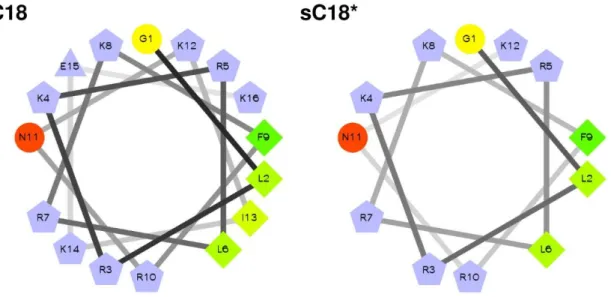
The design of the CPPs was based on the C-terminal domain of the cationic antimicrobial peptide CAP18 (sC18), which was modified in various ways. For instance, it could be demonstrated that dimerization of the sequence and the N-terminally introduction of nuclear targeting sequences led to higher uptake rates of the CPP. Furthermore, the branched variant (sC18)2 exhibited a certain cancer cell-selectivity and was able to disrupt the neoplastic plasma membranes resulting in higher cytotoxicity. Next to this, the combination of sC18 with N50 and NrTP - two sequences that specifically target the cell nuclei - are approving the intracellular trafficking into the cell nucleus in cancer cells. Furthermore, an application as drug delivery system in breast cancer cells was elucidated.
Krebs ist eine der häufigsten Todesursachen weltweit, weshalb die Forschung nach neuen Therapieansätzen zur Krebsbekämpfung eine zentrale Rolle annimmt. Die maßgeblichen Nachteile heutiger Therapien sind sowohl die ungezielte Ansteuerung von Krebszellen als auch eine ineffiziente Anhäufung der Medikamente in Tumoren. Das macht viele Therapeutika zu nicht zufriedenstellenden Lösungen, da diese ebenfalls gesunde Zellen angreifen und daher sehr viele Nebeneffekte mit sich bringen. Zurzeit sind viele andere Herangehensweisen, um Krebszellen spezifisch anzusteuern, im Fokus der Wissenschaft. Eine davon ist der Einsatz von zellpenetrierenden Peptiden (cell-penetrating peptides, CPPs), die in der Lage sind, sich selber in Zellen einzuschleusen und zusätzlich Wirkstoffe in diese zu transportieren. Kürzlich konnte für einige dieser CPPs gezeigt werden, dass sie Krebszellen spezifisch ansteuern und gleichzeitig als Transporter für Zytostatika dienen können. Außerdem haben einige Peptide die Eigenschaft, in speziellen Zellorganellen wie dem Zellkern zu akkumulieren, was nötig ist, um spezielle Medikamente an ihren Wirkungsort zu transportieren.
In dieser Arbeit sollten Peptide entwickelt werden, die selektiv gegenüber Krebszellen sind.
Weiterhin wurde untersucht, ob sich diese Peptide selektiv im Zellkern anlagern und als Transportsystem genutzt werden können.
Das Design der neuen CPPs basierte auf dem Peptid sC18, eine von der C-terminalen Domäne des antimikrobiellen kationischen Peptides CAP18 abgeleiteten Sequenz. Dieses zellpenetrierende Peptid wurden auf unterschiedlichen Wegen modifiziert. Es konnte gezeigt werden, dass eine Dimerisierung über die Seitenkette eines Lysins zu höheren Aufnahmeraten und einer Lokalisation der Peptide im Zellkern führt. Für das verzweigte (sC18)2 konnte eine erhöhte Krebsselektivität gezeigt werden. Das Peptid sorgt für eine Permeabilisierung der Zellmembran von Krebszellen und induziert dadurch eine gewisse Zytotoxizität.
Außerdem wurde sC18 durch das N-terminale Kuppeln von speziellen Zellkern-ansteuernden Sequenzen modifiziert. Die Kombination des Peptids mit N50 und NrTP – zwei Peptidsequenzen, die in der Lage sind, den Zellkern selektiv anzusteuern – führte zu einer erhöhten Anreicherung der Peptide im Nukleus von Krebszellen. Zudem konnte ein erfolgreicher Transport eines Therapeutikums in Brustkrebszellen gezeigt werden.
Abstract ... I Zusammenfassung ... II Table of contents ... III
1. INTRODUCTION ... 1
1.1 Overcoming cell membrane barriers ... 1
1.2 Commonly used cell-penetrating peptides ... 2
1.3 Cellular uptake mechanisms of cell-penetrating peptides ... 3
1.4 Cell-penetrating peptides for selective targeting of cancer cells ... 6
1.5 Application of CPPs as delivery vectors in cancer cells ... 7
1.6 Specific targeting of organelles using CPPs ... 8
1.7 Preliminary work ... 10
2. AIMS OF THE THESIS ... 12
3. MATERIALS AND METHODS ... 13
3.1 Materials ... 13
3.1.1 Chemicals ...13
3.1.2 Instrumentations...13
3.1 Methods ... 15
3.1.1 Peptide synthesis ...15
2.3.1.1Automated peptide synthesis ...15
2.3.1.2Coupling procedure with Oxyma/DIC ...16
2.3.1.3 Kaiser test (after Kaiser et al. 1970) ...16
2.3.1.4Cleavage of a Dde protecting group ...17
2.3.1.5Cleavage of an Fmoc protecting group ...17
2.3.1.6 Coupling of carboxyfluorescein ...17
2.3.1.7Coupling of DOTA and NODAGA ...18
2.3.1.8Sample cleavage ...18
2.3.1.9 Full cleavage ...18
2.3.1.10 Analytical high performance liquid chromatography- electrospray ionization mass spectrometry (HPLC/ESI-MS) ...19
2.3.1.11 Preparative reverse phase high performance liquid chromatography (RP- HPLC)...19
2.3.2 CD spectroscopy ...20
2.3.3.2Splitting and seeding cells ...21
2.3.3.3Freezing and thawing cells ...21
2.3.4 Cell viability assay based on resazurin ...22
2.3.5 Cell lysis assay based on LDH release ...22
2.3.6 Microscopy ...23
2.3.7 Flow cytometry ...23
2.3.8 Radiolabeling of the NODAGA-coupled peptides and uptake experiments ...24
2.3.9 Plasmid transformation in E.coli and plasmid isolation ...24
2.3.10 Electrophoretic mobility shift assay...25
2.3.11 Examination of transfection with pEGFP-N1 plasmid ...25
2.3.12 Examination of transfection with pGL4.13 plasmid ...25
2.3.13 Cargo delivery of actinomycin D or doxorubicin ...26
4 RESULTS AND DISCUSSION ... 27
4.1 Linear and branched dimeric versions of sC18 ... 27
4.1.1 CD spectroscopy of linear and truncated versions ...31
4.1.2 Uptake studies using different sC18 variants ...33
4.1.3 Cytotoxicity profile of linear and truncated versions ...35
4.1.4 Membrane interaction studies ...39
4.2 Characterization of a CPP with potential anticancer activity ... 43
4.2.1 Microscopic analysis of uptake at 4 °C ...43
4.2.2 Uptake studies using flow cytometry ...44
4.2.3 Uptake studies with radiolabeled NODAGA-coupled peptides ...45
4.2.4 Tracking the intracellular fate of the CPPs after cellular uptake ...47
4.2.5 Triggering endosomal release in HEK-293 cells ...49
4.2.6 Relevance of membrane composition ...52
4.2.7 Uptake and cytotoxicity screening on various cell lines ...56
4.2.8 Cargo delivery of actinomycin D ...60
4.3 Specific targeting of nuclei and nucleoli using modified CPPs ... 63
4.3.1 Peptide synthesis and analysis of the secondary structure ...63
4.3.2 Internalization rates of nuclei-targeting sequences ...65
4.3.3 Defining the intracellular localization cells after internalization...67
4.3.4 Microscopic analysis of uptake at 4 °C ...73
4.3.5 Defining the intracellular localization of CPPs ...74
4.3.7 Complexation capability with pEGFP-N1 and pGL4.13...77
4.3.8 Transfection of MCF-7 and HeLa cells ...78
4.3.9 Cargo delivery of Doxorubicin ...81
5 CONCLUSION AND OUTLOOK ... 86
5.1 Summary and conclusion of studies with linear and branched dimeric sC18 versions ... 86
5.2 Summary and conclusion of (sC18)2 studies ... 87
5.3 Summary and conclusion of N50 and NrTP studies ... 89
6 REFERENCES ... 91
7 ATTACHEMENT ... 101
7.1 List of abbreviations ... 101
7.2 List of figures ... 105
7.3 List of tables ... 107
7.4 Erklärung ... 108
1. INTRODUCTION
1.1 Overcoming cell membrane barriers
About four billion years ago, the first cells and with them, the primary cell membranes emerged.
Cellular plasma membranes have a lot of duties and functions towards the cell. It is a semi- permeable lipid bilayer, which defines the cell shape and separates the intracellular space from the surrounding environment, protecting the cell from invading pathogens and toxic substances. [1, 2] Due to their particular biological structure, the membrane is only permeable for small hydrophobic compounds with less than 1,000 Daltons [3]. Uptake of large or hydrophilic molecules can only occur through transmembrane protein channels and transporter molecules. Furthermore, cellular membranes are involved in various physiological processes such as signal transduction, energy synthesis and cell-cell communication. [4, 5]
Since the plasma membrane has extraordinary properties in protecting the cell from the surrounding environment, the problem occurs that delivery processes of drugs, proteins and nucleotides into cells and compartments are limited. Several studies about strategies to overcome membrane barriers have received major attention in the last years. Penetration of the cellular plasma membrane can either be achieved by the use of certain biological or chemical compounds, like prodrugs, carriers and permeation enhancers, or by physical units generating an energy-dependent disruption of the membrane. [2] Within this, cell membranes have already been permeabilized by sonoporation, resulting in a successful gene delivery to HeLa and PC-3 cells [6, 7]. However, for most of the physical methods, a power source is necessary. Therefore, they can be potentially expensive, which reduces the attraction of these methods. A variety of biological approaches like prodrugs, liposomes and nanoparticles were recently developed and investigated. In this context, liposomes were shown to transport anti- cancer, anti-fungal and antibiotic drugs as well as anti-inflammatory agents and gene medicines into a variety of cells [8]. Moreover, PEGylated nanoparticles have been used to deliver proteins and peptide-drugs in vivo [9].
Another group of transport vehicles has been discovered in the last decades; these are short peptides, able to cross membranes in a receptor-independent manner. These so-called cell- penetrating peptides (CPPs) have the ability to interact electrostatically with biological membranes and internalize into a number of different cell types, like microorganisms, fungi, mammalian and plant cells. [10, 11] In general, they are known to be usually positively charged, water soluble and partly hydrophobic. Advantages of these peptides are their small size, good biocompatibility and their easy and cost-effective synthesis. Furthermore, CPPs can be easily
modified concerning, for example their affinity, hydrophobicity, charge, solubility, and stability.
Additionally, they do usually not evoke an immune response. [12, 13] The most important feature of these recently discovered peptides is certainly their capability to efficiently transport different cargos into cells [14]. Anyway, also disadvantages of CPPs are known, like their low cell, tissue and organ specifity and their limited stability in vivo. Moreover, another problem to overcome is their often occurred internalization by endocytosis and the need of a subsequent endosomal release, essential for the transport of peptides and cargos to their targets. [15]
Nevertheless, cell-penetrating peptides represent a promising and precious tool for the transport and internalization of otherwise impermeable molecules.
1.2 Commonly used cell-penetrating peptides
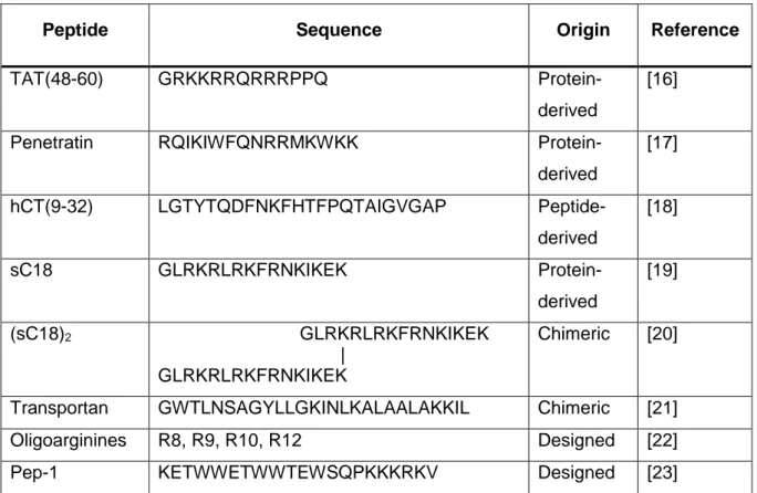
Cell-penetrating peptides can be divided into different subgroups based on their specific properties. One classification is based on the origin of the peptide sequence divided in protein- derived, chimeric or synthetic peptides. [10] An overview about some CPPs and their sequences is presented in table 1.
Table 1: Examples for commonly used cell-penetrating peptides and their origin.
Peptide Sequence Origin Reference
TAT(48-60) GRKKRRQRRRPPQ Protein-
derived
[16]
Penetratin RQIKIWFQNRRMKWKK Protein-
derived
[17]
hCT(9-32) LGTYTQDFNKFHTFPQTAIGVGAP Peptide-
derived
[18]
sC18 GLRKRLRKFRNKIKEK Protein-
derived
[19]
(sC18)2 GLRKRLRKFRNKIKEK |
GLRKRLRKFRNKIKEK
Chimeric [20]
Transportan GWTLNSAGYLLGKINLKALAALAKKIL Chimeric [21]
Oligoarginines R8, R9, R10, R12 Designed [22]
Pep-1 KETWWETWWTEWSQPKKKRKV Designed [23]
The peptides TAT and penetratin are examples for CPPs that are derived from natural proteins.
TAT(48-60) is a cationic and hydrophilic peptide derived from the transcription activating factor
of human immunodeficiency virus (HIV-1), that is able to translocate through the plasma membrane and accumulate in the cell nucleus. [16] Also penetratin is derived from a transcription factor, precisely from the Drosophila Antennapedia homeodomain, and is an extensively studied cell-penetrating peptide. Penetratin is declared to be a secondary amphipathic peptide, meaning that its amphipathic structure is only formed when the CPP is interacting with phospholipid membranes. [17] Furthermore, also a hydrophobic peptide, derived from the human peptide hormone calcitonin (hCT) has been reported to translocate membranes. The shorter fragment hCT(9-32) is able to transport small organic molecules as well as proteins into cells. The efficient delivery of DNA and RNA has been reported for hCT(18-32), which was further combined with a lysine- and arginine-rich sequence (k7), representing an example for a chimeric peptide built from a peptide-derived and designed sequence. [18, 19, 24] Another well-studied CPP is derived from a cationic antimicrobial protein from rabbit leucocytes, which is assumed to inactivate pathogens by permeabilizing their membranes. This cathelicidin-derived, short peptide sequence from the C-terminal domain is called sC18 and exhibits an α-helical structure. It was already demonstrated that its uptake rates are comparable to those of TAT(48-60) and hCT(18-32)-k7. [19, 25] Furthermore, also a branched, dimeric version of sC18 was examined by Hoyer et al. with even higher internalization capacity and an increased cytotoxicity. (sC18)2 is also able to efficiently transport cytostatic compounds into tumor cells. [20]
Transportan is classified as a chimeric CPP, which is composed of two sequences, mastoparan and galanin. Mastoparan is known as a wasp toxin that can penetrate and lyse cell membranes, while galanin is a neuropeptide, which inhibits the release of neurotransmitter from vesicles. Their chimeric combination connected via a lysine results in a cell-penetrating peptide with high internalization rates. [21] Examples for synthetically designed peptides are Pep-1 and oligoarginines. The amphipathic peptide Pep-1 was shown to deliver unbound active proteins or peptides into cells and different oligoarginines were reported to translocate through membranes due to their high hydrophilicity. [22, 23]
1.3 Cellular uptake mechanisms of cell-penetrating peptides
The cellular uptake of cell-penetrating peptides is a complex process, which is intensively studied. Unfortunately, the general uptake mechanism is still not fully understood, since the internalization of peptides can occur in several ways simultaneously. Moreover, the uptake is dependent on the peptide sequence, the concentration, temperatures as well as the cell type investigated. In addition, specific cargoes attached to the CPP can also influence the uptake mechanism. Generally, two different ways of CPP entry are discussed – the direct translocation over the membrane and the uptake via endocytosis. [26, 27]
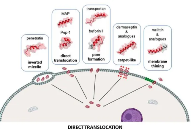
Direct penetration is an energy-independent process that can even occur at low temperatures when all endocytotic uptake routes are repressed. Hereby, it is thought that the mainly positively charged peptides interact electrostatically with negatively charged components of biological membranes, inducing finally the insertion of the peptide in the membrane and resulting in cell entry. In this context, the internalization by inverted micelles, direct translocation, pore formation, carpet-like model and membrane thinning can be differentiated as shown in figure 1.
Figure 1: Overview of the main mechanisms of cellular internalization used by cell-penetrating peptides among non-endocytotic direct penetration ways, with representative corresponding examples. [28]
For penetratin for example, the translocation via inverted micelles has been shown, whereby the membrane is destabilized and a curvature is induced. The formed micelle is then enclosing the peptides in its interior and the disruption of the inner membrane is resulting in the release of the CPPs into the cytoplasm. [29] Pore-formation, which is presumed for transportan can arise via the barrel stave or the toroidal model. The CPPs organize in barrel-like structures, where the hydrophilic residues of the sequences form the pore, or as in the toroidal model, the peptides and headgroups of membrane lipids built the pore together. [10]
Also the endocytic uptake pathway, which is an energy-dependent process, can be classified in various mechanisms like macropinocytosis, clathrin- or caveolin-dependent or -independent
endocytosis as depicted in figure 2. After endosomal uptake, the CPPs and CPP-cargo conjugates are enclosed in vesicles. The following endosomal release is a challenging mechanism, and also indispensable for the effect of many drugs taken up by endocytosis. [30]
Moreover, the uptake and release depends, among others, on the size and physicochemical nature of the cargo molecule [31].
Figure 2: Overview of the main mechanisms of cellular internalization used by cell-penetrating peptides among endocytotic-based mechanisms, with representative corresponding examples.
[28]
For the well-studied TAT(48-60) peptide, various uptake routes have been demonstrated including a direct penetration, but also macropinocytosis, as well as clathrin- or caveolin- independent internalization could be presented [28]. During macropinocytosis an inward folding of the plasma membrane to the outer surface takes place and results in a vesicle inside the cell [32]. The different uptake ways are dependent on concentration, time of incubation and composition of membrane phospholipids. [33, 34] NrTP analogues, which are also used in this thesis, crosses the cell membrane by clathrin-dependent endocytosis. [35]
For the cathelicidin-derived CPP sC18, an endocytotic uptake pathway is supposed, but a more precise declaration is still in study [19]. Also for the dimeric variant (sC18)2, internalization by endocytosis was postulated, which was further investigated in this study [20].
1.4 Cell-penetrating peptides for selective targeting of cancer cells
Cancer remains one of the main causes of death globally, and it is one of the most common in the United States responsible for nearly 25% of deaths. A majority of treatments includes the surgical removal of the tumor tissue. Furthermore, chemo- and radiotherapy are used to inhibit tumor growth – nevertheless, a major issue is that also healthy tissue is affected by these therapeutical approaches. [36, 37] The major drawbacks of cancer therapeutics are not only the lack of tumor specificity, but also drug resistance, inefficient drug accumulation and cancer cell heterogeneity. The traditional chemotherapeutic systems poorly distinguish between cancer and non-cancer cells, leading to non-negligible side effects. [12] Hence, a broad field of scientific studies try to find solutions to specifically target cancer cells and prevent the affection of healthy cells.
From this point of view, CPPs have been intensively studied and discussed, since for some of them showed a tumor targeting ability. Interaction, binding and translocation depends on the components of the extracellular matrix of the cell membrane as well as on their sugar and lipid composition. In fact, plasma membranes of cancer and non-cancer cells differ from each other, as neoplastic cell membranes have an increased amount of anionic molecules such as the phospholipid phosphatidylserine, sialylated gangliosides, O-glycosylated mucins and heparin sulfate. This results in a more negative net charge in contrast to healthy mammalian cell membranes, which occur mostly zwitterionic. [38, 39] Especially positively charged CPPs are able to interact with the more negatively charged cancer cell membranes, which is primarily responsible for their tumor cell selectivity. The positively charged, amphipathic peptide NK-2, which is derived from the cationic core region of an antibacterial effector protein from porcine immune cells, specifically attacks cancer cells that have a high content of phosphatidylserine.
Healthy human lymphocytes are not harmed by this membranolytic peptide. [40] Lim et al.
presented in 2013 a derivative of the anticancer peptide buforin IIb with extraordinary cancer- specificity. This peptide BR2 targets cancer cells by interaction with gangliosides, which are found in high amounts within tumor cells, and translocates the membrane via lipid-mediated macropinocytosis. [41]
Another strategy to specifically target cancer cells is the combination of cell-penetrating peptides with homing peptides, which are able to recognize particular cell types. Homing peptides are often membrane impermeable, which could be advanced by the conjugation to CPP sequences. Tumor homing peptides usually interact with receptors that are overexpressed on the cancer cell surface like integrins, somatostatin, transferrin or epidermal growth factor receptors (EGFR). [12] The conjugation of a CPP with S3, an EGFR-binding domain from vaccinia virus growth factor, increases the tumor selectivity extremely. The uptake
rates of the conjugate were 80-times higher than of the peptide alone, whereas the internalization in non-cancer cells remained at a low level. [42] The tripeptide Arg-Gly-Asp (RGD) is often used for the specific targeting of integrins because this sequence is specifically recognized by the transmembrane receptors. Recently, a study was published, whereby an RGD reverse sequence was conjugated to octaarginine. This conjugation peptide could specifically bind to integrin αvβ3, which is highly upregulated in some tumors, and translocate into various cancer cells with increased uptake rates compared to the controls. [43, 44]
1.5 Application of CPPs as delivery vectors in cancer cells
Very recently, in last August 2017, the company Novartis introduced the first chimeric antigen receptor T cell therapy on the market, which is based on gene transfer for the treatment of B-cell precursor acute lymphoblastic leukemia [https://novartis.gcs-web.com/novartis- receives-fda-approval-for-KymriahTM (29.09.2017)]. For this immunocellular therapy, named Kymriah, the patient's T-cells were taken and transduced with a lentiviral vector encoding a specific antigen receptor for the overproduced B-cells in this type of cancer. Transferring the cells back to the patient, they are able to express the antigen and the immune system starts to fight the cancerous B-cells. [45] In clinical phase II, 83% of the patients treated with the new therapy achieved complete remission, even if the treatment is associated to many side effects [Novartis media release, Basel, August 30, 2017].
A challenging process in this new treatment is the transfection of the T-cells with the lentiviral vector. In general, it is not just important to target specific cells with an application system, but also the transport capability of these systems should be reliable. Anyway, based on this example, there are some promising methods to treat cancer besides the use of conservative therapies like resection or chemotherapy.
Also cell-penetrating peptides received attention as potential candidates for anticancer drug delivery systems. [12] These peptides were shown to transport biologically active cargoes such as drugs, proteins, peptides, nanoparticles and nucleic acids into cells. CPPs were also used as anticancer peptidic vaccines in various approaches. [46, 47] Li et al. presented the amphipathic peptide CADY-1, which is able to form a stable complex with the cytotoxic anti- cancer drug doxorubicin (DOX) and triggers its transport over the cell membrane increasing its toxicity. This CADY-1/doxorubicin complex even exhibits anti-tumor activity in animals. [48]
Also TAT-DOX conjugates were investigated, showing a high decrease in viable cells compared to the incubation with TAT or DOX alone. They were even able to overcome the multidrug resistance of cervical cancer cells by circumventing drug efflux. [49] A human ovarian cancer cell line that is resistant to the chemotherapeutic agent taxol was treated efficiently with a conjugate of taxol and octaarginine. The approach to overcome this taxol resistance was
next to cell culture also observed in mouse models with ovarian cancer. [50] The CPP BP16 with delivery features for the anticancer drug chlorambucil was presented by Soler et al. The efficacy of chlorambucil increased between 6 and 9-times when linked to BP16 in various cancer cells. [51] Also other compounds used in tumor therapy were transported to target cells, for example organometallic compounds, like cisplatin, were efficiently carried by the well- studied CPP Transportan 10 to two different cell lines, whereas no internalization of the drug in non-cancer cells could be observed [52].
For the above described recently introduced immunocellular gene therapy, a delivery of nucleic acids, in this case a DNA vector, is necessary. In this topic, a high number of studies with successful delivery by CPPs could be published. Mussbach et al. demonstrated in 2011 that various cell-penetrating peptides in non-covalent complexes with nucleoside phosphates were able to transport them in high amounts into a cervix cancer cell line. Their binding to functional proteins in signal transduction or translation process can induce many intracellular reactions.
[53] The delivery of RNA or DNA is exceedingly challenging, because these nucleic acids need to be non-covalently bound to the CPPs and transported to the nuclei as well. This so-called transfection was first described in 1997 by Morris et al. using a 27 residue-long peptide sequence built from a hydrophobic domain of HIV gp41 and a hydrophilic domain from the nuclear localization sequence of SV40 T-antigen. They showed a rapid and efficient delivery into the nucleus of fibroblasts by the formation of peptide vector/oligonucleotide complexes.
[54] But also in cancer cells, transfection could be successfully conducted by CPPs. The basic amphiphilic peptide ppTG1 is efficiently triggering the transport of vector DNA into HeLa cells.
The luciferase reporter assay presented gene expression rates comparable to standard transfection reagents as polyethylenimine (PEI) and Lipofectin. [55] A short cell-penetrating peptides (RRWQW) from bovine lactoferrin was presented to deliver a plasmid, encoding the enhanced green fluorescent protein (EGFP) into human lung carcinoma cells, which was subsequently expressed. Also the efficiency of this peptide was comparable to the commonly used transfection reagent Lipofectamine. [56]
1.6 Specific targeting of organelles using CPPs
Many cargoes or drugs need in fact to be transported to specific targets within the cell. Some of them are depicted in figure 3. Especially organelles like mitochondria, the Golgi apparatus or the nucleus can be reached combining cell-penetrating peptides with a specific localization sequence. This intracellular targeting is a very promising feature of these peptides.
Figure 3: Uptake mechanisms and intracellular targeting of organelles. [15]
The so called mitochondria-penetrating peptides composed of a CPP and a mitochondria- targeting sequence are of great interest, since a lot of intracellular processes as intermediary metabolism, calcium signaling and apoptosis are controlled by these energy-generating organelles. Furthermore, a high number of human diseases such as Parkinson’s disease are caused by mitochondrial dysfunctions [57, 58]. The uptake and mitochondrial localization of a mitochondria-penetrating peptide in two cancer cell lines (HeLa and MCF-7) could already be demonstrated by Horton et al. [59].
Moreover, of a great interest is the targeting of the cell’s control center, the nucleus. This organelle contains the genetic information and controls cell growth, development and reproduction as well as many other intracellular processes. The nuclei are enveloped by a nuclear membrane that separates the nucleoplasm from the cytoplasm and protects the DNA from the surrounding cytoplasm [60, 61]. For some CPPs, an accumulation in the nuclei could be demonstrated. For example, the well-studied trans-activator of transcription TAT(48-60) is able to translocate through the plasma and nuclear membrane [16]. But also some nuclear localization sequences (NLS) were presented in the last years, natural as well as synthetic ones. NLS sequences can be found in cellular or viral proteins that need to be transported to the nucleus after translation or viral infection of a host cell. They often contain high amounts of lysines and arginines. After covalent attachment of a nuclear localization signal motif of the
Simian-Virus 40 (SV40)-T-antigen (PKKKRK) to a peptide, derived from dermaseptin, which failed to enter the nuclei, the conjugate was accumulating in this compartment of HeLa cells [62]. Lewis et al. showed that the synthesis of a novel CPP consisting of a glutamate-rich peptide (EEEAA) coupled to the nuclear localization signal of the Oct6 transcription factor results in nuclear localization as well as uptake in prostate and pancreatic cancer cell lines [63].
Nuclear factor-κB (NFκB) is a family of structurally related transcription factors bearing an essential role in inflammation, innate immunity and cancer development. The protein complex that controls DNA transcription is imported into the nucleus through the nuclear pores by importin. The NFκB1/p50 subunit contains a hydrophilic N50 motif patterned on its NLS region.
This N50 motif is known to bind the importin α and is therefore responsible for the efficient transport of the transcription factor into the cell nucleus. [64, 65] This N50 peptide sequence (VQRKRQKLMP) was used in this thesis for nuclear targeting of CPPs.
Another desirable target for intracellular delivery of drugs, transcription factors and siRNAs is the nucleolus. A cell contains between one and five of these dense granular structures in the nucleus, which are mostly composed of RNA and proteins. The ribosomal RNA transcription and synthesis of ribosome subunits takes plays in this compartment, and is involved in viral infection, regulation of oncogenesis and tumor suppression. [66, 67] For D-octa-arginine an accumulation in the nucleoli could be reported by Fretz et al. [68]. Lately, NrTP was designed by structural minimization of the rattlesnake toxin crotamine. The peptide sequence showed strong membrane-translocating properties and it was capable to penetrate various carcinoma cells and accumulate in their nucleoli. This sequence contains a lysine-rich palindrome hexad in the middle of the peptide (YKQCHKKGGKKGSG). Also the capability of NrTP for intracellular delivery of large molecules like β-galactosidase has been previously demonstrated. [35, 69]
The NrTP peptide sequence was used in this thesis to target cell nucleoli.
1.7 Preliminary work
This work is based on the studies that were performed during my master thesis. In this context, the dimeric cell-penetrating peptide (sC18)2 was investigated regarding its internalization, toxicity and membrane interaction in comparison to the monomeric sC18.
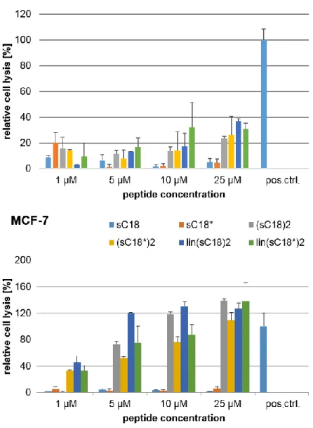
In this recent work, it could be shown that sC18 does not exhibit toxicity to three studied cancer cell lines (MCF-7, HeLa and HCT-15) and the non-cancer cell line HEK-293. In contrast, it could be executed that the toxicity of (sC18)2 is extremely increased when incubated with the cancer cell lines HeLa and MCF-7 compared to HCT-15 and HEK-293 cells. Moreover, (sC18)2
presumably acts via disrupting the plasma membranes of HeLa and MCF-7 cells.
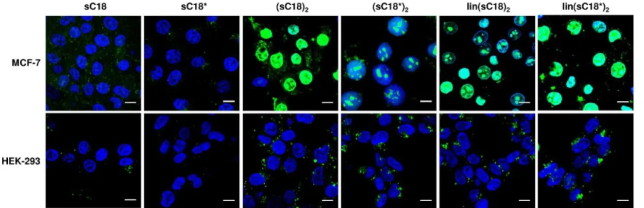
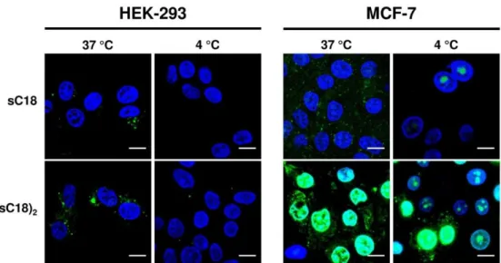
Also the internalization of the dimeric versus the monomeric peptide differed from each other, which was verified by microscopic studies. It was observed in all cell lines that the internalization rates of (sC18)2 were much higher compared to the uptake of the monomeric sC18. Furthermore, a detailed observation of a non-cancer and a cancer cell line presented, that sC18 and (sC18)2 seemed to internalize in HEK-293 cells via an endocytic uptake way.
Afterwards, the peptides were entrapped in the vesicles and did not appear in the nucleus. On the contrary, in MCF-7 cells, the peptides were distributed in the cytoplasm as well as in the nucleus, which could be explained by an endocytotic uptake with followed release from the endosomes or a direct translocation over the plasma membrane. It is also likely that both uptake ways occur simultaneously. The high internalization rates and the accumulation in the nuclei is probably correlated with the cytotoxic effect on MCF-7 cells. The difference in cancer and non-cancer cells was further investigated using an artificial membrane-like system, precisely different composed giant unilamellar vesicles (GUVs). In this context, it was demonstrated that the interaction of the peptides with negatively charged membranes is much higher than with GUVs composed of zwitterionic phospholipids, which is in accordance with the fact that the peptides showed increased penetration rates and lytic effects on the negatively charged cancer cell membranes.
2. AIMS OF THE THESIS
The main goal of this thesis was the development and characterization of cancer-selective and nuclei-targeting cell-penetrating peptides. Therefore, the well-studied CPP sC18 was chosen as candidate and modified in several ways.
In part I (“Linear and branched dimeric versions of sC18”) new variants of sC18 should therefore be developed and characterized. The focus was set on the dimerization of sC18 and its truncated form sC18* (lacking the four C-terminal amino acids). The aim was to investigate the impact of the dimerization strategy that yielded either linear or branched derivatives.
Therefore, all new peptides should be analyzed concerning their cell-penetrating and membrane damaging properties. In this regard, internalization studies via flow cytometer and microscopy, but also cytotoxicity experiments and furthermore, studies with artificial membrane systems should be performed.
The first part revealed that (sC18)2 showed the highest anticancer potential. Therefore, the focus was set on this peptide in part II of the thesis (“Characterization of a CPP with potential anticancer activity”). Within this part, the detailed uptake mechanism and cell selectivity were of great interest. Therefore, uptake studies in different cancer and non-cancer cell lines, studies about the uptake pathway and cargo delivery should be performed. The investigation should initiate with different uptake studies with various peptide concentrations and temperatures. Also flow cytometric analysis should be performed, as well as endosomal release experiments. Next to membrane interaction and disruption studies, also a screening on 16 different cell lines should be performed to further prove the cancer selectivity of the peptide. As a last approach, the drug transport capability should be investigated.
For the delivery of various cargoes, it is important that the transport is not just triggered over the plasma membrane. It is also necessary that the transport vehicle is able to deliver the cargo to its point of action, which could be for example the nucleus. In the third part (“Specific targeting of nuclei and nucleoli using modified CPPs”) the CPP sC18* should be combined with the two nuclear localization sequences N50 and NrTP. These fusion peptides should be investigated concerning their translocation efficiency, nuclear localization and toxicity in MCF-7 and HeLa cells. Furthermore, their capability to form complexes with plasmid DNA and the followed transfection ability should be elucidated, as well as the non-covalently conjugation of an anticancer drug and its delivery into cancer cells.
3. MATERIALS AND METHODS
3.1 Materials
3.1.1 Chemicals
All chemicals, reagents and consumables used in this work were purchased from Alfa Aesar (Karlsruhe, Germany), Applichem (Darmstadt, Germany), Fluka (Taufkirchen, Germany), Merck (Darmstadt, Germany), Roth (Karlsruhe, Germany) Sarstedt (Nürmbrecht, Germany), Sigma-Aldrich (Taufkirchen, Germany) and VWR (Darmstadt, Germany), if not specified otherwise. Nα-Fmoc protected amino acids were obtained from IRIS Biotech (Marktredwitz, Germany).
3.1.2 Instrumentations
Balances Analytical balance FA-210-4, Faust
Laboratory balance SBA52, Scaltec CD spectrometer Jasco Corp. J715
Cell culture clean bench Heraeus Herasafe HS12
Centrifuges Multifuge X1R, Thermo Scientific Pico 17, Thermo Scientific
CO2-Incubator BINDER
ESI mass spectrometer Bruker Esquire HCT
Flow cytometer Guava easyCyte, Merck
Hemocytometer Neubauer improved, Superiour Marienfeld, Germany, Depth 0.100 mm, 0.0025 m2
Heating block Thermomixer compact, Eppendorf
HPLC (analytic) Hewlett Packard Series 1100, Agilent 1100 Series column: Phenomenex Kinetex, 2,6 u, C18, 100A, 100 x 4,60 mm or Machery Nagel, C18ec Elite Lachrom Hitachi Pump L-2130, Elite
HPLC (preparative) Elite Lachrom Hitachi Pump L-2130, Elite Lachrom Hitachi Autosampler L-2200, Elite Lachrom Hitachi Diode Array Detector L-2455, Teledyne ISCO Fraction Collector Foxy R1, column: Phenomenex Jupiter, 4 u Proteo 90 A, 250 x 15 mm, 4 micron or Machery Nagel, C18ec
LC - mass spectrometer LC: Hewlett Packard Series 1100
MS: Finnigan MAT – LCQ or MS: Thermo Scientific LTQ-XL
Lyophilizer Lyovac GT2 with CertomaT SII and a Thermo Savant Refridgerated Vapor Trap RVT5105 or Christ; Alpha 2-4 LDplus
Microscope Inverted microscope (Motic AE31, Wetzlar) 20x objective (N.A. 0.25)) Confocal laser scanning system (Nikon D-Eclipse C1), inverted microscope (Nikon Eclipse Ti), 60x oil immersion objective (N.A. 1.4, Plan PO VC; Nikon)
Multipette® plus Eppendorf Research
NanoDrop NanoDrop™ 1000 Spectrophotometer, Thermo Scientific PCR Thermal Cycler FlexCycler analytikjena
Pipetting aid NeoLab
Pipettes Eppendorf Research
Plate reader Tecan infinite M200
Rotary Shaker KL-2 (Edmund Bühler GmbH)
Spectrophotometer Thermo Scientific Genesys 10S UV-Vis
Speed-Vac Thermo Scientific Speedvac Concentrator Savant SC210A, RVT5105 Refrigerated Vapor Trap VLP80 Vacuum Pump Synthesis robot MultiSynTech Syro I
Thermomixer compact Eppendorf
Vacuum pump VWR
Vortex Scientific Industries – Vortex Genie 2
Water bath Julabo SW22
XcelVapTM Horizon Technology
3.1 Methods
3.1.1 Peptide synthesis
All syntheses were performed in open polypropylene reactor vessels (2 ml syringes) stocked with a fritted filter disc. Manual coupling steps were performed under the hood and solvents were removed from the resin using a vacuum pump.
For each synthesis 15 µmol of resin were used and calculated according to the specific resin loading. The mentioned equivalents correlated with the amount 15 µmol per equivalent.
Following to all coupling steps a Kaiser test was performed to examine the presence of free amino groups and the completeness of a coupling. If the test was positive, the coupling step was repeated.
Since carboxyfluorescein is light sensitive, all experiments including CF-coupling, as well as all following steps were executed avoiding light, and all peptides were protected from light by covering with aluminum foil.
The theoretical molecular masses were calculated using the software Peptide Companion, Version 1.25, CoshiSoft/PeptiSearch or the peptide property calculator by Innovagen, 2015.
2.3.1.1 Automated peptide synthesis
For automated peptide synthesis, a multiple Syro I peptide synthesizer from MultiSynTech was used. The software SyroXP is able to calculate the amounts of amino acids that are needed to synthesize the peptides. All amino acids were dissolved in DMF except from phenylalanine that needed to be dissolved in NMP. After filling the reaction vessels with 15 µmol (30 mg) of Fmoc-Rink amide resin beads and starting the software, the robot performs all synthesis steps automatically using the Fmoc/t-Bu strategy. In the first step, the beads were swollen in DMF and the Fmoc protection group was cleaved from the resin using first a 40% piperidine solution in DMF and afterwards a 20% piperidine solution in DMF. After washing the beads four times
with DMF, coupling of the first Nα-Fmoc protected amino acid was performed using Oxyma and DIC. The amino acid was introduced in 8-fold excess and every coupling step was performed twice, each for 40 min respectively including four washing steps with DMF in between. Before repeating deprotection and coupling of the next amino acid, the sample was washed another time with DMF. In the last step, the vessels were again incubated with piperidine to cleave off all Fmoc protection groups and washed four times with DMF. After the complete synthesis, the samples were washed with CH2Cl2, MeOH and Et2O manually and the resin beads were dried in the Speedvac.
2.3.1.2 Coupling procedure with Oxyma/DIC
For the manual coupling of amino acids, the resin was swollen for 15 min in 1 ml DMF at RT shaking with 200 rounds/min. After removing the solvent with a vacuum pump, a solution containing the amino acid (7 eq. for general amino acids, 2 eq. for Dde-L-Lys-(Fmoc)-OH) and Oxyma (7 or 2 eq.) in 300 µl DMF and 7 or 2 eq. of DIC was added to the resin, respectively.
The solution was incubated for 2 h or o/N at RT and under shaking conditions. After washing the resin five times with DMF, the coupling procedure was repeated. In the end, the solvent was removed, the resin was washed five times with DMF, CH2Cl2, MeOH and Et2O, respectively, and afterwards it was dried for 10 min at low speed in the Speedvac.
2.3.1.3 Kaiser test (after Kaiser et al. 1970)
To monitor whether the coupling was successful and all free amino groups were successfully coupled with an amino acid, a protection group or carboxyfluorescein, a Kaiser test was performed on a few resin beads [70]. After transferring some beads into a 1.5 ml reaction vessel, one drop of the solutions I to III (table 2) was added in quick succession.
Table 2: Chemicals used for the Kaiser test.
Solution I 1.0 g ninhydrin in 20 ml EtOH Solution II 80 g phenol in 20 ml EtOH
Solution III 0.4 ml aq. KCN-solution (1 mM) in 20 ml pyridine
The mixture was incubated at 95 °C for 5 min in a Thermomixer. The test was defined positive, if the solution showed a blue color, indicating free amino groups. If the mixture was yellow, no free amino groups could be detected and the coupling was complete.
2.3.1.4 Cleavage of a Dde protecting group
To cleave the special Dde protecting group, the resin was swollen for 15 min in 1 ml DMF at RT. After removing the solvent, 1 ml of a 3% hydrazine solution in DMF was added to the resin and incubated for 10 min. The solvent was collected in a Falcon tube, the resin was washed twice with DMF and the flow through was harvested in the same Falcon tube. This procedure was performed ten times, collecting just the last phase. The amount of Dde in the first and the last sample was determined by measuring the absorption at 301 nm in a spectrophotometer.
A mixture of 2 ml DMF and 1 ml 3% hydrazine served as reference. If the absorption of the last washing step was lower than 0.1, the entire cleavage of the Dde group was successful. If it was higher than 0.1, a few cleavage steps with 3% hydrazine were repeated. Afterwards the resin was washed five times with DMF, CH2Cl2, MeOH and Et2O, respectively, and dried for 10 min at low speed in the Speedvac.
2.3.1.5 Cleavage of an Fmoc protecting group
Before coupling the following amino acid in the coupling procedure, the Fmoc protecting group had to be cleaved off. Therefore, the resin was swollen for 15 min in 1 ml DMF, the solvent was removed and 500 µl of a 30% piperidine solution in DMF was added. The resin was incubated shaking for 20 min at RT and afterwards washed five times with DMF. The procedure was done twice.
2.3.1.6 Coupling of carboxyfluorescein
For the modification of the peptide with the reporter group carboxyfluorescein, the resin was swollen as described previously and after removing the solvent, a solution was added that contained 10 eq. 5(6)-carboxyfluorescein and 10 eq. HATU in 300 µl DMF. Afterwards, 10 eq.
DIPEA were pipetted directly to the resin. The syringe was shaken at RT for 2 h. After washing the resin five times with DMF the coupling was repeated o/N using Oxyma and DIC as described above. Thereafter, the resin was washed five times with DMF, CH2Cl2, MeOH and Et2O respectively and dried for 10 min at low speed in the Speedvac. The successful coupling was verified by a Kaiser test.
If the test was negative, a polymer cleavage was performed to avoid CF polymers. Therefore, the resin was swollen in DMF, 1 ml of a 20% piperidine solution in DMF was added and the syringe was incubated shaking for 45 min at RT. In the end, the resin was washed five times with DMF, CH2Cl2, MeOH and Et2O, respectively, and dried for 10 min at low speed in the Speedvac.
2.3.1.7 Coupling of DOTA and NODAGA
The N-terminal coupling of the chelators DOTA (1,4,7,10-tetraazacyclododecane-1,4,7,10- tetraacetic acid – Sigma Aldrich) and NODA-Ga(tBu)3 (4-(4,7-bis(2-tert-butoxy-2-oxoethyl)- 1,4,7-triazonan-1-yl)-5-tert-butoxy-5-oxopentaoic acid – CheMatech / Dijon, France) was carried out in the same way as described for CF except from using 3 eq. of the substance to be coupled and activating with 3 eq. HATU/DIPEA in DMF under vigorous shaking for 2 h. A double coupling process was not desired.
After full cleavage and preparative HPLC (chapter 2.3.1.9 and 2.3.1.11), a salt exchange was done to eliminate the TFA salts in the peptide solution. Therefore, the lyophilized CPPs were dissolved in 5 ml of 80 mM HCl solution and incubated at RT shaking for 1 h. After freezing the sample for 30 min at - 80 °C, it was dried o/N by lyophilisation. The procedure was repeated two to four times, until no TFA adducts could be detected via analytical ESI-MS spectrometry anymore.
2.3.1.8 Sample cleavage
A sample cleavage was done to ensure that the manual coupling of amino acids, CF or chelator and the synthesis of the robot was successful. Some dry beads of the resin were transferred to a 1.5 ml tube and 2.5 µl of the scavenger triisopropylsilane mixed with 2.5 µl H2Odd was added. After the addition of 95 µl TFA using a multipette, the syringe was incubated at RT shaking for 3 h. Peptide sequences that contain cysteine, methionine or tryptophan were cleaved from the resin with 7 μl thioanisole, 3 μl 1,2-ethandithiole and 90 µl TFA. The peptide was precipitated in 1 ml of ice-cold diethyl ether and the mixture was incubated for at least 20 min at - 20 °C. The samples were centrifuged at 10,000 x g for 5 min at 4 °C. After removing the supernatant, the pellet was again resuspended in 1 ml ice-cold ether and centrifuged for 5 min at 10,000 x g at 4 °C. This procedure was repeated at least five times until no scavenger smell could be noted anymore. The pellet was then dried for 10 min at low speed in the Speedvac. After resuspending the peptide in 100 µl H2O or H2O/tBuOH (3:1 (v/v)) according to the solubility, the sample was centrifuged for 1 min at 10,000 x g to separate the beads from the peptide and the supernatant was analyzed via HPLC/ESI-MS (chapter 2.3.1.10).
2.3.1.9 Full cleavage
To cleave the peptides from the resin, a full cleavage was performed. A mixture of 25 µl of the scavenger triisopropylsilane and 25 µl H2Odd was added to the resin. After the addition of 950 µl TFA using a multipette, the syringe was incubated at RT shaking for 3 h. Peptide sequences that contain cysteine, methionine or tryptophan were cleaved from the resin with 70 μl
thioanisole, 30 μl 1,2-ethandithiole and 900 µl TFA. To precipitate the peptide, the solvent was pressed through the fritted disc in a 15 ml Falcon tube containing 10 ml of ice-cold diethyl ether. Furthermore, 200 µl of TFA were added to the resin and also harvested in the tube. After incubation for at least 20 min at - 20 °C to allow precipitation of the peptide, the sample was centrifuged at 5,000 x g for 5 min at 4 °C. The supernatant was removed, the pellet was resuspended in 10 ml ice-cold diethyl ether and the solution was again centrifuged. This procedure was repeated five times until no scavenger smell could be noted anymore. The pellet was then dried for 15 min at low speed in the Speedvac and afterwards resuspended in 2 ml H2O or H2O/tBuOH (3:1 (v/v)), respectively. The peptide solution was transferred into a balanced glass vessel and covered with paper. After freezing the sample for 30 min at - 80 °C, it was freeze-dried o/N by lyophilisation. The amount of the peptide could then be detected by weight and after subtraction of the TFA residues that bind to all free NH2-ends and the side chains of the positively charged amino acids arginine and lysine.
2.3.1.10 Analytical high performance liquid chromatography-
electrospray ionization mass spectrometry (HPLC/ESI-MS)
For the identification of the synthesized sequences, a high performance liquid chromatography/electrospray ionization mass spectrometry (HPLC/ESI-MS) method was performed. After sample or full cleavage from the resin, peptides were usually diluted 1:10 with 10% ACN/90% H2O containing 0.1% trifluoroacetic acid or formic acid, depending on the chosen method. Generally, the peptides were separated on the column with a flow rate of 6 ml/min via an increasing linear gradient from 10 – 60% ACN in H2O (inclusive 0.1% TFA or FA). After the HPLC, the sample passed the electrospray ionization source, whereby ions were generated and their m/z ratios were determined.
2.3.1.11 Preparative reverse phase high performance liquid chromatography (RP-HPLC)
After lyophilisation, the dry peptide, but not more than 30 mg, was dissolved in 960 µl of ACN/H2O (incl. 0.1% TFA) according to the initial ratio of gradient and centrifuged in a 1.5 ml tube for some seconds to separate the sample from possible debris. The supernatant was transferred to a bellied HPLC glass vessel.
The purification of the peptide was done with a preparative HPLC and the software EZChromElite. First, the column was equilibrated with ACN/H2O (incl. 0.1% TFA) according to the start gradient. Afterwards, the concentration of ACN was increased gradually and the peptide containing fractions were collected. The solvent was removed via evaporation in the XcelVap at 65 °C using a gradient of 880 – 1640 mbar twice for 30 min, respectively.
Afterwards, 15 µl were taken for the analytical HPLC and the rest was transferred to balanced glass vessels and covered with paper. After freezing the samples for 30 min at - 80 °C, they were freeze-dried by lyophilisation o/N.
According to the weight and the purity, the dried peptides were dissolved in H2Odd to a concentration of 1 mM regarding the accumulation of TFA on every arginine, lysine and free NH2-group at the terminal end.
2.3.2 CD spectroscopy
For the structural characterization of the synthesized peptides, circular dichroism spectra were recorded. The cell-penetrating peptides were analyzed in 10 mM potassium phosphate buffer (pH 7.0) with and without TFE (1:1 dilution), respectively. Therefore, 20 µM peptide solutions in the two different buffers were measured in 3 accumulations in the CD spectrometer according to the apparatus instructions. The peptides were measured in a 0.1 cm quartz cuvette with a sensitivity of 100 mdeg in the range from 260 to 180 nm in 0.5 nm intervals. The scanning mode was continuous and a scanning speed of 50 nm/min was chosen. The results of pure buffer were subtracted from the spectra of the peptides. The ratio between the molar ellipticity at 222 nm and 207 nm is a good indicator to confirm an α-helical structure of peptides.
For α-helices, these R-values should be around a value of one. [71]
2.3.3 Cell culture methods
All cell culture work was done in sterile conditions under the sterile hood wearing disinfected gloves. All working materials that were transferred under the hood were disinfected with 70%
EtOH before using. Autoclaved pipette tips and sterile serological pipettes were used for the cell culture work. A vacuum pump was utilized to remove media during washing or splitting steps.
All cell lines were grown in sterile culture dishes (diameter 10 cm) in a CO2-Incubator (5% CO2) at 37 °C. The cell line HEK-293 was grown in MEM containing 15% Fetal Bovine Serum (FBS) and 4 mM L-Gln. The cell lines HeLa, HCT-15 and MCF-7 were grown in RPMI-1640 medium containing 10% FBS and 4 mM L-Gln. For PC-3 cells, DMEM F-12 Ham’s medium was completed with 4 mM L-Gln and 5% FBS. HaCaT cells were grown in high glucose DMEM supplemented with 2 mM L-Gln and 10% FBS. Each medium was also prepared without FBS.
2.3.3.1 Mycoplasma testing
All used cell lines were tested at least once a year but anywhere after taking new cell lines in culture using the PCR Mycoplasma Test Kit II and the Superhot Taq DNA Polymerase provided
![Figure 3: Uptake mechanisms and intracellular targeting of organelles. [15]](https://thumb-eu.123doks.com/thumbv2/1library_info/3708599.1506295/17.892.143.757.130.560/figure-uptake-mechanisms-intracellular-targeting-organelles.webp)