Characterization of CAMTA1 and Nkx2.2 in the context of glioblastoma cancer stem cell biology
Dissertation zur Erlangung des Doktorgrades der
Naturwissenschaften (Dr. rer. nat.) der Fakultät für Biologie und Vorklinische Medizin der Universität Regensburg
vorgelegt von Ludwig Wankerl
aus Regensburg
im Jahr 2015
II
Das Promotionsgesuch wurde eingereicht am:
03. Juli 2015
Die Arbeit wurde angeleitet von:
Prof. Dr. rer. nat. Gunter Meister
Unterschrift:
III
Our greatest weakness lies in giving up.
The most certain way to succeed is always to try just one more time.
Thomas A. Edison
IV
V
Abstract
Glioblastoma multiforme (glioblastoma) is the most malignant type of tumor in the human brain. For glioblastomas and several other cancer types, the existence of so called
"cancer stem cells" was described. Cancer stem cells are characterized by stem cell-like traits such as the capacity of self-renewal and differentiation. Moreover, there is evidence that these cells contribute to cancer recurrence after therapy.
Several molecular-pathological factors are crucial for the emergence of glioblastomas in general and for glioblastoma cancer stem cells in particular. For example, certain microRNAs (miRNAs) such as the miRNA pair miR-9/9* are specifically enriched in glioblastoma cancer stem cells. This miRNA pair acts as oncogene for glioblastomas by repressing the tumor suppressor Calmodulin-binding transcription activator 1 (CAMTA1).
In this thesis, the upstream and downstream pathways of the CAMTA1 transcription factor were investigated in terms of their relevance in cancer biology. Therefore, siRNAs and antibodies against CAMTA1 were generated and established. Beyond that, a potential interaction partner of CAMTA1, Nkx2.2, was identified. Nkx2.2 is an important transcription factor for neural development and is also a potential tumor suppressor for glioblastomas. The tumor-suppressive effect of Nkx2.2 reported previously was further confirmed by this thesis. In this regard, it was shown that an overexpression of Nkx2.2 affected glioblastoma cancer stem cells negatively and impaired the proliferation of glioma cells. Additionally, a positive correlation between Nkx2.2 expression and survival of glioma patients was revealed.
Furthermore, it was hypothesized that Nkx2.2 and CAMTA1 act in concert to stimulate the transcription of NPPA, which encodes for the precursor of the natriuretic
VI – Abstract
peptide (ANP). This activation was thought to be the main mechanism for these two transcription factors to cause their tumor-suppressive effects. Consistently, it was shown that an overexpression of Nkx2.2 elevated the NPPA mRNA levels and that Nkx2.2 can strongly induce the NPPA promoter.
Nkx2.2 was further characterized in terms of additional interaction partners and a potential regulation by miRNAs.
The findings of this thesis allow to postulate a model which describes a possible regulatory network important for understanding glioblastomas, in particular the formation and traits of glioblastoma cancer stem cells. This will advance therapy strategies that "tackle" these persistent cells.
VII
Zusammenfassung
Glioblastoma multiforme (Glioblastom) ist eine Tumorform, die im Gehirn auftritt und unter den Gehirntumoren den höchsten Grad an Malignität aufweist. Für Glioblastome und diverse andere Krebsarten wurde die Existenz von sog. "Krebs-Stammzellen"
nachgewiesen. Krebs-Stammzellen zeichnen sich durch ähnliche Charakteristika aus wie Stammzellen von normalem, gesundem Gewebe, d.h. sie sind fähig, sich zu differenzieren und selbst zu erneuern.
Verschiedene molekular-pathologische Faktoren tragen zur Entstehung von Glioblastomen bzw. deren Krebs-Stammzellen bei. So sind beispielsweise bestimmte mikroRNAs (miRNAs) wie das bifunktionelle miRNA-Paar miR-9/9* vermehrt in diesen Krebs-Stammzellen exprimiert. Dieses spezielle miRNA-Paar reprimiert den Tumorsuppressor Calmodulin-binding transcription activator 1 (CAMTA1) und wirkt dadurch onkogen für Glioblastome.
In dieser Arbeit sollte das zelluläre Netzwerk von CAMTA1, durch welches die tumor- suppressive Wirkung hervorgerufen wird, näher untersucht werden. Dazu wurden siRNAs und Antikörper gegen CAMTA1 generiert und auf ihre Funktionalität überprüft. Darüber hinaus konnte der Transkriptionsfaktor Nkx2.2 als ein potentieller Interaktionspartner von CAMTA1 identifiziert werden. Nkx2.2 spielt bei der Entwicklung des zentralen Nervensystems eine wichtige Rolle und gilt ebenfalls als potentieller Tumorsuppressor für Glioblastome. Diese tumor-suppressive Wirkung von Nkx2.2 wurde in einer früheren Studie gezeigt und konnte durch diese Arbeit bestätigt werden: Eine Überexpression von Nkx2.2 beeinflusste Glioblastom-Krebs-Stammzellen negativ und verminderte deutlich die Proliferation von Glioma-Zellen. Zudem konnte
VIII – Zusammenfassung
ein Zusammenhang zwischen einer erhöhter Nkx2.2-Expression und einer höheren Überlebensrate von Patienten mit Gliomen festgestellt werden.
In dieser Arbeit wurde die Hypothese aufgestellt, dass CAMTA1 und Nkx2.2 miteinander interagieren, um die Transkription des Gens NPPA zu steigern. NPPA kodiert für den Vorläufer des atrialen natriuretischen Peptids (ANP). Die Aktivierung des NPPA-Gens stellt vermutlich einen zentralen Mechanismus dar, über den die beiden Transkriptionsfaktoren CAMTA1 und Nkx2.2 ihre tumor-suppressive Wirkung vermitteln. Es konnte gezeigt werden, dass eine Überexpression von Nkx2.2 tatsächlich zu einer Erhöhung der NPPA-Expression führte und dass Nkx2.2 in der Lage ist, den Promotor dieses Gens zu aktivieren.
Ferner wurde untersucht, ob für Nkx2.2 weitere potentielle Interaktionspartner identifiziert werden können und ob Nkx2.2 durch miRNAs reguliert wird.
Mit den in dieser Arbeit erbrachten Ergebnissen lässt sich ein Modell entwerfen, wie die Entstehung von Glioblastoma multiforme und dessen Krebsstam-Zellen durch einen Regulationsmechanismus potentiell unterdrückt wird. Dieses Modell kann dazu beitragen, die molekularen Mechanismen für die Entstehung von Glioblastomen, insbesondere von deren Krebs-Stammzellen, besser zu verstehen. Dadurch lassen sich möglicherweise neue Therapieansätze entwickeln.
IX
Contents
Abstract ... V Zusammenfassung ... VII Contents ...IX List of Figures ... XIV List of Tables ... XVI List of Abbreviations ... XVII
1. Introduction ... 1
1.1. Cancer stem cells ... 1
1.2. Glioblastoma multiforme ... 3
1.3. The role of microRNAs in glioblastoma biology ... 7
1.3.1. miRNA biogenesis and miRNA-directed transcript regulation ... 7
1.3.2. Several miRNAs are important for glioblastoma progression and glioblastoma cancer stem cell maintenance ... 8
1.4. The Calmodulin-binding transcription activator 1 is a tumor suppressor for glioblastomas ... 11
1.4.1. The CAMTA family of transcription factors: Protein organization ... 11
1.4.2. Function of CAMTA proteins from plants to mammals ... 14
1.4.3. CAMTA1 acts as tumor suppressor in neural tumors ... 16
1.5. Nkx2.2: A glioblastoma tumor suppressor candidate ... 19
X – Contents
1.5.1. The Nkx protein family: Transcription factors essential for development ... 19
1.5.2. Nkx2.2 is important for neural development and a potential tumor suppressor protein ... 21
1.6. Aims and working model for this thesis ... 24
2. Results ... 27
2.1. Characterization of CAMTA1 ... 27
2.1.1. Establishing of efficient CAMTA1 knockdown strategies ... 27
2.1.2. Polyclonal antibodies against CAMTA1 ... 29
2.1.3. Localization of CAMTA1 ... 37
2.1.4. A potential CAMTA1 interaction partner: Nkx2.2 ... 44
2.2. Characterization of Nkx2.2 ... 50
2.2.1. Nuclear localization of Nkx2.2 ... 50
2.2.2. Nkx2.2 interaction partners ... 51
2.2.3. Investigation of a putative regulation by microRNAs ... 54
2.2.4. The effect of Nkx2.2 on glioma tumor biology ... 60
2.2.4.1. Nkx2.2 overexpression leads to decreased neurosphere formation of primary glioblastoma cells ... 60
2.2.4.2. Nkx2.2 expression correlates with glioma outcome ... 63
2.2.4.3. Nkx2.2 overexpression shows an antiproliferative effect on stable glioma cells ... 66
2.2.5. NPPA as a potential Nkx2.2 target gene: Only Nkx2.2 with an intact homeobox binding domain induces the NPPA promoter... 68
3. Discussion ... 71
3.1. Characterization of CAMTA1 ... 71
3.1.1. Establishment of an siRNA-mediated CAMTA1 knockdown and a potential function for alternative CAMTA1 transcripts ... 71
Contents – XI
3.1.2. Functionality of the polyclonal antibodies against CAMTA1 ... 73
3.1.3. Interaction between CAMTA1 and Nkx2.2 ... 74
3.2. Characterization of Nkx2.2 ... 76
3.2.1. Nuclear localization of Nkx2.2 and identification of interaction partners ... 76
3.2.2. Is Nkx2.2 regulated by miRNAs? ... 77
3.2.3. Function of Nkx2.2 as a tumor suppressor for gliomas ... 80
3.3. Expanding the working model ... 88
4. Material and Methods ... 91
4.1. Materials ... 91
4.1.1. Consumables and Chemicals ... 91
4.1.2. Buffers ... 92
4.1.3. Enzymes ... 92
4.1.4. DNA-oligonucleotides ... 93
4.1.5. RNA-oligonucleotides ... 97
4.1.6. Plasmids ... 98
4.1.7. Antibodies ... 101
4.1.8. Bacterial strains and bacterial growth medium ... 101
4.1.9. Human cell lines ... 103
4.1.10. Software and databases ... 103
4.1.11. Devices ... 105
4.2. Methods ... 106
4.2.1. Working with DNA ... 106
4.2.1.1. Polymerase chain reaction ... 106
4.2.1.2. Molecular cloning and sequencing ... 108
4.2.1.3. Quantitative real-time polymerase chain reaction (qRT-PCR) ... 110
XII – Contents
4.2.2. Working with RNA ... 111
4.2.2.1. Isolation of RNA from cultured cells ... 111
4.2.2.2. Reverse Transcription ... 111
4.2.2.3. Annealing of siRNA-oligonucleotides for RNA interference ... 112
4.2.3. Working with proteins ... 112
4.2.3.1. Preparation of whole cell protein lysates ... 112
4.2.3.2. Immunoprecipitation ... 115
4.2.3.3. Generation of nuclear extracts ... 116
4.2.3.4. Determination of protein concentrations ... 118
4.2.3.5. SDS-polyacrylamide gel electrophoresis ... 119
4.2.3.6. Coomassie and silver staining of SDS-polyacrylamide gels ... 119
4.2.3.7. Western blot ... 120
4.2.3.8. Mass spectrometry ... 122
4.2.3.9. Purification of recombinant expressed GST-tagged proteins via fast protein liquid chromatography ... 123
4.2.3.10. Generation and purification of polyclonal antibodies ... 124
4.2.3.11. Immunofluorescence ... 126
4.2.4. Human cell culture ... 127
4.2.4.1. Cultivation of human cell lines ... 127
4.2.4.2. Transfection ... 128
4.2.4.3. Proliferation assays ... 130
4.2.4.4. Neurosphere assays using primary glioblastoma cell lines ... 130
4.2.5. Dual luciferase reporter assays ... 131
4.2.5.1. Validation of promoter regulation by transcription factors ... 131
4.2.5.2. Validation of miRNA binding sites in 3'-UTRs ... 132
Contents – XIII
4.2.6. Analysis of molecular-pathological data from glioma patients ... 133
5. References ... 135
Appendix ... 151
Danksagung ... 161
XIV
List of Figures
Figure 1.1: The concept of cancer stem cells (CSCs) ... 2
Figure 1.2: Suggested cellular origins of human brain tumors (medulloblastomas and gliomas) ... 5
Figure 1.3: Cultured primary glioblastoma cells form neurosphere-like clones when cultured under stem cell-like growth conditions ... 6
Figure 1.4: miRNA biogenesis and miRNA-induced transcript silencing ... 8
Figure 1.5: The CAMTA family of transcription factors ... 13
Figure 1.6: Nk-2 class of homeobox-binding domain containing transcription factors ... 20
Figure 1.7: Working model for this thesis. ... 25
Figure 2.1: Genomic locus of CAMTA1 and siRNAs against CAMTA1 ... 28
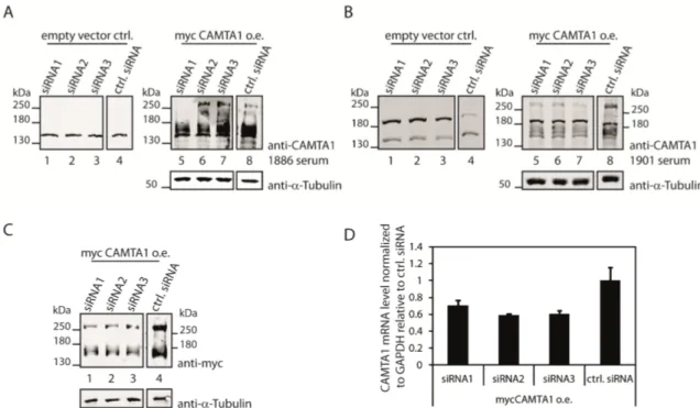
Figure 2.2: Testing the CAMTA1 antibodies 1886 and 1901 for their functionality to detect endogenous CAMTA1 in western blots ... 29
Figure 2.3: The CAMTA1 antibodies 1886 and 1901 can both detect overexpressed mycCAMTA1 .... 31
Figure 2.4: Generation of new CAMTA1 antibodies ... 33
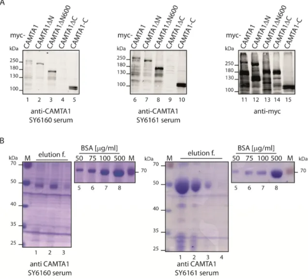
Figure 2.5: Testing and purifying the generated CAMTA1 antibody sera ... 34
Figure 2.6: Testing the purified new CAMTA1 antibodies for their functionality to detect endogenous CAMTA1 ... 35
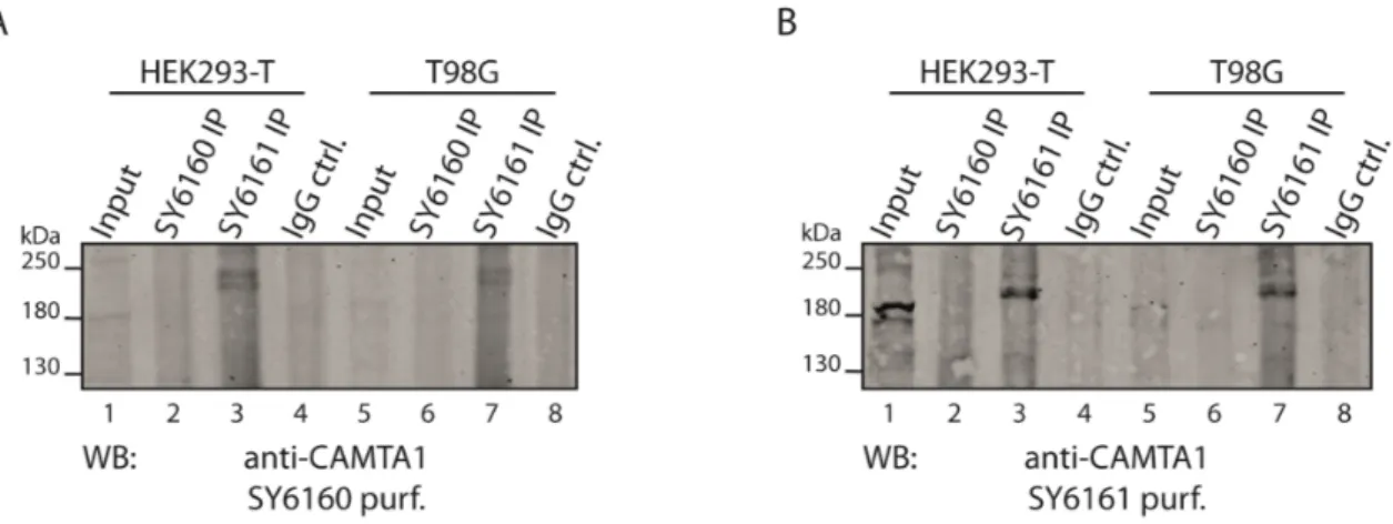
Figure 2.7: Immunoprecipitation of endogenous CAMTA1 from HEK293-T and T98G cells via the generated antibodies. ... 36
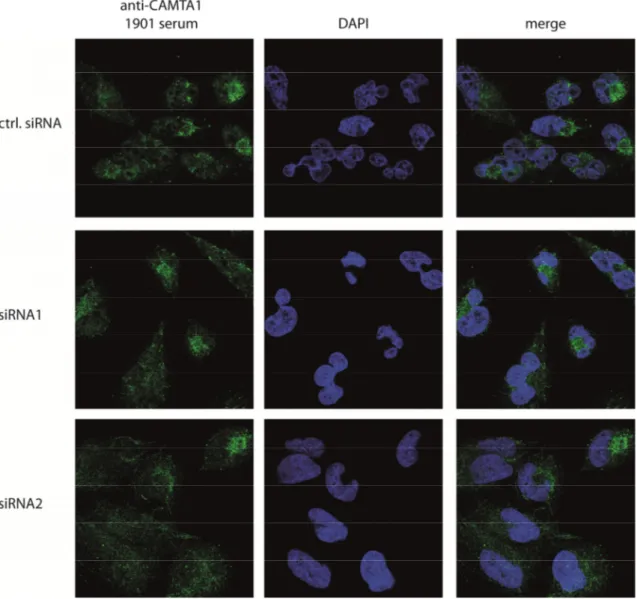
Figure 2.8: Immunofluorescence of endogenous and knocked down CAMTA1 using the 1886 antibody ... 38
Figure 2.9: Immunofluorescence of endogenous and knocked down CAMTA1 using the 1901 antibody ... 39
Figure 2.10: Immunofluorescence of endogenous and knocked down CAMTA1 using the purified SY6160 antibody. ... 40
Figure 2.11: Immunofluorescence of endogenous and knocked down CAMTA1 using the purified SY6161 antibody ... 41
List of Figures – XV
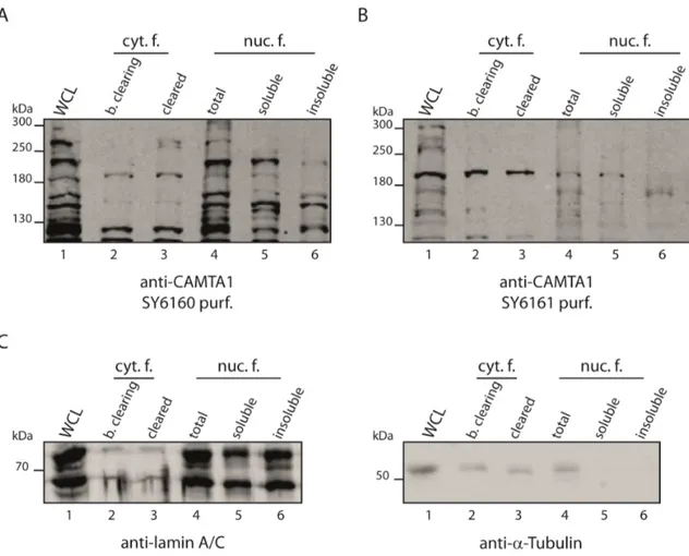
Figure 2.12: Subcellular fractionation of HEK293-T cells for CAMTA1 localization ... 43
Figure 2.13: Subcellular fractionation of HeLa-S3 suspension culture cells to detect endogenous CAMTA1 by the generated antibodies ... 44
Figure 2.14: Interaction between mycCAMTA1 and FLAG/HA-Nkx2.2 ... 45
Figure 2.15: Mapping the interaction between CAMTA1 and Nkx2.2 ... 47
Figure 2.16: Immunoprecipitation of endogenous CAMTA1 to investigate a potential interaction with Nkx2.2 in LNT229 cells ... 48
Figure 2.17: Immunoprecipitation of endogenous Nkx2.2 to investigate a potential interaction with CAMTA1 in LNT229 cells ... 49
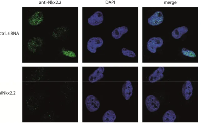
Figure 2.18: Nuclear localization of endogenous Nkx2.2 in LNT229 cells ... 51
Figure 2.19: Screening for potential Nkx2.2 interaction partners ... 53
Figure 2.20: Nkx2.2 as a potential target for miR-17-5p and miR-106b ... 56
Figure 2.21: Investigation via dual luciferase reporter assays if Nk2.2 is generally regulated by miRNAs in HeLa and T98G cells ... 59
Figure 2.22: Nkx2.2 reduces the formation neurosphere-like colonies ... 62
Figure 2.23: Effect of a mutated or deleted Nkx2.2 homeobox-binding domain on neurosphere formation of ZH161 cells ... 63
Figure 2.24: Nkx2.2 expression correlates with survival of glioma patients ... 65
Figure 2.25: Nkx2.2 causes an antiproliferative effect and elevates the expression of NPPA ... 67
Figure 2.26: Nkx2.2 is able to induce the NPPA promoter ... 69
Figure 3.1: The 3'-UTR of Nkx2.2 has additional binding sites for miR-26a and miR-182 ... 79
Figure 3.2: Expanded working model ... 89
XVI
List of Tables
Table 2.1: Results of the mass spectrometric analysis of the FH-Nkx2.2 IP ... 54
Table 4.1: Used kits listed with the corresponding application ... 92
Table 4.2: DNA-oligonucleotides used as primers for qRT-PCR ... 93
Table 4.3: DNA-oligonucleotides used for molecular cloning and sequencing ... 94
Table 4.4: RNA-oligonucleotides ... 97
Table 4.5: Used plasmids ... 99
Table 4.6: Cloned constructs ... 100
Table 4.7: Bacterial strains ... 101
Table 4.8: Used antibodies ... 102
Table 4.9: Human cell lines ... 103
Table 4.10: Software and databases ... 104
Table 4.11: Frequently used devices ... 105
Table 4.12: Templates used for PCR ... 107
Table A.1: Complete mass-spectrometric analysis of FH-Nkx2.2 (co-)immunoprecipitates. ... 151
XVII
List of Abbreviations
A
A (deoxy-)adenosine or ampere (context-dependent)
aa amino acid
AEBSF 4-(2-Aminoethyl) benzenesulfonyl fluoride hydrochloride
APS ammonium persulfate
Arabidopsis or at Arabidopsis thaliana AS antisense
ATP adenosine 5'-triphosphate
B
bp base pair(s)
BSA bovine serum albumin C
C (deoxy-)cytidine C. elegans or ce Caenorhabditis elegans
cDNA complementary DNA
cGMP cyclic guanosine monophosphate
CSC(s) cancer stem cell(s)
CSC(s) cancer stem cell(s)
C-terminus carboxyl-terminus D
d deoxy- or day(s) (context-dependent)
DAPI 4',6-diamidino-2-phenylindole DMEM Dulbecco’s Modified Eagle’s Medium
DMSO dimethyl sulfoxide
DNA deoxyribonucleic acid
DNase deoxyribonuclease Drosophila or dm Drosophila melanogaster
XVIII – List of Abbreviations
DTT dithiothreitol E
E. coli Escherichia coli
EDTA ethylenediaminetetraacetic acid
ERK extracellular-signal-regulated kinases F
F FLAG-tag
FBS fetal bovine serum
G
G (deoxy-)guanosine
g gram or standard gravity ( ≈ 9.81 m/s2) (context-dependent) GAPDH glyceraldehyde 3-phosphate dehydrogenase
GFP green fluorescent protein
GMP guanosine monophosphate
GST glutathione S-transferase
GTP guanosine 5'-triphosphate
H
H HA-tag HA hemagglutinin
HBS HEPES buffered saline
HEPES 4-(2-hydroxyethyl)-1-piperazineethanesulfonic acid HMGA2 high-mobility group AT-hook 2
hs or hsa Homo sapiens I
IgG immunoglobulin G
IF immunofluorescence IP immunoprecipitation IPTG isopropyl β-D-1-thiogalactopyranoside K
kb kilo bases (1,000 bp) L
LB lysogeny broth
M
MEK mitogen-activated protein kinase kinase
List of Abbreviations – XIX
mm Mus musculus
mRNA messenger RNA
N
N-terminus amino-terminus O
OD optical density
P
PA polyacrylamide
PABP poly(A)-binding protein
PACT protein activator of the interferon-induced protein kinase PBS phosphate buffered saline
PCR polymerase chain reaction
Q
qRT-PCR quantitative real-time PCR R
r ribo-
Ras rat sarcoma
RNA ribonucleic acid
RNase ribonuclease S
S sense SDS sodium dodecyl sulfate siRNA small interfering RNA T
T (deoxy-)tymidine
TBS TRIS-buffered saline
TEMED tetramethylethylenediamine TEV tobacco etch virus
TRBP transactivation-responsive RNA-binding protein TRIS tris(hydroxymethyl)-aminomethan
tRNA transfer RNA
U
U uridine or units (context-dependent)
UTR(s) untranslated region(s)
XX – List of Abbreviations
UV ultraviolet V
v/v volume per volume
W
w/v weight per volume
WB western blot
X
X-gal 5-bromo-4-chloro-3-indolyl-β-D-galactopyranoside
Units were abbreviated according to the international system of units (SI-units). For amino acids 3-letter or 1-letter abbreviations were used according to the recommendations of the International Union of Pure and Applied Chemistry (IUPAC).
1. Introduction
1.1. Cancer stem cells
Cancer describes a very diverse disease, which can emerge in almost every tissue of the human body. The progression of the several types of cancer is rather complex and involves disruptions of various cellular networks on different levels including alterations of the genome, transcriptome, proteome, and/or interactome.
In cancer research, it was thought for a long time that tumors are bulks of cells characterized by de-regulated proliferation. However, during the last twenty years, this simplified view of cancer has drastically changed: Many tumors form sophisticated and heterogeneous, almost organ-like, tissues which are sometimes also called "tumor microenvironment". This microenvironment is essential for the survival of the tumor and also for its distribution throughout the body by metastasis or migration. For example, it recruits blood vessels to supply tumor cells with nutrients and oxygen or it protects the tumor against the immune system (1).
But how can one single transformed cell or a small population of abnormal cells generate such a diversity? A likely explanation for this would be given by the existence of so-called cancer stem cells (CSCs) or tumor initiating cells (TICs). It was postulated that these CSCs behave like their normal counterparts in the body, i.e. these cells are capable of self-renewal and differentiation as normal stem cells are (Figure 1.1). By means of these stem cell-like abilities such cancer cells can constantly renew the tumor and generate the heterogeneous, differentiated tumor mass (1, 2).
The cancer stem cell hypothesis emerged at the end of the last century when for human acute myeloid leukemia a distinct, relatively small population of cells was identified (3, 4). The isolated cells differed from other cells of this cancer type regarding their
2 1. Introduction
phenotype and functionality: These cells expressed the same pattern of cell surface proteins as normal hematopoietic stem cells. Furthermore, they were capable of self- renewal and promoted the formation of human leukemia when these cells were injected into (immuno-deficient) mice (3, 4). In 2003, CSCs were also found for breast cancer (5) and for brain tumors (6). The latter CSCs were isolated from human brain tumors (gliomas and medulloblastomas) and characterized by the expression of the cell surface protein CD133 (6, 7), which is normally a specific marker for neural stem cells (8).
Remarkably, the injection of only 100 CD133-positve (CD133+) cells into brains of immuno-deficient mice was sufficient to initiate the formation of new tumors, whereas even the injection of 100,000 CD133-negative (CD133-) could not (7). Characteristics of CSCs in human brain tumors, particularly in glioblastoma multiforme, are described in more detail below (see paragraph 1.2.).
This hierarchical concept of a small subpopulation of CSCs and the residual differentiated tumor cells forming the bulk of the tumor was also reported for further types of cancer such as pancreatic (9), colon (10), or lung cancer (11). Hence, today, the cancer stem cell hypothesis is widely accepted.
Figure 1.1: The concept of cancer stem cells (CSCs). CSCs possess the abilities of self-renewal and to differentiate comparable to normal stem cells. In the tumor, CSCs represent a relatively small sub-population whereas the tumor mass is constituted of more differentiated cancer cells.
[Figure adapted and modified from (12)].
1.1. Cancer stem cells 3 Interestingly, signaling pathways such as Sonic Hedgehog (SHH) or Notch, which are important for differentiation from pluripotent stem cells and strictly regulated during normal development, are often found to be de-regulated in CSCs (2, 13). This also illustrates that CSCs seem indeed to be abnormal counterparts of normal stem cells. But what kind of cell is transformed into a CSCs? Up to now, this remains largely unknown and depends probably on the cellular origin from which the tumor emerges. It is generally assumed that cells in every phase of their individual differentiation process can possibly transform into a CSCs (Figure 1.1) (1, 2).
1.2. Glioblastoma multiforme
Origin, molecular characteristics, and therapy
Glioblastoma multiforme1 is not only the most frequent brain tumor in humans but also the most malignant. The World Health Organization (WHO) classifies human brain tumors into four grades due to their malignancy (14). In this regard, glioblastoma belongs to the highest WHO grade IV (lower-grade gliomas are for example astrocytomas) (14). Indeed, the median survival after diagnosis with best proceeding therapy is only 15 months, which drastically illustrates its malignancy. One reason for the fatal prognosis for glioblastomas is the fact that their cancer cells show a high degree of diffuse invasiveness into surrounding healthy brain tissue. Although the occurrence of glioblastomas is rare compared to other forms of cancer, glioblastomas are the most common type of all malignant gliomas (60 - 70 %) and they represent about 30 % of all human brain tumors (15-17).
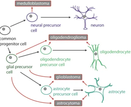
As the name "glioblastoma" already implicates, it is thought that the origin of this brain tumor are most likely glia cells or their progenitor cells (16). Figure 1.2 illustrates the potential lineage origin of human brain tumors including glioblastomas. It also shows that glioblastomas are histological heterogeneous due to their cellular lineage origins.
This heterogeneity can also be observed on the molecular-pathological level. In this
1 Hereinafter shortened called glioblastoma
4 1. Introduction
regard, Verhaak et al. classified glioblastomas, on the basis of genomic aberrations and corresponding alterations in expression levels of a large number of genes, into four distinct subtypes: classical, mesenchymal, proneural, and neural (18). The classical subtype is characterized by a massive EGFR amplification (encodes for the epidermal growth factor receptor). A hemizygous deletion of the NF1 containing locus (encodes for neurofibromin 1, a known tumor suppressor) is the main feature of the mesenchymal subtype (18). For the proneural subtype, a significant higher (compared to the other subtypes) amplification for Pdgfra (encodes for Pdgf receptor α, a receptor tyrosine kinase) as well as point mutations in IDH1 (encodes for isocitrate dehydrogenase 1) were observed (18). The neural subtype is characterized by the expression of certain neuron markers, e.g. neurofilament light polypeptide (NEFL) (18). Furthermore, Verhaak et al. linked all these characterized subtypes to cellular lineages of normal brain cell types: The proneural class reflected a oligodendrocytic signature but not the astrocytic signature which was in contrast associated with the classical type (18). The neural subtype showed both signatures as well as a neuron expression pattern and the mesenchymal type was associated with astroglial lineage (18).
Moreover, glioblastomas can be classified into primary and secondary glioblastomas:
Primary glioblastomas arise de novo (i.e. due to a transformation of a normal cell of the brain), whereas secondary glioblastomas originate from lower-grade gliomas such as astrocytomas. Most glioblastomas are primary (15, 16, 19). Although there is no difference between primary and secondary glioblastomas concerning their morphological and malignant features, distinctions were identified at the molecular level: For example, primary glioblastomas show a frequent amplification of EGFR, in contrast to secondary glioblastomas which are characterized by TP53 mutations (encodes for the tumor suppressor p53) (15, 16, 19).
Besides removal by surgery (which is often only possible to a limited extent without causing damage in healthy brain regions), combined radio- and chemotherapy applying the alkylating reagent temozolomide is the usual treatment (20). However, even with optimal therapy the survival of patients can only be extended for a few months.
1.2. Glioblastoma multiforme 5
Figure 1.2: Suggested cellular origins of human brain tumors (medulloblastomas and gliomas).
Red arrows indicate the possible transformation of a normal cell. Lineage origin based on histological and/or molecular features. It was suggested that glioblastoma emerge in the subventricular zone in the brain where (in adults) neural stem and progenitor cells can be found (21). [Figure adapted from (16)].
Glioblastoma cancer stem cells
As already mentioned in paragraph 1.1., CSCs expressing CD133 were identified about ten years ago in glioblastomas. In this work, Singh et al. showed that CD133-positive cells effectively induce the emergence of new tumors when they are injected into mouse brains. Notably, the newly formed tumors contained both CD133+ and C133- cells although only CD133+ cells were injected. This demonstrated that the CD133-positive cells can differentiate (7). Furthermore, Singh et al. showed a constant renewal and proliferation of the CD133+ pool (7). Besides their capability of self-renewal and differentiation, these cells are also more resistant to irradiation and chemotherapeutics such as temozolomide (22-24). These traits of glioblastoma CSCs gives a possible explanation for the poor therapy success for glioblastomas.
Furthermore, it was demonstrated by Bao et al. that these cells secret the vascular endothelial growth factor (VEGF) which causes angiogenesis, the formation of blood
6 1. Introduction
vessels by the surrounding tissue (25). This is essential for supplying the tumor with oxygen and nutrients.
The population of the CD133-positive CSCs within the tumor is small compared to the other differentiated cells of the tumor. Though, primary and secondary glioblastomas are divergent regarding their respective portion of CD133-positive cells:
Primary glioblastomas contain significantly more CD133-positive cells than secondary glioblastomas (26). However, also glioblastoma CSCs were identified which do not express the CD133 marker but show tumorigenicity to a similar extent as CD133- positive cells (26). Thus, also other (more general) markers were suggested to define glioblastoma CSCs, e.g. the stage specific embryonic antigen 1 (SSEA1 or CD15) (27) or Nestin (28).
When primary glioblastoma cells, in particular CD-133 positive cells, are cultured in vitro under normal stem-cell like growth conditions, they form non-adherent spheres, called "neurosphere-like clones/colonies" (Figure 1.3). In this way cultured glioblastoma CSCs maintain their stem cell-like features and their capability to induce tumor formation in vivo (26, 29, 30).
Figure 1.3: Cultured primary glioblastoma cells form neurosphere-like clones when cultured under stem cell-like growth conditions. A: Primary glioblastoma cells used in this thesis: (1) R11, (2) ZH161. B: Schematic illustration of a neurosphere-like colony.
1. Introduction 7
1.3. The role of microRNAs in glioblastoma biology
MicroRNAs (miRNAs, miRs) are small non-coding RNAs that regulate fundamental cellular processes and are hence crucial factors for development, differentiation, and cell metabolism (31-33). Since miRNAs are important regulators, a de-regulation of miRNA expression is often associated with several types of diseases including cancer.
Such miRNAs are also often referred to as "oncomiRs" (34, 35). On the one hand, oncomiRs can either act as tumor suppressors targeting oncogenes, on the other hand as oncogenes themselves inhibiting certain tumor suppressors (34, 35). Latter oncomiRs are often overexpressed and thus promote cancer progression. This paragraph focuses on several miRNAs (miR-26a, miR-17, miR-9/9*) which act as oncomiRs during glioblastoma progression. But first, the biogenesis of miRNAs and their mechanism of transcript regulation in mammals will be described.
1.3.1. miRNA biogenesis and miRNA-directed transcript regulation
MiRNA genes are normally transcribed by RNA-polymerase II, which leads to primary miRNA transcripts (pri-miRNAs) (36). These transcripts were first processed by a protein complex called microprocessor, which contains the RNase III Drosha and its co-factor DiGeorge syndrome critical region 8 (DGCR8). This processing step results in the miRNA precursor (pre-miRNA), which is characterized by a hairpin structure and a two nucleotide-overhang at the 3'-end (37-39). The precursor is afterwards exported by Exportin 5 from the nucleus into the cytoplasm (40-42), where the hairpin is cleaved off by another RNase III enzyme, Dicer. This yields a double-stranded miRNA (miRNA-duplex) with a length of about 22 nucleotides (43, 44). One strand of the duplex, the mature miRNA, is loaded into a protein of the Argonaute family whereas the other is usually degraded (referred to as miRNA*). Argonaute (Ago) proteins are the core components of the miRNA-induced silencing complex (miRISC) (45). The miRNA within the miRISC guides the silencing complex to the 3'-UTR of its target mRNA, whereas Ago recruits a member of the GW protein family (termed TNRC6A, B, and C in humans). TNRC6 proteins act by inhibiting translation or
8 1. Introduction
inducing mRNA degradation and are therefore important mediators of miRNA-guided target repression (46, 47). For the target recognition by the miRNA itself, the nucleotides 2 to 8 of the miRNA are important. This so called "seed sequence" is complementary to the target site within the particular mRNA (32). The biogenesis of miRNAs and its mRNA-mediated gene silencing are illustrated in Figure 1.4.
Figure 1.4: miRNA biogenesis and miRNA-induced transcript silencing. The first steps of the miRNA biogenesis take place in the nucleus and the maturation process is completed in the cytoplasm by transferring one strand of the miRNA duplex into an Ago protein. A rare event in mammals is a direct cleavage of mRNAs by cleavage-competent Ago2 loaded with a miRNA that is fully complementary to its target (45). This mechanism is comparable to an siRNA-directed knockdown.
1.3.2. Several miRNAs are important for glioblastoma progression and glioblastoma cancer stem cell maintenance
Various studies previously reported altered expression for many different miRNAs in gliomas, respectively in glioblastomas. This paragraph highlights four miRNAs important in glioblastoma biology.
1.3. The role of microRNAs in glioblastoma biology 9
miR-26a
In high-grade gliomas, miR-26a is frequently overexpressed (48). This results from a gene amplification of one of two miR-26a genomic loci (miR-26a-2) (48). Interestingly, this amplification is associated with a monoallelic deletion of PTEN (but was not observed for a homozygous PTEN deletion) (48). PTEN encodes for Phosphatase and Tensin homolog, which acts as a tumor suppressor and is a direct target of miR-26a in glioblastomas (48). It has been suggested that during glioblastoma progression, the monoallelic deletion of PTEN occurs before the gene amplification of miR-26a-2 (48).
The remaining PTEN is then post-transcriptionally repressed by the overexpressed miR-26a. This mechanism is basically equal to a homozygous loss of PTEN.
miR-17
Mammalian primary miRNA transcripts are often polycistronic, i.e. one primary transcript is transcribed from a cluster of various miRNA genes (49). This single primary transcript gives rise to several distinct mature miRNAs. MiR-17 is part of the miR-17~92 cluster. This particular miRNA is a well characterized oncogenic miRNA for several types of cancer (35, 50) and was found to be significantly higher expressed in glioblastomas (51, 52). Moreover, inhibition of all miR-17~92 cluster members in primary glioblastoma cells led to a decrease of proliferation on the one hand and had a stimulating effect on apoptosis on the other hand (52). In addition, miR-17-5p belongs to group of miRNAs, which are specifically enriched in CD133-positive glioblastoma cancer stem cells (53). An inhibition of this particular miRNA significantly reduces formation of neurosphere-like colonies of cultured CD133-postive glioblastoma cells (53). Other miRNAs showing also specific expression in CD133-positive cells are miR- 106b, which is part of a miR-17~92 paralogous cluster (miR-106b~25) and has the same seed sequence as miR-17-5p (50), and the miRNA pair miR-9/9* (53).
10 1. Introduction
miR-9/9*
The miR-9/9* duplex belongs to a type of miRNA duplexes characterized by a functional guide and passenger strand (54). Different from the usual miRNA biogenesis, where the miRNA* strand is normally degraded, both strands are loaded to Ago, yielding two distinct miRISCs each with particular targets.
MiR-9/9* is mainly expressed in the nervous system where it is an important factor for neurogenesis (54, 55). In this context, the transcription factor REST (repressor element-1 silencing transcription factor) was identified to be a target for miR-9 and its co-factor CoREST is regulated by miR-9* (54). REST and CoREST are highly expressed in neural stem cells, but not in differentiated neural cells. By repression of both factors via miR-9/9*, neural stem cells differentiate (54). Furthermore, the specific inhibition of miR-9 strongly decreased the proliferation rate of differentiating neural precursor cells and increased cell migration (55). Both effects are caused by stathmin, which is important for microtubule dynamics and a target of miR-9 (55).
Besides its role in neural development, a de-regulation of miR-9/9*, indicated by high expression levels, was observed in brain tumors like glioblastomas (56) and also in other cancers such as breast cancer (57). For glioblastomas, Schraivogel et al. showed that miR-9/9* is highly abundant in CD133-positive glioblastoma stem cells (53).
Importantly, a specific inhibition of miR-9 and miR-9* led in both cases to a drastic decrease of neurosphere-like clones and to a significant reduction of the CD133- positive cell population (53). Moreover, the inhibition also promoted differentiation of these cancer stem cells (53).
Taken together, these observations by Schraivogel et al., indicate that miR-9/9* are important for maintaining the stem cell character of glioblastoma cancer stem cells.
This raised the question for transcripts regulated by miR-9/9*. In this context, Schraivogel et al. identified the mRNA of the Calmodulin-binding transcription activator 1 (CAMTA1) as a potential target for miR-9/9* (53).
1. Introduction 11
1.4. The Calmodulin-binding transcription activator 1 is a tumor suppressor for glioblastomas
1.4.1. The CAMTA family of transcription factors: Protein organization
Human CAMTA1 belongs to a family of transcription factors, which can be found in plants as well as in animals. Members of this protein family were first described in plants about 15 years ago (58-60). The genome of Arabidopsis thaliana encodes six CAMTA proteins (atCAMTA1-6) whereas both in Caenorhabditis elegans and Drosophila melanogaster respectively one CAMTA protein could be identified (ceCAMTA and dmCAMTA) (60). Mice and humans have two representatives of this family (mmCAMTA1/hsCAMTA12 and mmCAMTA2/hsCAMTA22) (60, 61).
The large CAMTA proteins (e.g. human CAMTA1 has a molecular weight of 180 kDa) contain several domains with distinct functions. The organization of these domains within the protein and their amino acid sequence is highly conserved from plants to human (Figure 1.5) (60, 62). By bioinformatical analysis, the following domains were identified (from the N- to the C-terminus): A DNA-binding domain, called CG-1 domain, a TIG-domain, several ankyrin repeats, and IQ-domains (60, 62).
CG-1 domain
The CG-1 domain was first described for a DNA binding protein in parsley (63) and is characteristic for the CAMTA protein family (60, 62) . In Arabidopsis thaliana a target DNA sequence with a length of 6 bp was identified for atCAMTA1 and atCAMTA3 (59, 64-66). This target with a consensus sequence 5'-(A/C/G)CG(C/T)G(G/T/C)-3' was termed "CGCG-box" (59). Moreover, it was shown that in Arabidopsis abscisic acid-specific cis elements contain this CGCG-box (ABRE and its related coupling element ABRE-CE). These ABREs significantly occur upstream of calcium-induced genes suggesting that ABREs function as calcium-responsive cis elements (64, 67). Also in Drosophila melanogaster this CGCG-box was indentified within the promoter region
2 unless stated otherwise CAMTA1 or CAMTA2 refers always to the human proteins in this thesis
12 1. Introduction
of the dmCAMTA target gene dFbxl4 (68). Interestingly, the CG-1 domain of dmCAMTA mediates dimerization, which is important for nuclear localization of dmCAMTA (69). In mammals it was not known for a long time if the CG-1 domain of the mammalian CAMTA representatives binds to a similar target sequence. But recently, Long et al. identified a consensus sequence for murine CAMTA2 by in vitro binding assays (70). This sequence (5'-(T/C)GCA(G/A/T/C)TGCG-3') is also GC- rich as the consensus-sequence found in Arabidopsis but it differs from the plant sequence. Using reporter assays, Long et al. showed that mmCAMTA1 recognizes this motif and activates the reporter even stronger than CAMTA2 (70). The critical residue for DNA-binding seems to be a single lysine (K141 for mmCAMTA1 and K108 for mmCAMTA2) (70). Furthermore, this study suggests that mammalian CAMTA proteins bind probably as dimers to their target DNA sequence (70), which is in line with the previously reported dimerization of dmCAMTA (69). Bioinformatic analysis revealed a nuclear localization signal (NLS) within the CG-1 domain of almost all members of the CAMTA protein family (Figure 1.5) (60, 62). In contrast, for mmCAMTA2 the NLS was identified next to the C-terminus and a nuclear export signal (NES) in the CG-1 domain (61).
TIG domain
The organization of the TIG (transcription factor immunoglobulin) domain (as well as the ankyrin repeats) of the CAMTA proteins is similar to precursors p100 and p105 of the transcription factor NF-κB. The TIG domain mediates non-specific DNA contacts of NF-κB family members and is also responsible for dimerization of these proteins (71-74).
Ankyrin repeats
Ankyrin (ANK) repeats can be found in NF-κB family members (74), but also in many other eukaryotic proteins. Indeed, ANK repeats belong to the most abundant eukaryotic protein motifs and were identified in many proteins involved in a wide range of cellular functions (75, 76). One single ANK repeat has a length of about 30 amino acids and is
1.4. CAMTA1 is a tumor suppressor for glioblastomas 13 composed of two α-helices (77). The repeats are connected via a β-hairpin consisting of two β-sheets (78). These repeats are known as interfaces for protein-protein interactions (75, 76, 78). Due to their very frequent occurrence in many proteins with crucial cellular function, mutations disrupting protein-protein interactions via ANK repeats are often associated with human diseases such as cancer (75, 76).
Figure 1.5: The CAMTA family of transcription factors. The particular proteins are drawn to scale: For human (hs), Drosophila melanogaster (dm), Caenorhabditis elegans (ce), and Arabidopsis thaliana (at) CAMTAs according to (62), protein information for murine (mm) CAMTAs was retrieved from http://www.uniprot.org/ [(A2A891 (mmCAMTA1) and Q80Y50 (mmCAMTA2)]. The particular, highly conserved, domains are illustrated in different colors:
CG-1 DNA-binding domain, TIG transcription factor immunoglobulin domain (TIG), ankyrin repeats (ANK), IQ domain (IQ), calmodulin-binding domain (CaMBD), and nuclear localization signal (NLS).
IQ domain
The IQ domains, which are located next to the C-terminus of all CAMTA proteins, are known to bind Calmodulin (CaM) or CaM-related proteins, whereby binding can also occur in the absence of calcium (79, 80). These domains are characterized by the repetitive motif IQXXXRGXXXR, hence they are termed "IQ motifs" (79, 80). The number of IQ domains for the respective CAMTA proteins differs within the protein family: For ceCAMTA no IQ domain was identified, whereas atCAMTA6 contains five IQ domains (see also Figure 1.5) (60). But not only the number of IQ-domains is different, also the ability of CaM binding varies. AtCAMTA1 showed basically no
14 1. Introduction
binding of its IQ domains to Ca2+-CaM (60). In contrast, a Ca2+-independent CaM- binding motif within the IQ domain was characterized for dmCAMTA (68).
Up to now, it remains unknown if the IQ-motifs of the mammalian CAMTA representatives can also bind CaM (neither in presence nor absence of Ca2+). However, it was shown recently that an elevated intracellular calcium concentration obviously results in an increased CAMTA1 mRNA level in human mesenchymal stem cells (81).
This gives a first hint for a possible Ca2+-dependent transcription regulation of CAMTA1.
Other domains
Besides the highly conserved domains described before, some CAMTA proteins have less conserved domains. In this context, for atCAMTA1 (60), dmCAMTA (68), and mmCAMTA2 (61) a functional transcription activation domain (TAD) was identified.
In the case of atCAMTA1 and mammalian CAMTA2, this domain is located between their particular CG-1 and TIG domains (62). However, no consensus sequence for this domain was found via bioinformatic analysis (60).
1.4.2. Function of CAMTA proteins from plants to mammals
The function of CAMTA proteins in plants was extensively investigated during the last ten years. Obviously, plant CAMTA proteins act as key mediators of cellular response to signals caused by abiotic (e.g. wounding, drought, temperature extremes, salt concentration) and biotic (e.g. pathogens) stress (59, 65, 66, 82-85). These proteins function as relays between intracellular signal transducers such as calcium (stress signaling commonly results in an increased cytosolic Ca2+ concentration) and a comprehensive transcriptome response of the cell to stress (65, 66, 82-84). In this context, the CAMTA target sequence described above was found in many stress- responsive genes (65-67, 82, 83).
In contrast, the role of CAMTA proteins and the cellular processes controlled by these proteins in animals remain vague.
1.4. CAMTA1 is a tumor suppressor for glioblastomas 15 DmCAMTA1 is mainly expressed in photoreceptor cells where it functions as a important regulator of the visual process (68). As mentioned before, dmCAMTA stimulates the transcription of the F-box gene dFbxl4 which leads to a deactivation of light-induced rhodopsin, a G-protein coupled photoreceptor (68). For this transcription regulation dimerization-induced nuclear localization of dmCAMTA (69) and Ca2+- independent CaM-binding to dmCAMTA (68) are essential.
For mammalian CAMTA2 it was reported previously that it has an essential function during cardiac hypertrophy which describes the hypertrophic growth of heart muscle cells. Heart hypertrophy is caused for example by mechanical stress such as elevated blood pressure and calcium is an important intracellular transducer for this cellular stress (86, 87). Since murine and human CAMTA2 show a specific expression in cardiac myocytes of the adult heart (61), it appears to be likely that there is a calcium- dependent stress response mediated by CAMTA2 (maybe in similar way as observed in plants). Song et al. further demonstrated that CAMTA2 acts as a co-activator of the NPPA gene in cardiac myocytes (see also paragraph 1.5.1.) (61). This co-activation is blocked by HDAC5 (61), a member of class II histone deacetylases, which are known as negative regulators of cardiac hypertrophy (87). Thereby, HDAC5 directly interacts with the ankyrin repeats of CAMTA2 (61).
Mammalian CAMTA1 was found to be expressed mainly in the fetal heart and in the central nervous system, in particular in the brain (61, 88-91). During development of the murine brain, CAMTA1 is expressed ubiquitously, whereas later in development the expression is constricted to the cerebellum, hippocampi, and the olfactory bulbs (91). The cerebellum is important for motor coordination. Interestingly, it was reported before that heterozygous intragenic aberrations of the human CAMTA1 gene were associated with ataxia and in some cases with intellectual disability (91, 92). These aberrations concern exclusively the 5'-end of the CAMTA1 locus and are characterized by deletions of certain exons of which most are involved in coding for the CG-1 DNA- binding domain (91, 92). The described deletions lead either to frame shift mutations, and therefore to an early translation termination, or to non-functional protein (91, 92).
These findings are in line with a study recently published by Long et al., which describes
16 1. Introduction
an ataxic phenotype observed for a neural-specific CAMTA1 knockout in mice (70).
This phenotype was caused by a significant degeneration of Purkinje Cells in the cerebellum of CAMTA1-knockout mice. Since the organization of the cerebellum was normal in these mice, Long et al. concluded that CAMTA1 is less important for a proper development of the cerebellum but rather for the maintenance of cerebellar neurons (70). Moreover, Long et al. used microarray analysis to investigate which genes are de-regulated as a result of the CAMTA1 knockout. Among the down-regulated genes in CAMTA1-knockout mice are Snhg11, fbxl3a, and Gtl2. These genes contain the mammalian DNA-binding site for CAMTA proteins (70). The F-box gene fbxl3a is involved in protein degradation. The function of Snhg11 is still unknown but it shows a high expression level in Purkinje cells (70). Gtl2 encodes for a long non-coding RNA reported previously to act as a tumor suppressor (93). This might be of particular importance since CAMTA1 has also a function as a tumor suppressor (see paragraph 1.4.2.).
A further function suggested for mammalian CAMTA1 is a role in episodic memory performance. CAMTA1 expression is increased in memory-related human brain regions and a poorer memory performance seems to be associated with single nucleotide polymorphisms within the CAMTA1 allele (90).
Summing up, CAMTA1 seems be an important factor for normal function of the mammalian brain, in particular for a proper motor coordination. Furthermore, it was also reported before that it acts as tumor suppressor for neural tumors such as neuroblastoma and glioblastoma. This function will be described more closely in the next paragraph.
1.4.3. CAMTA1 acts as tumor suppressor in neural tumors
It was found that deletion located in the short arm of chromosome 1 is frequently associated with tumors originating from neural tissues such as neuroblastomas and glioblastomas (89, 94-103). This deletion is localized at position 1p36.3, which represents the genomic locus of CAMTA1 (89, 95-97, 99-102). Therefore, CAMTA1
1.4. CAMTA1 is a tumor suppressor for glioblastomas 17 was regarded as a potential tumor suppressor protein for neuroblastomas and glioblastomas for more than a decade. In 2006, it was shown for neuroblastoma patients carrying this specific deletion that a lower level of CAMTA1 expression correlates indeed with a poor prognosis for this tumor (99). Neuroblastomas originate from cells of the sympathetic nervous system and occur mainly during childhood (101). The above described 1p-deletion can be observed in about 30 % of all neuroblastomas and thereof mainly in high-malignant forms (94, 97). This corresponds to a significantly lower CAMTA1-expression in higher-stage neuroblastomas (99). A 2011 published study by Henrich et al. showed further that a constitutive overexpression of CAMTA1 in inducible neuroblastoma cell lines influenced the cell cycle significantly: The overexpression changed the cell population by decreasing the proportion of cells in the S-phase and increasing the proportion of cells in the G1/G0-phase (104). Moreover, this study demonstrated in vitro and in vivo an antiproliferative effect for overexpressed CAMTA1 (104). Increased CAMTA1 levels also cause induction of neural differentiation markers and, vice versa, an up-regulation of CAMTA1 was observed by Henrich et al. during neuroblastoma differentiation (104). Furthermore, microarray analyses conducted by this group revealed several genes which were de-regulated by CAMTA1 overexpression: In general, genes involved in differentiation and cell cycle inhibition showed up-regulation whereas genes promoting for example mitosis were down-regulated by CAMTA1 (104).
In gliomas the deletion of the 1p36-locus is often observed in oligodendrogliomas and is also associated with lower CAMTA1 expression (in an analogous manner as described for neuroblastomas): Oligodendrogliomas characterized by this deletion show a decrease of CAMTA1 expression of about 50 % compared to oligodendrogliomas without this deletion (89). However, the 1p36-deletion also occurs frequently in higher-grade gliomas such as glioblastomas (103). These glioblastomas often have hemizygous deletions at the CAMTA1-locus (103), which correlates with a lower CAMTA1- expression in glioblastomas compared to lower-grade gliomas such as astrocytomas (102). In 2011, Schraivogel et al. further supported the hypothesis that CAMTA1 is a tumor suppressor candidate for glioblastomas. As already mentioned in paragraph
18 1. Introduction
1.3.2., CAMTA1 was identified as a potential target for miR-9/9*. Indeed, miR-9/9*
predicted target sites in the 3'-UTR of the CAMTA1 mRNA were validated by Schraivogel et al. (53). Moreover, a CAMTA1 overexpression showed a tumor- suppressive effect in vitro and in vivo: The formation of neurosphere-like clones was significantly reduced by CAMTA1 overexpression in primary glioblastoma cells and the CD133+ cell population was also decreased drastically (53). An overexpression in these primary cells also led to a clear reduction of tumor growth when those cells were implanted in mouse brains (53). Interestingly, for the effect on primary glioblastoma cells the CG-1 DNA-binding domain of CAMTA1 is necessary since an overexpression of a CG-1 deletion mutant had no effect on these cells (53). As previously observed for CAMTA1 in neuroblastomas, an elevated CAMTA1 level correlates also here with patient survival: Patients with higher CAMTA1 levels survived significantly longer than patients with a normal or lower CAMTA1 expression.
Remarkably, a CAMTA1 overexpression in stable glioblastoma cell lines also distinctly resulted in elevated NPPA mRNA levels (53). Therefore, it seems likely that CAMTA1 acts as (co-)activator of the NPPA gene as previously reported for CAMTA2 in heart. Schraivogel et al. furthermore demonstrated a connection between up-regulated NPPA expression and patient survival whereupon patients with increased NPPA expression survived much longer (53).
1. Introduction 19
1.5. Nkx2.2: A glioblastoma tumor suppressor candidate
1.5.1. The Nkx protein family: Transcription factors essential for development
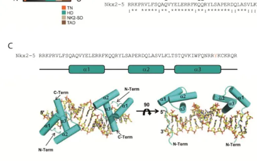
Nkx2.2 belongs to a family of vertebrate transcription factors characterized by a highly conserved homeobox-binding domain (HD). These transcription factors are known for more than twenty years to be important factors for proper development of different tissues. Together with other Nkx proteins such as Nkx2.1 or Nkx2.5, Nkx2.2 constitutes in vertebrates the Nk-2 class of transcription factors, whose homologs were first identified in Drosophila melanogaster (105, 106). In total, eleven Nk-2 class members are known and all of them show organ-specific expression patterns (106).
The HD of Nkx2.1, 2.2, and 2.5 binds to a DNA sequence (NK-response element, shortened NKE) which contains the core consensus sequence 5'-AAGTG-3' (Figure 1.6 C) and is highly conserved in mammals (107-110). For Nkx2.2, however, also an alternative core sequence (5'-GAGT-3') was postulated in 2011 (111). Besides the HD, Nk-2 class proteins feature other conserved domains: A transcriptional repression TN- domain which is located at the N-terminus, an Nk-2 specific domain (NK-2 SD), and a transcription activation domain (TAD) next to the C-terminus (see Figure 1.6 A) (105, 110). Thereby, the Nk-2 specific domain is thought to be an intrinsic repression domain for the TAD of the C-terminus due to its hydrophobic character (110).
Moreover, it was suggested to function as interface for specific protein-protein interactions (110).
Regarding their function as key factors for the development of different organs, Nkx2.1, 2.2, and 2.5 belong to the best studied members of the Nk-2 class proteins. Nkx2.1 is known for its important role during lung development (112-114). Consistently, Nkx2.1 knocked out mice die soon after birth from respiratory failure caused by malformed lungs (112, 113). Nkx2.1 was also linked to lung cancer biology: In several studies Nkx2.1 was reported both as suppressor of malignant progression and as oncogene promoting lung adenocarcinomas. The two-sided function of Nkx proteins is discussed more closely in paragraph 3.2.3.
20 1. Introduction
Figure 1.6: Nk-2 class of homeobox-binding domain containing transcription factors. A: Nkx2.2 with its Nk-2 class-typical domains (highlighted in different colors): Transcriptional repression domain (TN), homeobox-binding domain (HD), Nk2-specific domain (NK2-SD), and transcription activation domain (TAD). [(Illustration based on structural information available at http://www.uniprot.org/ for human Nkx2.2 (O95096)]. B: The HD of the Nk-2 class members Nkx2.1, Nk2.2, and Nkx2.5 is highly conserved. Sequence alignment was performed using ClustalW2 (http://www.ebi.ac.uk/). C: Structure of the Nkx2.5 HD. The domain consists of three α-helices of which α3 mediates specific interactions with the NK response element (NKE).
Within α3 a specific tyrosine (Y191 for Nkx2.5; highlighted in red in B and C) is essential for NKE recognition. Adopted from (115).
Nkx2.2 is essential for neural development, in particular for glia cell maturation, which is highlighted in the next paragraph (see 1.5.2.).
The crucial role of Nkx2.5 in heart development and maintenance of a normal heart function was extensively investigated in several studies before (109, 116-123). In this context, it was shown previously that a homozygous knockout of Nkx2.5 in mouse is embryonic lethal, because heart development is severely impaired (117, 118). As a transcriptional target for Nkx2.5, NPPA was identified already twenty years ago (108, 109). Nkx2.5 stimulates its expression via binding to the NKE sites in the NPPA promoter (108, 109, 115). NPPA in turn is the precursor of ANP, a secreted peptide
1.5. Nkx2.2: A glioblastoma tumor suppressor candidate 21 and a marker for cardiomyocyte differentiation (124). For the stimulation of the NPPA promoter, a member of the GATA family of transcription factors, GATA-4, was identified as an Nkx2.5 interaction partner during the maturation of cardiomyocytes (109, 123). Another co-inducer of NPPA, which can also interact with Nkx2.5, is the T-box transcription factor Tbx5 (119, 120).
As already mentioned in paragraph 1.4.2., CAMTA2 is also a co-activator of NPPA during cardiac hypertrophy. In this context, it was shown by Song et al. that CAMTA2 directly interacts with Nkx2.5 to stimulate the NPPA promoter (61). This interaction is mainly mediated by the CG-1 domain of CAMTA2 and the HD of Nkx2.5 (61). It is believed that these two transcription factors interact to promote cardiac hypertrophy as response to cellular stress.
Recently, the binding of the Nkx2.5 HD to the NKE site of its target NPPA was structurally resolved: Nkx2.5 binds as a homodimer to palindromically arranged NKE sites in the NPPA promoter (115). The HD itself consists of three α-helices connected by a loop (Figure 1.6 C). Thereby, helix α3 facilitates the majority of the HD-DNA interactions (115). The other helices seem to be important for the stability of the entire domain (115). Within helix α3, several residues are important for DNA-binding, but only a single tyrosine (Y191) is essential for the sequence-specific interaction (115).
Consistently, a mutation of this residue (Y191C) is frequently associated with human heart diseases (125). Furthermore, this tyrosine is highly conserved among other Nk-2 members, as illustrated in Figure 1.6 B.
1.5.2. Nkx2.2 is important for neural development and a potential tumor suppressor protein
The proper development of the central nervous system with its different classes of neurons and glia cells is very complex and requires a widespread, very accurately regulated network of signaling that turns certain transcription factors on or off at a precise time point and region during maturation of the different cell types.
22 1. Introduction
In this context, Nkx2.2 has an essential function for the differentiation of oligodendrocyte progenitor cells, which is best studied for the development of the mouse and chicken spinal cord (126-133). During dorsal-ventral pattering of the neural tube, Nkx2.2 expression emerges in a defined ventral region of the ventricular zone as a response to Sonic Hedgehog (SHH) signaling (126, 127). Together with its adjacent region which is characterized by the expression of the oligodendrocyte precursor- specific factor Olig2, this most ventral region is the major region for oligodendrocyte linage determination in the developing spinal cord (132). Nkx2.2 and Olig2 collaborate to activate the final maturation program during late oligodendrocyte development (131, 132, 134). In this regard, Nkx2.2 expression is inducible by Olig2 and Nkx2.2 also becomes a co-inducer of Olig2 during later maturation (131, 134), each probably in an indirect manner since both proteins are thought to act as transcriptional repressors during oligodendrocyte maturation (131).
Nkx2.2 also prevents the premature expression of the myelin basic protein (MBP) gene by binding directly to the MBP promoter and by association with a repressor complex that includes histone deacetylase 1 (HDAC1) (135). On the other hand, Nkx2.2 is also able to induce the gene of the proteolipid protein (PLP) (129, 132). Both, MBP and PLP, are essential proteins for the formation of myelin sheaths by the mature oligodendrocytes. By a similar mechanism as shown for MBP, via recruiting HDAC1, Nkx2.2 negatively affects expression of Sirt2, a protein which belongs to the Sirtuin class III deacetylases (136). Sirt2 is another important factor during oligodendrocyte maturation since it associates with myelin (137, 138). Recently, it was also shown that Nkx2.2 represses Pdgfra via direct binding to its promoter (133). Pdgfra (Pdgf receptor α) and Pdgf signaling via its receptor is important for early oligodendrocytes lineage determination and an interruption of Pdgfra leads to a premature differentiation of the oligodendrocyte precursors (139, 140). After terminal differentiation, Nkx2.2 is down- regulated in oligodendrocytes (128, 135).
Besides its function in oligodendrocytes development in the spinal cord and in the brain (128, 129), Nkx2.2 is also essential for the generation of v3 interneurons in the spinal cord and serotonergic neurons in the developing hindbrain (126, 127, 141).
1.5. Nkx2.2: A glioblastoma tumor suppressor candidate 23 In 2010, it was furthermore reported that Nkx2.2 acts as a tumor suppressor for glioblastomas. For this study, Muraguchi et al. generated a transgenic mouse, which develops glioblastomas characterized by a human phenotype (142). These tumors showed only a weak Nkx2.2 expression (142). Moreover, an overexpression of Nkx2.2 in cancer stem cells of these tumors and in primary human glioblastoma cells negatively affected their ability for self-renewal as it led to a significant reduction of neurosphere- like colonies (142). The overexpression in isolated glioblastoma cells from the introduced mouse model also resulted in a drastic decrease of Nestin and, consistently, in an elevation of mature oligodendrocytes markers such as NG2 (142). In addition, Muraguchi et al. also demonstrated a tumor suppressive effect of Nkx2.2 in vivo (142).
Taken together, the findings by Muraguchi et al. turn the important developmental transcription factor Nkx2.2 to a interesting tumor suppressor candidate in glioblastoma biology. A de-regulation of this factor could be important for progression of this type of brain cancer.
24 1. Introduction
1.6. Aims and working model for this thesis
The putative transcription factor CAMTA1 acts as a tumor suppressor for glioblastomas as well as for neuroblastomas. However, the exact mechanisms of how this protein carries out this function remains largely unknown.
Hence, it was the aim of this thesis to characterize CAMTA1 in detail and to gain insight into which cellular network CAMTA1 is involved in to promote its tumor suppressive function. To achieve this, it was intended to establish tools for a functional characterization of CAMTA1, including antibodies or siRNAs. With these tools it should be further possible to examine which genes are regulated by CAMTA1 and which other proteins it interacts with.
Based on previous findings about CAMTA proteins, a working model was developed for this thesis. As mentioned before, mammalian CAMTA2 interacts with Nkx2.5 in the heart to stimulate the expression of NPPA. Hence, it was assumed that a similar interaction might take place in neural cells between CAMTA1 and Nkx2.2. Moreover, both CAMTA1 and Nkx2.2 repress self-renewal and differentiation of glioblastoma CSCs. Therefore, one could speculate that these two proteins act in concert to suppress tumor formation and that a de-regulation of one or both proteins promotes glioblastoma progression. As mentioned before, CAMTA1 overexpression causes a significant elevation of NPPA mRNA levels. Remarkably, for the peptide hormone ANP (which arises from the precursor NPPA) an antiproliferative effect on glioblastoma cells was reported before (143) and, furthermore, NPPA expression correlates with survival of glioblastoma patients (53). Thus, it is likely that CAMTA1 and Nkx2.2 might interact to stimulate NPPA expression, which finally causes a tumor suppressive effect. This working model is illustrated in Figure 1.7.
Besides the investigation of a potential interaction between CAMTA1 and Nkx2.2, it was further intended to examine upstream and downstream pathways involving Nkx2.2 in the context of glioblastoma biology.
1.6. Aims and working model for this thesis 25
Figure 1.7: Working model for this thesis. It is hypothesized that in the mammalian brain CAMTA1 acts in concert with Nkx2.2 to stimulate the promoter of NPPA (in a similar manner as CAMTA2 and Nkx2.5 interacts in cardiac myocytes). The expression of NPPA, which is the precursor of ANP, would in turn cause tumor suppression. Furthermore, de-regulated miR-9/9*
would probably counteract this mechanism in glioblastoma CSCs by targeting the CAMTA1 mRNA. It is assumed, that this maintains the stem cells character of these CSCs and promotes glioblastoma progression.
26