Review
The Importance of Physioxia in Mesenchymal Stem Cell Chondrogenesis and the Mechanisms
Controlling Its Response
Girish Pattappa
1,* , Brian Johnstone
2, Johannes Zellner
1, Denitsa Docheva
1and Peter Angele
1,31
Laboratory of Experimental Trauma Surgery, Department of Trauma Surgery,
University Hospital Regensburg, Franz Josef Strauss Allee 11, 93053 Regensburg, Germany;
johannes.zellner@ukr.de (J.Z.); denitsa.docheva@ukr.de (D.D.); peter.angele@ukr.de (P.A.)
2
Department of Orthopaedics and Rehabilitation, Oregon Health & Science University, 3181 SW Sam Jackson Park Rd, Portland, OR 97239, USA; johnstob@ohsu.edu
3
Sporthopaedicum Regensburg, Hildegard von Bingen Strasse 1, 93053 Regensburg, Germany
* Correspondence: girish.pattappa@ukr.de; Tel.: +49-941-943-1743
Received: 14 December 2018; Accepted: 21 January 2019; Published: 23 January 2019
Abstract: Articular cartilage covers the surface of synovial joints and enables joint movement.
However, it is susceptible to progressive degeneration with age that can be accelerated by either previous joint injury or meniscectomy. This degenerative disease is known as osteoarthritis (OA) and it greatly affects the adult population. Cell-based tissue engineering provides a possible solution for treating OA at its earliest stages, particularly focal cartilage lesions. A candidate cell type for treating these focal defects are Mesenchymal Stem Cells (MSCs). However, present methods for differentiating these cells towards the chondrogenic lineage lead to hypertrophic chondrocytes and bone formation in vivo. Environmental stimuli that can stabilise the articular chondrocyte phenotype without compromising tissue formation have been extensively investigated. One factor that has generated intensive investigation in MSC chondrogenesis is low oxygen tension or physioxia (2–5%
oxygen). In vivo articular cartilage resides at oxygen tensions between 1–4%, and in vitro results suggest that these conditions are beneficial for MSC expansion and chondrogenesis, particularly in suppressing the cartilage hypertrophy. This review will summarise the current literature regarding the effects of physioxia on MSC chondrogenesis with an emphasis on the pathways that control tissue formation and cartilage hypertrophy.
Keywords: mesenchymal stem cells; chondrogenesis; hypoxia; cartilage; hypertrophy; hypoxia inducible factors; early osteoarthritis
1. Introduction
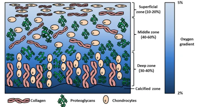
Articular cartilage is a tissue lining the surface of synovial joints that provides friction-free movement and facilitates load-bearing. Chondrocytes are the specialised cells within the tissue that create and maintain cartilage matrix that is primarily composed of aggrecan (a large proteoglycan) and collagen II. Chondrocyte orientation and matrix distribution within the tissue generates an anisotropic structure from the cartilage surface to the deep zone and outwards from the chondrocyte. These cartilage zones are described as the superficial, middle/transitional, deep and calcified zones; whilst the pericellular, territorial and interterritorial regions surround the chondrocyte. The differences in matrix distribution within these zones and regions contribute to cartilage biomechanics, enabling it to withstand high dynamic and compressive loads during joint loading [1,2].
Int. J. Mol. Sci.2019,20, 484; doi:10.3390/ijms20030484 www.mdpi.com/journal/ijms
However, articular cartilage is susceptible to progressive degeneration leading to changes in its structure and function. This degenerative disease is described as osteoarthritis (OA). Early observable events in OA include collagen fibrillation of the superficial layer that leads to loss of proteoglycans and other matrix molecules. Chondrocytes begin to proliferate and form clusters with increased matrix synthesis in response, whilst inflammatory cytokines stimulate the expression of matrix metalloproteinases (MMPs) and aggrecanases (ADAMTS) that facilitate cartilage degradation [3,4].
Eventually chondrocytes reduce their proliferative and anabolic response and the degeneration continues, whilst pain and changes in joint function become apparent and are clinical indicators for OA.
It has been noted that there is an increased risk of OA within the knee joint due to previous joint injury (e.g., anterior cruciate ligament (ACL) injury), excessive repetitive loading, joint dysplasia and meniscectomy. According to the German Cartilage Registry (Deutsche Gesellschaft für Orthopäedie und Unfallchirurgie (DGOU)) between October 2013–June 2014, 60% of treated cartilage defects were degenerative, whilst a recent multi-centre study showed that of 400 patients, approximately 40% had chondral injuries resulting from degenerative conditions [5,6]. In classifying the forms of OA that can be treated, recent studies have described the term “early OA” [7–9]. This latter condition may be amenable to regenerative medicine or tissue engineering therapies. An example of cartilage tissue engineering is autologous chondrocyte implantation (ACI) that is currently being used to treat focal cartilage defects. However, a high failure and re-operation rate has been observed when treating focal degenerative lesions compared with post-trauma defects. One potential reason for their poor outcome is the surrounding inflammatory environment that impairs cell-based solutions. In particular, IL-1β has been shown to be a negative predictor for ACI treatment post-surgery [5,10]. Thus, a primary goal for clinicians and scientists is to develop regenerative options that can be used to treat both focal and diffuse early OA in this challenging environment.
Autologous articular chondrocytes are an established cell-based tissue engineering strategy for treatment of large “focal traumatic” or “focal early OA” chondral or osteochondral defects of the knee joint [6,11,12]. Mesenchymal stem cells (MSCs) can be isolated from a variety of sources including bone marrow, adipose (liposuction or intrapatellar fat pad) or synovium with minimal donor site morbidity [13–16] and have been shown to have chondrogenic potential, initially in vitro with the creation of pellets or micromasses and the addition of the stimulatory growth factor, transforming growth factor-beta (TGF- β ) [17,18]. Scaffolds/biomaterials have been developed in which MSCs are seeded or encapsulated, and then chondrogenically differentiated to create clinically relevant chondrogenic implants that may be used to fill patient defects. However, in both pellets and scaffolds, markers of chondrocyte hypertrophy (collagen X, MMP13) have been detected and upon implantation in vivo, ectopic bone formation can occur [19]. Strategies to prevent hypertrophy and produce a stable articular chondrocyte phenotype with its defined extracellular matrix are the principle goals of in vitro cartilage tissue engineering.
Scientists have attempted to attain stable cartilage formation with environmental stimuli relevant to the in vivo situation, e.g., biomechanical stimulation, lower oxygen tension and/or addition of appropriate growth factors. The rationale for using low oxygen tension for cartilage tissue engineering is the oxygen level within articular cartilage that ranges from 2–5% oxygen (Figure 1) [20–24]. The use of growth factors (e.g., TGF- β ), low oxygen tension and other stimuli for MSC chondrogenesis can also be used to cause the redifferentiation of articular chondrocytes in either pellets or hydrogels. Adding TGF-β under physioxia upregulates chondrogenic gene (sex-determining region Y–box 9 (SOX9), collagen II alpha I (COL2A1) and aggrecan (ACAN)) expression and subsequent glycosaminoglycan (GAG) and collagen II accumulation, whilst downregulating the expression of catabolic genes (e.g., MMP9, MMP13), ADAMTS-4, ADAMTS-5) compared with cells in hyperoxic (atmospheric) conditions [25–32].
Similarly, OA chondrocytes cultured in the same low oxygen conditions also demonstrated an increase
in GAG deposition and a reduction in the expression of MMPs, although they had higher collagen
X alpha I (COL10A1) and MMP13 expression and lower GAG deposition compared with healthy
chondrocytes [30,31]. Additionally, low oxygen culture can also counter the inhibitory effects caused by the presence of inflammatory cytokines [29]. Thus, lowered oxygen tension could potentially help to induce a stable chondrogenic phenotype in MSC chondrogenesis, and many studies have been conducted to understand its effects on this process. The present review will summarise the literature and evaluate the effects of low oxygen tension or physioxia on MSC chondrogenesis, evaluating the outcomes of the various studies and the pathways that have been identified to be part of the cellular response to it. A Pubmed search was performed; the date of the last search was 31 October 2018. The keywords used for the gathering of relevant publications used the terms, “mesenchymal stem cells”
and “hypoxia” or “chondrogenesis” or “chondrocytes”. Publications since 2001 were evaluated for the purposes of this review.
Figure 1. Schematic diagram describing the zones within articular cartilage and the changes in oxygen tension from the superficial zone to calcified zone.
2. Physioxia and Cartilage
In vivo physiological oxygen tension (physioxia) within human articular cartilage ranges from 2–5%, whilst bone marrow physioxia has been measured at 7% oxygen (Figure 1) [20,21,23,24]. The atmospheric oxygen level (20% oxygen) at which most typical tissue culture incubators operate is actually non-physiological and represents hyperoxia [20,33].
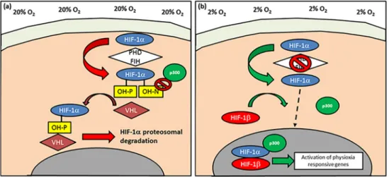
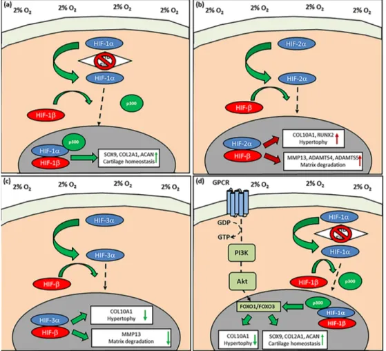
In all of the studies summarised for this review, incubator oxygen tension (20%; hyperoxia) is utilised as the ‘control’ for experiments done at physioxia even though the latter is closer to conditions found in vivo. An alternative method to mimic some of the effects of physioxia is through chemical induction, specifically on the pathway controlling the stability of hypoxia-inducible factor-1 alpha (HIF-1α) that has been found to be essential for chondrocyte survival and cartilage homeostasis (Figure 2) [34,35].
In chemically-induced physioxia, cobalt chloride is the most extensively studied compound. Under
hyperoxia, HIF-1α is hydroxylated by prolyl hydroxylases (PHDs) and factor inhibiting HIFs (FIH),
resulting in its proteosomal degradation (Figure 2a). The process is catalysed by molecular oxygen,
iron ions, ascorbic acid and 2-oxoglutarate [36–38]. Cobalt chloride competes with iron ions for the
active site of PHD and prevents HIF-1α hydroxylation, thereby stimulating a physioxic response in
hyperoxic oxygen conditions. Other hydroxylase inhibitors have also been used to create a physioxia
mimicking response, specifically dimethyloxalylglycine (a competitive inhibitor of PHDs and FIHs)
and desferrioxamine (sequesters iron ions). Although this review primarily focusses on experiments
that use altered incubator oxygen levels, studies utilising chemically-induced physioxic responses are discussed where appropriate [36,39,40].
Int. J. Mol. Sci. 2018, 19, x FOR PEER REVIEW 4 of 28
Figure 2. Schematic diagram describing the activation of hypoxia-inducible factor-1 alpha (HIF-1α). (a) Under normoxic/hyperoxic (20% oxygen) conditions, HIF-1α is hydroxylated by prolyl hydroxylases (PHDs) and factor inhibiting HIF (FIH) that enables HIF-1α to undergo proteasomal degradation by von-Hippel-Lindau (VHL) E3 ubiquitin ligase complex. In contrast, PHD and FIH activity is inhibited under (b) hypoxia/physioxia (2% oxygen), thus leading to nuclear translocation of HIF-1α that forms a complex with HIF-1β and co-factor p300, that results in upregulation of physioxia-responsive genes.
Arrows describe the flow and stages involved in HIF-1α behavior under (a) normoxia/hyperoxia and (b) hypoxia/physioxia (dotted arrow depicts HIF-1α nuclear translocation).
3. MSC Isolation and Expansion under Physioxia
Investigators examining MSC proliferation under physioxia have demonstrated that the initial plating of human bone marrow produced greater colony-forming units–fibroblasts (CFU-Fs) upon culture in a low oxygen environment [41–44]. Furthermore, MSCs were able to proliferate at a shorter cell doubling time and have consequent increased cell numbers in physioxia [43–46]. This pattern has also been replicated in the majority of studies for chondrogenic cells from different tissue sources (adipose and synovium) and other species (porcine, ovine and murine) [43,45,47–53].
Long-term culture under lowered oxygen conditions caused MSCs to reach greater population doublings with reduced cellular senescence [46,52,54,55]. The latter phenomena has been postulated to be due to the underlying cellular ATP metabolism. Under hyperoxia, MSCs have an increased oxygen consumption, thereby having a greater ATP production via oxidative phosphorylation. In comparison, physioxic MSC cultures had greater lactate production and reduced oxygen consumption compared with hyperoxic MSC cultures, resulting in greater energy production via glycolysis [54,56,57]. The increased utilisation of oxidative phosphorylation under hyperoxia generated reactive oxygen species that can enhance cellular senescence [58]. Furthermore, telomeres that control cellular aging were found to be longer for physioxia-cultured MSCs [46]. These investigations demonstrate a clear advantage of physioxia for MSC expansion.
Studies have also demonstrated that stem cell markers that are typically associated with either embryonic or induced pluripotent stem cells (e.g., octamer-binding transcription 4 (OCT-4), stage-specific embryonic antigen (SSEA-1) are expressed on MSCs expanded in physioxia [59,60].
Consistent with this, physioxia-expanded MSCs can undergo osteogenesis and adipogenesis at later population doublings compared with those expanded in hyperoxia. However, there are also studies showing that osteogenesis is inhibited under physioxia and with physioxia expanded MSCs differentiated under hyperoxia, indicating that differences in oxygen tension can affect differentiation towards different musculoskeletal lineages [48,54,59]. The present review focusses on how physioxia affects MSC chondrogenesis in terms of the amount of matrix formation that occurs and the types of matrix molecules that are expressed. The influence on the latter is of particular interest, as hyperoxic in-vitro MSC chondrogenesis results in the expression of hypertrophic markers (e.g., collagen X, MMP13, alkaline phosphatase) that upon implantation in a nude mouse model result in ectopic bone formation in vivo [19].
Figure 2. Schematic diagram describing the activation of hypoxia-inducible factor-1 alpha (HIF-1α).
(a) Under normoxic/hyperoxic (20% oxygen) conditions, HIF-1α is hydroxylated by prolyl hydroxylases (PHDs) and factor inhibiting HIF (FIH) that enables HIF-1α to undergo proteasomal degradation by von-Hippel-Lindau (VHL) E3 ubiquitin ligase complex. In contrast, PHD and FIH activity is inhibited under (b) hypoxia/physioxia (2% oxygen), thus leading to nuclear translocation of HIF-1α that forms a complex with HIF-1β and co-factor p300, that results in upregulation of physioxia-responsive genes.
Arrows describe the flow and stages involved in HIF-1α behavior under (a) normoxia/hyperoxia and (b) hypoxia/physioxia (dotted arrow depicts HIF-1α nuclear translocation).
3. MSC Isolation and Expansion under Physioxia
Investigators examining MSC proliferation under physioxia have demonstrated that the initial plating of human bone marrow produced greater colony-forming units–fibroblasts (CFU-Fs) upon culture in a low oxygen environment [41–44]. Furthermore, MSCs were able to proliferate at a shorter cell doubling time and have consequent increased cell numbers in physioxia [43–46]. This pattern has also been replicated in the majority of studies for chondrogenic cells from different tissue sources (adipose and synovium) and other species (porcine, ovine and murine) [43,45,47–53].
Long-term culture under lowered oxygen conditions caused MSCs to reach greater population doublings with reduced cellular senescence [46,52,54,55]. The latter phenomena has been postulated to be due to the underlying cellular ATP metabolism. Under hyperoxia, MSCs have an increased oxygen consumption, thereby having a greater ATP production via oxidative phosphorylation. In comparison, physioxic MSC cultures had greater lactate production and reduced oxygen consumption compared with hyperoxic MSC cultures, resulting in greater energy production via glycolysis [54,56,57]. The increased utilisation of oxidative phosphorylation under hyperoxia generated reactive oxygen species that can enhance cellular senescence [58]. Furthermore, telomeres that control cellular aging were found to be longer for physioxia-cultured MSCs [46]. These investigations demonstrate a clear advantage of physioxia for MSC expansion.
Studies have also demonstrated that stem cell markers that are typically associated with either
embryonic or induced pluripotent stem cells (e.g., octamer-binding transcription 4 (OCT-4), stage-specific
embryonic antigen (SSEA-1) are expressed on MSCs expanded in physioxia [59,60]. Consistent with
this, physioxia-expanded MSCs can undergo osteogenesis and adipogenesis at later population
doublings compared with those expanded in hyperoxia. However, there are also studies showing
that osteogenesis is inhibited under physioxia and with physioxia expanded MSCs differentiated
under hyperoxia, indicating that differences in oxygen tension can affect differentiation towards
different musculoskeletal lineages [48,54,59]. The present review focusses on how physioxia affects
MSC chondrogenesis in terms of the amount of matrix formation that occurs and the types of matrix molecules that are expressed. The influence on the latter is of particular interest, as hyperoxic in-vitro MSC chondrogenesis results in the expression of hypertrophic markers (e.g., collagen X, MMP13, alkaline phosphatase) that upon implantation in a nude mouse model result in ectopic bone formation in vivo [19].
4. Physioxia and MSC Chondrogenesis
4.1. Chondrogenic Matrix Formation
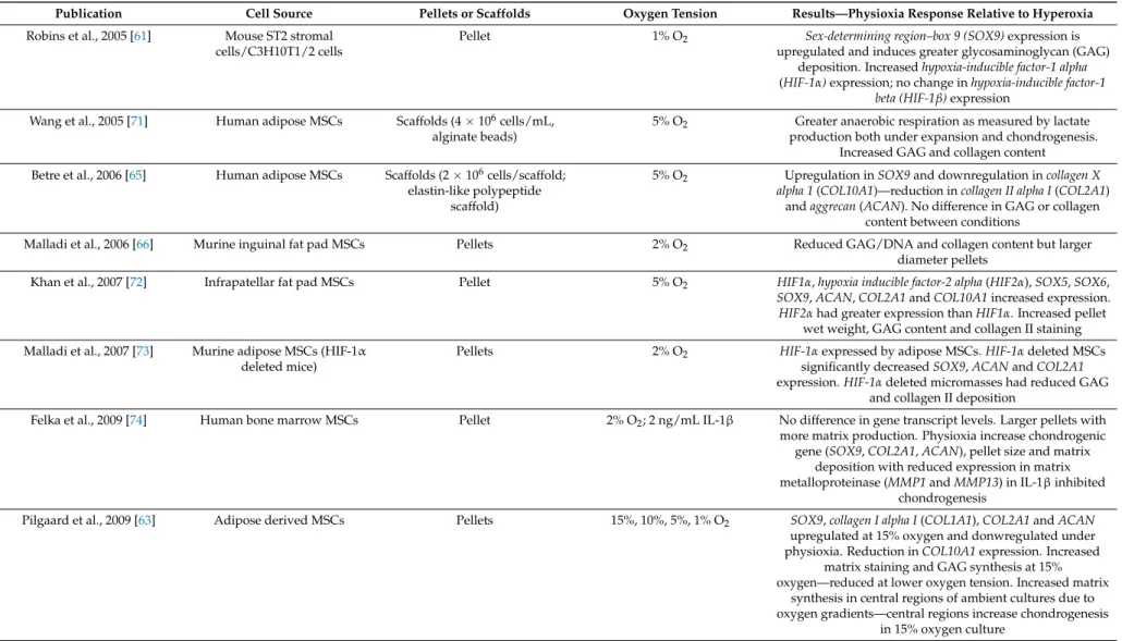
A summary of the outcomes for the effects of physioxia on MSC chondrogenic matrix formation are described in Tables 1 and 2. Mouse stromal cells in a pellet culture model demonstrated that physioxia (1% oxygen) upregulated SOX9 gene expression and increased GAG deposition in the matrix [61]. Later studies were in agreement with the described investigation, whilst also observing an increase in pellet wet weight and an upregulation in COL2A1 and ACAN expression [46,62]. However, there are also studies that found a downregulation in matrix gene expression (SOX9, COL2A1, ACAN) and no effect on matrix formation [44,51,53,54,63–69]. One study using adipose-derived murine MSCs cultured under physioxia demonstrated a reduction in both GAG and collagen II matrix content compared to hyperoxia, in spite of larger pellet diameter [66]. It was assumed in these cases that oxygen gradients had formed under hyperoxia, whereby a physioxic region develops within the central regions of pellets or scaffolds that correspond to greater GAG and collagen II matrix deposition [63,70].
Donor variability has been shown to influence chondrogenic potential, especially in response to low oxygen culture. Anderson et al. [33] investigated the effect of physioxia on MSC preparations with different levels of chondrogenic potential. Their intrinsic chondrogenic capacity was based on the amount of GAG produced relative to that formed by articular chondrocytes formed under normal atmospheric conditions. It was demonstrated that in preparation with low GAG, there was enhanced chondrogenesis under physioxia, whilst high GAG producing MSC preparations did not get a significant enhancement from lower oxygen. Thus, the differences described in studies displaying no influence of physioxia on MSC chondrogenesis could be related to the donor variability in chondrogenic potential.
With this as a caveat, the majority of studies of MSC chondrogenesis either in pellet or scaffold
cultures indicates an increase in chondrogenic genes (COL2A1 and ACAN) and matrix formation under
physioxia [33,41–43,47,49,50,52,55,62,71,72,76–79,82,86–88,90,92,93,95,97]. Furthermore, it has also
been shown that physioxia is a more potent promoter of chondrogenesis than dynamic (compressive)
loading with no synergistic effect upon combining the parameters [78,98].
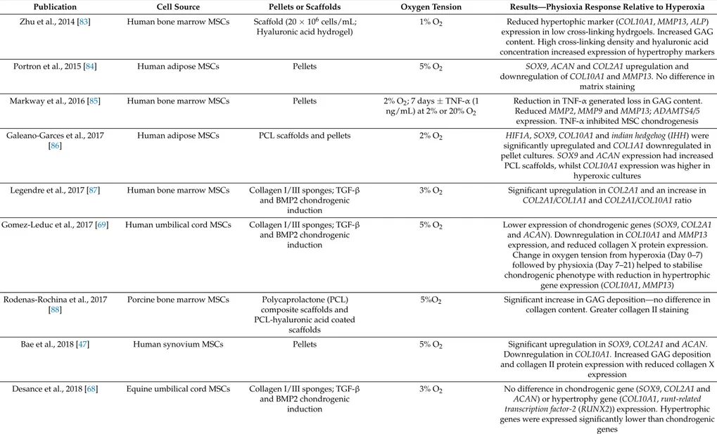
Table 1. Summary of the key findings from publications examining the effect of physioxia on mesenchymal stem cell (MSC) chondrogenesis. Findings are relative to hyperoxia.
Publication Cell Source Pellets or Scaffolds Oxygen Tension Results—Physioxia Response Relative to Hyperoxia
Robins et al., 2005 [61] Mouse ST2 stromal cells/C3H10T1/2 cells
Pellet 1% O2 Sex-determining region–box 9 (SOX9)expression is
upregulated and induces greater glycosaminoglycan (GAG) deposition. Increasedhypoxia-inducible factor-1 alpha (HIF-1α)expression; no change inhypoxia-inducible factor-1
beta (HIF-1β)expression Wang et al., 2005 [71] Human adipose MSCs Scaffolds (4×10⁶cells/mL,
alginate beads)
5% O2 Greater anaerobic respiration as measured by lactate production both under expansion and chondrogenesis.
Increased GAG and collagen content Betre et al., 2006 [65] Human adipose MSCs Scaffolds (2×10⁶cells/scaffold;
elastin-like polypeptide scaffold)
5% O2 Upregulation inSOX9and downregulation incollagen X alpha 1(COL10A1)—reduction incollagen II alpha I(COL2A1)
andaggrecan(ACAN). No difference in GAG or collagen content between conditions
Malladi et al., 2006 [66] Murine inguinal fat pad MSCs Pellets 2% O2 Reduced GAG/DNA and collagen content but larger
diameter pellets
Khan et al., 2007 [72] Infrapatellar fat pad MSCs Pellet 5% O2 HIF1α,hypoxia inducible factor-2 alpha(HIF2α),SOX5,SOX6, SOX9,ACAN,COL2A1andCOL10A1increased expression.
HIF2αhad greater expression thanHIF1α. Increased pellet wet weight, GAG content and collagen II staining Malladi et al., 2007 [73] Murine adipose MSCs (HIF-1α
deleted mice)
Pellets 2% O2 HIF-1αexpressed by adipose MSCs.HIF-1αdeleted MSCs
significantly decreasedSOX9,ACANandCOL2A1 expression.HIF-1αdeleted micromasses had reduced GAG
and collagen II deposition
Felka et al., 2009 [74] Human bone marrow MSCs Pellet 2% O2; 2 ng/mL IL-1β No difference in gene transcript levels. Larger pellets with more matrix production. Physioxia increase chondrogenic
gene (SOX9,COL2A1,ACAN), pellet size and matrix deposition with reduced expression in matrix metalloproteinase (MMP1andMMP13) in IL-1βinhibited
chondrogenesis
Pilgaard et al., 2009 [63] Adipose derived MSCs Pellets 15%, 10%, 5%, 1% O2 SOX9,collagen I alpha I(COL1A1),COL2A1andACAN upregulated at 15% oxygen and donwregulated under physioxia. Reduction inCOL10A1expression. Increased
matrix staining and GAG synthesis at 15%
oxygen—reduced at lower oxygen tension. Increased matrix synthesis in central regions of ambient cultures due to oxygen gradients—central regions increase chondrogenesis
in 15% oxygen culture
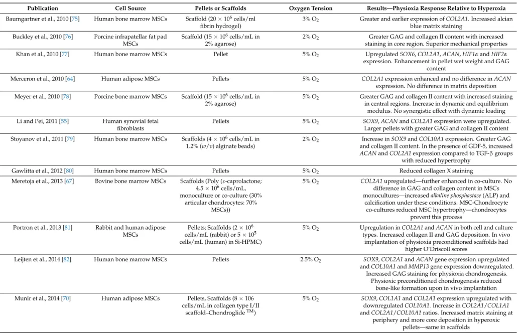
Table 1. Cont.
Publication Cell Source Pellets or Scaffolds Oxygen Tension Results—Physioxia Response Relative to Hyperoxia
Baumgartner et al., 2010 [75] Human bone marrow MSCs Scaffold (20×106cells/ml fibrin hydrogel)
3% O2 Greater and earlier expression ofCOL2A1. Increased alcian blue matrix staining
Buckley et al., 2010 [76] Porcine infrapatellar fat pad MSCs
Scaffold (15×106cells/mL in 2% agarose)
2% O2 Greater GAG and collagen II content with increased staining in core region. Superior mechanical properties
Khan et al., 2010 [77] Human bone marrow MSCs Pellet 5% O2 UpregulatedSOX6,COL2A1,ACAN,HIF1αandHIF2α
expression. Enhancement in pellet wet weight and GAG content
Merceron et al., 2010 [64] Human adipose MSCs Pellets 5% O2 COL2A1expression enhanced and no difference inACAN
expression. No difference in matrix deposition Meyer et al., 2010 [78] Porcine bone marrow MSCs Scaffold (15×106cells/mL in
2% agarose)
5% O2 Greater GAG and collagen II content with increased staining in central regions. Increase in dynamic and equilibrium
modulus. No synergistic effect with dynamic loading Li and Pei, 2011 [55] Human synovial fetal
fibroblasts
Pellets 5% O2 SOX9,ACANandCOL2A1expression were upregulated.
Larger pellets with greater GAG and collagen II content Stoyanov et al., 2011 [79] Human bone marrow MSCs Scaffolds (4×10⁶cells/mL in
1.2% (w/v) alginate beads)
2% O2 Increase inSOX9andCOL10A1expression. Greater GAG and collagen II content. In the presence of GDF-5, increased ACANandCOL2A1expression compared to TGF-βgroups
with reduced hypertrophy
Gawlitta et al., 2012 [80] Human bone marrow MSCs Pellets 5% O2 Reduced collagen X staining
Meretoja et al., 2013 [67] Bovine bone marrow MSCs Scaffolds (Poly (ε-caprolactone;
4.5×10⁶cells/mL, monoculture or co-culture (30%
articular chondrocytes: 70%
MSCs))
5% O2 COL2A1upregulated—further enhanced in co-culture. No difference in GAG and collagen content in MSCs monocultures—increasedalkaline phosphastase(ALP) and
calcification under these conditions. MSC-Chondrocyte co-cultures reduced MSC hypertrophy—chondrocytes
prevent this process Portron et al., 2013 [81] Rabbit and human adipose
MSCs
Pellets; Scaffolds (2×10⁶ cells/mL (rabbit) or 5×10⁵ cells/mL (human) in Si-HPMC)
5% O2 Upregulation inCOL2A1andACANin both cell and culture types. Increased collagen II and GAG deposition. In vivo
implantation of physioxia preconditioned scaffolds had higher O’Driscoll scores
Leijten et al., 2014 [82] Human bone marrow MSCs Pellets 2.5% O2 SOX9,COL2A1andACANgene expression upregulated
andCOL10A1andMMP13gene expression downregulated.
Increased GAG staining for physioxia chondrogenesis.
Physioxic preconditioned chondrogenesis reduced bone-like formation upon in vivo implantation Munir et al., 2014 [70] Human adipose MSCs Pellets, Scaffolds (8×10⁶
cells/mL in collagen type I/II scaffold–ChondroglideTM)
5% O2 SOX9,COL1A1andCOL2A1expression upregulated with downregulatedCOL10A1. Increase inCOL2A1/COL1A1 andCOL2A1/COL10A1ratios. Increased matrix staining at
periphery and more core deposition in hyperoxic pellets—same in scaffolds
Table 1. Cont.
Publication Cell Source Pellets or Scaffolds Oxygen Tension Results—Physioxia Response Relative to Hyperoxia
Zhu et al., 2014 [83] Human bone marrow MSCs Scaffold (20×106cells/mL;
Hyaluronic acid hydrogel)
1% O2 Reduced hypertophic marker (COL10A1,MMP13,ALP) expression in low cross-linking hydrgoels. Increased GAG
content. High cross-linking density and hyaluronic acid concentration increased expression of hypertrophy markers
Portron et al., 2015 [84] Human adipose MSCs Pellets 5% O2 SOX9,ACANandCOL2A1upregulation and
downregulation ofCOL10A1andMMP13. No difference in matrix staining
Markway et al., 2016 [85] Human bone marrow MSCs Pellets 2% O2; 7 days±TNF-α(1 ng/mL) at 2% or 20% O2
Reduction in TNF-αgenerated loss in GAG content.
ReducedMMP2,MMP9andMMP13;ADAMTS4/5 expression. TNF-αinhibited MSC chondrogenesis Galeano-Garces et al., 2017
[86]
Human adipose MSCs PCL scaffolds and pellets 2% O2 HIF1A,SOX9,COL10A1andindian hedgehog(IHH) were significantly upregulated andCOL1A1downregulated in pellet cultures.SOX9andACANexpression had increased
PCL scaffolds, whilstCOL10A1expression was higher in hyperoxic cultures
Legendre et al., 2017 [87] Human bone marrow MSCs Collagen I/III sponges; TGF-β and BMP2 chondrogenic
induction
3% O2 Significant upregulation inCOL2A1and an increase in COL2A1/COL1A1andCOL2A1/COL10A1ratio
Gomez-Leduc et al., 2017 [69] Human umbilical cord MSCs Collagen I/III sponges; TGF-β and BMP2 chondrogenic
induction
5% O2 Lower expression of chondrogenic genes (SOX9,COL2A1 andACAN). Downregulation inCOL10A1andMMP13 expression, and reduced collagen X protein expression.
Change in oxygen tension from hyperoxia (Day 0–7) followed by physioxia (Day 7–21) helped to stabilise chondrogenic phenotype with reduction in hypertrophic
gene expression (COL10A1,MMP13) Rodenas-Rochina et al., 2017
[88]
Porcine bone marrow MSCs Polycaprolactone (PCL) composite scaffolds and PCL-hyaluronic acid coated
scaffolds
5%O2 Significant increase in GAG deposition—no difference in collagen content. Greater collagen II staining
Bae et al., 2018 [47] Human synovium MSCs Pellets 5% O2 Significant upregulation inSOX9,COL2A1andACAN.
Downregulation inCOL10A1.Increased GAG deposition and collagen II protein expression with reduced collagen X
expression Desance et al., 2018 [68] Equine umbilical cord MSCs Collagen I/III sponges; TGF-β
and BMP2 chondrogenic induction
3% O2 No difference in chondrogenic gene (SOX9,COL2A1and ACAN) or hypertrophy gene (COL10A1,runt-related transcription factor-2(RUNX2)) expression. Hypertrophic genes were expressed significantly lower than chondrogenic
genes
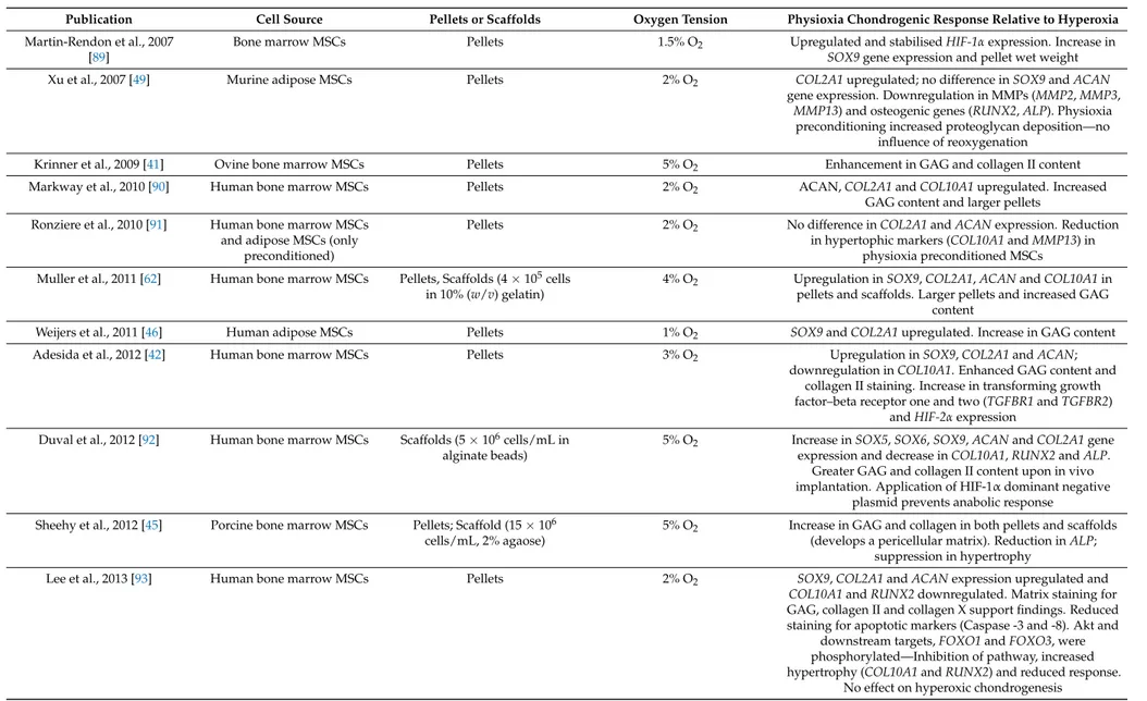
Table 2. Summary of key findings from publications examining the effect of physioxia preconditioning on MSC chondrogenesis. Findings are relative to hyperoxia.
Publication Cell Source Pellets or Scaffolds Oxygen Tension Physioxia Chondrogenic Response Relative to Hyperoxia Martin-Rendon et al., 2007
[89]
Bone marrow MSCs Pellets 1.5% O2 Upregulated and stabilisedHIF-1αexpression. Increase in
SOX9gene expression and pellet wet weight
Xu et al., 2007 [49] Murine adipose MSCs Pellets 2% O2 COL2A1upregulated; no difference inSOX9andACAN
gene expression. Downregulation in MMPs (MMP2,MMP3, MMP13) and osteogenic genes (RUNX2,ALP). Physioxia
preconditioning increased proteoglycan deposition—no influence of reoxygenation
Krinner et al., 2009 [41] Ovine bone marrow MSCs Pellets 5% O2 Enhancement in GAG and collagen II content
Markway et al., 2010 [90] Human bone marrow MSCs Pellets 2% O2 ACAN,COL2A1andCOL10A1upregulated. Increased
GAG content and larger pellets Ronziere et al., 2010 [91] Human bone marrow MSCs
and adipose MSCs (only preconditioned)
Pellets 2% O2 No difference inCOL2A1andACANexpression. Reduction
in hypertophic markers (COL10A1andMMP13) in physioxia preconditioned MSCs Muller et al., 2011 [62] Human bone marrow MSCs Pellets, Scaffolds (4×10⁵cells
in 10% (w/v) gelatin)
4% O2 Upregulation inSOX9,COL2A1,ACANandCOL10A1in pellets and scaffolds. Larger pellets and increased GAG
content
Weijers et al., 2011 [46] Human adipose MSCs Pellets 1% O2 SOX9andCOL2A1upregulated. Increase in GAG content
Adesida et al., 2012 [42] Human bone marrow MSCs Pellets 3% O2 Upregulation inSOX9,COL2A1andACAN;
downregulation inCOL10A1. Enhanced GAG content and collagen II staining. Increase in transforming growth factor–beta receptor one and two (TGFBR1andTGFBR2)
andHIF-2αexpression Duval et al., 2012 [92] Human bone marrow MSCs Scaffolds (5×10⁶cells/mL in
alginate beads)
5% O2 Increase inSOX5,SOX6,SOX9,ACANandCOL2A1gene expression and decrease inCOL10A1,RUNX2andALP.
Greater GAG and collagen II content upon in vivo implantation. Application of HIF-1αdominant negative
plasmid prevents anabolic response Sheehy et al., 2012 [45] Porcine bone marrow MSCs Pellets; Scaffold (15×106
cells/mL, 2% agaose)
5% O2 Increase in GAG and collagen in both pellets and scaffolds (develops a pericellular matrix). Reduction inALP;
suppression in hypertrophy
Lee et al., 2013 [93] Human bone marrow MSCs Pellets 2% O2 SOX9,COL2A1andACANexpression upregulated and
COL10A1andRUNX2downregulated. Matrix staining for GAG, collagen II and collagen X support findings. Reduced staining for apoptotic markers (Caspase -3 and -8). Akt and
downstream targets,FOXO1andFOXO3, were phosphorylated—Inhibition of pathway, increased hypertrophy (COL10A1andRUNX2) and reduced response.
No effect on hyperoxic chondrogenesis
Table 2. Cont.
Publication Cell Source Pellets or Scaffolds Oxygen Tension Physioxia Chondrogenic Response Relative to Hyperoxia O’HEireamhoin et al., 2013
[49]
Human infrapatellar fat pad MSCs
Pellets, scaffolds (20×106 cells/mL in 2% agarose or
fibrin)
5% O2 Increase in GAG and collagen II content in pellets. Only GAG deposition increased within scaffolds—reduced collagen X
staining
Pattappa et al., 2013 [54] Human bone marrow MSCs Pellets 5% or 2% O2 No difference in GAG content
Ranera et al., 2013 [94] Equine bone marrow MSCs Pellets 5% O2 SOX9,COL2A1,ACAN,cartilage oligomeric matrix protein (COMP) gene expression upregulated. Physioxia preconditioned cells enhanced GAG content.HIF-1α
expression increased with time
Boyette et al., 2014 [44] Ovine bone marrow MSCs Pellets 5% O2 Enhanced chondrogenesis in physioxia differentiated cells but inhibited differentiation for physioxia preconditioned cells
Kalpakci et al., 2014 [50] Dermis isolated MSCs Pellets 5% O2 Increased GAG and collagen content in physioxia
preconditioned MSCs; collagen II content was greater under hyperoxia
Bornes et al., 2015 [43] Ovine bone marrow MSCs Scaffolds (1×10⁷cells/cm2on either collagen type I and esterified hyaluronic acid
scaffolds)
3% O2 UpregulatedACANandCOL2A1gene expression and downregulation inCOL10A1. Enhanced GAG and collagen II
content in both scaffold types
Anderson et al., 2016 [33] Human bone marrow MSCs Pellets 2% O2 COL2A1andACANupregulated whilstCOL10A1and
MMP13downregulated depending upon chondrogenic capacity. Enhanced GAG production. MSCs with high chondrogenic capacity stained for collagen X inspite of
physioxic culture
Henrionnet et al., 2016 [95] Human bone marrow MSCs Alginate beads 5% O2 UpregulatedSOX9,COL2A1,ACANandCOMP,
downregulatedRUNX2andALPexpression. No change in COL10A1expression. Sequential hyperoxia then physioxia increasedCOL2A1andACANexpression with reduction in
COL10A1expression. No calcification
Hudson et al., 2016 [96] Human MSCs Collagen-alginate scaffold 5% O2 Greater GAG content and mechanical properties
Ohara et al., 2016 [53] Human synovial derived MSCs Pellets 5% O2 No difference in pellet wet weight or matrix staining Yasui et al., 2016 [52] Synovium MSCs Scaffolds (Sheet–like construct,
4×10⁵cells/cm2)
5% O2 Increase inSOX9,ACANandCOL2A1expression. Increased GAG and collagen II content
Bornes et al., 2018 [51] Ovine bone marrow MSCs HYAFF scaffolds 3% O2 No difference inCOL2A1andACANgene expression but significant increase in GAG content after 14 days culture.
Significant downregulation inCOL10A1with concomitant increase inCOL2A1/COL10A1ratio at day 4 and 14. No difference in cartilaginous tissue formation in preconditioned
chondrogenic MSCs upon in vivo implantation
Lee et al., 2018 [97] Human bone marrow MSCs Pellets 1% O2 Upregulation inSOX9,COL2A1andACANexpression.
Increase in GAG deposition