Dissection of piRNA precursor biogenesis and
the epigenetic mechanisms of the piRNA pathway
DISSERTATION ZUR ERLANGUNG DES DOKTORGRADES DER NATURWISSENSCHAFTEN (DR. RER. NAT.)
DER FAKULT ¨ AT F ¨ UR BIOLOGIE UND VORKLINISCHE MEDIZIN DER UNIVERSIT ¨ AT REGENSBURG
vorgelegt von
EVELYN STUWE
aus
SCHWETZINGEN
im Jahr 2015
Das Promotionsgesuch wurde eingereicht am:
13.02.2015
Die Arbeit wurde angeleitet von:
PROF. GUNTER MEISTER
Das Promotionskolloquium fand statt am:
24.07.2015
Zusammensetzung des Pr ¨ufungsausschusses:
Vorsitz: PD Achim Griesenbeck 1. Pr ¨ufer: Prof. Gunter Meister 2. Pr ¨ufer: Prof. Gernot L ¨angst 3. Pr ¨ufer: Prof. Klaus Grasser Vertretung: PD Attila Nemeth Unterschrift:
EVELYN STUWE ii
C ONTENTS
1 Summary 1
2 Introduction 3
2.1 Small RNAs in RNA silencing . . . 3
2.2 The discovery of small RNA silencing . . . 4
2.3 Argonaute proteins . . . 6
2.4 miRNAs . . . 8
2.4.1 miRNA biogenesis . . . 9
2.4.2 miRNA function . . . 13
2.4.2.1 Post-transcriptional gene silencing in the cytoplasm . . . 13
2.5 siRNAs . . . 14
2.5.1 siRNA biogenesis . . . 14
2.5.2 siRNA function . . . 14
2.5.2.1 siRNAs in plants . . . 15
2.5.2.2 Transcriptional gene silencing in the nucleus . . . 15
2.6 piRNAs . . . 17
2.6.1 piRNA biogenesis . . . 18
2.6.1.1 piRNA cluster architecture and transcription . . . 18
2.6.1.2 Biogenesis of primary piRNAs . . . 20
2.6.2 Nuclear and cytoplasmic function of the piRNA pathway . . . 24
2.6.2.1 The role of piRNAs in regulating chromatin structure . . 24
2.6.2.2 Ping-pong: the cytoplasmic function of the piRNA pathway 28 2.7 piRNAs are trans-generational epigenetic factors . . . 29
2.7.1 Introduction into epigenetic mechanisms . . . 29
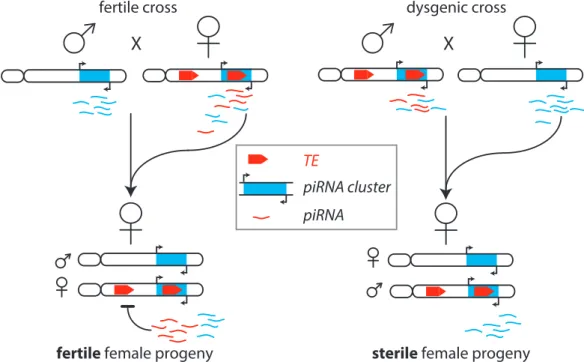
2.7.2 Trans-generational effects of piRNAs in Drosophila . . . 31
2.7.3 Hybrid dysgenesis - an old genetic phenomenon caused by piRNAs 33 3 Results 39 3.1 Characterization of piRNA precursors . . . 39
3.1.1 Objective . . . 39
3.1.2 Introduction into piRNA precursors . . . 39
CONTENTS
3.1.3 Cloning and sequencing of 5’ capped RNAs . . . 41 3.1.4 Single stranded piRNA cluster transcripts are capped and tran-
scribed from one defined start site . . . 42 3.1.5 Capped piRNA precursors from double stranded clusters originate
from internal transcription initiation . . . 46 3.1.6 piRNA precursors are 5’ mono-phosphorylated . . . 53 3.1.7 Approach for piRNA precursor detection . . . 53 3.1.8 piRNA precursors are 5’ mono-phosphorylated long RNAs . . . . 54 3.1.9 Identification of primary piRNA biogenesis factors involved in first
cleavage events . . . 57 3.1.10 The investigated factors affect precursor levels, but not the nu-
cleotide bias . . . 61 3.1.11 Efforts towards the elucidation of piRNA precursors to be co-transcriptionally
processed . . . 64 3.2 Transgenerational inheritance of piRNAs converts piRNA targets to piRNA
sources . . . 68 3.2.1 Objective . . . 68 3.2.2 Conversion of an inactive to an active piRNA cluster is accompa-
nied by acquisition of the H3K9me3 mark . . . 68 3.2.3 A paternally transmitted piRNA cluster does not recover piRNA
production . . . 72 3.2.4 The multiple effects of maternally inherited piRNAs . . . 76
3.2.4.1 Maternally inherited piRNAs are necessary for piRNA production in the next generation . . . 76 3.2.4.2 Inherited piRNAs induce primary piRNA biogenesis at
target loci . . . 80 3.2.4.3 piRNA targets are enriched in H3K9me3, Rhino and Cutoff 82 3.2.4.4 Read-through transcription on piRNA targets is increased 84 3.3 Transcriptional regulation of piRNA clusters . . . 86 3.3.1 Objective . . . 86 3.3.1.1 Rhino and Cutoff promote piRNA cluster transcription . 87 3.3.1.2 Cutoff is an anti-terminator and protects 5’ monophos-
phate ends . . . 92
4 Discussion 97
4.1 Tracing piRNA precursors . . . 97 4.1.1 Uni-stranded cluster transcripts are mRNA doppelganger . . . 98 4.1.2 Double stranded clusters do not have defined transcription start sites 99 4.1.3 piRNA precursors are processed in intermediate steps . . . 100 4.1.4 Zucchini’s role in piRNA precursor biogenesis . . . 101 4.2 Trans-generational effect of piRNAs on source and target loci . . . 103 4.2.1 maternally inherited piRNAs ensure piRNA biogenesis in progeny 103 4.2.2 Maternally inherited piRNAs induce ping-pong amplification . . . 104 4.2.3 piRNA clusters rely on inherited piRNAs to stay active . . . 104 4.2.4 Maternally inherited piRNAs act as an epigenetic switch . . . 106 4.2.5 H3K9me3 attracts piRNA biogenesis factors . . . 107
Contents
4.2.6 Cutoff acts as an anti-terminator . . . 107
4.2.7 Cutoff protects free 5’ monoposhate ends . . . 108
5 Conclusions 111 6 Methods and Materials 113 6.1 Standard prcedures . . . 113
6.1.1 RNA isolation . . . 113
6.1.1.1 Classic Phenol/Chloroform Extraction . . . 113
6.1.1.2 RNA extraction using Ribozol Reagent . . . 113
6.1.2 Quantification of nucleic acids . . . 114
6.1.2.1 Spectrophotometric DNA and RNA quantification . . . 114
6.1.2.2 Fluorometric DNA and RNA quantification . . . 114
6.1.3 PCR . . . 115
6.1.4 DNase treatement . . . 115
6.1.5 Reverse transcription . . . 116
6.1.6 Quantitative PCR (qPCR) . . . 116
6.1.7 Strand specific RT and qPCR for detection of read through tran- scription . . . 117
6.1.8 DEPC treatment of water . . . 117
6.1.9 T7 RNA transcirption . . . 117
6.2 Specific Experimental Methods . . . 119
6.2.1 Chromatin Immunoprecipitation - ChIP . . . 119
6.2.2 Nuclear Run-On / GRO-Seq . . . 119
6.2.2.1 Preparations . . . 119
6.2.2.2 Nuclei isolation . . . 120
6.2.2.3 nuclear run on reaction . . . 120
6.2.2.4 Immunoprecipitation of run-on RNA . . . 120
6.2.3 5’ capped RNA cloning . . . 121
6.2.3.1 Removal of RNAs shorter than 200 nt . . . 121
6.2.3.2 CIP treatment . . . 122
6.2.3.3 TAP treatment . . . 122
6.2.3.4 5’ adapter ligation . . . 122
6.2.3.5 Introduction of the 3’ adapter . . . 123
6.2.3.6 Linear PCR . . . 123
6.2.3.7 Final library amplification . . . 124
6.2.4 5’ mono-phosphate RNA cloning . . . 125
6.2.5 Nascent transcript isolation . . . 125
6.2.5.1 Nuclei isolation . . . 125
6.2.5.2 Chromatin precipitation and nascent RNA isolation . . . 125
6.2.6 Oligo(dT) beads assay to determine polyA containing RNAs . . . 126
6.2.7 Bioinformatic analysis of RNA sequencing data . . . 126
6.2.7.1 RNA mapping . . . 126
6.3 Methods related to work with Drosophila . . . 127
6.3.1 LacZ staining in Drosophila ovaries . . . 127
6.3.2 Fly crosses and stocks . . . 127
CONTENTS
6.4 Materials . . . 133
7 Acknowledgement 141
8 Appendix 145
8.1 Publications related to this thesis . . . 145 8.2 Curriculum Vitae . . . 146
L IST OF F IGURES
2.1 Schematic view of an Argonaute protein . . . 7
2.2 miRNA and siRNA biogenesis pathways . . . 10
2.3 piRNA clusters in flies and mouse . . . 19
2.4 piRNA biogenesis pathways . . . 22
2.5 Schematic of Hybrid Dysgenesis . . . 32
2.6 Transgeneational effect of piRNAs . . . 34
2.7 Multi-generation experiment by Bucheton . . . 36
3.1 Scheme of cloning capped RNAs . . . 43
3.2 5’Cap RNA annotations . . . 44
3.3 5’Cap RNA genic signal . . . 45
3.4 RPKMS for 5’ capped RNA libraries . . . 46
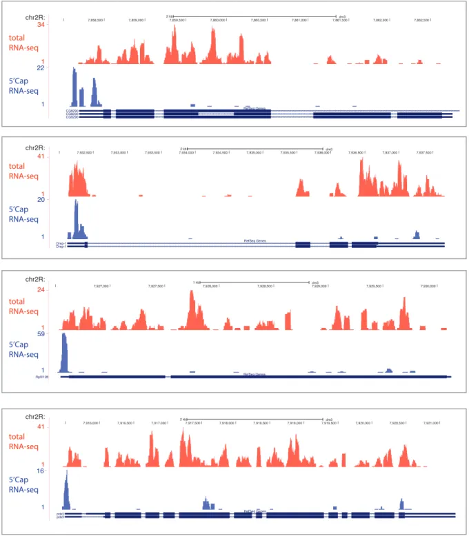
3.5 UCSC tracks for flamenco . . . 47
3.6 UCSC tracks for cluster 20A . . . 48
3.7 UCSC tracks for cluster 42AB . . . 49
3.8 UCSC tracks for cluster 80EF . . . 50
3.9 UCSC tracks for clusters 38C.1 and 38C.2 . . . 51
3.10 UCSC tracks for cluster flamenco . . . 52
3.11 Distribution of reads on 5’P and 5’OH spikes . . . 55
3.12 Gene ontology analysis for 5’P libraries . . . 56
3.13 RPKMS for 5’P RNA libraries . . . 57
3.14 Nucelotide distribution in piRNA precursors from wild type Drosophia . 58 3.15 qPCRs in sh knockdowns . . . 60
3.16 total RNA sequencing in sh knockdown lines . . . 62
3.17 piRNA precursor abundance in sh knockdown lines . . . 63
3.18 5’nucleotide distribution for piRNA precursors . . . 64
3.19 scheme of assays to assess purity of nascent transcripts . . . 66
3.20 PAT-Assays . . . 67
3.21 T1-BX2 scheme . . . 70
3.22 T1-BX2 H3K9me3 ChIP . . . 71
3.23 T1-BX2 nuclear run-on . . . 71
LIST OF FIGURES
3.24 Schematic overview of crosses to follow a paternally transmitted piRNA
producing locus . . . 73
3.25 T1-BX2 LacZ silencing . . . 75
3.26 H3K9me3 levels on the LacZ transgene after paternal transmission and in subsequent generations . . . 76
3.27 Schematic of P1152/BC69 fly system . . . 78
3.28 P1152 and BC69 piRNA production . . . 79
3.29 BC69 unique region spanning piRNAs . . . 81
3.30 P1152 targeted endogenous loci . . . 83
3.31 H3K9me3, Rhino and Cutoff ChIPs upon maternal deposition . . . 84
3.32 Read through transcription upon maternal deposition . . . 85
3.33 Schematic overview over nuclear run-on procedure . . . 88
3.34 GroSeq signal in shRhi, shCuff and control . . . 89
3.35 GroSeq signal in piRNA clusters . . . 90
3.36 GroSeq density in double-stranded clusters . . . 91
3.37 GroSeq density in single-stranded clusters . . . 91
3.38 Cutoff structure model . . . 93
3.39 piRNA precursor levels in shCuff, shRat1 and shWhite . . . 95
3.40 Cuff acts as anti-cleavage factor . . . 96
4.1 Model of uni-stranded and double-stranded cluster transcription . . . 100
4.2 Model for effect of Zucchini knockdown . . . 102
4.3 Model for effects of maternally inherited piRNAs . . . 110
6.1 Strand specific RT for read-throug-PCRs . . . 118
L IST OF T ABLES
3.1 piRNA cluster coordinates . . . 40
3.2 Read numbers and mapping statistics of 5’Cap RNA libraries . . . 42
3.3 Sequencing statistics of two 5’P RNA replicates . . . 56
3.4 Gro-Seq sequencing statistics . . . 88
6.1 Drosophila strains . . . 129
6.2 List of buffers . . . 134
6.3 List of antibodies . . . 137
6.4 List of enzymes . . . 137
6.5 List of equipment . . . 137
6.6 List of reagents . . . 138
6.7 List of reagents . . . 139
6.8 List of kits . . . 139
CHAPTER 1
S UMMARY
RNA interference (RNAi) is a cellular mechanism where small RNAs (20 to 35 nucleotides in length) pair with proteins to build effector complexes guiding them to RNA targets through homologous base pairing.
The piRNA pathway is a germ line specific RNAi pathway that protects the gonads of animals from harmful genetic sequences, termed transposable elements (TEs). TEs are viral DNA remnants that can autonomously mobilize within genomes and cause damage through insertion into new genomic loci and double-strand breaks in DNA. TE activ- ity in germline cells ultimately leads to sterility and its suppression through the piRNA pathway is therefore essential for propagation. While the piRNA pathway shares common characteristics with the two major RNAi pathways (miRNAs and siRNAs), it additionally exhibits several unique features, particularly during biogenesis. piRNAs originate from large non-genic regions, so called piRNA clusters, or from mRNAs of transposable ele- ments. Neither of these RNA transcripts contain the requisite structural features capable of triggering small RNA production, seen in both siRNAs and miRNAs. Consequently, piRNAs are produced in a Dicer independent manner. Therefore, it is not yet understood, how target transcript are recognized by the piRNA pathway.
Furthermore, the extent of piRNA functionality in vivo are not well defined, as they seem to act on multiple levels to protect the germline genome from the propagation of transposable elements. piRNAs lead to direct cleavage of TE mRNAs, but also seem to change the chromatin landscape of regions expressing RNA targets. Additionally, the effect of piRNAs reaches into the next generation through a transgenerational epigenetic mechanism.
CHAPTER 1. SUMMARY
This study focuses on the characterization of the early stages in piRNA biogenesis, starting at their transcription through the first processing steps. The data presented suggest that the two different piRNA cluster types, uni-stranded and double-stranded clusters, differ greatly in the properties of their transcription. Uni-stranded clusters exhibit a defined transcription start site, giving rise to 5’ m7G-capped piRNA precur- sor molecules that resemble canonical mRNAs. Whereas, double-stranded clusters do not show a distinct start site and can be transcribed through internal initiation or read through transcription from neighboring genes. The work furthermore reveals the ex- istence of piRNA intermediates: they are over 200 nucleotides long, which contain a 5’
monophosphorylated terminus and already exhibit the nucleotide biases similar to mature piRNAs.
After processing of piRNA precursors into mature piRNAs, their effects can reach into subsequent generations. To investigate the trans-generational aspect of piRNAs, I utilized transgenic fly models. The results reveal that piRNAs are epigenetic factors influencing a plethora of mechanisms in the piRNA pathway: ensuring a strong piRNA production in the next generation through maintenance of both piRNA production pathways (pri- mary and secondary piRNA production). Furthermore, they maintain the characteristic structural chromatin features of piRNA clusters in the next generation. However, most strikingly piRNAs are capable of transforming regions expressing targeted transcripts into loci producing primary piRNAs. Therefore, the latter result informs a revision to the current definition of piRNA sources and their subsequent targets. piRNA sources and targets might not be strictly distinct, but any piRNA target acquires piRNA cluster signatures and starts producing piRNAs.
The discovery that inherited piRNAs transform a piRNA target into a source, allowed the investigation of the molecular changes at loci targeted by the piRNA pathway. These newly established piRNA clusters acquire a specific chromatin configuration consisting of the histone mark H3K9me3 and two piRNA protein factors Rhino and Cutoff. Together, these factors correlate with increased transcription of their targeted loci. Cutoff appears to be the key factor in piRNA production, through both promotion of read-through transcription beyond stop signals and protection of RNA termini against exonucleolytic degradation.
Overall, the presented data contribute to an understanding of the molecular mecha- nisms in piRNA biogenesis, from the initial processing steps through the trans-generational effect of piRNAs. It suggests that the piRNA system is organized as a giant feed-forward loop, where piRNAs themselves reach into the next generation to ensures reliable propa- gation of the system and effective silencing of deleterious TE sequences.
CHAPTER 2
I NTRODUCTION
2.1 Small RNAs in RNA silencing
Since the discovery of the first small RNAs in the early 1990 (Lee et al., 1993) and the later realization that those small RNAs play a pivotal role in gene expression and genome stability, research on small RNA silencing has been an ever expanding universe (reviewed by (Ghildiyal and Zamore, 2009)). It has been less then 20 years ago, when Fire and Mello discovered that double-stranded RNA molecules can induce gene silencing. This phenomenon was termed RNA interference (RNAi) and the elucidation of its mechanisms, as well as its implementation as a molecular tool in everyday research, have changed the understanding of gene regulation. Small RNA silencing mechanisms can be found in plants, eucaryotes and even some procaryotes and they take part in a variety of cellular processes. They regulate and shape gene expression, cell growth and differentiation, they maintain genome stability and defend cells against invading viruses and mobile genetic elements. The mechanisms of their actions influence chromatin structure, chromosome segregation, transcription, RNA processing, RNA stability and translation (Ghildiyal and Zamore, 2009; Castel and Martienssen, 2013; Meister, 2013). At the core of this pathway a small RNA of 20-30 nucleotide length pairs with a protein of the Argonaute family to form the effector of RNA silencing: the RNA-induced silencing complex (RISC). In this complex the small RNA provides sequence specificity through complementary base pairing with a specific target, whereas the Argonaute exerts the function. Small RNAs are divided into several classes, depending on their origin, length, mode of targeting and type of Argonaute protein they associate with. Three major classes of small RNAs were
CHAPTER 2. INTRODUCTION
defined to date: siRNAs, miRNAs and piRNAs. They all share common features of RNA silencing mechanisms, but every class has distinct characteristics.
2.2 The discovery of small RNA silencing
Our understanding of small RNA silencing emerged from the efforts of many different research groups in seemingly unrelated fields. Several phenomena of gene expression regulation through RNA had been described in plants and animals, before some hallmark discoveries linked those observations to our current understanding of small RNA silencing mechanisms.
One of the first observations of small RNA silencing was made in plants. Napoli and coworkers (Napoli et al., 1990) described, how introduction of an extra copy of the chalcone synthase transgene to enhance pigmentation of petunia flowers led to a surprising effect. Instead of enhancing the color of the flowers through increased production of the coloring pigment, the flowers showed variegated colouring, or were white altogether, suggesting the silencing of the endogenous locus. In the same year, van der Krol and coworkers (van der Krol et al., 1990), also investigating the chalcone synthase pathway, introduced an antisense chalcone synthase gene into petunia and observed silencing of the endogenous gene. Even though RNA interference with an antisense RNA had been used before to manipulate gene expression in cells (Izant and Weintraub, 1984), the common assumption was, that an antisense RNA molecule directly inhibits targeted mRNAs through hybridization. Through dissection of the requirements for the silencing effect, though, van der Krol and colleagues concluded that the mechanism of antisense inhibition reaches beyond RNA duplex formation of sense and antisense RNA.
Several other labs observed that plants, infected with a replicating RNA virus, were able to suppress symptoms of a viral infection and at the same time exhibited silencing of endogenous genes with sequence homology to viral RNA(Dougherty et al., 1994; An- gell and Baulcombe, 1997; Ruiz et al., 1998). An observation similar to the transgene silencing in petunia, made by Napoli and colleagues, was made in the filamentous fungus Neurospora crassa, where the introduction of an exogenous sequence led to the decreased expression of the corresponding edogenous gene (Romano and Macino, 1992). Those observations were termed ’quelling’ in fungus, and post-transcriptional gene silencing (PTGS) in plants.
Only a few years after the first reports of transgene silencing in plants, animals and fungi, Andy Fire and Craig Mello conducted hallmark experiments inCaenorhabditis ele- gans, which would pave the way to understanding the principles underlying the observed phenomena (Fire et al., 1998). They reported that double-stranded RNA leads to inher-
2.2. The discovery of small RNA silencing
itable gene silencing in Caenorhabditis elegans and that this silencing effect of double- stranded RNAs was orders of magnitudes more potent than purified single stranded RNA.
It became apparent that something about the double-strandedness of the RNA leads to a
”catalytic or amplification component in the interference process” (Fire et al., 1998). The phenomenon that a double stranded RNA was able to induce silencing of any homologous sequence was named RNA interference (RNAi).
Originally, the term RNAi was used to describe silencing through siRNAs, the class of small non-coding RNAs discovered by Fire and Mello. With our growing understanding about mechanistic details of different RNA silencing pathways, however, clear distinctions between different pathways became less apparent. They are partially overlapping and converging, and thus the term ”RNAi” is now broadly used for silencing through small RNAs.
The discovery of RNAi changed and advanced our understanding of fundamental cellular principles and it became an invaluable tool in basic and applied research. It was rewarded with a Nobel Prize for Fire and Mello in 2006.
At the same time of the first phenomenological reports of transgene silencing in plants and fungi, the first small RNA, the miRNA lin-4, was discovered in 1993 by Victor Ambros and Gary Ruvkun (Lee et al., 1993). They found a transcript from the lin-4 locus to be a negative regulator of the protein LIN-14. Cloning and investigation of this locus showed that it was not coding for a protein, but produces two small RNA species, a 61 nucleotide (nt) long lin-4L and a 22 nt long lin-4S molecule. The sequence of this lin-4 transcript is highly conserved between differentCaenorhabditis elegans species and has a 10 nt long block of complementarity to a repeated sequence element in the 3’ untranslated region (UTR) of lin-14 mRNA. This publication presents hallmarks in miRNA mechanisms. It described the miRNA precursor lin-4L and suggested its stem- loop structure. Furthermore, it described the first miRNA lin-4S and proposed that it regulates its targets through complementary binding in its 3’UTR. The 10 nt block of complementarity is today known as ’the seed’ of miRNAs.
The second essential component of RNA silencing are Argonaute proteins. They were first described in plants and Drosophila melanogaster (Lin and Spradling, 1997; Cox et al., 1998; Bohmert et al., 1998) and their name stems from the phenotype of AGO1 mutants inArabidopsis thaliana. The unusual appearance of mutant plants reminded the researchers of a squid, and thus they named the gene after the pelagic octopus, Agonauta argo (Bohmert et al., 1998). Those first reports on Argonaute connected it to a function in stem cell differentiation and growth. Later, genetic and biochemical studies on RNAi established them as the second essential component of small RNA mediated silencing.
They were found in all eucaryotes with an RNAi system, as well as in some archaea and
CHAPTER 2. INTRODUCTION
bacteria (Makarova et al., 2009).
Even though the first structural insights into the Argonaute architecture originated from procaryotic Argonautes, their procaryotic function still remains unclear. While RNAi is the crucial defense mechanism of many organisms against invasive genetic el- ements, procaryotes protect their genome through the analogous, but not homologous CRISPR-associated system. Only recently some reports suggest that the function of bacterial Argonautes might be a second line of defense against phages and plasmids, ad- ditionally to the CRISPR system (Carmell et al., 2002; Olovnikov et al., 2013; Swarts et al., 2014; Vogel, 2014).
2.3 Argonaute proteins
Proteins of the Argonaute family are found in plants, animals and fungi (Hutvagner and Simard, 2008; Meister, 2013). As the central effector proteins of RNA silencing they are diversified in certain aspects, but their core domains remain conserved throughout evo- lution. Argonautes can be divided into three subfamilies, the Ago, WAGO and the Piwi subfamily. Argonautes of the Ago clade are expressed ubiquitously and are involved in various aspects of RNA silencing pathways such as miRNA or siRNA mediated silencing;
WAGOs are worm specific Argonautes. Piwi Argonautes are specific to animals and their expression is mainly restricted to germline cells. They were first described as factors for germline maintenance in the Drosophila germline (Lin and Spradling, 1997; Cox et al., 1998). Since the mutation of the germline specific Argonautes leads to small gonads due to derepression of transposable elements, they were dubbed ”P-Element Induced Whimpy Testis”. The name PIWI persisted for the germline specific Argonautes, and one of the main protein domains of all Argonautes is called PIWI.
Throughout evolution, Argonaute proteins underwent significant gene duplications and functional diversification. In yeast, for example, Saccharomyces pombe carries only one Argonaute protein, which covers all RNAi related functions from PTGS to hete- rochromatin silencing (Sigova et al., 2004). Saccharomyces cervisiae lost its Argonaute protein with the accompanied RNAi machinery altogether. To the other extreme, the Caenorhabditis elegans genome encodes 27 distinct Argonautes, where specific proteins conduct distinct aspects of RNA silencing (Buck and Blaxter, 2013). 18 of the 27 Arg- onautes in Caenorhabditis elegans are worm specific, and they are thus called WAGOs.
In between those extremes, plants have ten Argonautes (Mallory and Vaucheret, 2010), humans have four Argonautes and four Piwis andDrosophila melanogaster has two Arg- onautes and three Piwis.
The basic common architecture of Argonaute proteins is their division into four do-
2.3. Argonaute proteins
small RNA guide target RNA
SLICER catalytic site
PAZ
5’
3’
mid N-term
PIWI
A
B C
N-term L1 PAZ L1 mid PIWI
mid
PAZ
N-term L1
L2
PIWI
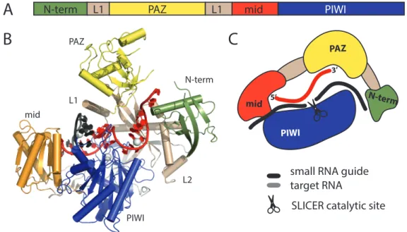
Figure 2.1: Architecture of an Argonaute protein. (A) Domains of Argonaute proteins. (B) Structure of the human Argonaute hAgo2 in complex with a micro RNA (PDB code 4F3T). (C) Schematic representation of an Argonaute protein in a cartoon.
Domain colors are green for the N-terminal domain, yellow for the PAZ domain, red for the mid domain, blue for the Piwi domain and wheat for the two linker domains L1 and L2. The small RNA guide (red) is anchored in the PAZ domain with its 3’ and and in the MID domain with its 5’ end. In case of catalytically active Argonautes, slicing of the target (black) is facilitated by the Piwi domain.
mains: the N domain, the PAZ (Piwi-Argonaute-Zwille) domain, the MID (middle do- main) and the PIWI (P-element-induced wimpy testes) domain. Crystal structures of procaryotic (archea and eubacteria), yeast and other eucaryotic Argonautes, provided insights into the overall architecture and molecular mechanisms of their functions (Song et al., 2004; Parker et al., 2005; Ma et al., 2005; Wang et al., 2009, 2008; Schirle and MacRae, 2012; Nakanishi et al., 2012; Elkayam et al., 2012)).
The PAZ domain harbors the anchor point for the 3’ end of the small RNA. It provides a binding pocket, which specifically recognizes the single stranded 3’ overhang of a double stranded RNA molecule. Those 3’ overhangs are the characteristic product of upstream processing by the RNAseIII endonucleases Drosha and Dicer. The MID domain anchors the 5’ end of the small RNA (Boland et al., 2010; Frank et al., 2010) and in some cases, as seen for the hAgo2 MID domain, it seems to have a preference for certain nucleotides, leading to a bias for 5’ nucleotides of Argnaute bound small RNAs. The PIWI domain has an RNase-H-like fold. RNase-H endonucleases generally cleave RNAs guided by a DNA template. In some Argonautes, the endonucleolytic activity of the PIWI domain is conserved and those Argonautes are commonly referred to as ”Slicers”. Slicers use the
CHAPTER 2. INTRODUCTION
small RNA as a guide and cleave the targeted RNA, leaving a phosphate at the 5’ end.
Only a few Argonautes are cleavage-competent. In Arabidopsis thaliana only two (Ago1 and Ago4) out of the ten Argonautes are slicers (Baumberger and Baulcombe, 2005; Qi et al., 2005, 2006), in Drosophila melanogaster both Argonautes (Ago1 and Ago2), and all three Piwis are cleavage-competent. In human, only Ago2 is an active Slicer, while Ago1, 3 and 4 are not (Meister et al., 2004; Liu et al., 2004). Studies on those human Argonautes revealed the structural requirements for an Argonaute to be an active slicer.
Sequence alignments and later structural studies revealed that the catalytic center of the hAgo2 PIWI domain contains a DEDH tetrade, which is mutated in the other, non- slicing Argonautes. This catalytic center, however, is not the only requirement to make an Aronaute an active slicer. The close investigation of the PIWI domain showed that besides the DEDH tetrade, there are other crucial sequence requirements for the PIWI domain to be active. Most likely, those regions are responsible for the proper orientation of the catalytic center. Domain swap, mutagenesis and DNA shuffling experiments revealed that also the N domain of an Argonaute needs to meet certain sequence requirements in order to make an Argonaute a Slicer (Hauptmann et al., 2013; Faehnle et al., 2013;
Schurmann et al., 2013; Hauptmann et al., 2014).
2.4 miRNAs
Until now, miRNAs were found in the plant and animal branches of the tree of eukaryotic organisms. They are 21-25 nt long and originate from double-stranded RNA molecules.
The precursor RNAs for miRNA production (pri-miRNAs) are usually transcribed by RNA Polymerase II (Ghildiyal and Zamore, 2009) and they originate from intergenic regions, 3’ UTRs or introns. Pri-miRNAs contain a 5’ Cap and a poly-adenylate tail (Kim, 2005). The defining feature for miRNA production is a stem-loop structure, where the stem is 33 nt long and consists of imperfectly paired bases (Bartel, 2004). One pri-miRNA transcript can contain only one, or a cluster of several distinct miRNAs. In animals, maturation of the pri-miRNA into a 20-25 nt miRNA is facilitated sequentially in the nucleus and in the cytoplasm by two RNAse III endonucleases with their double- stranded RNA binding domain (dsRBD) partner proteins. In plants, this two-step process occurs entirely in the nucleus, carried out by only one RNAse III endonuclease (Zhu, 2008).
2.4. miRNAs
2.4.1 miRNA biogenesis
In animals, the first step of miRNA processing takes place in the nucleus. Initially, the dsRBD DGCR8 (in mammals) or Pasha (in flies), recognizes the stem-loop structure and attracts its partner RNAse III endonuclease Drosha. In this complex, which is called Micorprocessor, DGCR/Pasha positions Drosha precisely at the stem of the stem-loop and Drosha catalyzes the excision of the RNA stem-loop (Gregory et al., 2006). The resulting product is called pre-miRNA and it has a two-nucleotide overhang at its 3’ end and a 5’ phosphate group. The 3’ end of this product will not be further trimmed, and thus Drosha defines the 3’ end of miRNAs.
CHAPTER 2. INTRODUCTION
GW182
siRNA duplex endo-siRNA exo-siRNA
RISC loading complex Dicer-2
AGO2
HEN1 SAM
SAH 2’-OCH3 AGO2
AGO1
2’-OCH3 AGO2 AGO2
RISC
target cleavage translational repression
AGO1 RISC Dicer-1
Dicer-1 TRBP
AAAAn
EXP-5
7mGppp Pasha Spliceosome
siRNA
A B
long dsRNA
Gene
Pseudogene inverted repeats
convergent transcripts miRNA
Drosha
pre-miRNA pri-miRNA
miRNA/miRNA*
Argonaute loading
Microprocessor Complex
intron 3’UTR
3’
LOQS
AGO1 GW182 AAAAn
7mGppp
AAAAn
7mGppp
Dicer-2
R2D2
LOQS-PD R2D2
Dicer-2
Figure 2.2:
miRNA and siRNA biogenesis pathways (partially adapted from (Ghildiyal and Zamore, 2009). [Legend see next page]
2.4. miRNAs
Figure 2.2: miRNA and siRNA biogenesis pathways. [The figure is on the previous page]
Biogenesis pathways for miRNAs and siRNAs exemplified with Drosophila melanogster proteins. (A) miRNAs are transcribed from intergenic regions, 3’ UTRs or introns into primary miRNAs (pri-miRNA).
The stem-loop structure is recognized by Pasa and cleaved by Drosha. Together, they build the Micro- processor complex. An alternative route is the excission of the stem-loop through the pre-mRNA splicing pathway. The pre-miRNA is exported into the cytoplasm through Exportin-5 and loaded into Dicer- 1, assisted by the double-stranded RNA binding protein LOQS. The miRNA/miRNA* duplex is then loaded into AGO1, aided by TRBP and possibly other co-factors. After passenger strand degradation and association with the cofactor GW182, the guide miRNA leads AGO1 RISC to its targets.
(B) siRNAs are derived from long, double-stranded precursor molecules, which get processed into siRNA dublexes by Dicer-2. To detect a dsRNA as a substrate, Dicer-2 relies on a double-stranded RNA binding protein. In Drosophila, those are Loqs-PD for exo-siRNAs and R2D2 for endo-siRNAs. Dicer together with the processed siRNA dublex and the respective double-strand RNA binding protein forms the RISC loading complex and transfer the dublex into AGO2. The passenger strand is degraded and the guide strand is methylated on its 3’ terminus through HEN1 to increase stability. Association with GW182 forms the AGO2 RISC, which is guided to its targets by the siRNA.
CHAPTER 2. INTRODUCTION
In worms, flies and mammals, a Microprocessor independent route has been described for the nuclear processing of so-called mirtrons. In the case of mirtrons, the excision of the stem-loop is facilitated by the nuclear pre-mRNA splicing pathway (Okamura et al., 2007; Ruby et al., 2007; Berezikov et al., 2007; Babiarz et al., 2008). In animals, pre- miRNAs are exported into the cytoplasm through Exportin-5 (Yi et al., 2003; Bohnsack et al., 2004; Lund and Dahlberg, 2006). The next cut to define the 5’ end of the mature miRNA is performed in the cytoplasm by Dicer, assisted by its dsRBD partner protein TRBP (in mammals) or Loquacious (LOQS) (in flies). Dicer binds the 3’ end of the pre-miRNA with its PAZ-domain, which is a common feature of Dicer and Argonautes.
Cleavage is facilitated through its RNAse III catalytic site. The catalytic site is positioned two helical turns (or 22 bp) away from the PAZ domain and consists of a cleft, formed by an intramolecular dimer involving the RNAse III domains. Each RNAse III domain cleaves one of the two RNA strands, which results in an RNA duplex with a 2 nt 3 overhang. It is thus the distance of Dicer’s PAZ domain to its catalytic site, which makes it act as a molecular ruler for miRNAs. The two resulting strands are called miRNA and miRNA*.
However, to this general principle of miRNA biogenesis, there are some exceptions.
In zebrafish and mouse, a specific miRNA, miR-451, has been described to be processed independently of Dicer by Ago2 (Cifuentes et al., 2010; Cheloufi et al., 2010). A class of miRNAs derived from small nucleolar RNAs, seem to be processed in a Dicer dependent, but DGCR8/Drosha independent way. (Ender et al., 2008)
In plants, the processing of pri-miRNAs into miRNA-miRNA* duplexes is not di- vided into a nuclear and a cytoplasmic phase, but takes place entirely in the nucleus.
There, Dcl1 (Dicer-like protein), together with its dsRBD partner HYL1 and the C2H2 Zn-finger protein SE, performs the cleavages solo (Kurihara and Watanabe, 2004; Dong et al., 2008). Another distinction between miRNA biogenesis in plants and animals is the 2’-O-methylation of the 3’ ends of plant miRNAs by HEN1. (Park et al., 2002; Yu et al., 2005; Yang et al., 2006). This modification is believed to enhance stability of miR- NAs by preventing 3’ uridinylation, which acts as a signal for degradation (Li et al., 2005).
One of the two strands from a miRNA- miRNA* duplex guides the Argonaute to its targets. This chosen strand is called ”guide”, whereas the other strand will ultimately be degraded and is called ”passenger”. Which of the strands is chosen to be the guide, depends on the relative thermodynamic stability of the duplex - in most cases the 5’ end is chosen from the end with the less stable base-pairing (Khvorova et al., 2003; Schwarz et al., 2003).
The Argonaute together with its small RNA ultimately forms the RNA-induced Si-
2.4. miRNAs
lencing Complex (RISC). Loading of a miRNA from Dicer into an Argonaute depends on many accessory co-factors, which furthermore differ between organisms.
The current model is that a RISC loading complex consists of Dicer, TRBP and Argonaute. After degradation of the passenger strand, the loaded Argonaute together with GW182 forms the mature RISC. Functional studies show that GW182 is necessary and sufficient to induce Ago mediated gene silencing in humans, Caenorhabditis elegans and Drosophila (Liu et al., 2005; Ding and Han, 2007; Eulalio et al., 2008; Carthew and Sontheimer, 2009; Ding and Grosshans, 2009).
2.4.2 miRNA function
In the miRISC complex each function is clearly assigned to one component. The miRNA acts as a sequence specific guide, while the Argonaute protein provides the function.
Gene silencing through small RNAs interferes with gene expression on different levels. A nuclear pathway silences on a transcriptional level (transcriptional gene silencing, TGS), whereas a cytoplasmic pathway targets mRNAs in the cytoplasm (post-transcriptional gene silencing PTGS). The cytoplasmic PTGS through miRNAs will be described subse- quently, while principles of the nuclear TGS will be addressed in section 2.5.2.2.
2.4.2.1 Post-transcriptional gene silencing in the cytoplasm
Targeted mRNAs undergo three possible fates, depending on the type of Argonaute protein and the degree of complementarity to the miRNA. They can be either directly cleaved, channeled into a degradation pathway, or be translationally suppressed.
Cleavage of the target is generally induced by perfect or almost perfect complemen- tarity between miRNA and target. High degree of complementarity is mainly observed in plant miRNAs, and this mode of action is restricted to RISC complexes containing a Slicer.
The two other processes - degradation and translation inhibition - are initiated through imperfect miRNA-target complementarity and can be performed by any Argonaute pro- tein. They seem to be the prevalent mode of regulation in animals.
The details of non-cleavage mediated silencing is intensively studied and several, mutu- ally not exclusive mechanisms are proposed (Gu and Kay, 2010). Detection of the miRNA target sites in the 3’ UTR of an mRNA can lead to deadenylation and degradation of the target. Alternatively, initiation of translation can be inhibited through prevention of 5’ m7G-cap recognition or through prevention of ribosome assembly. Some data suggest that even after translation initiation, protein synthesis can be repressed by slowed-down elongation or by dissociation of the ribosome. One key player in translation-inhibition is
CHAPTER 2. INTRODUCTION
GW182 (Ding and Han, 2007; Zekri et al., 2009), which interacts with Argonaute pro- teins, as well as with the poly-A binding protein (PABP). It localizes RISC comlpexes into specific cytoplasmic compartments, P-bodies, which are centers for mRNA regulation and degradation.
On the other hand, Argonaute proteins show specific interaction with 5’ m7G-caps and thus could compete with eIF4E, the cap binding factor necessary for translation initiation. Since a proper interaction between the 5’ cap and the polyA tail of an mRNA is required for translation initiation, it is conceivable that miRNA mediated translational repression is a consequence of interference with those basic processes.
2.5 siRNAs
siRNAs are produced from linear, perfectly base paired precursor molecules and they can be subdivided into two groups, depending on the origin of the precursor. If the long dou- blestranded RNA (dsRNA) originates from an exogenous source, like in an experimental laboratory setup, or a virus, they are called exo-siRNAs. If the dsRNA stems from en- dogenous sources, such as transposable elements, repetitive sequences, pseudogenes or convergent transcription, they are called endo-siRNAs (Golden et al., 2008).
2.5.1 siRNA biogenesis
Like miRNAs, siRNAs are produced from double-stranded precursor RNA by Dicer, leaving a 5’ phosphorylated end and a 3’ OH end with a 2 nt overhang. In mammals and Caenorhabditis elegans, one Dicer produces miRNAs as well as siRNAs. Other organisms have designated Dicers, for instance inDrosophila Dicer-1 makes miRNAs, whereas Dicer- 2 produces siRNAs. To detect a dsRNA as a substrate and channel it into the siRNA pathway, Dicer relies on double-strand binding proteins (dsRBPs). In Caenorhabditis elegans this is RDE-4, inDrosophila R2D2 and Loqs-PD. (Liu et al., 2003; Parker et al., 2006; Mirkovic-Hosle and Forstemann, 2014; Hartig et al., 2009; Hartig and Forstemann, 2011). In human cells, Dicer can associate with two different dsRBPs, protein activator of PKR (PACT) and trans-activation response RNA-binding protein (TRBP). (Lee et al., 2013). Loading and maturation of the siRISC is in many parts convergent with the miRNA pathway and relies on mainly the same components (see figure 2.2).
2.5.2 siRNA function
siRNAs are distinguished from miRNAs based on their origin, but also through their perfect complementarity to their targets. Perfect base pairing between small RNA and
2.5. siRNAs
target triggers direct cleavage of the target, as opposed to a non-cleavage dependent repression. Perfect complementarity is especially observed in plants, where the siRNA pathway is a powerful defense against viral infections. It appears that the absence of a protein-based immune system in plants expanded the breadth of siRNAi. In most animals, the mechanisms between siRNA and miRNA mediated silencing are converging.
In plants and worms, however, the response to long dsRNAs differs from most other organisms. They possess a system, in which they can amplify the silencing.
2.5.2.1 siRNAs in plants
Exogenous dsRNAs trigger processing into siRNAs through Dicer. Those so called pri- mary siRNAs lead RISC to the target, which is cleaved by the Argonaute. Plants and worms encode an RNA dependent RNA polymerase (RdRP), which uses this cleaved transcript as a template and synthesizes a second strand, resulting in a long dsRNA.
The plant, which is widely used for a model organism to investigate plant RNAi, is Ara- bidopsis thaliana. Arabidopsis thaliana expresses 4 different Dicers and one of them is specialized to process the products of RdRP into secondary siRNAs (Bologna and Voin- net, 2014). The resulting siRNA can once more be loaded into Argonautes and direct nuclear or cytoplasmic silencing. In Caenorhabditis elegans the mechanism to produce secondary siRNAs is sligthly different. Detection of the target RNA by the Argonaute RDE-1 attracts the RdRP, which produces small RNAs from the target template.
2.5.2.2 Transcriptional gene silencing in the nucleus
Nuclear RNA silencing mechanisms have first been described in yeast, ciliates and plants, where they have been found to modify the chromatin structure of targeted genomic regions (Castel and Martienssen, 2013). Animals also seem to have nuclear RNA silencing pathways, but mechanistic insights are still sparse.
The fission yeast Saccharomyces pombe only has one Argonaute protein, Ago1, and its main function appears to be nuclear - formation of heterochromatin on centromers (Moazed et al., 2006). Those regions consist of repetitive sequences, which are bidirec- tionally transcribed into long dsRNAs. Those dsRNA molecules are diced into siRNAs and loaded into Ago1 to form the RITS complex (RNA-induced transcriptional silencing).
The siRNA brings RITS to nascent transcripts of the centromeric region, where it ensures the maintenance of heterochromatin through different mechanisms. RITS attracts the Histone-methyl transferase Clr4, which methylates histone H3 lysine 9. This chromatin mark attracts the HP1 homolog Swi6, which establishes heterochromatin at this locus.
At the same time, RITS recruits the RNA-dependent RNA polymerase Rdp1, which am-
CHAPTER 2. INTRODUCTION
plifies the siRNA signal through further generation of dsRNA from this locus. (Motamedi et al., 2004) Further, genetic studies showed that Ago1, Dicer and Rdp1 are crucial for heterochromatin formation (Volpe et al., 2002), that Clr4 is required for siRNA produc- tion (Noma et al., 2004) and that Swi6 is required for Rdp1 localization. This whole system is organized in a feed-forward loop, where siRNA production, RITS localization and H3K9 methylation are reinforcing each other.
In Arabidopsis thaliana the establishment of pericentromeric heterochromatin also depends on Argonaute proteins and small RNAs. Yet, the platform for heterochromatin formation does not rely on histone tail modification, but rather on DNA methylation (Matzke et al., 2009). The centromeric repetitive regions are transcribed by the plant specific RNA polymerase IV (Pontier et al., 2005; Kanno et al., 2005) and the resulting transcripts are templates for the RNA dependent polymerase RDR2, which produces the dsRNA for siRNA production through Dicer. Those siRNAs are loaded into Ago4 in the cytoplasm and are re-imported into the nucleus, where they recognize nascent transcripts from the second plant-specific RNA polymerase V (Pontier et al., 2005; Kanno et al., 2005). Upon detection, the PolV transcribed region gets repressed through DNA methylation by the DNA methyltransferase DRM2.
The two mechanisms of RNAi mediated pericentromeric heterochromatin formation necessitate an intricate regulation: heterochromatin is presumably silenced, but still needs to be transcribed in order to be established. The nuclear role of RNAi in fission yeast and Arabidopsis thaliana seems to be mainly focused on establishment of a structural pericentromeric heterochromatin.
In animal somatic cells evidence for nuclear RNAi is getting denser. The molecular mechanism, however, is far less understood (Ohrt et al., 2012; Schraivogel and Meister, 2014). Argonautes have been reported to shuttle between the cytoplasm and the nucleus (Weinmann et al., 2009) and Dicer, too, can localize into the nucleus (Doyle et al., 2013).
For both proteins the nuclear levels seem to be tightly regulated. Other reports sug- gest the presence of a complete nuclear RNAi machinery containing all components that are essential in cytoplasmic RNAi. (Gagnon et al., 2014). In analogy to Saccharomyces pombe and Arabidopsis thaliana, mammalian RNAi also was implicated in heterochro- matin formation of structural heterochromatin on satellites, different other repeat-rich loci and transposable elements (Peng and Karpen, 2007; Fagegaltier et al., 2009; Desh- pande et al., 2005). One report suggested a direct interaction of Ago1 and Dicer2 with chromatin and RNA polymerase II in somaticDrosophila cells (Kavi and Birchler, 2009).
Another study showed a direct association of Dicer with Polymerase II on actively tran- scribed genes and suggests transcriptional silencing (White et al., 2014). To understand
2.6. piRNAs
the full extend of nuclear RNAi functions in mammals, further studies will be required.
2.6 piRNAs
The piRNA pathway is an RNA silencing pathway, which is specialized in repression of transposble elements. piRNAs associate with the Piwi-clade Argonautes, which is specific to metazoans and show gonad-specific expression (Houwing et al., 2007a; Lin and Spradling, 1997; Kuramochi-Miyagawa et al., 2001, 2004; Cox et al., 1998; Carmell et al., 2002).
The domain structure of Piwi Argonautes is very similar to canonical Argonautes, with the MID domain anchoring the 5’ end of a piRNA, and the PAZ domain lodging the 3’ end. In Drosophila all three Piwi proteins have slicer activity. Slicer deficient Piwi, however, does not result in any phenotype (Sienski et al., 2012; Darricarrere et al., 2013), whereas Aub and Ago 3 absolutely require their slicer activity. What distinguishes Piwi clade Argonautes from other Argonautes, is the presence of Arginine-rich motifs near their N-termini. These residues are post-translationally dimethylated and arginine methylation allows Piwi proteins to interact with Tudor family proteins, which are key components of the piRNA pathway (Vagin et al., 2009; Nishida et al., 2009; Kirino et al., 2009; Liu et al., 2010).
The eponymous Tudor domain binds methylated arginines on Piwi proteins. Since many Tudor proteins often have several Tudor domains, they can act as a scaffold for the formation of higher-order complexes and possibly coordination of piRNA biogenesis and function (Mathioudakis et al., 2012; Huang et al., 2011).
In germ cells ofDrosophila melanogaster and in mouse testes, recognition of arginine- methylated effector proteins seems to compartmentalize the piRNA pathway into distinct perinuclear granules, which have been described by cell biologists as ”nuage” (for their clowd-like morphology, ’nuage’ in french means ’cloud’). They are similar to P-bodies and harbor multiple components of the piRNA pathway (Brennecke et al., 2007; Aravin et al., 2008, 2009; Malone et al., 2009)
With a typical size between 24 and 30 nucleotides piRNAs are longer than siRNAs and miRNAs. They are generated either from RNA transcripts of active TE copies or from transcripts originating from specialized loci in the genome, called piRNA clusters.
Clusters are intergenic regions harboring defunct remnants of TEs, forming the basis of germline defense against TE propagation (Brennecke et al., 2007; Aravin et al., 2007, 2008). piRNAs that are generated from piRNA clusters are mostly antisense to TE mRNA sequences and thus can guide the piRISC to mRNAs of active TEs through complementary base pairing.
CHAPTER 2. INTRODUCTION
The piRNA pathway has often been compared to an adaptive immune system, as it conveys memory of previous transposon invasions by storing TE sequence information in piRNA clusters. By amplifying piRNAs that are complementary to the active transposon sequence, the piRNA pathway can respond quickly and specifically to acute TE activation.
In addition to targeted cleavage of TE mRNAs within the cytoplasm, Piwi proteins also function on the chromatin level to silence TE transcription. Silencing is mediated through formation of repressive chromatin on TE copies (Rozhkov et al., 2013; Sienski et al., 2012;
Le Thomas et al., 2013).
2.6.1 piRNA biogenesis
2.6.1.1 piRNA cluster architecture and transcription
piRNA populations are highly complex: deep sequencing of piRNAs from mouse and fly revealed millions of individual, distinct piRNA molecules. Neither piRNAs, nor their precursor sequences show any structural motif or sequence bias except for a preference for Uracil as the first 5’ nucleotide (1U bias) of the piRNA. However, when mapped to the genome, this highly diverse population of piRNAs falls into a few discrete genomic loci, called piRNA clusters (Ro et al., 2007; Brennecke et al., 2007; Lau et al., 2006;
Grivna et al., 2006; Aravin et al., 2007; Houwing et al., 2007b; Aravin et al., 2006).
Clusters are up to 200 kilobases long, mostly located in pericentromeric and subtelomeric heterochromatin and are highly enriched in transposable element sequences (Brennecke et al., 2007). Upon invasion of a new transposon the TE eventually jumps into one of the clusters leaving a memory of the invasion. Once a transposable element leaves a trace in a cluster, it can be targeted by the piRNA machinery, which will limit further propagation of the TE. TEs insert into different genomic sites in a non-random fashion (Huang et al., 2012; Yamanaka et al., 2014). Studies on the P-element transposon established that those elements preferably jump into the immediate 5’ region of genes or into 5’ exons (Spradling et al., 2011; Yamanaka et al., 2014). Another transposable element, piggyBac, was also shown to preferably insert near promoter regions (Thibault et al., 2004; Yamanaka et al., 2014). Hence, TEs seem to require an open chromatin structure, which is characteristic for actively transcribed regions (Yamanaka et al., 2014).
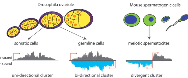
In fly ovaries, piRNA clusters are expressed in two cell types: cells of germline origin that include the developing oocyte and associated nurse cells, and somatic support cells called follicular cells. Interestingly, the structure of clusters differs depending on where they are expressed: Germline clusters are transcribed bi-directionally, generating both sense and antisense piRNA in relation to the transposon mRNA. Conversely, somatic clusters appear to be transcribed uni-directionally, producing mostly piRNA that are
2.6. piRNAs
somatic cells germline cells
Drosophila ovariole
uni-directional cluster + strand
- strand
bi-directional cluster
Mouse spermatogenic cells
meiotic spermatocites
divergent cluster
Figure 2.3: piRNA clusters Drosophila and mouse. In the Drosophila ovaiole germline cells (in yellow) are surrounded by a layer of somatic cells (called follicular cells, in pink). The somatic cells express uni-stranded piRNA clusters, in germline cells the majority of piRNA clusters is transcribed in a bi-directional manner, giving rise to piRNAs mapping to both strands. In meiotic spermatocites of mouse testes, most piRNA clusters are transcribed divergently from a central promoter region.
antisense to TE coding regions (Brennecke et al., 2007) (see figure 2.3). Overall, the cellular source of piRNA cluster transcripts destines how they are processed.
In mouse testes, spermatogenic cells have two kinds of piRNA clusters: one class is transcribed during embryonic development, and like inDrosophila melanogaster, piRNAs derived from these clusters defend the germline against transposable elements. A second type of piRNA cluster is expressed in spermatogenic cells of adolescent mice during the first division of meiosis. These are called pachytene clusters, since piRNAs derived from these clusters are highly abundant in the pachytene stage of meiosis. Pachytene piRNAs are not enriched in transposon sequences and to date their function is unknown. An interesting feature of many pachytene clusters is that they are transcribed divergently (i.e. in both directions) from a central promoter (figure 2.3) (Aravin et al., 2006; Houwing et al., 2007b) .
To date, the transcriptional regulation of piRNA clusters in flies and in embry- onic stages of mouse spermatogenesis remains elusive. Chromatin immunoprecipitation (ChIP) analysis on histone modifications in the silkworm ovary-derived BmN4 cell line (this cell line contains a functional piRNA pathway and is used as an analogous model for theDrosophila piRNA pathway) revealed that piRNA clusters display features of eu- chromatin. They are enriched for histone modifications associated with transcriptional activity such as H3K4 di- and tri-methylation in addition to H3K9 acetylation. At the same time, they are devoid of repressive histone H3K9 di- and tri-methyl marks (Kawaoka et al., 2013). In ChIP performed on Drosophila ovaries, however, H3K9me3 marks are
CHAPTER 2. INTRODUCTION
present on clusters and transposon loci (Rangan et al., 2011). The heterochromatin pro- tein (HP1) homolog of Drosophila, Rhino, seems to bind those histone marks on piRNA clusters and is essential for piRNA cluster transcription (Klattenhoff et al., 2009). Still, it remains unresolved if these chromatin marks are a cause or consequence of cluster transcription.
In summary, data about piRNA clusters in Drosophila suggests that they are similar to pericentromeric heterochromatin in Saccharomyces pombe, where a specific chromatin configuration is required for generation of small RNAs.
2.6.1.2 Biogenesis of primary piRNAs
piRNAs in flies are 23-25nt in length, whereas in mouse they are slightly longer, averaging 25-28nt. Similar to other small RNA classes, piRNAs are processed from longer precur- sors. In contrast to miRNAs and siRNAs, however, the precursors are single stranded transcripts without obvious hairpin structures and they are produced in a Dicer inde- pendent pathway. It is thought that piRNA biogenesis begins with the endonucleolytic cleavage of the long precursor transcript, generating shorter piRNA precursors. This cleavage event possibly specifies the 5’ end of the future piRNA (figure 2.4). Genetic screens and structural studies suggest that the endonuclease, Zucchini (Zuc), might con- duct this first cleavage of piRNA precursors (Voigt et al., 2012; Nishimasu et al., 2012;
Ipsaro et al., 2012; Olivieri et al., 2010; Haase et al., 2010; Muerdter et al., 2013; Handler et al., 2013; Czech et al., 2013). Endonucleolytic cleavage of ssRNA in vitro through Zuc- chini results in a 5’ phosphate end and a 3’ hydroxyl end. (Nishimasu et al., 2012; Ipsaro et al., 2012). Since mature piRNAs possess a 5’ phosphate, Zucchini seems a good candi- date for this cleavage step. Nishimasu and colleagues investigated the cleavage activity of Zucchini on heterogeneous and poly(U) ssRNA (Nishimasu et al., 2012) and found that it exhibits no strong bias towards any nucleotides. It is therefore unclear, how the 1U bias of primary piRNAs is generated. A bias for the first nucleotide in Argonaute bound small RNAs has been observed in many systems. miRNAs, for example, exhibit a strong bias for Uridine at the 5’ end inCaenorhabditis elegans (Lau et al., 2001) andDrosophila (Czech et al., 2009; Ghildiyal et al., 2010). siRNAs in flies and in plants tend to have a 1C bias (Lee et al., 2010; Ghildiyal et al., 2008; Kawamura et al., 2008). Structural studies on the MID domain of human andArabidopsis thaliana Argonautes showed that a so-called ”specificity loop” seems to be a determinant for 5’ nucleotide specificity (Frank et al., 2010, 2012) for miRNAs and siRNAs and it is suggested that this bias is detected during loading of the double-stranded Dicer product. The aminoacid sequence of the nucleotide specificity loop of Piwi Argonautes is different from hAGO2 and AtAGO1, and still all those Argonautes exhibit a 1U selectivity. This suggested that the specificity
2.6. piRNAs
loop interacts with the 5’ nucleotide through hydrogen bonds and not through specific aminoacid side chains. Studies in Drosophila melanogaster embryo lysate showed that the 5’ nucleotide of miRNAs and siRNAs seems to be sensed and monitored during RISC loading and unwinding of the passenger strand (Kawamata et al., 2011). After cleavage of the piRNA cluster transcript, the 5’ end of piRNA precursors gets loaded into a Piwi protein. In flies, primary piRNA are loaded into two of the three Piwi proteins, Piwi and Aubergine (Aub). The factors that constitute the piRNA loading machinery are currently unknown, however several studies have identified the proteins Shutdown (Shu), Vreteno (Vret), Brother-of-Yb (BoYb) and Sister-of-Yb (SoYb) as components involved in loading of Piwi and Aub with primary piRNA (Olivieri et al., 2010; Handler et al., 2013). Loading of Piwi seems to require some additional factors including Armitage (Armi) and Zucchini (Zuc) and in somatic ovary cells, the Tudor domain protein, Yb (Szakmary et al., 2009;
Olivieri et al., 2010).
CHAPTER 2. INTRODUCTION
PiwiPiwi U
Aub HEN1
A a
a b
b
c c
d
e Germline cells
B Follicular cells
Zuc
piRNA precursor
piRNA precursor
piRNA precursor
nucleus
cytoplasm
nucleus
cytoplasm precursor
cleavage
trimmer U
Transposon mRNA Transposable element
Zuc precursor cleavage
Ping-Pong Cycle
target cleavage
primary piRNA
maternally deposited piRNA 3’ end
modification secondary
piRNA
Aub
trimmer
Aub
Aub
Piwi
AGO3
AGO3
Transposable element piRNA cluster
Pol2 Pol2
piRNA cluster Pol2 Pol2
U
Aub
U
HEN1 3’ end modification
U
U
A
A 10
5’ 10
5’
Piwi
2’-OCH3 U
U mit
ochondr ion
UA
2’-OCH3
2’-OCH3
Figure 2.4:
piRNA biogenesis pathways in germline cells and somatic follicular cells of the Drosophila ovary. [Legend see next page]
2.6. piRNAs
Figure 2.4: piRNA biogenesis pathways in germline cells and somatic follicular cells of the Drosophila ovary. [The figure is on the previous page]
Biogenesis of piRNA in flies begins in the nucleus and initiates the ping-ping cycle in the cytoplasm. (A) piRNA pathway in germline cells. a) Precursor transcripts originate from bi-directionall piRNA clusters, containing TE fragments in both orientations (grey arrows). They are exported from the nucleus into the cytoplasm and processed into smaller fragments, possibly by the mitochondrial-associated endonuclease Zucchini. b) Aub binds precursor piRNA fragments with a preference for fragments that contain a 5 terminal uracil (1U). The 3’ end of piRNA precurosrs is generated when an unknown 3- 5 exonuclease trims the precursor piRNA fragment bound to Aub and Hen1 catalyzes the 2-O methylation. This cluster derived piRNA is called primary piRNA. c) When mRNA of active transposons is exported from the nucleus, Aub loaded with a piRNA complementary to this element is guided to the transcript and cleaves it. d) The cleaved transcript is loaded into Ago3, anchored with its 5’ end, followed by trimming and 2’ O-methylation of its 3’ end, to generate a mature secondary piRNA. e) Ago3 loaded with the secondary piRNA is then believed to target and cleave newly generated precursor transcripts that are loaded into Aub to initiate a new round of ping-pong. (B) piRNA pathway in somatic follicular cells.
a) Uni-stranded clusters containing TE fragments mainly in antisense orientation are transcribed into piRNA precursor molecules and exported into the cytoplasm, where they are cleaved through Zucchini.
b) Piwi incorporates the piRNA precursor with a prefrernce for 1U, which gets trimmed and methylated at its 3’ end. The Piwi-piRNA complex re-enters the nucleus and finds transcriptionally active TEs through complementarity to the nascent transcript. There it silences the TE on a transcriptional level.
CHAPTER 2. INTRODUCTION
While Dicer determines the size of miRNAs and siRNAs, piRNAs are sized according to the Argonaute, they associate with. Once loaded into Piwi proteins, the piRNA precursor is trimmed at its 3’ end by an unknown 3’ - 5’ exonuclease. This nuclease is tentatively named ”Trimmer”. The final length of the piRNA is determined by the piRNA binding pocket of the Piwi protein: Piwi associated piRNAs are 25 nt long, whereas piRNAs associated with AUB or AGO3 are 24 nt or 23 nt long, respectively.
After trimming, piRNAs acquire a characteristic 2’ methyl modification at their 3’ end that is introduced by the piRNA methyl-transferase Hen-1 and leads to stabilization of the small RNA (Kawaoka et al., 2011). Overall, while the exact biochemistry and the involved factors are still not fully known, piRNA biogenesis consists of multiple consecutive steps from transcription of precursors, through precursor cleavage and loading, to trimming and 2’ O-methylation. Defects in any of these steps leads to diminished piRNAs, upregulation of transposons and sterility.
2.6.2 Nuclear and cytoplasmic function of the piRNA pathway
Transposons are selfish genetic elements that can be classified into DNA or retrotrans- posons based on the mechanism by which they transpose in the genome. DNA trans- posons mobilize by a ’cut and paste’ mechanism that is accomplished with the help of an encoded transposase protein. Retrotransposons propagate by reverse transcription of an RNA intermediate. The required enzymes are encoded by the transposable elements.
Another class of TE is the semi-autonomous sequences, such as Alu repeats, that require the protein machinery of other transposons for their replication. In either case the trans- posable element needs to be transcribed as an mRNA in order to produce the enzymes necessary for its transposition. Accordingly, transposition can be regulated on multiple levels, from the efficiency of transcription, to transcript stability and rate of translation of required factors, or by interfering with the process of re-integration into the genome. The piRNA pathway seems to target two steps that are required for all transposons: In the nucleus, piRNAs are implicated in the regulation of chromatin structure that can affect transcription of targeted loci. In the cytoplasm, the piRNA pathway directly targets and destroys RNAs of transposons that escaped transcriptional silencing.
2.6.2.1 The role of piRNAs in regulating chromatin structure
Transcriptional regulation of specific loci is influenced by the overall chromatin archi- tecture. In mammals this regulation is achieved on two levels: through methylation of DNA and through modifications of histone proteins or even incorporation of special hi- stone variants. In Drosophila melanogaster, histone modifications and histone variants
2.6. piRNAs
are mainly responsible for defining the properties of chromatin. In both flies and mice, one of the three Piwi proteins localizes to the nucleus, suggesting a nuclear role for the piRNA pathway (Cox et al., 1998; Aravin et al., 2008). Such a mechanism is further strengthened by the observation that in flies Piwi shows a banding pattern on polytene chromosomes of nurse cells, suggesting a direct interaction with chromatin (Le Thomas et al., 2013).
Nuclear piRNA effects in mouse
In mouse, DNA methylation plays an essential role in imprinting and in silencing of trans- posable elements (TEs). Male germ cells are established in a narrow developmental win- dow during embryonic germ cell development, following global genome de-methylation.
This is a critical point in the fate of spermatogenic cells, since the failure to methylate sequences of retrotransposons scattered throughout the genome leads to their activation and subsequent meiotic failure and sterility (Bourc’his and Bestor, 2004). Several lines of evidence indicate that piRNAs are responsible for directing DNA methylation to its genomic targets. First, several proteins involved in the piRNA pathway have been de- scribed to localize to the nucleus: Miwi2, Tdrd9, and Mael (Soper et al., 2008; Shoji et al., 2009; Aravin et al., 2008, 2009). While Tdrd9 and Mael are expressed through- out male germ cell development, Miwi2 is expressed in a narrow developmental window exactly at the same time of de novo DNA methylation in the embryonic spermatocyte.
Additionally, genetic data implicate piRNAs in establishing de novo DNA methylation patterns in the mouse male germline on regulatory regions of TEs (Kuramochi-Miyagawa et al., 2008; Carmell et al., 2007; Aravin et al., 2008). Dnmt3L is an accessory factor for de novo DNA methyltransferases Dnmt3a and Dnmt3b, and is essential forde novo DNA methylation. The defects observed in Dnmt3L knock-out animals are strikingly similar to those observed in animals deficient in many piRNA pathway components, including the two Piwi proteins, MILI and MIWI2: all result in meiotic arrest of spermatogenesis and apoptosis of germ cells. Furthermore, methylation patterns were not reestablished on retrotransposon sequences in mice lacking Mili or Miwi2 (Kuramochi-Miyagawa et al., 2008), whereas piRNAs are still produced in Dnmt3L mutants (Aravin et al., 2008).
A second mouse Piwi protein, MILI is concomitantly expressed in the embryonic stage of spermatogenesis. While it localizes to the cytoplasm, its expression is necessary for piRNA loading and nuclear localization of MIWI2. The phenotype of Mili mutants is similar to that of Miwi2 suggesting that piRNAs serve as sequence-specific guides that direct Miwi2 to sites where DNA methylation is established (Kuramochi-Miyagawa et al., 2001, 2004, 2008; Aravin et al., 2008).
Indeed, piRNA directed DNA methylation is one mode of regulation for transposable