(PROF. DR. DR. MED. LUKAS PRANTL) DER FAKULTÄT FÜR MEDIZIN DER UNIVERSITÄT REGENSBURG
PDGF regulated migration of mesenchymal stem cells towards malignancy acts via the PI3K signaling
pathway
Inaugural – Dissertation zur Erlangung des Doktorgrades
der Medizin
der
Fakultät für Medizin der Universität Regensburg
vorgelegt von Sonia Salha
2019
Dekan: Prof. Dr. Dr. Torsten E. Reichert 1. Berichterstatter: Prof. Dr. Dr. med. Lukas Prantl 2. Berichterstatter: PD Dr. med. Markus Loibl Tag der mündlichen Prüfung: 22.10.2019 (Regensburg)
24.10.2019 (Zürich)
Inhaltsverzeichnis
Zusammenfassung ... 3
Fragestellung ... 3
Methoden ... 5
Ergebnisse ... 10
Diskussion ... 14
Referenzen ... 17
Publikation als Erstautor ... 21
Erklärung ... 30
Curriculum Vitae ... 31
Zusammenfassung
Fragestellung
Adulte Stammzellen spielen eine entscheidende Rolle für die Regeneration und Reparation im Rahmen von Verletzungen oder des programmierten Zelltodes (i.e.
Apoptose) [1]. In den letzten Jahren konnten in den verschiedensten Geweben multi- potente Zellpopulationen nachgewiesen werden, die eine Zelldifferenzierung unter anderem in Knorpelzellen [1], Herz-und Skelettmuskelzellen [2,3] aber auch in neuro- ektodermale Zellen wie Astrozyten [4,5] oder endodermale Zellen wie Hepatozyten [6,7] zeigten. Es wurde jedoch auch berichtet, dass stromale Zellen von Krebszellen aktiv rekrutiert werden und dadurch ein vermehrtes Tumorwachstum nachgewiesen werden konnte [8,9]. Eine große Anzahl von Myo-Fibroblasten sind im Bindegewebe von invasivem Brustkrebs zu finden, welche jedoch nicht im gesunden Brustgewebe vorhanden sind [10]. Es gibt Hinweise, dass diese Cancer-Associated Myo- Fibroblasten (CAF) von rekrutierten, zirkulierenden Knochenmarksstammzellen abstammen [11,12]. Darüber hinaus gibt es Daten die zeigen, dass auch lokales Fettgewebe der Brust eine Quelle für diese mesenchymale Stammzellen sein kann [13]. Des Weiteren konnte in Brustkrebsmodellen gezeigt werden, dass Mesenchymale Stammzellen (MSC) zum Tumor wandern und sich in dem tumor- assoziiertem Stroma integrieren und dabei das Tumorwachstum, die Metastasefähigkeit erhöhen und zur Gefäßneubildung des Tumors beitragen [14–16].
Die Migration von MSC in Richtung von Tumoren wird durch eine große Anzahl verschiedener bioaktiver Moleküle dirigiert (e. g. Wachstumsfaktoren, Cytokine, Chemokine) welche abhängig von der Art der Tumorzellen und deren Nische ist [17–
20]. Aber auch topographische Mikrostrukturen des Tumorumgebenen Stromas
spielen eine Rolle [21] Neuere Untersuchungen deuten darauf hin, dass PDGF von
Brustkrebszellen einen signifikanten Einfluss auf das Migrationsverhalten von Stammzellen hat [22]. Erst kürzlich konnte gezeigt werden, dass eine Aktivierung des PDGFR-β eine entscheidende Rolle bei der Rekrutierung von MSC in Richtung des Tumors spielt [23]. Ausserdem ist bekannt, dass Brustkrebszellen PDGF exprimieren, wobei Strahlentherapeutische Interventionen eine gesteigerte Freisetzung von PDGF- B durch Tumorzellen induzieren kann. Dieses zusätzliche PDGF-B kann nicht nur neue MSC in Richtung Tumor dirigieren, sondern auch eine Differenzierung von rekrutierte MSC in Pericyten unterstützen, welche die Gefässneubildung und das Tumorwachstum unterstützt [24].
Durch mehrere Studien wurde belegt, dass korrespondierende Rezeptoren für PDGF Liganden durch MSC exprimiert werden und das die Interaktion von PDGF-BB und PDGFR- β in der Gefäßbildung eine entscheide Rolle spielt ist [25–27]. Es konnte unteranderem gezeigt werden, dass während der Gefässneubildung endotheliale Zellen (endothelial cells, ECs) PDGF-BB sezernieren, welches nicht nur die Motilität der Pericyten verstärkt sondern auch einen chemotaktischen Gradienten generiert, der Pericyten rekrutieren kann [28,29]. Darüber hinaus haben erst kürzlich mehrere Studien zeigen können, dass der PDGF-BB Signalweg mit der Remodellierung von Gefässen in Tumoren zusammenhängt und die Expression von transgenem PDGF- BB in Tumoren mit einer erhöhten Pericyten Dichte einhergeht [30–32].
Unsere Arbeitsgruppe konnte kürzlich zeigen, dass Fettgewebe multipotente
Stammzellen enthält, die in Myo-Fibroblasten transdifferenzieren können, wenn sie
dem Einfluss von Tumorzellen produziertem TGF ausgesetzt sind [33]. Dies unterstützt
andere Ergebnisse die zeigten, dass a) Pericyten spezifische Marker sowohl in vitro
als auch in vivo von ASC (Adipose tissue-derived stem cells) exprimiert werden, b)
eine Steigerung der Migration von ASC in vitro durch PDGF-BB erreicht wird, c) ASC
eine peri-vaskuläre Morphologie annehmen wenn sie in vivo injiziert werden und d) die
mikrovaskuläre Densität während der Angiogenese erhöhen indem ASC durch vorhandene Blutgefässe migrieren [34].
Unsere Arbeitsgruppe konnte ausserdem zeigen, dass ASC in Tumorgefässe inkorporiert werden [15,35] und die Differenzierung von Myo-Fibroblasten induzieren können [33]. Allerdings ist bis heute nicht vollständig geklärt durch welche Signalkaskade PDGF-BB an ASCs wirkt und deren Migration reguliert.
In der vorliegenden Arbeit soll untersucht werden, welchen Einfluss die einzelnen Iso- Formen von PDGF (PDGF-AA, PDGF-BB, PDGF-CC) auf die Migration von mesenchymalen Stammzellen hat und ob die Signalvermittlung über den ERK1/2 Signalweg allein oder downstream auch von dem mitogen-activated protein kinase (MAPK) Signalweg abhängig ist.
Methoden
Zelllinien und Zellkultur Techniken
Murine 4T1 Brustkrebszellen wurden von der Amerikanischen Typ Cultur Collektion (ATCC) gekauft und in RPMI 1640 Medium (Cellgro) kultiviert, wobei sie Supplementierung mit 10% fetalen Kälberserum (FBS, Atlanta Biologicals), L- glutamine (Cellgro) und Penicillin-Streptomycin (Cellgro) erfolgte. Die Zellkulturen wurden bei 37° in einem 5% CO 2 Inkubator aufbewahrt.
Isolation von murinen ASCs
Perirenales, pelvines und subkutanes Fettgewebe wurde von Mäusen (BALB/c)
extrahiert und in kleine Stücke zerschnitten [36]. Das Fettgewebe wurde dann mit
Blendzyme III (Roche Diagnostics, Basel, Switzerland) (2U/mL) für 30 Minuten bei
37°C auf einem Rüttler bei 100 rpm inkubiert. Anschließend wurde die Suspension
durch einen 100 μm Steriflip (Milipore) gefiltert und bei 450g für 10 Minuten
zentrifugiert. Der Überstand wurde verworfen und die Zellen mit 40ml Hanks Balanced Salt Solution (Cellgro) zweimal gewaschen und zentrifugiert. Die Zellen wurden in MEM alpha 1 (Cellgro) mit 20% FBS, Penicillin (100 U/ml) und 100 µg/ml Streptomycin suspendiert und kultiviert.
ELISA für PDGF-BB
Serumfreies Medium (RPMI 1640) wurde durch 4T1 über 48 Stunden konditioniert und anschliessend bei 1500 U/min für 5 Minuten zentrifugiert. Abschliessend wurde der Überstand durch einen 0.45 µm Filtersystem (Millipore) geleitet. Das Konditionierte Medium wurde frisch hergestellt und für das Migration Assay benutzt oder bei -20°C für den ELISA Test aufbewahrt. Der ELISA Test wurde mit dem PDGF- BB Quantikine Kit (R&DSystems) entsprechend der Herstellerangaben durchgeführt.
Überexpression und Inaktivierung von PDGF-B in 4T1 Zellen
Ein lentiviraler Vektor zur Überexpression von murinen PDGF-B (mPDGF-B/PLVX) wurde durch Klonierung der murinen PDGF-B Sequenz (Invivogen) in den PLVX Leervektor (Clontech) generiert, wobei die Restriktionsenzyme EcoRI und XbaI verwendet wurden. Eine spezifische Kozak Sequenz (GTCGGC) für das PDGF-B Gen wurde dabei vor die Gensequenz eingefügt, um eine höhere Protein Expression in den Zielzellen zu erreichen. Eine Präzisions PCR (high fidelity PCR, Roche) kam dabei mit folgenden Primern zum Einsatz:
sense 3’ CTATAAATAAGGAATTCGTCGGCATGAATCGCTGCTGGGCGC 5’;
antisense 3’ GTTTATATAATCTAGAGGCTCCGAGGGTCTCCTTCAGGGCC 5’
4T1 Zellen wurden mit lentiviralen shRNA Konstrukte (Sigma) infiziert, die als
spezifisches Ziel die PDGF-B mRNA hatte und damit die Proteintranslation in der
Zielzelle verhinderte, mit konsekutiver Inaktivierung der PDGF-B Sekretion. Ein
Leervektor des pLVX Konstruktes und unspezifische shRNA Konstrukte wurden verwendet, um entsprechende Kontrollzelllinien zu generieren, welche keine spezifische Veränderung der PDFG-BB Sezernierung zeigten (Figur 1e).
Figur 1: PDGF-BB Protein Gehalt für verschiedene Zellklone nach erfolgreicher lentiviraler
Transfektion für die Inaktivierung (a) und Überexpression (b) von PDGF-BB. Es zeigten sich
keine signifikanten Unterschiede zwischen den transfizierten Zelllinien und den entsprechenden
Kontrollzelllinien bezüglich Apoptose (c) oder Proliferation (d). Lentivirale Konstrukte für die
Inaktivierung der PDGF-BB mRNA in Brustkrebszellen basierte auf dem pLKO.1 Vektor wobei
die Überexpression von PDGF-BB durch das Klonen einer spezifischen Sequenz in einen pLVX
Leervektor erfolgte (e).
Lentivirale Transfektion
Lentivirale Vektoren wurden durch die Calcium Phosphate vermittelte Transfektion von 293T Zellen hergestellt wie kürzlich durch unsere Arbeitsgruppe detailliert beschrieben [37]. Dafür wurde eine Ko-Transfektion von 3 Plasmiden auf HEK 293T mit 40 µg der spezifischer Vektor DNA (für die Überexpression oder die Inaktivierung für PDGF-BB), 30 µg pCMV- ΔR8.91 (The Broad Institute, MA, USA), und 10 µg pMD2.G (Addgene, clone 12259) unter Verwendung eines Calcium-Phosphate Transfektion-Kits (Invitrogen) benutzt. Die Lentivirale Vektor Konzentration wurde mit einem p24 ELISA (Cell Biolabs) bestimmt. 4T1 Zellen wurden mit 2.56 x 10 5 TU/ml Virus und Polybrene (8 µg/ml) (Chemicon) infiziert. Nach 8 Stunden wurde das Medium mit frischem DMEM Medium ersetzt. Eine antibiotische Selektion (1.3 µM Puromycin) wurde nach 24h für insgesamt 10 Tagen begonnen. Die stabile Überexpression und Inaktivierung von PDGF-BB in Tumorzellen wurde durch einen entsprechenden ELISA (RnD System) bestimmt.
Proliferation und Apoptose Assay
Die Zellproliferation wurde mit einem Zellproliferations-Kit (Roche) überprüft. Hierzu wurden 1x10 3 lentiviral transfizierte Zellen in 96-Loch Zellkulturplatten gesät, wobei 5 Replikate pro Gruppe verwendet wurden. Den Zellen wurden für 24 Stunden bei 37°
inkubiert, um sich auf die Zellkulturplatten festsetzen zu können. Danach wurden die Zellen in frischem Medium für 24 Stunden inkubiert und die Analyse der Zellproliferation nach Herstellerangaben durchgeführt. Die Absorption wurde direkt unter der Verwendung eines Mikroplatten Spektrophotometer (BioTek) durchgeführt.
Dabei wurden Messungen bei 492nm (Test Wellenlänge) und zur Korrektur bei 620nm
(Referenz Wellenlänge) gemessen.
Die Apoptose wurden mit einem Zelltod-Detektion ELISA plus Kit (Roche) bestimmt, wobei die DNA Fragmentation bestimmt wurde. Alle lentiviral infizierten Zellen wurden dabei in 24-Loch Zellkulturplatten gesät mit einer Konzentration von 1 × 10 4 /cm 2 und bis zur Konfluenz für 72 Stunden inkubiert. Die Zellen wurden von der Zellkulturplatte durch eine Trypsin-EDTA Inkubation gelöst und anschliessend abzentrifugiert. Das Apoptose Assay wurde nach Herstellerangaben durchgeführt. Jeder Ansatz wurde dabei in mindestens 3-facher Repliation durchgeführt. Die Reaktionsprodukte wurden in einem 96-Loch Zellkulturplatte an einem BioTek Mikroplattenleser bestimmt.
Migration Assay
Ein Transwell Migration System (BD Biosciences) mit einer 3-µm Poren Größe wurde
für die Migrationsexperimente verwendet. ASC wurden dabei in der oberen Kammer
ausgesät. Die Migration von ASC in Richtung PDGF-Isoformen wurde unter
Verwendung von rekombinantem PDGF-AA, PDGF-BB und PDGF-CC (RnDSystems)
untersucht. Außerdem wurde die Migration gegen Tumorkonditioniertes Medium
untersucht, welches in der unteren Kammer vorhanden war. Es wurden zusätzlich
LY294002 (Cell Signaling) der ein PI-3 Kinase spezifischer Blocker ist und der ATP
Bindungsstelle agiert in einer Endkonzentration von 50 µM verwendet. Darüber hinaus
wurde PD 98059 (Cell Signaling) verwendet, der ein spezifischer Inhibitor der
MAP/ERK Kinase-1 ist und selektiv die ERK MAPK Aktivität inhibiert, ohne dabei einen
Effekt auf die Aktivität anderer Serine Threonine Protein Kinasen (wie z.B. Raf1, p38
or JNK MAPK) zu haben. Der Inhibitor wurde mit einer Endkonzentration von 10 µM
verwendet. Nach 8 h Migration wurden die Zellen auf der Membran fixiert und mit
Calcein gefärbt. Die Zellen wurden nach Fixierung in zufällig gewählten Feldern (n=3)
pro Ansatz (N=3) unter einem Fluoreszenz Mikroskop gezählt (Nikon TE-2000U),
dokumentiert und schließlich mit einer entsprechenden Software ausgewertet (ImageJ)[38]. Unbehandelte Zellen dienten als entsprechende Kontrollen.
Revers Transkription Polymerase Kettenreaktion
Die gesamt RNA wurde unter Verwendung des RNAqueous kit (Ambion) entweder aus der Ursprungszellline oder aus transfizierten Tumorzelllinien extrahiert. Die RNA wurde nachfolgend mittels des iScript TM cDNA Synthese Kits (Biorad) in copy-DNA zurückgeschrieben. Die Höhe der Konzentration von PDGF-BB und GAPDH mRNA wurde anschliessend mittels Real-Time PCR an einem iCycler (Biorad) unter Verwendung eines SYBRGreen Assays (Applied Biosystem) bestimmt.
Ergebnisse
Konzentrationsabhängige Migration von ASC
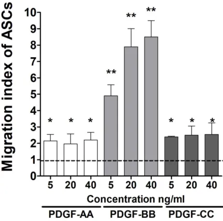
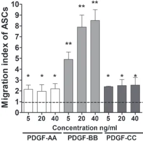
Die PDGF abhängige Migration von ASC wurde in einem in vitro Migrations-Assay
untersucht. Dabei zeigte sich eine signifikante Steigerung der Migration von ASC
schon bei einer Konzentration von 5ng/ml PDGF-BB, wobei eine Erhöhung der
Konzentration ebenfalls mit einer Steigerung der Migration von ASC einherging. Im
Gegensatz zeigte PDGF-AA und PDGF-CC nur eine geringe Steigerung der Migration
auch bei vielfach höherer Konzentration im Vergleich zu PDGF-BB (Figur 2).
Figur 2: Konzentrationsabhängig zeigte sich nur für PDGF-BB eine signifikante Steigerung der Migration von ASC (**p<0.0001). Im Gegensatz veränderte sich das Migrationsverhalten von ASC nicht für steigende Konzentrationen von PDGF-AA und PDGF-CC aber zumindest eine signifikante Steigerung zur Kontrollgruppe (*p<0.01). Zusätzliche Steigerung der Migration unter PDGF-AA oder PDGF-CC war nur bei Konzentrationen über 500ng/ml möglich (Daten nicht gezeigt).
Inaktivierung und Überexpression von PDGF-BB in Brustkrebszellen
PDGF-B wurde in der Brustkrebszelllinie (4T1) über-exprimiert oder inaktiviert, in dem entsprechende lentivirale Konstrukte verwendet wurden (Figur 1). Anschliessend wurden Einzelklone gewonnen und in der Kultur expandiert. Die PDGF-BB Expression, die Proliferation und Apoptose der Einzelklone wurde bestimmt und mit der Ausgangszelllinie verglichen. Dabei zeigte sich kein signifikanter Unterschied
zwischen den untersuchten Zelllinien bezüglich Apoptose und Proliferation
Figur 1c, d).
Der PI3-Kinase Signalweg
Die Zellmigration konnte signifikant durch PDGFR-α Antikörper blockiert werden, jedoch nur wenn PDGF-AA oder PDGF-CC im Medium verwendet wurden. Im Unterschied dazu zeigte die Blockade des PDGF-β Rezeptor eine signifikante Reduzierung der Migration unter PDGF-BB und PDGF-CC Zugabe, nicht jedoch für PDGF-AA. Diese Ergebnisse konnten durch die gemeinsame Blockade der PDGF Rezeptoren mit entsprechendem neutralisierendem Antikörper für PDGF-AA, PDGF- BB oder PDGF-CC bestätigt werden (Figur 3a).
Ausserdem konnte nur der PI3K spezifische Inhibitor (LY294002) nicht aber der MAPK
Inhibitor (PD98059) eine signifikante Verminderung der Migration der ASC in Richtung
der Tumorzellen bewirken (Figur 3b). Die Western Blot Analyse von ASC zeigte eine
starke Expression von PDGFR-β und nur eine sehr schwache Expression von PDGFR-
α im Vergleich zu einer entsprechenden Kontrollzelllinie (Figur 3c), wie schon in einer
früheren Studien von unserer Arbeitsgruppe gezeigt werden konnte [39].
Figur 3: (a) Neutralisierende Antikörper gegen PDGF-AA und PDGF-CC zeigten keinen Effekt auf die Migration von ASCs unter gleichzeitiger Blockierung des PDGF-α Rezeptors (n.s., nicht signifikant). Dahingegen zeigte sich eine signifikante Reduktion der Migration von ASC unter Blockierung des PDGF-β Rezeptors (**p<0.0001). (b) Der spezifische PI3K Inhibitor LY294002 konnte die Migration von ASC in Richtung Tumorzell-Medium signifikant reduzieren (**p<0.0001).
Im Gegensatz dazu hatte der MAPK Inhibitor (PD98059) keinen Effekt auf die Migration von ASC.
(c) Die Western Blot Analyse von ASC zeigte eine starke Expression von des PDGF-β Rezeptors
and eine nur schwache Expression des PDGF-α Rezeptors im Vergleich zur Kontrollzelllinie
(Brustkrebszellen).
Diskussion
Die vorliegende Studie zeigt, dass die Migration von ASC hauptsächlich durch PDGF- BB über den PI3K Signalweg beeinflusst wird. Die aktuellen Daten belegen, dass der PI3K Signalwege eine entscheidende Rolle für die Migration von ASC in Richtung von Tumoren spielt und daher weiter untersucht werden sollte, um gerade die Tumorprogression in frühen Krankheitsstadium zu verstehen und gegebenenfalls beeinflussen zu können.
Die Rolle von Mesenchymalen Stammzellen bei der Brustkrebserkrankung wird schon seit vielen Jahren untersucht. So konnte zum Beispiel gezeigt werden, dass Stammzellen das Tumorwachstum beeinflussen, die Angiogenese des Tumors unterstützen können und selbst bei dem Prozess der Metastasierung agieren [14–16].
Aktuelle Studien weisen darauf hin, dass Mesenchymale Stammzellen durch Tumore
angelockt werden und nachfolgend in Perizyten oder Tumor assoziierte Fibroblasten
(Cancer associated fibroblasts, CAFs) transdifferenzieren [15]. Stammzellen
akkumulieren im Tumor und in dessen Nähe, wobei sie durch die Sekretion von
verschiedenen Chemokinen und Zytokinen (z.B. PDGF) ein entsprechendes Milieu
erzeugen, welches das Tumorwachstum begünstigt [40]. In der aktuellen Studie
konnten wir zeigen, dass die Migration von ASC in Richtung Tumorzellen eine
Konzentrationsabhängigkeit von PDGF-BB zeigt. Ein kürzlich veröffentlichte Studie
bestätigt den Zusammenhang der Migration von Stammzellen und Tumor, denn es
wurde gezeigt, dass eine Bestrahlung von Brustkrebstumore eine zusätzliche
Freisetzung von PDGF-BB zur Folge hatte, die mit einer vermehrten Migration von
Stammzellen einherging [41]. Darüber hinaus unterstreicht eine Tierstudie die
Bedeutung des PDGF-BBs, da gezeigt werden konnte, dass die Zahl der
Mesenchymalen Stammzellen in Glioma Tumoren abhängig von der Expression des PDGF-BB war [42].
Die Ergebnisse der vorliegenden Studie zeigen das PDGF-BB der Tumorzellen die Migration von ASC über den PDGF-β Rezeptor steuern. Untersuchungen mit neutralisierenden Antikörpern konnten nur einen signifikanten Effekt auf die Migration von ASC bewirken, wenn der PDGF-β Rezeptor adressiert wurde. Diese Ergebnisse decken sich mit einer aktuellen Studie bei der der PDGF-β Rezeptor auf Progenitorzellen blockiert und dadurch die PDGF-BB induzierte Migration von Mesenchymalen Stammzellen unterbrochen wurde [43]. Darüber hinaus gibt es Studienergebnisse über ein Nuklease-resistentes RNA Aptamer (Gint4.T), welches an die extrazelluläre Domäne des PDGF-β Rezeptors bindet und damit die Liganden abhängige Kommunikation zwischen Stammzellen und Tumorzellen sowohl in vitro als auch in vivo unterbrechen konnte [44–46].
Wir haben ausserdem das Downstream Signal des PDGF-β Rezeptors untersucht, indem wir einen PI3K spezifischen Inhibitor (LY294002) und einen MAPK spezifischen Inhibitor (PD98059) verwendet haben. Dabei zeigte sich, dass nur die Verwendung des LY294002 mit einer signifikanten Beeinträchtigung der Migration von ASC in Richtung Tumorzellen einherging. Diese Ergebnisse werden durch Experimente unterstützt, die bei einer transgenen Maus mit einer Mutation im PDGF-β Rezeptor eine PI3K abhängige Signaltransduktion zeigt [47]. Ausserdem wurde in einer erst kürzlich veröffentlichen Studie berichtet, dass in einem Kolo-rektalen Xenograft Tiermodell Stammzellen das Tumorwachstum und die Tumor-Vaskularisation stimulieren, wobei Akt und ERK in Endothelzellen aktiviert wird und dadurch die Rekrutierung und deren Angiogenese Potential erhöht wird [48].
Durch unsere vorliegende Studie und die in der Literatur vorhandenen Datenlage
besteht aktuell kein Zweifel, dass Mesenchymale Stammzellen einen enormen Effekt
auf das Mikroenvironment von Tumoren bezüglich Angiogenese, Tumorwachstum und
Metastasierung hat. Es ist daher von entscheidender Bedeutung, dass die Affinität von
Mesenchymalen Stammzellen zu Brustkrebzellen oder anderen Tumorzellen
untersucht und verstanden wird, um einen therapeutischen Ansatz zu entwickeln, der
das Verhalten von Mesenchymale Stammzellen in diesem Prozess kontrolliert oder
beeinflussen kann.
Referenzen
[1] Caplan AI. Mesenchymal stem cells. J Orthop Res. 1991;9(5):641–50.
[2] Dezawa M, Ishikawa H, Itokazu Y, Yoshihara T, Hoshino M, Takeda S, et al. Bone marrow stromal cells generate muscle cells and repair muscle degeneration.
Science. 2005;309(5732):314–7.
[3] Kawada H, Fujita J, Kinjo K, Matsuzaki Y, Tsuma M, Miyatake H, et al.
Nonhematopoietic mesenchymal stem cells can be mobilized and differentiate into cardiomyocytes after myocardial infarction. Blood. 2004;104(12):3581–7.
[4] Kopen GC, Prockop DJ, Phinney DG. Marrow stromal cells migrate throughout forebrain and cerebellum, and they differentiate into astrocytes after injection into neonatal mouse brains. Proc Natl Acad Sci USA. 1999;96(19):10711–6.
[5] Sanchez-Ramos J, Song S, Cardozo-Pelaez F, Hazzi C, Stedeford T, Willing A, et al. Adult bone marrow stromal cells differentiate into neural cells in vitro. Exp Neurol.
2000;164(2):247–56.
[6] Chagraoui J, Lepage-Noll A, Anjo A, Uzan G, Charbord P. Fetal liver stroma consists of cells in epithelial-to-mesenchymal transition. Blood. 2003;101(8):2973–
82.
[7] Lee K-D, Kuo TK-C, Whang-Peng J, Chung Y-F, Lin C-T, Chou S-H, et al. In vitro hepatic differentiation of human mesenchymal stem cells. Hepatology.
2004;40(6):1275–84.
[8] Bhowmick NA, Neilson EG, Moses HL. Stromal fibroblasts in cancer initiation and progression. Nature. 2004;432(7015):332–7.
[9] Muehlberg FL, Song Y-H, Krohn A, Pinilla SP, Droll LH, Leng X, et al. Tissue- resident stem cells promote breast cancer growth and metastasis. Carcinogenesis.
2009;30(4):589–97.
[10] Sappino AP, Skalli O, Jackson B, Schürch W, Gabbiani G. Smooth-muscle differentiation in stromal cells of malignant and non-malignant breast tissues. Int J Cancer. 1988;41(5):707–12.
[11] Chauhan H, Abraham A, Phillips JRA, Pringle JH, Walker RA, Jones JL. There is more than one kind of myofibroblast: analysis of CD34 expression in benign, in situ, and invasive breast lesions. J Clin Pathol. 2003;56(4):271–6.
[12] Ishii G, Sangai T, Oda T, Aoyagi Y, Hasebe T, Kanomata N, et al. Bone-marrow- derived myofibroblasts contribute to the cancer-induced stromal reaction. Biochem Biophys Res Commun. 2003;309(1):232–40.
[13] Jotzu C, Alt E, Welte G, Li J, Hennessy BT, Devarajan E, et al. Adipose tissue
derived stem cells differentiate into carcinoma-associated fibroblast-like cells under
the influence of tumor derived factors. Cell Oncol (Dordr). 2011;34(1):55–67.
[14] Karnoub AE, Dash AB, Vo AP, Sullivan A, Brooks MW, Bell GW, et al.
Mesenchymal stem cells within tumour stroma promote breast cancer metastasis.
Nature. 2007;449(7162):557–63.
[15] Muehlberg FL, Song Y-H, Krohn A, Pinilla SP, Droll LH, Leng X, et al. Tissue- resident stem cells promote breast cancer growth and metastasis. Carcinogenesis.
2009;30(4):589–97.
[16] Kamat P, Schweizer R, Kaenel P, Salemi S, Calcagni M, Giovanoli P, et al.
Human Adipose-Derived Mesenchymal Stromal Cells May Promote Breast Cancer Progression and Metastatic Spread. Plast Reconstr Surg. 2015;136(1):76–84.
[17] Lejmi E, Perriraz N, Clément S, Morel P, Baertschiger R, Christofilopoulos P, et al. Inflammatory Chemokines MIP-1δ and MIP-3α Are Involved in the Migration of Multipotent Mesenchymal Stromal Cells Induced by Hepatoma Cells. Stem Cells Dev. 2015;24(10):1223–35.
[18] Lourenco S, Teixeira VH, Kalber T, Jose RJ, Floto RA, Janes SM. Macrophage Migration Inhibitory Factor–CXCR4 Is the Dominant Chemotactic Axis in Human Mesenchymal Stem Cell Recruitment to Tumors. The Journal of Immunology.
2015;194(7):3463–74.
[19] Dwyer RM, Potter-Beirne SM, Harrington KA, Lowery AJ, Hennessy E, Murphy JM, et al. Monocyte chemotactic protein-1 secreted by primary breast tumors stimulates migration of mesenchymal stem cells. Clin Cancer Res.
2007;13(17):5020–7.
[20] Johann PD, Müller I. Multipotent Mesenchymal Stromal Cells: Possible Culprits in Solid Tumors? Stem Cells Int. 2015.
[21] Li Z, Xu X, Wang W, Kratz K, Sun X, Zou J, et al. Modulation of the
mesenchymal stem cell migration capacity via preconditioning with topographic microstructure. Clin Hemorheol Microcirc. 2017;67(3–4):267–78.
[22] Gehmert S, Gehmert S, Prantl L, Vykoukal J, Alt E, Song Y-H. Breast cancer cells attract the migration of adipose tissue-derived stem cells via the PDGF- BB/PDGFR-beta signaling pathway. Biochem Biophys Res Commun.
2010;398(3):601–5.
[23] Ellis SJ, Pines M, Fairchild MJ, Tanentzapf G. In vivo functional analysis reveals specific roles for the integrin-binding sites of talin. J Cell Sci. 2011;124(11):1844–56.
[24] Zhu W, Huang L, Li Y, Zhang X, Gu J, Yan Y, et al. Exosomes derived from human bone marrow mesenchymal stem cells promote tumor growth in vivo. Cancer Lett. 2012;315(1):28–37.
[25] Lindahl P, Johansson BR, Levéen P, Betsholtz C. Pericyte loss and
microaneurysm formation in PDGF-B-deficient mice. Science. 1997;277(5323):242–
5.
[26] Soriano P. Abnormal kidney development and hematological disorders in PDGF
beta-receptor mutant mice. Genes Dev. 1994;8(16):1888–96.
[27] Dwyer RM, Potter-Beirne SM, Harrington KA, Lowery AJ, Hennessy E, Murphy JM, et al. Monocyte chemotactic protein-1 secreted by primary breast tumors stimulates migration of mesenchymal stem cells. Clin Cancer Res.
2007;13(17):5020–7.
[28] Enge M, Bjarnegård M, Gerhardt H, Gustafsson E, Kalén M, Asker N, et al.
Endothelium-specific platelet-derived growth factor-B ablation mimics diabetic retinopathy. EMBO J. 2002;21(16):4307–16.
[29] Hellström M, Kalén M, Lindahl P, Abramsson A, Betsholtz C. Role of PDGF-B and PDGFR-beta in recruitment of vascular smooth muscle cells and pericytes during embryonic blood vessel formation in the mouse. Development. 1999;126(14):3047–
55.
[30] Abramsson A, Lindblom P, Betsholtz C. Endothelial and nonendothelial sources of PDGF-B regulate pericyte recruitment and influence vascular pattern formation in tumors. J Clin Invest. 2003;112(8):1142–51.
[31] Furuhashi M, Sjöblom T, Abramsson A, Ellingsen J, Micke P, Li H, et al. Platelet- derived growth factor production by B16 melanoma cells leads to increased pericyte abundance in tumors and an associated increase in tumor growth rate. Cancer Res.
2004;64(8):2725–33.
[32] McCarty MF, Somcio RJ, Stoeltzing O, Wey J, Fan F, Liu W, et al.
Overexpression of PDGF-BB decreases colorectal and pancreatic cancer growth by increasing tumor pericyte content. J Clin Invest. 2007;117(8):2114–22.
[33] Jotzu C, Alt E, Welte G, Li J, Hennessy BT, Devarajan E, et al. Adipose tissue- derived stem cells differentiate into carcinoma-associated fibroblast-like cells under the influence of tumor-derived factors. Anal Cell Pathol (Amst). 2010;33(2):61–79.
[34] Amos PJ, Shang H, Bailey AM, Taylor A, Katz AJ, Peirce SM. IFATS collection:
The role of human adipose-derived stromal cells in inflammatory microvascular remodeling and evidence of a perivascular phenotype. Stem Cells.
2008;26(10):2682–90.
[35] Pinilla S, Alt E, Abdul Khalek FJ, Jotzu C, Muehlberg F, Beckmann C, et al.
Tissue resident stem cells produce CCL5 under the influence of cancer cells and thereby promote breast cancer cell invasion. Cancer Lett. 2009;284(1):80–5.
[36] Gehmert S, Gehmert S, Prantl L, Vykoukal J, Alt E, Song Y-H. Breast cancer cells attract the migration of adipose tissue-derived stem cells via the PDGF- BB/PDGFR-beta signaling pathway. Biochem Biophys Res Commun.
2010;398(3):601–5.
[37] Gehmert S, Wenzel C, Loibl M, Brockhoff G, Huber M, Krutsch W, et al. Adipose tissue-derived stem cell secreted IGF-1 protects myoblasts from the negative effect of myostatin. Biomed Res Int. 2014;2014:129048.
[38] Schneider CA, Rasband WS, Eliceiri KW. NIH Image to ImageJ: 25 years of
image analysis. Nat Meth. 2012;9(7):671–5.
[39] Gehmert S, Gehmert S, Hidayat M, Sultan M, Berner A, Klein S, et al.
Angiogenesis: the role of PDGF-BB on adipose-tissue derived stem cells (ASCs).
Clin Hemorheol Microcirc. 2011;48(1):5–13.
[40] Nwabo Kamdje AH, Kamga PT, Simo RT, Vecchio L, Seke Etet PF, Muller JM, et al. Mesenchymal stromal cells’ role in tumor microenvironment: involvement of signaling pathways. Cancer Biol Med. 2017;14(2):129–41.
[41] Klopp AH, Spaeth EL, Dembinski JL, Woodward WA, Munshi A, Meyn RE, et al.
Tumor irradiation increases the recruitment of circulating mesenchymal stem cells into the tumor microenvironment. Cancer Res. 2007;67(24):11687–95.
[42] Hata N, Shinojima N, Gumin J, Yong R, Marini F, Andreeff M, et al. PDGF-BB Mediates the Tropism of Human Mesenchymal Stem Cells for Malignant Gliomas.
Neurosurgery. 2010;66(1):144–57.
[43] Fiedler J, Etzel N, Brenner RE. To go or not to go: Migration of human mesenchymal progenitor cells stimulated by isoforms of PDGF. J Cell Biochem.
2004;93(5):990–8.
[44] Camorani S, Hill BS, Fontanella R, Greco A, Gramanzini M, Auletta L, et al.
Inhibition of Bone Marrow-Derived Mesenchymal Stem Cells Homing Towards Triple- Negative Breast Cancer Microenvironment Using an Anti-PDGFRβ Aptamer.
Theranostics. 2017;7(14):3595–607.
[45] Camorani S, Esposito CL, Rienzo A, Catuogno S, Iaboni M, Condorelli G, et al.
Inhibition of receptor signaling and of glioblastoma-derived tumor growth by a novel PDGFRβ aptamer. Mol Ther. 2014;22(4):828–41.
[46] Monaco I, Camorani S, Colecchia D, Locatelli E, Calandro P, Oudin A, et al.
Aptamer Functionalization of Nanosystems for Glioblastoma Targeting through the Blood-Brain Barrier. J Med Chem. 2017;60(10):4510–6.
[47] Rodt SA, Ahlén K, Berg A, Rubin K, Reed RK. A novel physiological function for platelet-derived growth factor-BB in rat dermis. J Physiol. 1996;495(Pt 1):193–200.
[48] Huang W-H, Chang M-C, Tsai K-S, Hung M-C, Chen H-L, Hung S-C.
Mesenchymal stem cells promote growth and angiogenesis of tumors in mice.
Oncogene. 2013;32(37):4343–54.
IOS Press
PDGF regulated migration of mesenchymal stem cells towards malignancy acts via the PI3K signaling pathway
Sonia Salha a , c , Sebastian Gehmert b , c ,∗ , Vanessa Br´ebant c , d , Alexandra Anker c , d , Markus Loibl c , e , Lukas Prantl c , d and Sanga Gehmert c , f
a Department of Orthopedics and Traumatology, University Hospital Basel, Switzerland
b Department of Orthopedics, University Children’s Hospital Basel, Switzerland
c Applied Stem Cell Research Center, University Medical Center Regensburg, Germany
d Department of Plastic, Hand-and Reconstructive Surgery, University Medical Center Regensburg, Germany
e Department of Trauma Surgery, Regensburg University Medical Center, Regensburg, Germany
f Department of Gynecology and Obstetrics, Kantonsspital Baselland, Liestal, Switzerland
Abstract.
INTRODUCTION: Mesenchymal stem cells (MSCs) have been described in breast cancer models to migrate towards carcinoma and integrate into tumor associated stroma supporting tumor growth, increasing their metastatic potency and contributing to tumor-angiogenesis. Platelet-derived growth factor (PDGF) isoforms (AA, BB, CC) stimulate growth, survival and motility of MSCs and certain other cell types. Noteworthy, breast carcinomas are known to express PDGF. We aim to further shed light on i) the relevance of the different PDGF isoforms on adipose tissue derived stem cells (ASCs) migration and ii) the underlying pathway dependent on PDGF stimulation.
MATERIALS AND METHODS: Breast cancer cell lines were purchased and ASC’s were isolated from murine subcuta- neous adipose tissue. The transmigration of ASC’s towards the PDGF-isoforms was assessed by using recombinant human PDGF-AA, PDGF-BB and PDGF-CC in a trans-well culture dish system. Transmigrated ASC’s were quantified in 5 randomly selected fields per condition using fluorescence microscopy after calcein-staining. PDGF-BB depended transmigration of ASC’s was verified by downregulation and overexpression of PDGF-BB in breast cancer cell line using lentiviral vectors.
In addition, a PI3-kinase inhibitor (LY294002) and a MAP-kinase inhibitor (PD98059) were used to identify the pathway involved in the PDGF-BB mediated migration of ASC’s towards tumor.
RESULTS: ASC’s transmigration significantly increased towards PDGF AA at 50 ng and only showed further increase by 500 ng which was similar to cell behavior when exposed to PDGF CC. In comparison, PDGF-BB significantly increased ASC’s transmigration already at a low level of 5 ng with further significant increase for 20 ng and 40 ng. Cell transmigration was blocked with PDGFR-␣ antibodies but only for PDGF-AA and PDGF-CC whereas PDGFR- blockage showed a significant effect on transmigration for PDGF-BB and PDGF-CC but not for PDGF-AA. Neutralizing antibodies in combination with PDGF receptor blockage confirmed findings. In addition, only PI3-kinase inhibitor but not the MEK-1 selective inhibitor caused a significant decrease of transmigration for ASCs towards breast cancer cells.
DISCUSSION: The transmigration of ASC’s is most significantly enhanced by PDGF-BB via the PI3-kinase pathway. This data support that PI3-kinase is an important key player for MSC migration towards malignancy which need further research to prevent tumor progression in early disease stage.
Keywords: Mesenchymal stem cells, migration, breast cancer, PDGF-BB, PI3K pathway
∗