Characterization of four functionally distinct human B-cell subsets that are defined by the expression of CD21 and
CD86
Inaugural-Dissertation zur Erlangung des Doktorgrades
der Mathematisch-Naturwissenschaftlichen Fakultät der Universität zu Köln
vorgelegt von
María Alejandra García Márquez aus Maracay, Venezuela
Köln 2015
Berichterstatter: PD. Dr. Thomas Wunderlich Prof. Dr. Björn Schumacher
Tag der mündlichen Prüfung: 19 June, 2015
Para mis padres Luis y Pilar
I
DANKSAGUNG
I would like to thank everyone who has supported me and has contributed to the success of this work.
My thanks go to Prof. Hallek, for providing helpful advice, comments, or suggestions through the years. Many thanks to my thesis committee members Dr.
Thomas Wunderlich, Prof. Björn Schumacher, Prof. Ines Neundorf and Dr. Isabell Witt for their kind willingness to support this work.
I would like to make a special recognition to my advisor Prof. Michael von Bergwelt-Baildon who gave me the opportunity to research in his group and provided me his guidance throughout my graduate studies. I am grateful for all his support, encouragement and enthusiastic discussions sustained through the years. It is also important to mention and express my gratitude to Dr. Alexander Shimabukuro- Vornhagen for his helpful advice, critiques and insightful comments as well as his expert and technical assistance. I will continue to learn and grow as I pursue my career with the foundation that Michael and Alex helped to build. Thanks also to Rieke Fischer for trusting me to continue with her research project.
I cannot forget Kerstin and Juliane, who created a supportive and fun working environment over the years. They have been not only my colleges but also my friends and they have always being there for me, lending me their assistance and advice in every possible way. You will forever have a place in my heart. Thanks Kerstin for your comments, suggestions, and proofreading of my thesis.
I would like to further thank Michael, Kerstin, Juliane, Martin, Sabrina, Anne, Hans S., Hans B., Sebastian, Alex, Sacha, Geothy, Rieke, Steffi and Claudia, for the great time spent in the laboratory, the entertaining lunch breaks, dinners and lab excursions. Those good memories and the time we spent together will remain with me forever.
Quiero agradecer a Helmut, Luz Angela, Helmut papá, Erich, Dani, Ricardo, Max, Zivile, Adrian, Kati, Marina, José Daniel, Ingrid, Felipe, Andy y Santi por darme una familia en Alemania y por hacerme sentir en casa.
Helmut, tú me has acompañado desde el principio en este camino, gracias por tu apoyo incondicional, por tu paciencia en esos días en los que no estuve tan segura de si conseguiría esta meta. Por hacerme reír cuando estaba triste, por saber escucharme y saber cómo aligerar mis cargas, gracias. Por ser mi “fiel chofer” y llevarme a todos lados, y por acompañarme en las madrugadas y fines de semanas en los que tuve que estar en el lab. Infinitas gracias.
II
Por último pero no menos importante, quiero agradecer a mi familia. A mis papas Luis y Pilar por siempre apoyarme, por estar orgullosos de mi, por acompañarme desde la distancia. Gracias a ustedes es que soy quien soy, este título es de ustedes. Ustedes son mi fuerza y aunque no los pueda ver todos los días siempre los llevo en mi corazón. A mis hermanos Luijo e Isa, por apoyarme y ayudarme siempre que los necesito. Los amo con todo mi corazón. A mis abuelitas Margoth y Teresa por las conversaciones de los fines de semana que siempre me alegran el día y a mis abuelitos Luis y Darío que desde el cielo siempre me acompañan.
III
ZUSAMMENFASSUNG
Inzwischen ist bekannt, dass B-Lymphozyten phänotypisch und funktional heterogen sind. Zusätzlich zu ihrer wesentlichen Funktion als Antikörper- produzierende B-Zellen, dienen sie als Antigen-präsentierende Zellen und regulieren Immunreaktionen, unter anderem durch die Sekretion von Cytokinen und Chemokinen. Obwohl bereits mehrere Subpopulationen identifiziert worden sind, ist ein tieferes Verständnis für die Rolle der B-Zellen in der Pathophysiologie bei menschlichen Erkrankungen durch einen Mangel an exakt definierten B-Zell- Subpopulationen behindert worden. In der vorliegenden Arbeit wurden vier B-Zell- Subpopulationen mit sehr verschiedenen phänotypischen und funktionellen Eigenschaften durch eine umfassende phänotypische und funktionierende Charakterisierung mittels Expression von CD21 und CD86 definiert. Die CD21pos CD86low B-Zell-Untergruppe zeigte einen naiven, IgD+ IgM+ Phänotyp, der durch die fehlende Expression von Costimulatorischen- und Gedächtnis-Markern charakterisiert war. Zusätzlich wies sie ein niedriges Calcium- und Phosphorilierungs- Niveau sowie eine starke Reaktion nach Stimulierung des B-Zellrezeptors auf.
Darüber hinaus induzierte diese B-Zell-Untergruppe nur eine sehr langsame Aktivierung in autologen T-Zellen nach Cokultivierung. Die CD21pos CD86pos B-Zellen- Untergruppe zeigte einen aktivierten, IgD+ IgM+ CD27+ Gedächtnis-Phänotyp und eine moderate Fähigkeit zur Induktion einer T-Zellreaktion. Die CD21low Population, die in andere Untersuchungen in verschiedenen Erkrankungen als „ermüdet“ und wenig immunstimulatorisch beschrieben worden ist, konnte durch die unterschiedliche Expression von CD86 in zwei Untergruppen aufgeteilt werden. Die CD21low CD86pos B- Zell-Untergruppe zeigte einen aktivierten, IgD- IgM- CD27+ Gedächtnis-Phänotyp, eine starke immunangregende Fähigkeit und eine beeinträchtigte Reaktion des B-
IV
Zellrezeptors. Diese konnte jedoch durch die synergistische Stimulierung mit dem CD40-Liganden wieder hergestellt werden. Im Gegensatz dazu war die CD21low CD86neg B-Zellen-Untergruppe funktionell beeinträchtigt und wies Merkmale auf, die zu einem anergischen oder „ermüdeten“ Zustand passten.
In der vorliegenden Arbeit wurden bemerkenswerte Unterschiede in der Frequenz dieser B-Zell-Subpopulation in verschiedenen Erkankungen fest gestellt.
Dies lässt darauf schließen, dass die homöostatische Balance zwischen diesen Subpopulationen den funktionellen Zustand des gesamten B-Zell-Kompartiments definieren könnte. So hatten Patienten mit traumatischen Verletzungen beispielsweise einen erhöhten Prozentsatz an CD21pos CD86pos B-Zellen im Vergleich zu gesunden Spendern und Patienten mit rheumatischer Arthritis wiesen eine höhere Frequenz an CD21pos CD86pos und CD21low CD86pos B-Zellen auf. In Patienten mit Darmkrebs waren CD21low CD86pos B-Zellen im Tumorgewebe angereichert im Vergleich zu peripherem Blut von Darmkrebspatienten und gesunden Spendern.
Zusammenfassend lässt sich fest halten, dass die verschiedenen B-Zell-Untergruppen unterschiedliche funktionelle Eigenschaften aufweisen, und dass sie eine Rolle in Autoimmun- und Krebserkankungen zu spielen scheinen. Dies legt den Schluss nahe, dass eine stärkere Fokussierung auf die spezifischen B-Zellen-Untergruppen eine viel versprechende therapeutische Strategie repräsentieren könnte.
V
ABSTRACT
It is now recognized that B lymphocytes are phenotypically and functionally heterogeneous. In addition to their essential role as antibody producing B cells, they serve as antigen-presenting cells, contribute to immunoregulation and represent an important source of cytokines and chemokines. Although several subpopulations have been identified, a deeper understanding of the role of B cells in the pathophysiology of human diseases has been hampered by the lack of well-defined functional B-cell subsets. In this study, a comprehensive phenotypic and functional characterization of B cells defined by expression of CD21 and CD86, revealed four B- cell subpopulations with very distinct phenotypical and functional properties. CD21pos CD86low B-cell subset had a naïve, non-switched phenotype characterized by the absence of expression of costimulatory and memory markers. Additionally, it was characterized by low basal calcium and phosphorylation levels and robust response after B-cell receptor stimulation. Furthermore, when cultivated with autologous T cells they induced very low stimulation. The CD21pos CD86pos B-cell subset had an activated, non-switched-memory phenotype and moderated immunostimulatory capacity. The CD21low "exhausted" population described by others in several diseases could be partitioned in two subsets based on the expression of CD86. CD21low CD86pos B-cell subset had an activated, class-switched-memory phenotype, potent immunostimulatory capacity and impaired B-cell receptor signaling that was restored by synergistic stimulation with CD40 ligand. On the other hand, CD21low CD86neg B- cell subset was composed of functionally impaired B cells with features consistent of an anergic or exhausted state.
Since striking differences were detected in the frequency of these subsets in several medical conditions, it appears that the homeostatic balance between these
VI
subpopulations could describe the functional state of the B-cell compartment. For instance, patients with traumatic fractures contained an increased percentage of CD21pos CD86pos B cells compared to healthy donors. Patients with active rheumatoid arthritis had a higher frequency of CD21pos CD86pos and CD21low CD86pos B cells.
Patients with colorectal cancer revealed that CD21low CD86pos B cells were enriched within the tumor tissue compared to the peripheral blood of colorectal cancer patients and healthy controls. Taken together, the observation that the different B- cell subsets display distinct functional features and that the different subsets are associated with immune-related diseases suggests that a more specific targeting of B-cell subsets could represent a promising therapeutic strategy.
VII
ABBREVIATIONS
Acronym Expansion
AM Acetoxymethyl esters
APCs Antigen-presenting cells
ATP Adenosine triphosphate
B10 cells IL-10 producing regulatory B cells BAFF B cell-activating factor
BAPC
antigen-presenting
CD21low CD86posB cellsBconv conventional CD21pos CD86negB cells
BCR B cell receptor
Binact Inactivated CD21low CD86negB cells
Bm B mature
Breact Recently activate CD21pos CD86posB cells
BSA Bovine serum albumin
°C Degrees Celsius
Ca2+ Calcium ions
CaCl2 Calcium chloride
CD40B cell CD40-activated B cell
CD40L CD40 Ligand
CFSE 5-(and-6)-carboxyfluoresceindiacetate, succinimidyl ester
CLP Common lymphoid progenitor
CO2 Carbon dioxide
VIII
CR Complement receptor
CRC Colorectal cancer
CSR Class switch recombination
CVID Common variable immunodeficiency
D Diversity gene
DC Dendritic cell
DMSO Dimethyl sulfoxide
DNA Deoxyribonucleic acid
EBV Epstein-Barr virus
EDTA Ethylenediaminetetraacetic acid
ER Endoplasmic reticulum
ERK Extracellular signal regulated kinase FACS Fluorescence activated cell sorting
FBS Fetal bovine serum
FoxP3 Forkhead box P3
FS Forward scatter
FSS Forward scatter signal
G Gravity
GC Germinal center
Gy Gray
H2O2 Hydrogen Peroxide
HCV Hepatitis C virus
HIV Human immunodeficiency virus
HSCs hematopoietic stem cells
IX
Ig Immunoglobulin
IgLC Immunoglobulin light chain IƙBα Inhibitor of ƙ B α
IKK IƙB kinase
IL- Interleukin
IFN Interferon
IP3 Inositol-1,4,5-trisphosphate
ITAM Immunoreceptor tyrosine-based activation motif ITIM Immunoreceptor tyrosine-based inhibitory motif
J Joining gene
JAK Janus kinase
Jnk
c-Jun N-terminal kinase
L Liter
LFA-1 leukocyte function antigen-1
M Molar
mAbs Monoclonal antibodies
MALT Mucosa-associated lymphoid tissue MAPKs
Mitogen activated protein kinases
MFI Mean fluorescence intensity
Mg Milligram
MHC Major histocompatibility complex
mIg Membrane-bound immunoglobulin
mL Mililiter
MLR Mixed lymphocyte reaction
X
mM Milimolar
Mm Millimeter
µHC Immunoglobulin heavy chain
µL Microliter
µm Micrometer
µM Micromolar
µSM µ-suppressed mice
NF Nuclear factor
NIK Nuclear ƙ B inducing kinase
NK Natural killer
PBMCs Peripheral blood mononuclear cells
PBS Phosphate-buffered saline
PCs plasma cells
PFA Paraformaldehyde
Pg Picogram
PhosphoFlow Multiplexed phospho-specific flow cytometric profiling PI3K Phosphoinositol 3 kinase
PIP2 Phosphatidylinositol 4,5-bisphosphate
PLCγ2 phospholipase Cγ2
pro-B cells Progenitor B cells
PTPs Protein tyrosine phosphatases RAG Recombination-activating gene rCD40L Recombinant CD40 ligand
rh human recombinant
XI
RNA Ribonucleic acid
RT Room temperature
sCD40L Soluble CD40 ligand
SD Standard deviation
SEM Standard error of the mean
SHM Somatic hyper mutation
SLC Surrogate light chain
SLE Systemic lupus erythematosus
SPADE Spanning-tree progression analysis of density-normalized events
SS Side scatter
STAT Signal transducer and activator of transcription TCR T cell antigen receptor
TGF Transforming growth factor
Th T helper cell
TLR Toll-like receptors
TNF Tumor necrosis factor
TOF Time of flight
TRAF
TNF receptor associated factor
Treg Regulatory T cell
U Units
V Variable gene
w/w Weight of solute over weight of solution
XII
TABLE OF FIGURES
Figure 1. B cell development and differentiation subsets ... 8
Figure 2. B cell functions ... 18
Figure 3. Interaction between B and T lymphocytes ... 21
Figure 4. CD40 signaling ... 26
Figure 5. CD21 down-regulation and CD86 up-regulation as features of long-term activated B cells ... 31
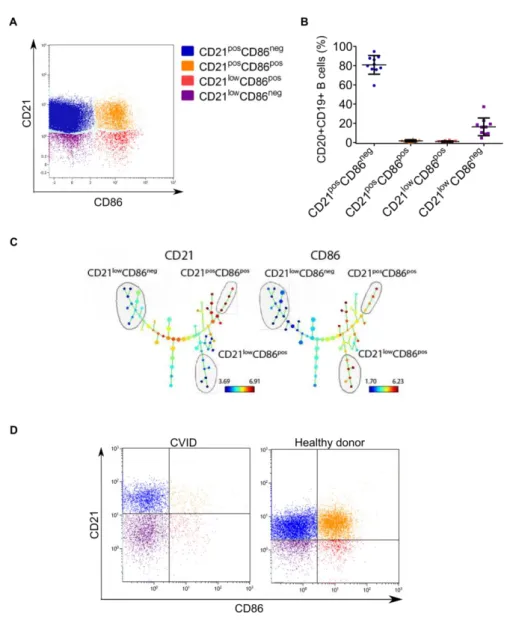
Figure 6. CD21 and CD86 defined four B-cell subsets in peripheral blood from healthy donors. ... 33
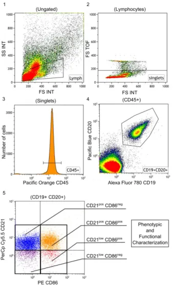
Figure 7. Gating strategy for flow cytometry analysis ... 52
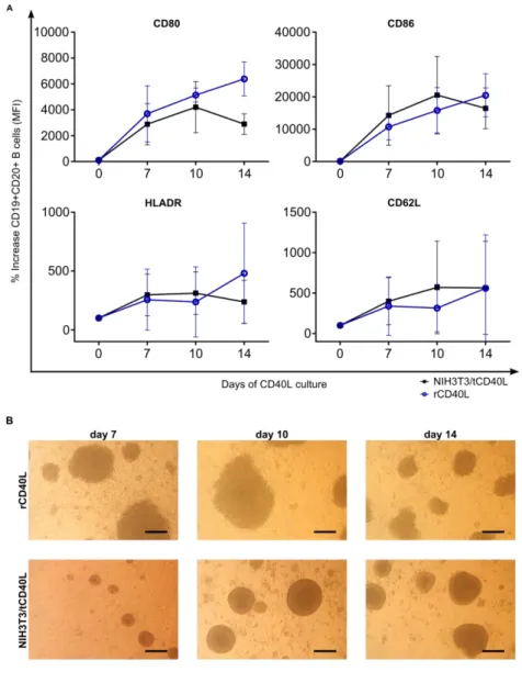
Figure 8. Ligation of B cells by CD40L induced activation and homotypic cluster formation ... 61
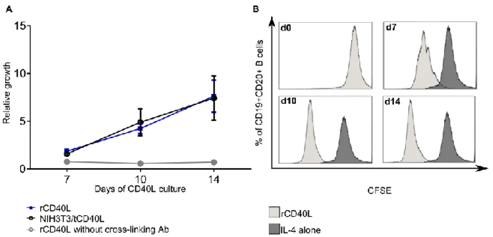
Figure 9. B cell proliferation in response to CD40 ligation... 63
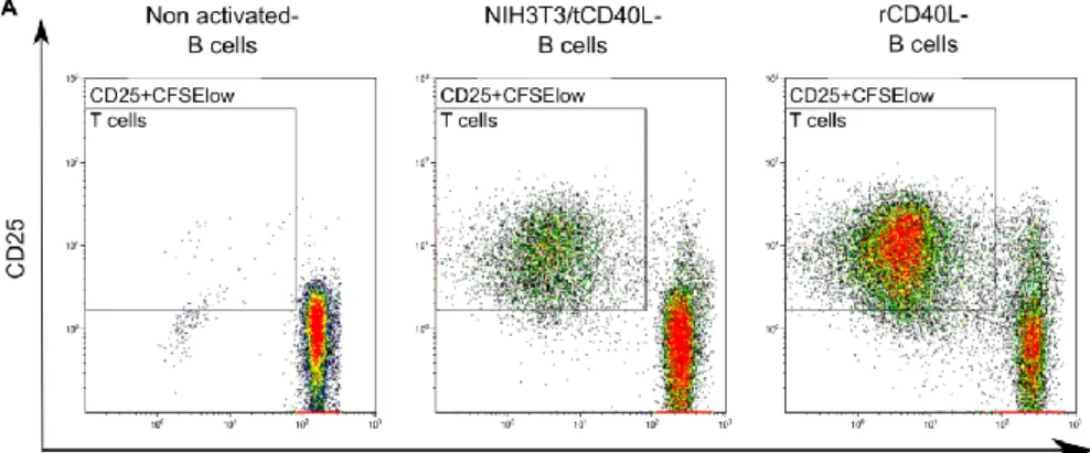
Figure 10. CD40 activated B cells induced strong proliferation of allogeneic T cells 64 Figure 11. Frequency of B-cell subsets in human peripheral blood, bone marrow and lymphoid tissue. ... 66
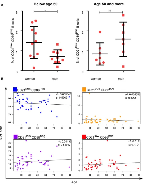
Figure 12. Individual variation in the frequency of B-cell subsets ... 68
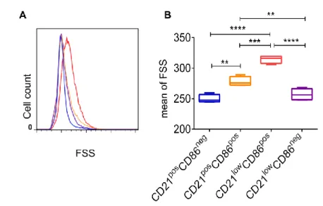
Figure 13. CD21low CD86pos B cells were larger than B cells from the other three cell subsets... 70
Figure 14. CD21low CD86pos B-cell subset had a higher proportion of proliferating cells ... 72
XIII
Figure 15. CD21low CD86pos subset consisted primarily of IgMneg IgDneg class-switched B cells ... 73 Figure 16. B-cell subsets differed in terms of expression of several cell surface
molecules ... 75 Figure 17. CD21low CD86pos B cells expressed lower levels of the inhibitory receptors CD22, and CD32 ... 76 Figure 18. Expression of molecules involved in cell trafficking ... 78 Figure 19. The majority of CD21low CD86pos and CD21pos CD86pos B cells expressed
CD43 on their surface ... 80 Figure 20. Isolation on B-cell subsets via FACS ... 81 Figure 21. Mixed lymphocyte reaction with purified B-cell subpopulations and
autologous T cells ... 83 Figure 22. Baseline cytosolic calcium levels and response to BCR stimulation in the
four B-cell subsets ... 86 Figure 23. BCR-triggering induced calcium flux on CD40B cells ... 87 Figure 24. CD40L stimulation induced no calcium flux in B-cell subsets ... 89 Figure 25. Addition of extracellular calcium induced signaling after CD40 ligation on B
cells ... 90 Figure 26. Addition of extracellular calcium induced signaling after CD40 ligation on B-cell subsets ... 91 Figure 27. CD40B cells were unresponsive to brief CD40L stimulation ... 93 Figure 28. BCR induced signaling ... 95
XIV
Figure 29. Elevated basal levels of phospho-proteins in CD21pos CD86pos B-cell subset ... 96 Figure 30. BCR induced signaling was impaired in all but CD21pos CD86neg B-cell subset
... 98 Figure 31. Impaired BCR-induced signaling in CD86pos subsets was not restored by
inhibition of H2O2 - sensitive phosphatases ... 100 Figure 32. Pervanadate was required for pZAP/SYK phosphorylation in CD21low
CD86pos subset ... 101 Figure 33. Baseline phosphorylation of NF-ƙB-p65 ... 102 Figure 34. Impaired phosphorylation of pERK and NF-ƙB-p65 after CD40L stimulation
in CD21low B-cell subsets ... 104 Figure 35. Combination of BCR and CD40 ligation restored signaling on CD21low
CD86pos B cells ... 106 Figure 36. Hypothetical model for the function-based categorization of B-cell subsets
divided by the expression of CD21 and CD86 ... 108 Figure 37. BAPC were increased following vaccination ... 109 Figure 38. BAPC were increased in patients with EBV reactivation following allogeneic
stem cell transplantation ... 112 Figure 39. Breact were increased in patients with acute traumatic fractures ... 113 Figure 40. Breact and BAPC were increased in patients with rheumatoid arthritis... 114 Figure 41. Breact, BAPC and Binact were increased in tumors specimens of CRC patients
... 116
XV
LIST OF TABLES
Table 1: Chemicals ... 36
Table 2: Reagents ... 36
Table 3: Buffers and solutions... 37
Table 4: NIH3T3 Standard medium ... 38
Table 5: Human CD40B wash medium ... 39
Table 6: Human CD40B culture medium ... 39
Table 7: Calcium flux medium ... 39
Table 8: Kits ... 40
Table 9: Tools and instrumentation ... 40
Table 10: Consumables ... 41
Table 11: Software ... 42
Table 12: Anti-Human antibodies ... 50 Table 13: Dyes used for the detection of free intracellular Ca2+ by flow cytometry 54
XVI
TABLE OF CONTENTS
Danksagung ... I Zusammenfassung ... III Abstract ... V Abbreviations ... VII Table of Figures ... XII List of Tables ... XV Table of Contents ... XVI
1 Introduction... 2
1.1 Immune system ... 2
1.2 Innate and adaptive responses ... 3
1.3 T lymphocytes ... 4
1.3.1CD8+ cytotoxic T Cells ... 4
1.3.2CD4+ helper T Cells ... 5
1.4 B lymphocytes ... 6
1.4.1B cell origin and development ... 6
1.4.2Circulating peripheral blood B-cell subsets ... 10
1.4.3B cells in health and disease ... 13
1.4.4Molecular architecture of B cells ... 14
XVII
1.4.4.1The complement receptor (CR) 2: CD21 ... 15
1.4.5B cell functions ... 17
1.4.5.1How B cells capture, process and present antigens? ... 20
1.4.5.1.1BCR internalization and signaling... 21
1.4.5.2CD40-CD40L interaction ... 23
1.4.5.2.1Signaling through CD40L ... 24
1.4.6Generation of CD40-activated B cells in vitro ... 27
1.4.6.1DCs vs. CD40B cells as antigen presenting cells ... 29
1.5 CD21low CD86pos cells: an in vivo counterpart to in vitro generated CD40B cells? ... 30
1.6 Aim of the study ... 34
2 Materials and Methods ... 36
2.1 Materials ... 36
2.1.1 Chemicals ... 36
2.1.2 Reagents ... 36
2.1.3 Buffers and solutions ... 37
2.1.4 Cell culture media ... 38
2.1.5 Kits ... 40
2.1.6 Cell lines ... 40
2.2 Methods ... 42
XVIII
2.2.1 Blood samples ... 42
2.2.2 Cell subset enrichment ... 43
2.2.2.1 B cell purification ... 43
2.2.2.2
...
B-cell subset isolation by fluorescence activated cell sorting (FACS) ... 442.2.2.3 CD3+ T cell isolation ... 44
2.2.2.4 Cell isolation from human tumor tissue ... 45
2.2.3 Cell culture ... 45
2.2.3.1 Counting of cells – Trypan Blue exclusion test ... 46
2.2.3.2 Cryopreservation and thawing of cells ... 46
2.2.3.3 Trypsinization of adherent cells ... 46
2.2.3.4 Culture of NIH3T3/tCD40L cell line ... 47
2.2.3.5
...
Generation of CD40-activated B cells with NIH3T3/tCD40L cell line ... 472.2.3.6 Generation of CD40-activated B cells with soluble CD40L... 48
2.2.3.7 Assessment of cell morphology by microscopy ... 48
2.2.4 Phenotype analysis of single cells by FACS ... 49
2.2.5 Surface staining with monoclonal antibodies ... 49
2.2.6 Human monoclonal antibodies used for frequency and conventional phenotype analysis ... 50
2.2.7 Gating strategy for flow cytometry analysis ... 51
XIX
2.2.8 Autologous and allogeneic MLR for T cell proliferation analysis ... 53 2.2.9 Analysis of Ca2+ mobilization. ... 54 2.2.10 Multiplexed phospho-specific flow cytometric profiling (PhosphoFlow) ... 55 2.2.10.1 Stimulants used in PhosphoFlow experiments ... 55 2.2.10.2 Cell stimulation ... 55 2.2.10.3 Staining and fluorescent-cell barcoding ... 56 2.2.11 Statistics ... 56 3 Results ... 59 3.1 Generation of highly proliferative antigen presenting B cells was achieved by
activation via soluble as well as membrane bound CD40L ... 59 3.2 Similar distribution of B-cell subsets was found in human peripheral blood, as well as in primary and secondary lymphoid organs ... 65 3.3 Phenotypic characterization of the four B-cell subsets ... 69 3.3.1 CD21low CD86pos B cells were significantly larger than the other subsets ... 69 3.3.2
CD21pos CD86pos and CD21low CD86pos B-cell subsets were actively proliferating ... 70 3.3.3 CD21low CD86pos B-cell subset contained mostly antigen experienced IgMneg
IgDneg class-switched B cells ... 72 3.3.4 Expression of cell surface molecules among B-cell subsets ... 73 3.3.5 Expression of molecules involved in cell trafficking among B-cell subsets ... 77 3.4 Isolation of B-cell subsets via fluorescence activated cell sorting (FACS) ... 79
XX
3.5 CD21pos CD86pos and CD21low CD86pos B cells were potent antigen-presenting cells ... 81 3.6 Triggering of BCR poorly induced calcium flux in CD21low B-cell subsets ... 84 3.7 CD21pos CD86pos subsets exhibited increased constitutive SYK, BTK and ERK
phosphorylation and decreased response to BCR mediated stimulation ... 94 3.8 Impaired CD40 signaling in CD21low B-cell subsets ... 102 3.9 Combined CD40 engagement and BCR stimulation restored signaling in CD21low
CD86pos subset ... 105 3.10 CD21 and CD86 delineated four functionally distinct peripheral blood B-cell
subsets... 107 3.11 BAPC were increased following vaccination ... 109 3.12 EBV-infected human B-lymphocytes gain a CD21low CD86pos phenotype in vivo
... 110 3.13 The balance of CD21low B cells was altered in patients with inflammatory
diseases ... 112 4 Discussion ... 118 4.1 Soluble rCD40L system to generate antigen-presenting B cells ... 118 4.2 The CD21low population ... 120 4.3 Functional and phenotypic characterization of B cells classified by the
expression of CD21 and CD86 revealed striking differences among subsets 121 4.3.1 CD21pos CD86neg Bconv subset ... 121 4.3.2 CD21pos CD86pos Breact subset ... 121
XXI
4.3.3 CD21low CD86pos BAPC subset ... 123 4.3.4 CD21low CD86neg Binact subset ... 128 4.4 Calcium flux signaling in B cells after CD40L stimulation ... 131 4.5 B-cell subset homeostasis is disturbed in some diseases... 132 4.6 Conclusion ... 136 References ... 139 5 Attachments ... 181 5.1 Erklärung § 4 Abs. 1 Nr. 9 ... 181 5.2 Teilpublikationen ... 181 5.3 Lebenslauf ... 182 5.4 Publikationen ... 184
1
Introduction
2
1 INTRODUCTION
In addition to their recognized role in humoral immune response, B cells have several other functions less well understood. This research project seeks to contribute to a deeper understanding of the role that B cells play in antibody- independent human responses. In order to get a better understanding of the alterations in B cell homeostasis and composition in physiologic as well as in the context of immune-related diseases, a detailed analysis of the B-cell compartments of healthy and patient samples was performed.
1.1 Immune system
Humans and other higher-order animals are constantly exposed to a broad range of microorganisms that might threaten their survival. As a result, the immune system has evolved in a complex array of protective mechanisms, which recognize, control and eradicate such threats and preserve the homeostasis and normal function of the host. Using an interactive network of physical and chemical barriers, lymphoid organs, immune cells and secreted factors, the immune system is able to fight and discriminate whether these microbes are pathogenic or beneficial commensal organisms. More importantly, it can distinguish self from non-self. These functions become more obvious when the immune system is not functioning properly. As a consequence, immunocompromised patients are prone to development of severe infections and tumors while failure of tolerance mechanisms leads to allergic reactions and autoimmunity.
3
1.2 Innate and adaptive responses
The immune system is composed of two main branches referred to as innate and adaptive immunity. Although determined by the speed and specificity of the response, they are not mutually exclusive and act together (Parkin and Cohen 2001). The innate response is the first line of host defense while the adaptive response is more prominent after a few days when B and T lymphocytes have undergone clonal expansion. Innate immunity includes common physical and chemical barriers to prevent the entry of harmful organisms. Moreover, cells such as neutrophils, monocytes and macrophages equipped with highly conserved recognition molecules identify the majority of pathogens, thereby ensuring a rapid and unspecific host reaction (Chaplin 2010).
It is not surprising that in order to defend the host against the overwhelming amount of microorganisms that are constantly changing and evolving, the immune system needs to evolve as well. The adaptive immune response is the answer to this challenge. The hallmark of adaptive immunity is that it relies mainly on the recognition of specific antigens by receptors expressed on B and T lymphocytes.
Hence, adaptive immune responses are highly specific. In contrast to the germline- encoded recognition receptors found on cells of the innate immune response, B and T lymphocytes express antigen-specific receptors, whose genes are assembled by somatic recombination of germline gene segments. The rearrangement of these elements results in a vast variety of intact T or B-cell receptor genes, each with a potentially unique specificity for a different antigen. This process ensures that at least few naïve B and T cells will have a high-affinity receptor to bind virtually any pathogenic antigen. Thus, the encounter with the specific antigen will lead to clonal expansion and generation of immunological memory for a rapid response in case of re-exposure (Bonilla and Oettgen 2010).
4
1.3 T lymphocytes
T cells develop from progenitor cells in the bone marrow that migrate to the thymus at early stage of their development. Naïve T lymphocytes that have not yet encountered their antigen traffic to secondary lymphoid tissues (lymph nodes, tonsils, mucosa-associated lymphoid tissue -MALT- and spleen). These areas provide the appropriate microenvironment in which lymphocytes and cells of the innate immune response, such as antigen-presenting cells (APCs) interact. Trafficking of lymphocytes through lymphoid organs is regulated by an array of adhesion molecules and chemokines, which brings cells in contact and increases the chances to recognize their specific antigen. Binding of antigen by the specific T cell in the context of costimulation by activated APCs leads to cell priming, activation and differentiation (Parkin and Cohen 2001).
T lymphocytes bind antigen through the T cell antigen receptor (TCR). This type of receptor recognizes antigenic peptides that have been processed and presented in the context of major histocompatibility complex (MHC) molecules on the cell surface of APCs. After antigen recognition, T cells proliferate, migrate to antigenic sites and perform effector functions such as direct killing of antigen- expressing cells (CD8+ cytotoxic T cells) or release of cytokines to modulate the immune response (CD4+ helper T cells) (Broere et al. 2011).
1.3.1 CD8
+cytotoxic T Cells
CD8+ cytotoxic T cells recognize 8-10 amino acid long peptides presented on the surface of their target cells in combination with self-MHC class I molecules (Klein and Sato 2000). Among their effector functions are the secretion of interferon (IFN)-γ and apoptosis-inducing molecules, such as perforin and granzyme B, and the up-
5
regulation of Fas ligand. Moreover, specific antigen binding induces differentiation of memory T cells, which are faster and more efficient responders upon antigen re- exposure (Weninger et al. 2002).
1.3.2 CD4
+helper T Cells
CD4+ T helper cells recognize 10-34 amino acid long peptides from exogenous proteins presented in combination with MHC class II molecules by APCs (Klein and Sato 2000). CD4+ T cells can differentiate into several functionally distinct subsets depending on the context of antigen encounter. According to the cytokine profile secreted, helper T cells can be divided broadly into type 1 (Th1), type 2 (Th2), Th17 and regulatory T cells (Tregs). The cytokine environment plays a critical role in this lineage commitment process, for example interleukin-12 (IL-12) promotes Th1 differentiation, whereas IL-4 promotes Th2 differentiation (O'Garra 1998). Th1 cells produce the proinflamatory cytokine IFN-γ and are responsible for regulating cellular immunity (Cohen et al. 2011). On the other hand, Th2 secrete IL-4, IL-5 and IL-13 which favors humoral immune responses by B cells, while inhibiting Th1 cellular immune responses (Chang et al. 2009).
A novel family of CD4+ helper T cells was identified during the last years, which is essentially characterized by the production of IL-17 and was therefore named “Th17” (Cua et al. 2003, Harrington et al. 2005). Th17 cells exist both in mice and humans, but their phenotypic and functional characteristics, as well as the mechanisms responsible for their development in the two species, appear to be different (Romagnani 2008). In humans they have been found after polyclonal stimulation in peripheral blood and in gut from healthy individuals or patients with Crohn's disease (Acosta-Rodriguez et al. 2007, Annunziato et al. 2007).
6
Finally, Tregs are characterized by the constitutive expression of CD25 and the transcription factor forkhead box P3 (FoxP3) (Wing and Sakaguchi 2010). Tregs are key controllers of peripheral tolerance to self-antigens and alloantigens (Sakaguchi et al. 1995).
1.4 B lymphocytes
B lymphocytes are cells of the acquired immune response. They develop from progenitor cells in the bone marrow and remain there through their development. B cells recognize intact antigens through the B-cell receptor (BCR) that consists of an immunoglobulin (Ig) bound to the cell membrane (mIg). Activation of B cells is triggered by the binding of antigen to the BCR, thus initiating a cascade of signaling events that lead to antigen processing and presentation to T cells (Treanor 2012).
1.4.1 B cell origin and development
During early fetal development, Pre-B cells can be found in several tissues such as fetal liver (Gathings et al. 1977) and fetal omentum (Solvason and Kearney 1992). However, after this stage and throughout life, B lymphopoiesis occurs only in the bone marrow (Figure 1).
B lineage cells develop from hematopoietic stem cells (HSCs) in fetal liver or adult bone marrow. Once HSCs undergo an asymmetrical division to generate one stem cell and one differentiating cell, it gives rise to progenitor cells that undertake lineage commitment to originate lymphoid or myeloid/erythroid progenitors. The common lymphoid progenitor (CLP) has the capacity to develop into T, B or natural killer (NK) cells but low or no capacity to generate cells of myeloid/erythroid lineage.
7
The model proposes that CLP can differentiate into two types of intermediate lymphoid progenitors: early-B cells or T/NK/dendritic cell (DC) tri-lineage cell (LeBien 2000). Early-B cells that have not yet started BCR rearrangement are named progenitor B cells (pro-B cells) (Figure 1). During differentiation from CLP to pro-B cells recombination enzymes such as recombination-activating gene (RAG)-1, RAG-2 and deoxynucleotidyl transferasa are activated to promote the ordered rearrangement of variable (V), diversity (D) and joining (J) gene segments to form the BCR (Ichii et al. 2014).
Pre-B cells arise from pro-B cells with the expression of the so-called pre- BCR. The pre-BCR consists of the immunoglobulin heavy chain (µHC) pair with the surrogate light chain (SLC) components (VpreB and λ5) and the transmembrane proteins Igα and Igβ (also known as CD79a and CD79b) (Brouns et al. 1995). Pre-BCR expression is a pre-requisite for B cell development and also a signal for its own internalization and degradation (Burrows et al. 2002) as well as for inactivation of genes encoding the SLC components. Therefore, as cells proliferate, less pre-BCR complex can be formed on the surface. If a cell fails to generate a viable immunoglobulin light chain (IgLC) to associate to its µHC, it undergoes cell arrest and eventually death (Lam et al. 1997). Moreover, pre-BCR signaling induces rearrangements of the V and Jlight genes and allelic excision at µHC (Ichii et al.
2014). At this stage IgLC assembles with the µHC resulting in an immunoglobulin of the IgM class, which is transported to the cell surface. Although BCR rearrangement is complete, cells at this stage are called immature B cells since they only express IgM and their ability to bind self-antigens is still to be tested (Figure 1).
8
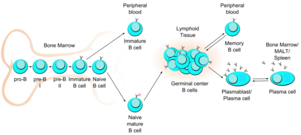
Figure 1. B cell development and differentiation subsets
Human B cell development in the bone marrow, differentiation subsets in peripheral blood and secondary lymphoid tissues: spleen and mucosa-associated lymphoid tissues (MALT). Modified from (Perez-Andres et al. 2010).
Immature B cells are particularly sensitive to tolerogenic signals, a feature that is lost after maturation and differentiation into B-cell subtypes with unique functions (Allman and Northrup 2007). In this respect, immature B cells undergo a receptor-mediated negative selection. During the process, only 20% of the cells survive and can emigrate from the bone marrow. Self-reactive B cells undergo negative selection through three known mechanisms: deletion, anergy and receptor editing in which auto-reactive cells undergo a further gene rearrangement to modify their receptors (Wang and Clark 2003). The fate of self-reactive immature B cells depends on several factors, including receptor affinity, IgM expression, stage of maturation and site of ligand encounter. It has been shown that binding of high- avidity ligands leads to deletion (Erikson et al. 1991, Chen et al. 1994) while immature B cells that bind lower-avidity ligands become anergic (Goodnow et al.
9
2009). Level of IgM expression has also been related to how B cells respond to antigen. In vitro experiments done by Melamed et al, demonstrated that IgMlow immature B cells undergo receptor editing after BCR stimulation. In contrast, BCR ligation on IgMhi B cells induces apoptosis (Melamed et al. 1998). The microenvironment surrounding the place where immature B cells find the antigen is also affecting the outcome; while binding in the bone marrow induces receptor editing, ligation in the spleen induces deletion (Wang and Clark 2003). These mechanisms of tolerance diminish to a minimum the risk of reaction of B cells to tissues of the host organism.
As soon as the small proportion of cells that escape negative selection leave the bone marrow or fetal liver to populate peripheral blood and lymphoid tissues, B cells start to diversify the constant region of the membrane bound immunoglobulin (Figure 1). Via the process known as class switch recombination (CSR), maturing B cells start to express on the surface a second isotype with the same specificity, IgD.
Isotype switching takes place through a mechanism of alternative splicing of a large primary messenger ribonucleic acid transcript containing both µ and δ constant region genes. After splicing, the recombined VDJ gene contains the exon coding δ heavy chain segment to produce mIg of the IgD isotype (Geisberger et al. 2006). At this stage naïve B lymphocytes are characterized by their short-life span, their dominance during early phases of reconstitution of the peripheral B-cell pool and their susceptibility to undergo apoptosis rather than to proliferate in response to BCR ligation (Allman et al. 2001).
Naïve B cells recirculate through peripheral blood and enter lymph nodes, spleen and MALT (Figure 1). If they do not encounter antigen, they will continue recirculating until they die after a couple of days due to failed positive selection.
10
Those naïve B cells that bind their cognate antigen in the secondary lymphoid tissue will be primed and start the germinal center (GC) reaction in cooperation with CD4+ and CD8+ antigen-specific T cells (Figure 1) (Allen et al. 2007). In the GC, B cells are constantly migrating between the dark zone (centroblasts) and the light zone (centrocytes). In the dark zone, B cells undergo clonal expansion, CSR and somatic hypermutation (SHM) of variable heavy-chain region genes. By the SHM process, non-templated point mutations are introduced in the variable region of rearranged immunoglobulin heavy and light chain genes. Then, in the light zone, they re- encounter the antigen and undertake affinity maturation of the BCR (Allen et al.
2007, LeBien and Tedder 2008). This results in the preferential outgrowth of B cells expressing an immunoglobulin that has high affinity for its cognate antigen (Odegard and Schatz 2006).
GC-B cells are characterized by up-regulation of CD10, CD38, CD95 and HLA- DR and decreased expression of CD44 and bcl2 and heterogeneous expression of CD27 which differentiated them from naïve, memory B cells and plasmablasts (Allen et al. 2004, Allen et al. 2007). After undertaking several rounds of proliferation and affinity maturation, these cells become memory and pre-effector (plasmablasts) B cells (Allen et al. 2007). Quiescent memory B cells remain recirculating in peripheral blood or through tissues, where they can find antigen, while plasmablasts migrate to bone marrow or MALT to complete the differentiation and become antibody- producing B cells (plasma cells -PCs-) (Figure 1) (Perez-Andres et al. 2010).
1.4.2 Circulating peripheral blood B-cell subsets
B lymphocytes are phenotypically and functionally heterogeneous. During the past years, several subpopulations of B cells have been discovered. The use of
11
monoclonal antibodies (mAbs) and multicolor flow cytometry combined with functional assays has facilitated this work. The first attempt to classify human B cells was the introduction of the B mature (Bm) system (Liu and Arpin 1997). Tonsillar B cells were classified according to the surface expression of IgD and CD38. Five major subsets were reported: naïve B cells (IgDpos CD38neg) that can be further subdivided in Bm1 (CD23neg) and Bm2 (CD23pos) subsets, GC-B cells (IgDneg CD38pos), which were separated by CD77 into centrocytes and centroblasts (Bm3 and Bm4) and the double negatives (IgDneg CD38neg), which were described to be memory B cells (Bm5). This was the first classification of B cells. Although extremely useful, it is now clear that
binary
classification is not enough to discriminate between phenotypically different subpopulations that share the same surface profile.In this way, IgM and/or CD27 are included nowadays to further categorize the populations (Weller et al. 2004). Therefore, new subgroups have been identified within the above subsets, i.e. Bm5 cells can be subdivided into memory B cells, which express CD27, and PCs, which do not (Youinou 2007). Moreover, later work demonstrated that CD77 does not discriminate centrocytes and centroblasts (Hogerkorp and Borrebaeck 2006).
Human naïve B cells comprise most of the peripheral blood B cells; they co- express IgM and IgD and have unmutated Ig variable regions. On the other hand, memory B cells consist of about 20 to 30% of all peripheral blood B cells and have undergone SHM, affinity maturation and about half also underwent CSR (Klein et al.
1998, Tangye et al. 1998). Naïve and memory B cells also differ in terms of in vitro responsiveness to stimulation, mimicking the primary and secondary response in vivo. Therefore, memory B cells rapidly enter the cell cycle and undertake more rounds of division. A higher proportion of them become antibody-secreting PCs
12
(Fecteau and Neron 2003, Good et al. 2009). Both naïve and memory B cells recirculate in peripheral blood and lymphoid tissues. However, many of the memory B cells inhabit places of antigen draining such as the marginal zone of the spleen and the mucosa epithelium of the tonsils (Klein et al. 1998, Tangye and Tarlinton 2009).
Plasmablasts and PCs can be found in low frequencies in peripheral blood from healthy donors (Caraux et al. 2010). They are responsible for high affinity antibody secretion and humoral memory, providing a high quality defense when reencountering antigen. They represent 1-3% of total peripheral blood B cells and might be newly generated plasmablasts that are migrating from secondary lymphoid tissues to a niche in bone marrow or inflamed tissues (Brandtzaeg and Johansen 2005). Higher frequency of PCs in peripheral blood has been associated with bacterial infection and systemic lupus erythematosus (SLE) (Ten Boekel et al. 2007).
Another minor subset of circulating immature B cells has been described in peripheral blood from healthy donors. In addition to expression of the B cell-specific marker CD19, this transitional B-cell subset can be identified using expression of the developmentally regulated markers CD24 and CD38. Transitional B cells make up around 2% of B cells, express high levels of both CD24 and CD38 (CD24bright CD38bright), and co-express IgM and IgD and lack CD27 (Carsetti et al. 2004).
Additionally, recently multiple regulatory B-cell subsets that suppresses cellular immune responses through the production of immunomodulatory cytokines have been identified. IL-10-producing B cells (B10 cells) are the most widely studied regulatory B cells (Yanaba et al. 2008, DiLillo et al. 2010, Mauri and Bosma 2012).
These rare cells are named B10 cells to highlight that their regulatory function is mediated solely by its IL-10-dependent regulatory properties, and to distinguish
13
them from other B-cell subsets that regulate immune responses through different mechanisms (Ray et al. 2012).
In recent years, the development of multi-parameter flow cytometry coupled with improvements in the ability to purify minor cellular subsets using cell sorting has enabled the identification and characterization of novel B-cell subsets that can only be identified using a complex set of surface markers.
1.4.3 B cells in health and disease
Immunity is coordinated by a complex network of cells that are continuously changing in response to the signals they receive. Therefore, there is a high degree of interdependency among the components, and any disruption in the tightly regulated process might result in a failure of the protective mechanisms of the immune system.
The introduction of B cell-depleting agents has helped to uncovered the role of B cells in the pathogenesis of several immune-related diseases (Edwards et al. 2004, Looney et al. 2004, Sanz et al. 2007). Interestingly, depletion of B cells not only improved symptoms in diseases typically considered of B cell origin (such as SLE, idiopathic autoimmune thrombocytopenia, dermatomyositis and autoimmune blistering diseases) but also diseases in which B cells where not thought to play a major role (rheumatoid arthritis, multiple sclerosis and type 1 diabetes) (Edwards et al. 2004, Looney et al. 2004, Hauser et al. 2008, Xiu et al. 2008, Mei et al. 2012, Fuertes et al. 2013). Studies done in mice might explain the phenomenon, since they showed that mAb Rituximab (anti-CD20) therapy leads to B cell depletion by monocyte-mediated antibody-dependent cellular cytotoxicity (Uchida et al. 2004) and that amelioration of symptoms might be due to regulation of CD4+ T cell expansion, thus delaying and controlling the acute inflammatory phase that leads to
14
tissue damage (Bouaziz et al. 2007). Moreover, it might be that the therapeutic effect seen by this intervention is to a large extent related to the disturbance of the functional balance of B-cell subsets (Sanz and Lee 2010).
Malfunction in B-cell compartment influences development of immunodeficiencies, autoimmunity or hematologic malignancies. For instance, a typical immunodeficiency generated by disturbances in later stages of B cell development is common variable immunodeficiency (CVID). It is a disease that can be manifested at any time of life and is probably influenced by genetic or environmental factors. Patients exhibiting this disease have low serum immunoglobulin and are more susceptible to infections. Additionally, they have low levels of B cell memory, CSR and B cell activation (LeBien and Tedder 2008). It is important to stress that disruption in tolerance checkpoints in this disease results in a loosening of tightly- controlled screening process for reactivity to self-antigens and predisposes to autoantibody production and autoimmunity. However, the role of B cells in the pathogenesis of autoimmune diseases is not restricted to autoantibody production but also contributes to autoantigen presentation to T cells and through proinflamatory cytokine production (Shlomchik 2008). In the case of B cell-related cancer, disruption of differentiation at any stage of development (Figure 1) can lead to the expansion of a malignant counterpart of dominant subclones that ultimately derive in leukemia and lymphoma.
1.4.4 Molecular architecture of B cells
It was not until 1980, with the use of mAbs that the molecular constitution of the B cell surface started to be identified. B cell molecules that have been characterized so far are associated with BCR signaling, development, function,
15
adhesion and communication with the extracellular environment (LeBien and Tedder 2008).
B lymphocytes express on their surface several molecules that are shared with many other leukocyte types. For instance, B cells proliferate in response to bacterial deoxyribonucleic acid (DNA) due to signaling through Toll-like receptors (TLR), which are also known to be expressed by multiple leukocyte lineages (LeBien and Tedder 2008). As discussed earlier, CD38 and CD27 molecules are essential to define B-cell subsets although they are not B-cell lineage restricted (Jackson et al.
2008).
On the other hand, several molecules are preferentially expressed by B cells.
Among them are CD19, which can be found in virtually all B-lineage cells, CD20 that functions as a calcium (Ca2+) channel embedded in the membrane, CD21 (also known as complement receptor 2), CD22 and CD72 that negatively regulate BCR signaling, CD23 a low-affinity receptor for IgD, CD24 – a molecule with unknown function - and CD40 that serves as a survival factor for GC-B cells when binding to its ligand on T cells (CD40 Ligand -CD40L- also known as CD154) (LeBien and Tedder 2008).
1.4.4.1 The complement receptor (CR) 2: CD21
It is well known that in order to generate an appropriate immune response, B cells require not only BCR cross-linking but also additional signals through coreceptors such as CR1, CR2 and Fcɣ (FcɣRI, FcɣRII and FcɣRIII) receptors (Erdei et al.
2009).
CR2, also known as CD21, is a glycoprotein receptor expressed by B cells, T cells and follicular DCs. On B cells it appears at the mature stage and disappears after PC differentiation. In humans, it is composed of 15 to 16 short consensus repeats, a
16
transmembrane domain and a short cytoplasmic tail and is generated from the CD21 gene which encodes a single RNA transcript (Erdei et al. 2009). The interaction of CD21 with its ligands is known to require the first 2 consensus repeats (Lowell et al.
1989, Szakonyi et al. 2001). CD21 binds C3 degradation products C3d, iC3b and C3b (Boackle et al. 1997). In addition it also binds CD23 (Aubry et al. 1992) and IFN-α (Asokan et al. 2006) and serves as entry receptor for Epstein-Barr virus (EBV) by binding pg350/220 glycoprotein on EBV (Nemerow et al. 1987). CD21, as a receptor of complement fragments facilitates the cross-talk between the innate and adaptive branches of the immune response.
CD21 is expressed on the surface of B cells either as part of a trimolecular complex of CD21, CD19 and CD81 or associated with CR1 (CD35); very few reside alone (Tuveson et al. 1991, Grattone et al. 1999). As part of the B-cell coreceptor complex, cross-linking of CD21 and BCR by antigen-complex augments and potentiates activation through recruitment of CD19 and CD81, and helps to initiate downstream stimulatory events (Rickert 2005). In fact, C3d fragments deposit on antigen increases the immunogenicity by recruiting both proteins and inducing phosphorylation not only in CD19 but also in CD21 cytoplasmic tail (Dempsey et al.
1996, Barrault and Knight 2004). In summary, there is substantial evidence that supports the in vivo significance of CD21 providing an important link between the innate and adaptive immune responses (Dempsey et al. 1996, Fearon and Carroll 2000).
Most of circulating B lymphocytes expresses high levels of CD21. However, there is a small fraction of naïve and memory B cells with low levels of CD21 expression. Although to date little information is known about the role of CD21low B cells, in diseases such as CVID and human immunodeficiency virus (HIV) infection, the
17
frequency of this subset is increased (Moir et al. 2008, Rakhmanov et al. 2009).
1.4.5 B cell functions
During the past years, the old concept that B cells are exclusively committed to produce antibodies has been reconsidered. Nowadays, it is apparent that in addition to their essential role in humoral immunity, B cells accomplish many other tasks crucial for immune homeostasis (Figure 2). Of great importance is the requirement of B cells to initiate T cell immune responses. The first in vivo evidence for this assumption came from studies done by Ron et al., in which mice were depleted of B cells by administration of anti-µ antibodies from birth (µ-suppressed mice (µSM)) (Ron et al. 1981, Ron et al. 1983). After antigen inoculation with complete Freund's adjuvant, µSM had a severe impairment in CD4+ T cell activation.
In addition, these mice were also more susceptible to bacterial and viral infection (Cerny et al. 1988, Cerny et al. 1988), had a reduced delayed-type hypersensitivity reaction to soluble antigens (Herrmann et al. 1988) and were resistant to diabetes by abrogating completely the development of insulitis and sialitis (done with µSM- Nonobese diabetic mice) (Noorchashm et al. 1997). When mice were injected with B cells prior to antigenic challenge, the observed defects in µSM were largely overcome (Janeway et al. 1987, Ron and Sprent 1987, Noorchashm et al. 1997). All this evidence suggests that CD4+ T cell could not be efficiently primed in the absence of B cells and that B cells play a direct role as APCs.
18 Figure 2. B cell functions
Selected examples of the multifunctional attributions of B cells and how they regulate immune homeostasis. Most of the functions of B cells are independent from antibody production. Modified from (LeBien and Tedder 2008).
APC function by B cells became controversial with the introduction of B-cell deficient mice generated by gene targeting either of the µ-chain or Ig heavy chain genes (Kitamura et al. 1991, Chen et al. 1993). Experiments done by independent groups with these B-cell deficient mice range from complete deficiency to almost normal T cell response (Epstein et al. 1995, Liu et al. 1995, Phillips et al. 1996, Macaulay et al. 1998). A more recent study using several B-cell deficient mice strains
19
generated by the same gene targeting technique demonstrated that in all but one strain (C57BL/6 background; which were used for most of the previous studies) T cell responses were impaired (Rivera et al. 2001). They showed that T cell priming of B- cell deficient mice of C57BL/6 background were much more variable, explaining the controversial results.
In addition, by breeding chimeras it was possible to show that the congenital absence of B cells also plays a role during immune system development, which is evidenced by a diminished amount and diversity of thymocytes, defects within DC and T cell compartments, Peyer patch organogenesis and impaired chemokine expression by macrophages (LeBien and Tedder 2008).
Moreover, B cells are also important for regulation and maintenance of the immune system by cytokine secretion. Cytokine-producing B cells can be divided into regulatory B cells, which secrete IL-10 (B10 cells) or transforming growth factor (TGF)β-1 and effector B cells that produce IL-2, IL-4, tumor necrosis factor (TNF)α, IL- 6 or INFγ, IL-12 and TNFα (Lund 2008). By releasing cytokines, B cells can influence T cells, DCs, APC functions, regulate lymphoid tissue organogenesis and wound healing, modulate transplant rejection, tumor development and tumor immunity (LeBien and Tedder 2008). Moreover, the profile of cytokine released by effector B cells can influence the polarization of the T cell response (Harris et al. 2000). Additionally, cytokine production and antigen presentation by B cells have been associated to influence a range of diseases such as atherosclerosis, insulin resistance, HIV, graft- versus-host disease, and allograft rejection (Moir and Fauci 2009, Shimabukuro- Vornhagen et al. 2009, Ait-Oufella et al. 2010, Winer et al. 2011).
20
1.4.5.1 How B cells capture, process and present antigens?
In order to confer effective protection against the huge variety of pathogenic antigens that can face the immune system throughout the body, the response must be tightly harmonized. Therefore, antigenic interaction occurs mostly in defined sites (secondary lymphoid organs) such as lymph nodes and spleen. The lymphoid tissue possesses a highly compartmentalized microarchitecture that is essential for cellular interactions and initiation of optimal immune responses. In order to get activated and to exert APC function, B cells require two main signals.
First, B cells recognize and capture the specific antigen by their BCR, which induces growth, proliferation, survival and expression of the costimulatory molecule CD86 on the B cells (Lenschow et al. 1994). Then, the antigen is degraded into peptide fragments that are loaded onto MHC class II molecules that are next presented on the cell surface to specific CD4+ T cells. Interaction with CD4+ T cells provides the second stimulatory signal mediated by CD40-CD40L binding and secretion of IL-4 by T cells (Figure 3) (Ranheim and Kipps 1993, Evans et al. 2000). T and B cell cooperation is required for proper B cell activation, GC formation, differentiation into high-affinity antibody-producing PC, generation of memory B cells (Yuseff et al. 2013) and proper APC function by up-regulation of the expression of MHC class I and II and costimulatory molecules CD80 and CD86 (Kennedy et al. 1994, Faassen et al. 1995).
21 Figure 3. Interaction between B and T lymphocytes
Model for interaction between CD4+ helper T cell and B cell after antigen encounter.
The antigen bound to the B-cell receptor (BCR) of the specific B cell is processed and the peptides of the processed antigen are presented in the context of MHC class II (MHC II) molecules to the helper CD4+ T cells via the T cell receptor (TCR). Optimal T- cell activation takes place when the costimulatory molecules CD80 and CD86 interact with CD28, providing the necessary costimulation. The activated CD4+ helper T cells upregulate CD40L and secrete IL-4, which results in contact-dependent B-cell activation through CD40 and through IL-4 signaling. The functional end point after antigen encounter is differentiation of effector T and B cells. Modified from (Guttormsen et al. 1999).
1.4.5.1.1 BCR internalization and signaling
Receptor endocytosis and initiation of signaling events required for B cell activation are induced by both soluble and membrane-bound antigens binding to BCR. Signaling through BCR and coreceptor starts with the recruitment and activation of tyrosine kinases. Critical for signaling is the transmembrane heterodimer Igα/β, which is associated noncovalently with the BCR coreceptor complex (Campbell et al.
22
1991). Upon antigen binding, mIg and Igα/β heterodimer, which contain highly conserved phosphorylation motifs (immunoreceptor tyrosine-based activation motif or ITAMs) are phosphorylated. Upon phosphorylation the BCR complex translocates to the glycolipid and cholesterol-rich membrane microdomains where proteins of the non-receptor protein tyrosine kinase Src family including Lyn, Fyn and Blk are constitutively present to initiate the down-stream cascade of activation signaling (Rickert 2005). Once phosphorylated, Igα/β ITAMs recruit and activate the tyrosine kinase SYK (Kurosaki et al. 1995). Subsequently, SYK and Src family kinases initiate separate but inter-related signaling pathways. While Src family kinases serve to phosphorylate nuclear factor (NF)-ƙB (Saijo et al. 2003), CD22 (Fujimoto et al. 1999) and BAM32 (Niiro et al. 2002), SYK phosphorylates BLNK (Fu et al. 1998), which in turns coordinates the assembly and activation of a receptor-retained signalosome containing phospholipase Cγ2 (PLCγ2), Vav, BTK, Nck, and Grb2 (Chiu et al. 2002).
Concurrent with signal initiation, the majority of BCR-antigen complexes are rapidly cleared from the cellular membrane. In fact, BCR-antigen engagement induces maturation of late endosomal and lysosomal compartments, into which BCR complexes are internalized and antigen is depredated by the acidification of the vesicles (Siemasko et al. 1998). Initial activation events in response to membrane- bound antigen induce a biphasic spreading and contraction reaction on B cells. First, in the rapid spreading phase B cells extend lamellipodia across the follicular DC loaded with antigen increasing the amount of BCR-antigen interactions depending on the reorganization of the actin cytoskeleton. This dynamic structure, in addition to integrins such as leukocyte function antigen-1 (LFA-1), promotes the adhesion of B cells to APCs lowering the threshold for activation (Carrasco et al. 2004).
Subsequently and more slowly, the B cell contracts cumulating antigen into a central cluster that acts as a platform for antigen internalization (Fleire et al. 2006).
23
Collectively the spreading and contraction responses determine how much antigen is acquired and presented by the B cell, and therefore determine the outcome of B cell activation (Depoil et al. 2008).
In addition, secretion of molecules of both endocytic and exocytic processes takes place in the immunological synapse. It is believed that proteases and hydrolases released in the vesicles are required to free the membrane-bound antigens from APCs and to start antigen degradation at the extracellular space (Yuseff et al. 2011). Some antigenic peptides released are directly loaded onto MHC class II molecules in the extracellular space probably at the same time that occurs in the main intracellular pathway. On the other hand, B cells also extract tethered antigen by trogocytosis, which involves the exchange of membrane fragments between cells (Aucher et al. 2008).
Synthesis of MHC class II and the costimulatorymolecules CD80 and CD86 are also enhanced by BCR signaling (Reth and Wienands 1997, Zimmermann et al.
1999). Internalization of the BCR is regulated by clathrin (Stoddart et al. 2002) and depends on the ubiquitylation of BCR-antigen complexes that occur downstream of SYK-dependent signaling (Katkere et al. 2012) providing evidence for the relationship between signaling and endocytic trafficking. Propagation of the signal continues within endocytic compartments, with further kinase phosphorylation that ultimately leads to transcription of genes required for B cell activation (Chaturvedi et al. 2011).
1.4.5.2 CD40-CD40L interaction
CD40 is a type 1 transmembrane protein member of the TNF-receptor family. It was first described in 1985 as a molecule constitutively expressed on B cells throughout development and differentiation (van Kooten and Banchereau 2000).
24
Furthermore, it is expressed by a variety of immune and non-immune cells including APCs such as macrophages, DCs and endothelial cells (Schonbeck et al. 2000). Its natural ligand, CD40L, is expressed mainly on activated CD4+ T cells (van Kooten and Banchereau 2000). It is a type 2 transmembrane protein of the TNF family of ligands and forms homotrimers on the cell membrane (Schonbeck et al. 2000). Moreover, CD40L is also released in a soluble form (sCD40L) retaining its function and the trimeric structure of the membrane-bound protein (Mazzei et al. 1995). As described in section 1.4.5.1, interaction between CD40 and CD40L results in proliferation, differentiation and increased expression of surrogate molecules of APC function, which enhance efficacy of their antigen presentation capacity (Banchereau et al.
1994, Yellin et al. 1994, von Bergwelt-Baildon et al. 2002). Antigen presentation in the context of MHC class II on the surface of APCs together with costimulation through receptors and ligand pairs, such as CD40-CD40L, enhances T cell activation and differentiation modulating the immune response (Figure 3) (Bretscher 1999).
1.4.5.2.1 Signaling through CD40L
Ligation of CD40 by its ligand induces the activation of several signaling pathways (Figure 4). The cytoplasmic signaling domain of CD40 lacks intrinsic catalytic activity, therefore upon activation relies on several proteins of the TNF receptor associated factor (TRAF) family that interact with different recognition motif in the cytoplasmic domains (Harnett 2004). Association of TRAF with CD40 leads to activation of MAPKs (mitogen activated protein kinases) ERK (extracellular signal regulated kinase), Jnk (c-Jun N-terminal kinase) and p38 and the canonical and noncanonical NF-ƙB pathways. ERK, p38 and Jnk are activated by a cascade of MAP kinases and result in the activation of transcription factors, among them Elk-1, ATF1 and AP-1 (Fos/Jun), respectively (van Kooten and Banchereau 2000). Binding of