Improving cargo delivery in cancer therapy with the help of cell-penetrating peptides
Inaugural-Dissertation
zur
Erlangung des Doktorgrades
der Mathematisch-Naturwissenschaftlichen Fakultät der Universität zu Köln
vorgelegt von
Lucia Feni aus Chiari
Köln
2019
Berichterstatter: Prof. Dr. Ines Neundorf
Prof. Dr. Norbert Sewald
Tag der mündlichen Prüfung: 29. März 2019
Die im Rahmen der vorliegenden Arbeit durchgeführten Experimente und Untersuchungen
wurden im Zeitraum von September 2015 bis Dezember 2018 am Institut für Biochemie der
Universität zu Köln unter der Anleitung von Frau Prof. Dr. Ines Neundorf durchgeführt.
Corpora non agunt nisi ligata
I
Abstract
A major obstacle of many active pharmaceutical compounds is their low ability to cross body barriers, especially cell membranes. Cell permeability of a drug is therefore considered as a key step for therapeutic efficacy. Over the last decades, different approaches to overcome this limitation have been studied intensively. Among these are so-called cell-penetrating peptides (CPPs). CPPs are able to autonomously internalize into cells without the need for auxiliary proteins. However, not only the cellular uptake is important but also cell selectivity has to be addressed. Over the past two decades, cancer research has dramatically evolved, particularly with the appearance of targeted molecular therapies and advances in antibody engineering that allowed the discovery and validation of innovative molecules, more effective and less harmful than conventional chemotherapy. Especially small molecule-drug conjugates, like peptide-drug conjugates, became of particular interest since they combine several advantages as deep tissue penetration, possibility of cell organelle targeting and relatively easy access by chemical synthesis.
This work focuses on the design and synthesis of an array of tumor-targeting peptide-drug conjugates combining known tumor-homing peptides with a well-described CPP and potent cytotoxic drugs. The development of these hybrids was followed by a validation of the model
via in vitro studies where their selectivity towards different cell lines was evaluated. Twotargeting ligands (GnRH-III and c[DKPf3RGD]) were employed for the conjugation to the CPP sC18 and a very straightforward synthesis could be developed in both cases. The conjugates maintained a remarkable binding affinity in low nanomolar range towards GnRH and α
vβ
3integrin receptors, respectively, and for further
in vitro experiments, the expression of thereceptors in different cell lines was explored. For the investigation of the final compounds, a new
in vitro model based on a short contact time with the cells was established in order toemphasize the role of the fast CPP-mediated internalization after reversible binding to the receptors. While for the GnRH-III-conjugates a selectivity was difficult to detect, the c[DKPf3RGD] was identified as very effective targeting moiety for the synthesis of an efficient drug delivery system. Different drugs were attached to the CPP and daunorubicin turned out to be the most advantageous in terms of simple synthesis and stability. Fluorescence analysis demonstrated that the internalization was mainly mediated by the CPP but that the ligand had an important role in targeting the surface of the cells overexpressing the receptor. The selectivity could also be proved by anti-proliferative assays providing another demonstration that with this approach it would be possible to overcome the drawbacks of CPP-mediated drug transport leading to higher target selectivity and better bioavailability.
In the second part of the thesis, cyclic CPPs with peculiar diketopiperazine scaffolds (trans
DKP3 and
cis DKP1) were synthesized starting from the sequence of a truncated variant ofII
sC18. An optimized cyclization strategy could be developed and the secondary structure of
these peptides was analyzed and compared with the linear counterparts by different
techniques. The biological activity of these compounds was also evaluated in cell systems
where the ability to transport cytotoxic drugs inside the cells was explored by using both a
non-covalent as well as covalent drug coupling approach. Notably, the cycle actually showed
a higher ability to increase the activity of daunorubicin than the linear CPP, proving that
cyclization via a diketopiperazine scaffold is a promising strategy to improve CPP-mediated
drug delivery.
III
Zusammenfassung
Viele aktive pharmazeutische Verbindungen sind nicht in der Lage Barrieren, insbesondere Zellmembranen, ohne Hilfe zu überwinden, um ihren spezifischen Wirkort zu erreichen. Aus diesem Grund gilt diese Zellpermeabilität eines Arzneimittels als Schlüsselschritt für die therapeutische Wirksamkeit. Um diese Verbindungen zu transportieren, wurden bereits verschiedene Strategien etabliert, unter denen sich auch sogenannte zellpenetrierenden Peptide (CPP, cell-penetrating peptides) einreihen. CPPs sind in der Lage von einer Vielzahl von Zellen aufgenommen zu werden, ohne dabei auf Hilfe von Transportproteinen angewiesen zu sein. Aber nicht nur die zelluläre Aufnahme steht im Fokus der Forschung, insbesondere die Zellselektivität ist von großem Interesse. In den letzten zwei Jahrzehnten hat sich die Krebsforschung stark weiterentwickelt, vor allem durch gezielte molekulare Therapien und Fortschritte in der Antikörperentwicklung, die die Entdeckung und Validierung innovativer Moleküle ermöglichten, die dadurch sowohl wirksamer als auch weniger schädlich als konventionelle Chemotherapien sind. Besonders kleine Molekül-Wirkstoff Konjugate, wie zum Beispiel Peptid-Wirkstoff Konjugate, sind vielversprechend, da sie sich durch tiefe Gewebepenetration und mögliches Ansteuern verschiedenster Zellkompartimente auszeichnen. Desweitern sind diese Konjugate relativ einfach herzustellen und so einfach zugänglich.
Diese Arbeit konzentriert sich auf das Design und die Synthese von Krebs spezifischen Peptid- Wirkstoff-Konjugaten, bei der ein Peptidfragment, das in der Lage ist ein bestimmte Tumorart anzusteuern, mit einem bekannten CPP und einem Zytostatikum kombiniert wird. Nach der Herstellung dieser Hybride folgte eine Validierung ihrer Aktivität durch verschiedenste in vitro Studien. Zwei spezifische Ziel-Liganden (GnRH-III und c[DKPf3RGD]) wurden für die Konjugation an das CPP sC18 verwendet, und in beiden Fällen konnte eine optimierte Syntheseroute entwickelt werden. Die Konjugate zeigten eine bemerkenswerte Bindungsaffinität im niedrigen nanomolaren Bereich zu GnRH bzw. α
vβ
3Integrin-Rezeptoren.
Für weitere
in vitro Experimente wurde außerdem die Expression der Rezeptoren inverschiedenen Zelllinien untersucht. Für die Analyse der Verbindungen wurde ein neues
in vitro Modell etabliert, das auf einer kurzen Kontaktzeit mit den Zellen basiert, um die Rolle derschnellen Internalisierung durch das CPP nach der Bindung zu den Rezeptoren zu
untersuchen. Während für die GnRH-III-Konjugate eine Selektivität schwer nachzuweisen war,
wurde c[DKPf3RGD] als sehr wirksame Zielgruppe für die Synthese eines effizienten
Transportsystems identifiziert. Verschiedene Toxine wurden an das CPP gebunden, wobei
sich Daunorubicin im Hinblick auf die einfache Synthese und Stabilität als das
vielversprechendste erwies. Aufnahmestudien zeigten, dass die Internalisierung hauptsächlich
durch das CPP vermittelt wurde, der Ligand jedoch möglicherweise eine wichtige Rolle beim
IV
Binden an die Oberfläche der Zellen hatte, die den Rezeptor überexprimieren. Die Selektivität konnte auch durch antiproliferative Versuche nachgewiesen werden. Somit liefert der hier vorgestellte Ansatz eine mögliche Lösung CPPs mit einer Zellselektivität auszustatten.
Im zweiten Teil dieser Doktorarbeit wurden verschiedene zyklische CPPs synthetisiert. Dafür
wurden spezielle Bausteine, basierend auf Diketopiperazinen (trans DKP3 und
cis DKP1),verwendet. Es konnte eine optimierte Zyklisierungsstrategie entwickelt werden und die
Sekundärstruktur dieser neuen Peptide wurde durch verschiedene Techniken analysiert und
mit den linearen Versionen verglichen. Die biologische Aktivität dieser Verbindungen wurde in
Zellen getestet, dabei stand besonders im Vordergrund, zytotoxische Wirkstoffe in Zellen zu
schleusen. Die Wirkstoffe wurden dabei sowohl nicht-kovalent als auch kovalent an das Peptid
gekuppelt. Das zyklische Peptid war in der Lage die Aktivität von Daunorubicin deutlich zu
verbessern im Vergleich zum linearen Peptid. Dieses Ergebnis unterstreicht, dass die
Zyklisierung mittels eines Diketopiperazingerüstes zu neuen CPPs mit sehr
vielversprechenden Aktivitäten führt.
V
Table of contents
1. Introduction ... 1
1.1. Traditional chemotherapy versus targeted chemotherapeutics ... 2
1.2. Cytotoxic payloads ... 5
1.2.1. Daunorubicin ... 5
1.2.2. Chlorambucil ... 6
1.2.3. Cryptophycin ... 6
1.3. Antibody-drug conjugates (ADCs) ... 7
1.4. Small-molecule drug conjugates (SMDC) ...10
1.4.1. GnRH receptors ...10
1.4.2. Integrin receptors and integrin ligands ...12
1.5. Receptor-mediated uptake ...16
1.6. CPPs as carrier molecules and their role in cancer therapy ...17
1.7. Rational for cyclic peptides ...21
1.7.1. Previous work...22
1.7.2. DKP scaffolds...22
2. Aims of the thesis ...24
2.1. Receptor-targeted CPPs for selective delivery of anticancer therapeutics ...24
2.2. Cyclic CPPs for cargo delivery ...24
3. Novel CPP-drug conjugates bearing a targeting moiety ...26
3.1. GnRH-III as targeting moiety and daunorubicin as cytotoxic payload ...26
3.1.1. Synthetic strategy ...27
3.1.2. Triptorelin binding assay ...29
3.1.3. Cell viability assays ...30
3.2. DKP3RGD as targeting moiety ...36
3.2.1. Synthesis and biological evaluation of the drug delivery system ...37
3.2.2. Development of cryptophycin conjugates ...47
3.2.3. Development of chlorambucil conjugates and their biological evaluation ...50
3.2.4. Development of daunorubicin conjugates and their biological evaluation ...52
4. Head-to-tail cyclization of a cell-penetrating peptide through DKP scaffolds ...66
4.1. Novel cyclic peptides bearing a DKP scaffold ...66
4.1.1. Synthetic strategy ...66
4.1.2. Circular dichroism ...68
4.1.3. NMR-based structure elucidation ...69
4.1.4. Biological evaluation...72
VI
4.2. Drug delivery with cyclic peptides ...73
5. Conclusion and Outlook ...80
5.1. Receptor-targeted CPPs for selective delivery of anticancer therapeutics ...80
5.2. Cyclic CPPs for cargo delivery ...85
6. Material and Methods ...88
6.1. Materials ...88
6.1.1. Chemicals and consumables ...88
6.1.2. Media and solutions for cell culture ...88
6.1.3. Equipment ...89
6.2. Methods ...91
6.2.1. Automated Solid Phase Peptide Synthesis ...91
6.2.2. Fmoc-cleavage ...92
6.2.3. Manual coupling ...92
6.2.4. Coupling of 5(6)-carboxyfluorescein (CF) and polymer cleavage...93
6.2.5. Kaiser test ...93
6.2.6. Boc protection ...93
6.2.7. Dde-cleavage ...93
6.2.8. Sample cleavage ...94
6.2.9. Full cleavage ...94
6.2.10. Coupling of daunorubicin ...95
6.2.11. Copper(I)-catalyzed azide alkyne cycloaddition (CuAAC) ...95
6.2.12. Synthesis of compound V ...95
6.2.13. Synthesis of compound VI ...96
6.2.14. Synthesis of compound VII ...96
6.2.15. Azide reduction ...96
6.2.16. Cyclization ...96
6.2.17. Synthesis of the cyclic peptide conjugated to daunorubicin ...97
6.3. Peptide analysis ...97
6.3.1. Analytical HPLC-MS ...97
6.3.2. Preparative HPLC ...98
6.3.3. Circular dichroism spectroscopy ...98
6.4. Biological methods ...98
6.4.1. Cell lines and cell culture conditions ...98
6.4.2. Freezing and thawing cells ...99
6.4.3. Cell viability assays ... 100
6.4.3.1. Resazurin-based cytotoxicity assay ... 100
VII
6.4.3.2. MTT-based cytotoxicity assay ... 100
6.4.4. Internalization studies ... 100
6.4.4.1. Flow cytometry ... 100
6.4.4.2. Confocal laser scanning microscopy ... 101
6.4.5. Integrin expression on cell surface ... 101
6.4.6. Solid-phase integrin binding assay ... 102
7. References ... 103
8. Attachment ... 111
8.1. List of abbreviations ... 111
8.2. Attachment of supplementary data: spectra, chromatograms and figures ... 116
8.3. List of figures ... 149
8.4. List of tables ... 152
8.5. Acknowledgment... 154
8.6. Statement about collaborations ... 158
8.7. Declaration... 159
1
1. Introduction
Over the last year, 18.1 million new cases of cancer have been reported worldwide. In other words, one in five men and one in six women is diagnosed with cancer and these numbers increase constantly. These data from the Globocan report have been published in the journal
CA: A Cancer Journal for Clinicians and represent a "photograph" of the diagnoses of cancerregistered globally. The statistics show an increase in cancer diagnosis, which may be due to several factors from the aging of the population to the precarious conditions of social and economic development that are recorded in different areas of the planet. This second aspect also affects cancer-caused mortality, which in 2018 should be almost about ten million, while over 43 million people are expected to live within the five-year prevalence.
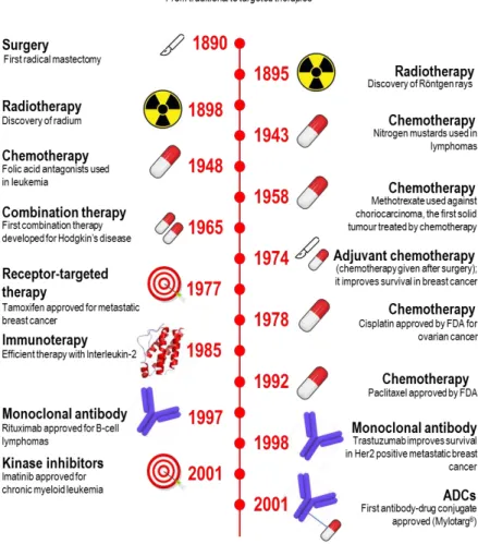
[1]In fact, research has led to increasingly effective therapies with fewer side effects, which in many cases are able to reduce mortality. The progresses made in the last century are highlighted in Figure 1.
The main treatments of tumors are represented by surgical resection,
[2]chemotherapy,
[3-4]radiation therapy
[5-6]but also by the more innovative hormone therapies,
[7-9]targeted therapies,
[10-11]immuno-oncology and gene therapy, used individually or in combination.
[12-13]Figure 1. Timeline: milestones in cancer therapy. From traditional to targeted therapies. Adapted from DeVita et al. and Chabner et al. [14-15]
2
1.1. Traditional chemotherapy versus targeted chemotherapeutics
Conventional chemotherapy is still widely applied in cancer treatment. 5-fluorouracil/leucovorin and oxaliplatin, for example, are still the gold standard for colorectal cancer,
[16]while a regimen of cisplatin or carboplatin combined with paclitaxel, gemcitabine or docetaxel is currently used for the treatment of non-small cell liver cancer at stage IV.
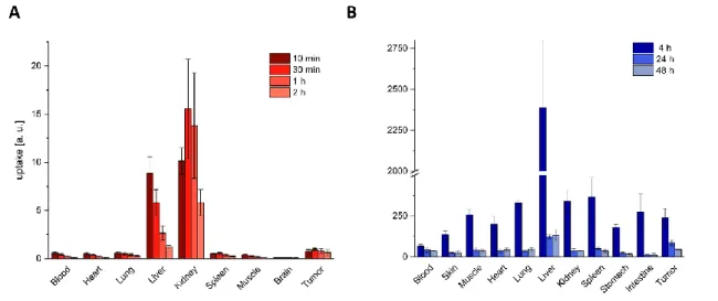
[17]However, the compounds used as chemotherapeutic agents localize with low efficiency in solid tumors. This unfavorable biodistribution profile, exemplified in Figure 2, combined with a mechanism of non-selective action, causes serious side effects and prevents a dose increase at therapeutically active regimens.
[18]Figure 2. Unfavorable biodistribution profile of traditional chemotherapeutic drugs. A: Tissue distribution of 5-Fluorouracil (5-FU) in ascitic hepatoma bearing rats. B: Tissue distribution of 3H-Paclitaxel in Balb/c nude mice bearing MDA-MB-435 tumors (melanoma). Adapted from Abe et al. and Cao et al. [19-20]
Two main approaches have been followed to reach the final goal of widening the therapeutic window: the combination of two or more cancer drugs without overlapping mechanism and/or toxicity
[21]and the introduction of more potent drugs administrated at lower dosage.
[22]Both strategies could lead to encouraging results in terms of efficacy, although an absolutely positive safety profile could not be achieved. Researchers understood that the unique key to completely avoid a systemic toxicity was the enhancement of selectivity. Targeted therapies, for instance, are interfering in a much more directed way with a molecule or a specific process of cell growth, not causing damage to healthy tissues, thus reducing side effects.
[23-24]In fact, they selectively act on specific cell receptors, hence improving the tolerability of the treatment, to the benefit of the patient and his quality of life.
[25]Targeted therapies represent one of the most important tools of personalized medicine, since
the treatment is no longer chosen only based on the development of the tumor, but also in
relation to its molecular characteristics and expression of biomarkers, which can be different
from patient to patient.
[26-30]Many efforts have been made in this field and the results obtained
3
in the last few years are exciting. Even if novel targeted therapies are pioneering and reserved for particular types of cancer, they already display an important role in the fight against this disease. In fact, many data show that they have prolonged survival and improved the quality of life of many patients.
[31-33]Over the years a number of directed agents have been used and these therapies are able to:
- manipulate the endocrine system through external administration of precise hormones or drugs that inhibit their production (hormone therapy for hormone-dependent tumors, e.g. anti-estrogens,
[34-36]aromatase inhibitors,
[37]GnRH agonists,
[38-39]anti- androgens
[40-41]);
- stimulate the immune system to identify and destroy cancer cells (immuno-oncology);
[42-43]
- hinder angiogenesis,
[44-45]inhibit tumor-related kinases
[46]and other oncoproteins (targeted therapy);
- selectively release toxic substances that act on cancer cells through different ways, e.g promoting apoptosis or decreasing their uncontrolled ability to grow and divide (targeted delivery).
[47-48]The last example, in particular, has been proposed as an alternative method to overcome the limits of classic chemotherapy, carrying powerful cytotoxic compounds at the tumor site after conjugation to ligands that are specific towards tumor-associated targets.
[24]The conjugation of these pharmacodelivery vehicles with a cytotoxic drug realizes the concept of "magic bullet"
as it was conceived more than a century ago by Paul Ehrlich (1854-1915), who was awarded
the Nobel Prize for Medicine in 1908.
[49-50]In Figure 3 the peculiar and advantageous
characteristic of these new compounds is illustrated with a schematic representation of their
mode of action. The selectivity of these drug-delivery systems is driven by the high binding
affinity towards particular tumor cell substrates resulting in a moderate occurrence of
undesirable effects since the healthy cell lines should not be affected.
4
Figure 3. Traditional chemotherapy vs targeted delivery of chemotherapeutics. A: General strategy of traditional chemotherapy; B: The “magic bullet” concept.
Antibody-drug conjugates (ADCs) and small molecule-drug conjugates (SMDCs) represent
two innovative classes of biopharmaceutical products, designed to selectively bring cytotoxic
agents to the tumor tissue. They combine the best features of two therapeutic modalities. In
particular, antibodies and small ligands that display target specificity but limited antitumor
activity are conjugated to cytotoxic agents, very potent but with poor safety and pharmaceutical
profiles. The following sections will first focus on three commonly used drugs, which were also
employed in this work, and will further highlight two novel delivery strategies.
5
1.2. Cytotoxic payloads
1.2.1. Daunorubicin
Daunorubicin and doxorubicin are the parent compounds of the anthracycline antibiotics.
Mostly isolated from natural sources (Streptomyces peucetius),
[51]they are extensively used for the treatment of cancer alone or in combination and widely investigated as cytotoxic payloads in conjugation with tumor homing peptides
[52-54]or antibodies.
[55-56]In fact, they are very effective but also very toxic, as they do not discriminate between malignant and healthy cells, leading in particular to cardio-toxicity
[57-58]and myelosuppression. From the structural point of view, anthracycline antibiotics are characterized by a planar tetracyclic portion, glycosidically linked to an aminosugar (daunosamine). The molecular structures of daunorubicin and doxorubicin differ only in one of the terminal substituents, as it is shown in Figure 4.
Figure 4. Structures of the two anthracycline parent compounds: daunorubicin and doxorubicin. Red box:
planar tetracyclic portion; blue box: daunosamine; green: hydroxyl group substituent in doxorubicin that is missing in daunorubicin.
Although small, this structural difference has important consequences on the activity spectrum of the two cytotoxic antibiotics. Doxorubicin, in fact, has significant clinical applications especially in solid tumors,
[59-62]while the main indication of daunorubicin is acute leukemia.
[63]The current tendency is to consider the DNA intercalation as necessary but not sufficient for
anti-tumor action.
[64]In fact, numerous results have indicated topoisomerase II as the main
anthracycline target. This nuclear enzyme relaxes the supercoiled DNA by the formation of a
phosphodiester bond between the OH group of its active-site tyrosyl residue and the
phosphoric group of DNA. This allows the free end of the nucleic acid to rotate, solving the
supercoiling. At this point the OH group at the free end of the DNA can restore the continuity
of the helix by attacking the activated phosphate.
[65]At present, it is known that anthracyclines,
after intercalating in the double helix, stabilize a ternary cleavage complex between the DNA,
tied to the enzyme, and the drug. Therefore the action of the drug leads to irreversible cuts in
DNA that open the way to the programmed cell death in cancer cells.
[66]Two further
mechanisms were identified as responsible for toxicity, notably the production of free oxygen
radicals through an enzymatic reduction process
[67]and induction of histone eviction from open
chromatin.
[68]6
1.2.2. Chlorambucil
Chlorambucil is a chemotherapeutic agent belonging to the class of the so-called alkylating drugs, in particular deriving from nitrogen mustards.
Figure 5. Structure of chlorambucil.
At physiological pH, chlorambucil forms a very reactive cyclic intermediate (aziridinium ion) which attacks the nitrogen in position 7 of a guanine, present in the DNA chain, building a covalent bond. The same process takes place on the other chain (ClCH
2CH
2N-) of the chlorambucil, which in turn will interact with a new guanine, present in the same or in the other DNA helix. Inter or intra helix bridges do not allow anymore DNA to perform its biological functions (duplication and transcription).
[69-70]The alteration that the chlorambucil induces in the DNA prevents the cancer cell from dividing, forcing it to undergo apoptosis.
[71]It is mainly used for the treatment of chronic lymphocytic leukemia,
[72-73]normally in combination with other chemotherapeutic agents. As previously described in the case of daunorubicin, chlorambucil provokes the common side effects of the non-targeted therapies; hence, its conjugation to targeting moieties has been studied and researched intensively.
[74-77]1.2.3. Cryptophycin
Cryptophycins are 16-membered macrocycles with bacterial origin composed by four units,
[78]having potent activity towards cancer cells and MDR (multi-drug resistant) cancer cells (IC
50in
the pM range), as the human cervical carcinoma cell line KB-V1.
[79-80]They are able to
coordinate to β-tubulin interacting with the
vinca domain. In particular, they inhibit tubulinpolymerization, inflicting a conformational change on tubulin dimers and depolymerize
microtubules
in vitro, reducing microtubule dynamics. [81]This leads to mitotic arrest and
apoptosis.
7
Figure 6. Structure of cryptophycin-52 and derivatives of cryptophycin-55 glycinate conjugated to a targeting peptide and a mAb. Blue: unit A; black: unit B; yellow: unit C; pink: unit D. Adapted from Weiss et al. [82]
Cryptophycin-52 is the lead compound within this class and was tested in clinical Phase II, where it unfortunately showed a lack of
in vivo efficacy and high toxicity (in particularneurotoxicity since neurons are the main tubulin producers for the transport of neurotransmitters). Cryptophycin-55 (the chlorohydrin of Cry-52) and its glycinate correspondent derivative were described as highly active
in vivo in preclinical models,displaying a better pharmacokinetic profile.
[83-84]Under physiological conditions the chlorohydrins are converted to the original epoxides, hence they are considered as prodrugs of the epoxides. After esterification with the glycine, improvement in water solubility and stability was also reached and this most importantly allowed the conjugation to homing peptides, like octreotide, but also antibodies
[82, 85]and other ligands, e.g. acetazolamide
[86](Figure 6).
1.3. Antibody-drug conjugates (ADCs)
An antibody-drug conjugate (ADC) is the unique combination of a monoclonal antibody, a linker
and a potent cytotoxic agent. It is designed to provide therapeutic potency to the antibody and
specificity to the anti-cancer agents, which can be directed to the tumor cell in a targeted way
8
to limit systemic exposure.
[87]This idea dates back to the early eighties. However, the first products did not obtain the desired results due to a series of technological limits, inadequate knowledge of the receptor target, use of insufficiently potent drugs and instability of the linker in biological fluids.
[88]Notable improvements in the conjugation technology associated with a greater understanding of the biology of the system led to the discovery of a second generation of ADCs. These new therapeutic agents have better stability in biological fluids and allow an appropriate release of the toxic agent to the target cell.
[89]ADCs include some of the most promising molecules in the oncology pipeline of large pharmaceutical companies. The peculiarity of ADCs is their long half-life (over a week), no systemic toxicity in circulation and activity only upon binding to tumors. All these characteristics lead to a maximum efficiency in administering the cytotoxic substance to the tumor cells in a perfectly selective way.
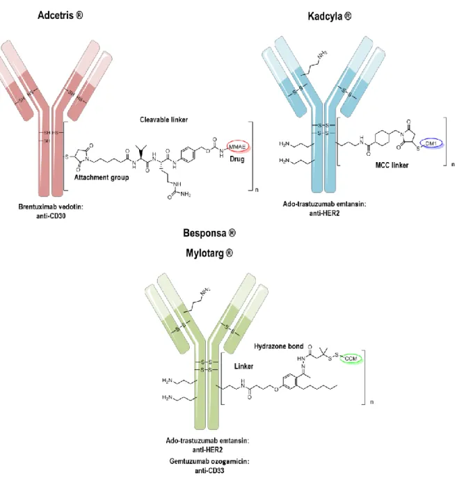
[90]To date, there are four ADCs approved by the FDA: Adcetris ® for the treatment of refractory Hodgkin's lymphoma
[91]and anaplastic large cell lymphoma,
[92-94]Kadcyla ® for the treatment of HER2-positive metastatic breast cancer,
[95-96]Mylotarg ® for adulte acute myeloid leukemia
[97-98]
and Besponsa ® for adult acute lymphoblastic leukemia,
[99-100]as represented in Figure 7. Auristatin (MMAE), maytansine (DM1) and N-acetyl-gamma-calicheamicin (CCM) are used as cytotoxic agents, respectively. On average, two to four toxins are conjugated to the mAb.
In addition to the four commercial ADCs, nearly forty are under investigation in many different
types of cancer.
[101]9
Figure 7. FDA approved ADCs. Drug-to-Antibody ratio is different, dependently from the conjugation strategy. n=4 (Adcetris ®), n=3.5 (Kadcyla ®), n=2.5 (Besponsa ® and Mylotarg ®).
Despite their growing success, the clinical advances of ADC products may be restricted by some limitations. Among these, heterogeneity is a crucial drawback leading to analytical and process challenges
[102]and difficulties in administrating these compounds to patients.
[103-104]The optimal design to obtain homogeneous compounds with the same average and distribution
of payloads is a key point to achieve product safety and it is being addressed by researchers
with new methods involving site-specific conjugation like engineering of the antibody
sequence,
[105]site-selective conjugation strategies
[106]or modular approaches.
[107]A high
Drug-to-Antibody Ratio (DAR) is also affecting the propensity of the ADCs to aggregate,
especially after being conjugated to very hydrophobic linker-drugs, so that the introduction of
hydrophilic molecules could help to solve this major problem.
[108]Besides that, increased
stability against extracellular proteases needs to be reached to avoid dangerous off-site toxicity
10
after administration if the linker is not stable enough to prevent the premature release of the drug in the circulation before being introduced in the tumor cell.
[109]Last but not least, the slow extravasation that characterizes immunoglobulins (IgG) is hindering their activity against solid tumors leading to the need of optimizing the format of the targeting moiety like for example by the introduction of small immune proteins (SIP) distinguished by a fast clearance, good extravasation and very good tumor/blood and tumor/organ ratio.
[110]1.4. Small-molecule drug conjugates (SMDC)
SMDC products, in particular peptide-drug conjugates, have been proposed to overcome some typical limits of antibodies.
[111]In fact, small organic ligands can penetrate into depth of solid organs and tumors within a few minutes after administration, conveying the drug to the tumor with greater efficiency, and are typically characterized by rapid excretion from circulation.
[112]In addition, they are easily developed, obtainable with inexpensive production and are easy to manipulate. However, they are often less selective than the correspondent antibodies.
[47, 113]Some of the prerequisites for a peptide to become a receptor-mediated carrier are:
- overexpression of the receptors on the surface of tumor cells;
- knowledge of SAR to efficiently synthesize peptide analogues;
- high affinity for the target;
- efficient internalization (to provide the introduction of the drug).
Two examples of receptor targeting moieties are gonadotropin-releasing hormone (GnRH) agonists and integrin ligands, whose biological background is introduced in the following paragraphs.
1.4.1. GnRH receptors
GnRH and its analogs have been widely used in clinical medicine since they were identified and synthesized in 1971 by Schally’s group.
[114]The native GnRH-I, also called luteinizing hormone-releasing hormone (LHRH), is a decapeptide (pGlu-His-Trp-Ser-Tyr-Gly-Leu-Arg- Pro-Gly-NH
2), produced and released in a pulsatile manner by the hypothalamus. After binding to specific receptors (GnRH-I R) on the plasma membrane of the gonadotrophs (Figure 8), it stimulates them to secrete follicle-stimulating hormone (FSH) and luteinizing hormone (LH) with a consequent gonadal response.
[115]A second form of GnRH (GnRH II) is ubiquitous and maintained in its structure throughout different species. Nevertheless, the GnRH-II receptor has not been identified yet in humans, even if there is strong evidence that this receptor exists.
[116]
GnRH receptors are highly expressed on various cancer cells and apart from pituitary cells
and reproductive organs, they are present in a very limited number in healthy tissues. They
are for this reason a good target for selective cancer chemotherapy.
[117-118]11
Figure 8. Binding of GnRH to its receptor. [119] IC: intracellular; EC: extracellular
In order to increase the power and duration of action of the GnRH-I, through modifications of the molecular structure of this decapeptide, many analogs have been synthesized, and are available for clinical use. The substitution with
D-amino acids in position 6 (involved in the enzymatic cleavage) or 10 (important for the tridimensional structure) resulted in analogs with agonist activity with a greater potency and a longer half-life than the native GnRH-I. These produce an initial stimulation of the pituitary cells followed by the down-regulation and inhibition of the hypophysis-gonadal axis. GnRH-I analogs are powerful therapeutic agents, proved to be very useful in various clinical indications, including the therapy of some hormone-dependent tumors, like prostate or breast cancer. In this context, doxorubicin was conjugated by an ester bond to a GnRH-I agonist called zoptarelin.
[120-121]The final conjugate AEZS-108 was tested till Phase III of clinical trials on endometrial cancer but failed to extend survival in the advanced disease.
[122]Taking advantage of these findings, some researchers are paying a special attention on the improvement of GnRH-based drug delivery systems, by starting from the sequence of a third form of GnRH (GnRH-III) discovered in sea lamprey, able to selectively bind to GnRH receptors but with a lower endocrine effect compared to GnRH-I agonists.
[123-127]In the group of Prof.
Mező (ELTE University, Budapest) the native sequence was modified in order to obtain a
higher stability and allow drug-conjugation and many strategies have been pursued.
[128-130]In
particular Ser
4was substituted with a butyrylated lysine, Lys(Bu), while Lys
8, functionalized
with an aminooxyacetic acid linker, was conjugated to daunorubicin by an oxime linkage, in
order to prevent the enzymatic cleavage of the ester bond by carboxylesterases (Figure 9).
[131]12
Figure 9. Optimization of the GnRH sequence. Pink: changes in the sequence; Green circle: conjugation point for the attachment of the drug. Bu: butyryl.
The conjugates are very stable in human serum from the chemical and enzymatic point of view but in presence of lysosomal homogenate they are degraded with subsequent release of different metabolites. H-Lys(Dau=Aoa)-OH was recognized as the smallest drug containing metabolite which is still able to bind to DNA
in vitro. [132]The stability and selectivity of the conjugate have been favored at the expense of the cytotoxicity that is in fact lower than the free drug. The introduced peptide was chosen in this work for further studies.
1.4.2. Integrin receptors and integrin ligands
Adhesive contacts with neighboring cells and with the extracellular matrix (ECM) control cell behavior and development. These interactions are mediated by proteins of the cell surface, called cell adhesion receptors, divided in four groups: cadherins, selectins, immunoglobulin superfamily and integrins.
[133]Integrins form the largest and most versatile receptor family, being implied in both cell-ECM
and cell-cell interactions. They are transmembrane heterodimeric glycoprotein receptors found
in mammals in 24 combinations, constituted from 18 different α- and β-subunits.
[134]The
subunits are not covalently linked and consist of an ectodomain, a transmembrane region and
a short unstructured cytoplasmic tail (Figure 10).
[135]The N-terminal regions of all α subunits
contain seven repeating sections, folded in the form of a seven-bladed β-propeller, supported
by a thigh and two calves. The β-chains of α
vintegrins present a domain I for the interaction
with the matrix followed by a hybrid and a PSI (plexin-semaphorin-integrin) domain, four
cysteine-rich EGF-like repeats and a β-tail.
[136]A conserved region called MIDAS (metal ion-
dependent adhesion site) in the β-I domain, is particularly important for the sequence
recognition, because it binds divalent cations (e.g., Ca
2+, Mg
2+, Mn
2+) that can possibly
coordinate a carboxylic acid residue of the ligand, e.g., aspartic acid residue of the RGD
pattern.
[137]13
Figure 10. Integrin structure and activation. A: α and β subunits of an integrin receptor. B: Outside-in and inside- out signaling pathways. Adapted from Shattil et al. [138] EC: extracellular; IC: intracellular.
The connection between ECM and cytoskeleton provided by integrins is highly dynamic and involves a bidirectional transfer of information: while the cytoskeleton regulates the affinity of the integrin extracellular domain, the binding of ECM proteins or cell-surface ligands to integrins alter the arrangement of the cytoskeletal system.
[139]In the pathway of signal transduction from the cell interior to outside (inside-out signaling), the integrin ectodomain initially adopts a collapsed configuration stabilized by a salt bridge between the cytoplasmic domains of the two subunits. This arrangement is susceptible to unfold after the recruitment of certain cytosolic proteins, especially talin and kindlin that bind to the
β-subunit, stimulating the integrin to assume an activated form. [135, 140]Subsequently, interaction sites with the extracellular matrix are revealed allowing the interaction with the appropriated ligand. At the same time, the interaction of integrins with their extracellular ligands changes the conformation of the integrin generating signals in the inside of the cell (outside-in signaling). The formation of integrin clusters stimulated by multimeric ligands causes an increased ligand-receptor interaction inducing the formation of focal adhesion complexes and causing a transfer of stronger signals.
[135]The two processes are reciprocally influencing each other: while the integrin activation can promote ligand binding, concurrently interaction with the ligand can generate intracellular signals.
[138]The activation of these receptors can control the change of the cell shape, migration and tissue organization playing a crucial role in many physiological but also pathological processes like cancer development because of their role on the one hand in tumor cells (development of metastases) and, on the other hand, in endothelial cells (neo-angiogenesis). For this reason, the pharmacological targeting of these receptors for these indications has been the subject of numerous studies.
[141]In this work, the integrin α
vβ
3, will serve as a target in the development of tumor therapeutics,
therefore this class of integrins will be characterized here in more detail. The α
vβ
3integrin is
14
overexpressed on tumor cells rather than on healthy tissues, e.g. melanoma, breast cancer and glioblastoma cells, which is why it is also used as an indicator of the invasive phase of tumors.
[141]Ligand oriented design was the starting point for the synthesis of new selective compounds: each integrin is able to recognize well-defined ligands at the level of extracellular matrix and the integrin α
vβ
3recognizes the motive Arg-Gly-Asp (RGD) on fibronectin and some other proteins. Pierschbacher and Ruoslathi found the tripeptide sequence RGD in fibronectin in 1984.
[142]This was identified as the minimal fragment for stimulating cell adhesion and called 'universal' cell recognition motif, as it is present in about half of the matrix proteins and it is recognized by eight members of the 24 membered integrin family.
[143]Starting from this natural binding sequence, a variety of peptidic and non-peptidic integrin ligands were developed, resulting in different receptor affinity, selectivity and bioavailability, partly superior to the natural ligand.
[144]Conformational restriction is a way to achieve superactivity and selectivity of sub-type recognition; in peptides, cyclization and peptidomimetic constraints help to pin-down the active conformation.
[145]It could be shown for example that flanking amino acids of the recognition sequence and their conformation are essential for the integrin-ligand interaction, the ligand affinity and selectivity. In the research group of Prof. Kessler, the incorporation of
D
-amino acids
[146]and N-methylation
[147-148]resulted in the first synthetic, metabolically stable
α
vβ
3-selective ligand c[RGDf(N-Me)V], called cilengitide.
[149]Unfortunately, it failed in Phase
III of clinical trial because it did not meet its primary endpoint of significantly increasing overall
survival when added to the current standard chemoradiotherapy regimen.
[150]NMR
spectroscopic studies combined with molecular dynamics simulations revealed that this
particular conformational restriction was crucial to achieve maximal binding affinity and this
was finally confirmed by the crystal structure of the α
vβ
3integrin in complex with cilengitide,
reported by Arnaout and coworkers.
[151]As schematically illustrated in Figure 11 and 13, the
side chains of arginine and aspartic acid come thereby in an optimal orientation for interaction
with the α
vβ
3integrin receptor (with Asp
218/Asp
150and with the metal ion in the MIDAS region,
respectively) and have been identified as essential groups. The aromatic residue, adjacent to
the carboxylic group, increases binding affinity by π-interaction with the receptor (Tyr
122). The
role of the glycine imposing steric restrictions in the relatively flat binding pocket is also
fundamental, thus analogous RAD peptides possess a much lower affinity and can be used as
controls. On the contrary, the valine residue does not interact with the receptor, which is why
it can be replaced through different amino acids without loss of integrin affinity and selectivity.
15
Figure 11. Interactions of cilengitide with the RGD binding pocket. Red circle: π-interaction; blue circle: ionic interactions; yellow circle: steric restriction; green arrow: no interaction. Adapted from Mas-Moruno et al. [149]
After the structure of the complex between receptors and ligands has been analyzed in detail, further optimization could be done via rational structure oriented design. This is what they tried to study and efficiently succeeded in the research groups of Prof. Gennari and Prof. Piarulli. In this case, important peptidomimetic variations, helping to optimize biological activity and selectivity between subtypes, were introduced in the new developed constrained peptides containing the RGD motif. They understood that to prepare effective compounds they had to work on the conformation; for this reason, various ligands were screened, which differed from each other because of the peculiar DKP (diketopiperazine) scaffold used to close the ring, each functionalized with a carboxylic acid and a Boc-protected amino group as showed in Figure 12 and obtained with good overall yields.
[152-154]Figure 12. DKP library developed in the research groups of Prof. Gennari and Prof. Piarulli. Red box: elected DKP scaffold for the synthesis of a DKP-RGD ligand with high affinity and selectivity towards the receptor sub-type αvβ3.
Binding affinity studies on the purified α
vβ
3and α
vβ
5integrin receptors showed very promising
results: the affinity rate was in general in nM range a part from the DKP1-containing ligand,
characterized by a non-extended arrangement. DKP3RGD was chosen for further studies,
since it showed a high affinity in low nanomolar range, comparable to cilengitide but contrarily
to the latter, a much higher selectivity for α
vβ
3(for the binding to α
vβ
5IC
50values in micromolar
16
range were measured). These results were confirmed also by the investigation of ligand- integrin interactions. The best pose of the ligand into the crystal structure of α
vβ
3binding site was overlaid on cilengitide during docking studies and showed that all the important interactions of the X-ray complex were conserved, in particular the distance between Arg and Asp was maintained (Figure 13).
[153]Figure 13. Docking into αvβ3 binding site. All the important interactions are conserved. The metal ion in the MIDAS region is represented by a blue sphere. Green: cilengitide. Grey: c[DKP3RGD]. [155]
Since 2012 a functionalized version of the integrin ligand c[DKP3RGD] was employed as tumor-homing device for site-directed delivery of paclitaxel,
[156-157]SMAC (second mitochondrial-derived activator of caspases) mimetic proapoptotic compounds
[158]and antiangiogenic helical peptides targeting VEGF receptors.
[159]In
in vivo tumor-targetingexperiments the paclitaxel conjugate exhibited a superior activity than the free drug despite the lower molar dosage used.
[156]These results could demonstrate that the position of the functionalization was ideal not to interfere with the binding to the integrins and that very likely integrin-mediated endocytosis occurred.
1.5. Receptor-mediated uptake
Receptor-mediated uptake is a type of endocytosis where specific ligands are combined with receptor proteins of the cell membrane and subsequently internalized. These receptors are localized and concentrated in particular areas of the membrane called coated pits or migrate in these zones after binding to the molecule, which should be transported. The coated pits are characterized by the presence of a layer of peripheral membrane proteins known as clathrins.
Once the receptors are bound to specific molecules, the dimple folds back into the cell and forms a vesicle covered by the clathrin layer and containing the substance of interest.
Subsequently the vesicle loses the clathrin becoming an endosome, which then forms two
vesicles: one containing the receptors and the other containing the ligand. The receptors are
17
recycled and return to the plasma membrane while the ligand-containing vesicle merges with the lysosome to form a secondary lysosome whose content, once digested, is released into the cell (Figure 14).
[160]Figure 14. Receptor mediated uptake of SMDCs. After internalization, the linker is cleaved in the lysosome, the free drug is released from the ligand and it can express the activity on the particular target (tubulin, DNA, neighbouring cells). Adapted from Khalil et al. [160]
An integrin heterodimer can follow more than one internalization route. Both proteins caveolin and clathrin are able to interact with the tails of α
vβ
3integrin and trigger the vesicles formation.
[161-163]
In this context, Coll and coworkers investigated the integrin mediated-internalization pathway of a multimeric cRGD ligand showing that this was able to bind to two integrins at the same time favoring clustering and subsequent internalization via clathrin coated vesicles.
[164]However, issues concerning the respective contributions made by integrin dependent vs independent endocytosis remain largely unresolved.
[165-166]For the GnRH receptors, it has been demonstrated that ligand binding induces receptor dimerization and the formation of small receptor groups, which are internalized. Following the internalization, the hormone-receptor complex undergoes degradation in the lysosomes and a fraction of the receptors is recovered on the plasma membrane, thus participating in a recycling process strongly related to the up-regulation of receptors after GnRH stimulation. The agonists are internalized very fast: after 15 minutes, the complexes are already transferred to the lysosomes and after 30 minutes the shift is completed.
[167]1.6. CPPs as carrier molecules and their role in cancer therapy
The bioavailability and efficiency of many biological therapeutic molecules is frequently
restricted by their chemical features, in particular their large size and hydrophilicity, which
18
contrast with the ability to passively diffuse through the membrane and internalize in the cell reaching their site of action. This issue consequently leads to a diminished therapeutic effect or even complete loss of activity. The conception of an efficient drug delivery in living cells is therefore an essential challenge in the development of new drugs.
[168]To overcome this problem, during the last years, many research groups have been working on new transport vectors called cell-penetrating peptides (CPP). These are short peptides, up to 30 amino acids, with low cytotoxicity and exceptional translocation properties being able to pass cell membranes without destroying membrane integrity.
[169-170]Since the discovery of TAT in 1988, originated from a transactivating regulatory protein in HIV,
[171-172]
and penetratin a few years later, derived from the
Drosophila antennapediahomeodomain,
[173-174]the development of innovative CPPs has rapidly expanded.
[175]Other members included in the class of natural CPPs were identified later, as for example VP22 from virus Herpes simplex
[176]and pVEC,
[177]a peptide of 18 amino acids derived from the cadherin of murine vascular endothelium. Based on these discoveries and on SAR studies showing that the amino acid arginine plays a fundamental role in the uptake, various synthetic CPPs have been also developed and comparable results in cell internalization were obtained. The most known representatives of this group are the synthetic oligopeptides R8/9 consisting in poly- arginine sequences displaying maximum translocation efficiency.
[178-179]In our research group the CPP sC18 was developed.
[180]It derives from the
C-terminal domain of the cationicantimicrobial protein CAP18, consists of 16 amino acids and belongs to the group of amphipathic CPPs. Its internalization is time- and concentration- dependent and mainly occurs through endosomal pathways, when cargoes are attached.
[180-182]Furthermore, the C-terminal truncated fragment sC18*, lacking the last four amino acids, also shows a cell-penetrating ability, although weaker than sC18, probably due to the two missing positive charged lysine residues.
[183-184]These two CPP variants have been used in this work. In Table 1, important members of the CPP family are listed with their correspondent amino acid sequences and origin.
Table 1. Name, sequence and source of some important CPPs.
Name Sequence Origin Reference
TAT GRKKRRQRRRPPQ HIV-1 [171-172]
polyarginine Rn Synthetic [178, 185]
pVEC LLIILRRRIRKQAHAHSK VE-cadherin [177, 186]
Penetratin RQIKIWFQNRRMKWKK Drosophila antennapedia [173-174]
VP22 NAATATRGRSAASRPTQR VHS [176]
sC18 GLRKRLRKFRNKIKEK CAP18 [180-181, 187]
sC18* GLRKRLRKFRNK [183]
19
Notably, even if the sequence and secondary structure of these peptides are divergent, the mechanism of transfer within the cells seems to be quite similar. Thanks to the favorable attributes of these peptides, they can transport inside the cell covalently or electrostatically bound cargoes, from small therapeutic molecules to plasmid or nanoparticles that otherwise could not pass through the cell membrane.
[188-189]In order to ensure an efficient drug delivery into the target cell, it is fundamental to understand the uptake mechanism. So far, the exact process has not been definitively disclosed yet and contrasting data are described even if it is believed that the internalization always starts by interacting with the components on the surface of the plasma membrane (proteoglycan, phospholipids). The main uptake route for CPPs occurs
via energy-dependent endocytosis, although direct translocation also exists undercertain conditions and it cannot be excluded that the different internalization mechanisms are concomitantly used (Figure 15). In particular, the type of internalization depends on a variety of factors such as the type of CPP, the peptide concentration, the type of cargo molecule, the cell type, and the lipid composition.
[190]Figure 15. Cellular uptake mechanisms of CPPs. Adapted from Mickan et al. [191]
The internalization of CPPs
via endocytosis is divided into different subclasses, includingclathrin, caveolae, lipid-raft mediated endocytosis and macropinocytosis.
[192-193]Differently, the
uptake via direct translocation involves several models, primarily based on the interaction of
the negatively charged membrane and the positively charged CPP sequence. The
inverted micelle model, originally proposed for penetratin, describes the uptake of CPPs caused by thestrong attractive potential between positive residues, in particular arginine, and the anionic
phospholipids. After merging with the membrane, a subsequent interaction of the hydrophobic
amino acids with the hydrophobic tails of the phospholipids occurs, resulting in the
reorganization of the bilayer and formation of inverse micelles that release the CPPs into the
intracellular space. The
pore formation model describes the generation of transient poresresulting from the bundles originated from the amphipathic α-helical structure of CPPs, where
the hydrophobic residues interact with the lipid tails of the phospholipids while the hydrophilic
side chains are directed towards the lumen of the pore. Differently, in the carpet model, the
20
entry into the cell is facilitated by the parallel alignment of the CPP sequence on the membrane surface in a carpet-like manner until a maximal concentration is reached. This provokes the rotation of the peptide with the subsequent interaction with the hydrophobic core of the membrane leading to its destabilization and the subsequent penetration of the CPP.
[194-198]The indisputable efficiency of this drug delivery system in cancer therapy is hindered by the lack of selectivity so that many researchers had to deal with some new strategies in order to overcome this disadvantage, which could lead to unwanted toxicity and side effects on healthy cell lines.
[197]One approach is to combine CPPs with homing peptides targeting particular receptors overexpressed on the surface of cancer cells. This strategy is depicted in Figure 16 where some examples of active targeted CPPs are exemplified.
Figure 16. Different approaches to develop selective CPPs. A: Conjugation of a homing peptide to the CPP sequence; B: The CPP is masked by a negatively charged sequence and the construct is selectively directed towards tumor cells by the homing peptide. In the tumor environment, the linker will be cleaved by MMP-2 and the CPP will restore its penetration ability. C: The targeting moiety is represented by a mAb, conjugated to a molecule of heparin, which is electrostatically interacting with the CPP. Adapted from Martin et al. and Kurrikoff et al. [199-200]
For example, Langel and coworkers conjugated the two homing peptides PEGA and CREKA
to the CPP pVEC to carry the cytotoxic payload chlorambucil in breast tumor cells. In both
cases the system improved the cytotoxicity of the drug and the selectivity of the first compound
could be even demonstrated in vivo.
[74, 201]CPP-drug conjugates with monomethylauristatin E
have been designed by Crisp
et al. to selectively target tumor cells overexpressing integrinreceptors, by adding the ligand cRGD.
[202]This strategy involved the preparation of an
activatable CPP attached to a negatively charged sequence that should prevent the anticipated
internalization of the construct on healthy cell lines. These two elements are in fact combined
via a MMP-2 cleavable linker that would be selectively cleaved in the tumor environment where21
these enzymes are overexpressed. Also in this case,
in vivo studies could demonstrate animproved tumor targeting. However, not only homing peptides could be used as targeting moieties. An example is the employment of a mAb conjugated to heparin and further complexed with a TAT-gelonin construct as described by Shin et al., which was also validated in several in vivo models.
[203]A part from selectivity, blood stability is also a very important attribute that a drug should possess in order to reach the target without being degraded by blood proteases before arriving to the tissue.
[204]This obstacle can be circumvented applying different shielding strategies in order to protect the structure of the CPPs till reaching the desired tissue and utilizing for instance more stable
D-amino acid configurations or
[205]backbone cyclization.
[183, 206-207]Further development of CPPs through cyclization strategies will be highlighted in the following section.
1.7. Rational for cyclic peptides
Cyclic peptides are an unusual class of compounds, first discovered in microorganisms,
[208]and subsequently object of great interest from the scientific community due to their attractive biological activities.
[209]Among them are antibiotics, such as bacitracin
[210]and polymyxin B,
[211]
immunosuppressive agents as cyclosporine A,
[212]or also toxins such as α-amanitin, the poison of the mushroom
Amanita phalloides. [213]All of these compounds have been very actively investigated as potential sources for new drugs and antibiotics. The three-dimensional conformation of these peptides is more rigid than that of their linear analogues, which could partly explain the observed increase in receptor selectivity and biological activity.
[214]Moreover, one of their most interesting features is the enhanced resistance to proteolytic enzymes in comparison to correspondent linear peptide chains, reaching a higher stability in the human body.
[215-219]In addition, such cyclic peptides often include unusual amino acids, further enhancing their resistance against proteolytic degradation and improving their bioavailability.
[220-222]The concept of cyclization has been found wide acceptance with respect to modulate the
biological activity of peptides including peptide carriers, such as CPPs. Their cyclization has
been demonstrated to be an effective strategy for enhancing their proteolytic stability, cellular
uptake rates and promoting endosomal escape, thus cytoplasmic distribution.
[223-224]Indeed,
endosomal escape is a decisive concern, since for many CPPs the main entry pathways
proceed via endocytic mechanisms. In fact, the CPP construct must be internalized by cells,
but more importantly, cargoes have to be released and reach their extra-endosomal targets in
the cytosol or in the nucleus.
22
1.7.1. Previous work
Recently, triazole-bridged cyclic peptides were synthesized and characterized in our research group.
[206]In more detail, a fragment of the cell penetrating peptide sC18 (GLRKRLRKFRNK, namely sC18*) was cyclized via chemoselective copper-catalyzed azide-alkyne cycloaddition (CuAAC) in three different ways, yielding three different ring sizes.
Figure 17.Structures of the sC18*-derived cyclic peptides synthesized in our group. [184]
These cyclic CPPs were evaluated regarding cellular uptake, toxicity and interaction with lipid systems. It has been observed that the internalization rate was strongly associated with the number of arginine residues included in the cycle. The peptides contain respectively one, three or five arginine residues and show an improved cell internalization in this order. The rigid presentation of guanidinium groups leading to the enhancement of the internalization efficiency has been already described by Lättig-Tünnemann
et al. in 2011: when guanidinium groupswere forced into maximally distant positions by peptide cyclization higher uptake rates have been registered.
[225]Also in this case, the improved interaction with negatively charged constituents of the membrane played an important role in cell entry. A certain cancer selectivity was demonstrated too, since the internalization pattern in MCF-7 breast cancer cells was mainly cytosolic and nuclear, speaking for a direct penetration and good membrane permeability, differently from the endosomal distribution observed in HEK-293 (human embryonic kidney) healthy cells. Our results let conclude that particularly cyc-3 benefited from cyclization, since it demonstrated improved lipid-peptide interaction and thus, cellular uptake properties.
1.7.2. DKP scaffolds
Peptide cyclization can be achieved in different ways, commonly divided into two groups: head-
to-tail (C-terminus to N-terminus) and side chain-to-side chain cyclization, the latter involving
various strategies, like thioether and disulfide bond formation, lactone/lactame formation, ring
closing metathesis and the previously mentioned CuAAC.
[226-227]Encouraged by the previously
23

![Figure 15. Cellular uptake mechanisms of CPPs. Adapted from Mickan et al. [191]](https://thumb-eu.123doks.com/thumbv2/1library_info/3708593.1506294/31.892.113.781.518.754/figure-cellular-uptake-mechanisms-cpps-adapted-mickan-et.webp)
![Figure 17.Structures of the sC18*-derived cyclic peptides synthesized in our group. [184]](https://thumb-eu.123doks.com/thumbv2/1library_info/3708593.1506294/34.892.260.633.275.525/figure-structures-sc-derived-cyclic-peptides-synthesized-group.webp)