Pharmaco-fMRI Challenges before and after short-term Treatment of Major Depression with Escitalopram,
Mirtazapine, Agomelatine or Placebo and the Relation to the Hypothalamus-Pituitary-Adrenal-Axis Activity
Somayeh Mohammadi Jooyandeh
Pharmaco-fMRI Challenges before and after short-term Treatment of Major Depression with Escitalopram, Mirtazapine, Agomelatine or Placebo and the
Relation to the Hypothalamus-Pituitary-Adrenal-Axis Activity
Inaugural-Dissertation zur Erlangung der Doktorwürde
der Philosophischen Fakultät II (Psychologie, Pädagogik und Sportwissenschaft) der Universität Regensburg
vorgelegt von
Somayeh Mohammadi Jooyandeh, M.Sc. Psychology
aus Teheran, Iran
2019
Gutachter (Betreuer): Prof. Dr. rer. nat. Mark W. Greenlee
Gutachter: Prof. Dr. med. Rainer Rupprecht
Contents
Contents ... 3
1. Introduction ... 6
1.1. Major Depressive Disorder (MDD) ... 6
1.2. HPA axis activity ... 6
1.3. Antidepressant treatment ... 8
1.3.1.Antidepressants ... 8
1.3.2.Psychotherapy ... 11
1.4. The processing of facial expressions ... 12
1.5. Influence of Major Depression on processing of facial expressions ... 12
1.6. Magnetic resonance imaging and functional MRI ... 13
1.6.1.fMRI and pharmacological MRI ... 13
1.6.2.Brain regions involved in the processing of facial expression ... 14
1.6.2.1. Amygdala ... 14
1.6.2.2. Hippocampus ... 15
1.6.2.3. Insula ... 16
1.6.2.4. Motor cortex and prefrontal cortex ... 17
1.6.2.5. Fusiform gyrus ... 18
1.6.2.6. Summary of brain regions involved in the processing of facial expression in MDD ... 18
1.7. Research questions ... 20
2. Methods ... 21
2.1. Study design ... 21
2.1.1.Study sample and patient selection ... 21
2.1.2.Study design and drug treatment ... 24
2.1.3.Functional magnetic resonance imaging data acquisition ... 25
2.1.4.Functional MRI experimental task ... 25
2.1.5.Dex/CRH Test ... 27
2.1.6.Clinical assessment and psychiatric ratings ... 28
2.1.7.Statistical analyses ... 28
2.1.7.1. Demographic and clinical variables ... 28
2.1.7.2. fMRI data analysis ... 29
2.1.7.2.1. Preprocessing ... 29
2.1.7.2.2. Statistical analysis of the images - First level (individual) analysis ... 29
2.1.7.2.3. Contrasts computed on the individual analysis level ... 30
2.1.7.2.4. Group analysis ... 31
2.1.7.2.5. Second level analysis ... 31
2.1.7.2.6. Region of Interest (ROIs) analysis ... 32
2.1.7.2.7. The MarsBaR / SPM interface ... 32
2.1.7.2.8. Final statistical evaluation ... 33
3. Results ... 34
3.1. Demographic and clinical variables ... 35
3.2. Pharmaco-fMRI challenge before and after antidepressant treatment ... 36
3.2.1.BOLD % signal change in different ROIs in all MDD patients before and after treatment ... 36
3.2.1.1. BOLD % signal change in the left and right amygdala ... 36
3.2.1.2. BOLD % signal change in the left and right DLPFC ... 39
3.2.1.3. BOLD % signal change in the left and right fusiform gyrus ... 41
3.2.1.4. BOLD %signal change in the left and right hippocampus ... 42
3.2.1.5. BOLD %signal change in the left and right insula ... 43
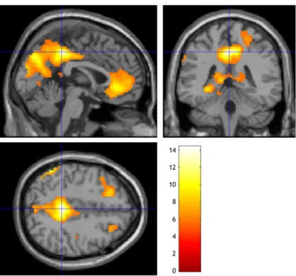
3.2.2.Peak activation and exploratory whole brain analysis in all MDD patients before and after treatment ... 43
3.3. BOLD % signal change in MDD patients after short term treatment with antidepressants or placebo ... 50
3.3.1.BOLD % signal change in the different ROIs of MDD patients: comparison
3.3.1.2. BOLD % signal change in the left and right DLPFC ... 52
3.3.1.3. BOLD % signal change in the left and right fusiform gyrus ... 53
3.3.1.4. BOLD % signal change in the left and right insula ... 54
3.3.2.BOLD % signal change in different ROIs of MDD patients: comparison of four treatment groups receiving agomelatine, escitalopram, mirtazapine or placebo ... 55
3.3.2.1. BOLD % signal change in the left and right amygdala ... 55
3.3.2.2. BOLD % signal change in the left and right DLPFC ... 61
3.3.2.3. BOLD % signal change in the left and right fusiform gyrus ... 62
3.3.3.Correlation of the BOLD % signal change with the severity of depression (HAM-D21) in all ROIs and during all contrasts ... 62
3.3.4.Correlation of the BOLD % signal change with the HPA-axis activity (cortisol levels) in all ROIs and during all contrasts ... 65
4. Discussion ... 74
4.1. Facial processing in major depressed patients ... 74
4.2. Effects of antidepressant medication versus placebo treatment ... 79
4.3. Effects of agomelatine, escitalopram, mirtazapine or placebo treatment ... 80
4.4. Correlation BOLD % signal change with severity of depression ... 83
4.5. Correlation BOLD % signal change with HPA-axis activity ... 85
4.6. Limitations and strengths of the study ... 87
5. Summary ... 89
6. References ... 91
7. Abbreviations ... 104
8. List of tables ... 107
9. List of figures ... 107
10. Acknowledgments ... 110
1. Introduction
1.1. Major Depressive Disorder (MDD)
According to the World Health Organization (WHO) depressive disorders are of outstanding health-economic importance as they are the psychiatric disorders that most frequently cause psychosocial disability (WHO 2017). Intensive biologically oriented psychiatric research over the last decades contributed to a deeper insight in miscellaneous pathophysiologic mechanisms playing a role in the etiology of Major Depressive Disorder (MDD) (Schüle, Baghai et al. 2007).
Nevertheless, the etiology of depressive disorders still is not fully understood. A multifactorial genesis is supposed and has been elucidated in increasing detail. Besides psychological and social factors biological variables apparently play a major role, which lead in their whole to a disturbed central nervous homeostasis. MDD is a chronic stress related disorder and a complex clinical syndrome characterized not only by depressed mood but also by vegetative and cognitive symptoms. Moreover, genetic, neuroendocrine and neurochemical biomarkers may predict impaired processing and regulation of emotions related to major depression as well as antidepressant treatment response in general. In patients suffering from acute depression a wide range of negative influence on the processing of emotional information is manifested. This is believed to contribute to the etiology and maintenance of the depressed state (Beck 2008).
1.2. HPA axis activity
A dysregulation of the activity of the hypothalamic-pituitary-adrenocortical (HPA) system is one of the major neuroendocrine abnormalities in major depressive disorder (Holsboer and Barden 1996). These include elevated circulating plasma levels of both corticotropin (adrenocorticotropic hormone, ACTH) and cortisol (Holsboer and Barden 1996) in addition with elevated levels of corticotropin releasing hormone (CRH) in the cerebrospinal fluid (Nemeroff, Widerlov et al. 1984). One of the first neuroendocrine function tests investigating HPA axis dysregulation in depression was the dexamethasone suppression test (DST). In contrast to the suppressibility of ACTH and cortisol secretion after administration of the synthetic glucocorticoid dexamethasone (Dex) in healthy volunteers there was an enhanced proportion of patients suffering from affective disorders with escape from adequate cortisol suppression (Carroll 1982). In addition, a change of the DST results in dependency from the
assessment of the HPA system dysregulation in depression available so far. A test sensitivity of 80 up to 90% has been shown in depressed patients (Heuser, Yassouridis et al. 1994). It has been suggested that the hyperactivity of the HPA system during depression may be considered as a neuroendocrine sign of a disturbed HPA system homeostasis which is rather used as a state than as a trait marker of diagnostic entities (Heuser, Yassouridis et al. 1994). Despite partly conflicting results, the amygdala and the hippocampus seem to have faciliatory or regulatory roles in the generation of HPA axis responses, at least this was shown in animal experiments (Goursaud, Mendoza et al. 2006). Also, a relationship between hypercortisolemia and cognitive dysfunction in acutely depressed patients has been published some years ago (Behnken, Bellingrath et al. 2013). An involvement of the amygdala in the processing of facial emotional expressions such as anger, sadness and disgust was hypothesized. Porter and Gallagher described a dysfunction of monoaminergic transmitter systems in depression including cognitive impairment which is facilitated by the HPA-system (Porter and Gallagher 2006).
Adolphs described an activation of the HPA system during the perception of fear. This results in an activation of the hypothalamus after perception of the threatening emotional information by projection of the stress-signaling impulses via the amygdala (Adolphs 2008). Thus, it is still discussed, whether the specific role of the amygdala is only the processing of fear or whether it is responsible for the accurate identification of facial emotional expressions in general (Loughead, Gur et al. 2008). There exists strong evidence for the very important role of the amygdala in regulating the response of the HPA axis to stress, but also to positive emotional faces (van Marle, Hermans et al. 2009), although the exact kind of emotion processing by the amygdala is less clear up to now (Ellenbogen, Carson et al. 2010). In MDD both types of processing are abnormal, HPA axis function (investigated using the DST) and facial emotion processing (Maes, Calabrese et al. 1994). Patients suffering from MDD demonstrate a biased and increased attention during the processing of negative facial expressions (Bourke, Douglas et al. 2010). This seems to be true even for non-affected high risk probands for depression, in first-degree relatives of patients with MDD who – in addition – show an elevated HPA axis activity (Ising, Lauer et al. 2005, Le Masurier, Cowen et al. 2007). Therefore, in depression an interdependency between the perception of emotional facial expressions and the HPA-axis regulation, which in turn appears to be influenced by major depression, has been confirmed by these earlier studies.
1.3. Antidepressant treatment 1.3.1. Antidepressants
A multifactorial genesis of MDD is supposed and has been elucidated in increasing detail during the last years. Accordingly, treatment of depression uses also multilayer approaches. In case of moderate depression national and international guidelines recommend either antidepressants or psychotherapy respectively, in case of severe or treatment resistant depression both approaches are recommended (DGPPN 2015). In case of medium to severe depression the combination of antidepressants with psychotherapy, psychoeducation and social support is associated with the highest probability of a fast response to the treatment.
Over the last decades, our understanding of the neurochemical mechanisms of antidepressant drug action has advanced considerably, but, although the knowledge about the pharmacodynamic mechanisms of action of antidepressants increased steadily, there is still a lack of information about the exact mechanisms of action in the human brain and which of these are mandatory for the antidepressant efficacy. Moreover, it remains to be elucidated, how far changes in emotional processing may account for the resolution of this multiple symptom domains during successful treatment with antidepressants. Recent fMRI neuroimaging studies suggest that a change in emotional processing could also explain partly the global improvement because attention and appraisal are of fundamental importance in brain function. The influence of antidepressant treatments on HPA axis activity and the potential to predict treatment response with repeated Dex/CRH tests are well known (Schule, Baghai et al. 2009), nevertheless, it remains unclear up to now whether the activity in central areas such as amygdala or hippocampus or other brain regions have the potential to contribute to an earlier and more reliable prediction of treatment effects. For some classes of antidepressants, e.g. for selective serotonin reuptake inhibitors (SSRIs) changes in the emotion processing circuitry and area specific changes in the brain activation were associated with treatment response (Anderson, McKie et al. 2008) or efficacy (Cipriani, Furukawa et al. 2009). However, most pharmaco- fMRI studies deduced effects in brain activation by antidepressants from the investigation in healthy subjects, not in depressed patients who showed dysfunctions in emotional and cognitive processing in the brain associated with depressive disorders (Akimova, Lanzenberger et al.
2009, Lanzenberger, Wadsak et al. 2010). A huge number of studies have considered the effect of a single dose of an SSRI on emotional processing. These data are useful in addressing the
Bhagwagar et al. 2003, Browning, Reid et al. 2007, Grillon, Levenson et al. 2007). Importantly, in addition to this anxiogenic-like effect, positive changes in emotional bias are also observed in single dose studies, in terms of attentional bias to positive words (Browning, Reid et al. 2007) and increased recognition of happy faces (Harmer, Bhagwagar et al. 2003). This suggests that a single administration of an SSRI produces a mixture of negative and positive emotional effects in cognitive models. However, after seven days of SSRI treatment there is evidence for a decreased fear response in term of diminished recognition of fearful faces and reduced emotional startle reaction (Harmer, Shelley et al. 2004). This fits well with the clinical observation that acutely administered SSRIs may first increase anxiety, while repeated treatment has sustained anxiolytic effects.
There is a growing body of evidence that antidepressants can affect emotional processing very early after starting the treatment and independently from changes in subjective mood. For example, after a one-week treatment with the SSRI citalopram or the selective noradrenaline reuptake inhibitor (NARI) reboxetine in healthy volunteers a change in emotional processing could be seen which indicated responses in the opposite direction of the changes seen generally in depressive disorders (Harmer, Shelley et al. 2004). Moreover, according to studies in healthy volunteers, antidepressant drugs have some effects on emotional processing very quickly after administration in the absence of discernible changes in mood. One study shows that short term SSRI treatment was associated with reduced blood oxygenation level-dependent (BOLD) response in the amygdala to negative facial expressions presented outside of conscious awareness (Harmer, Mackay et al. 2006). Such a finding suggests thinking about the role of the amygdala in antidepressant treatment, because the amygdala plays a key role in the processing of threat or fear-relevant cues as described before.
Taken together, findings from the single dose and seven-day pharmaco-fMRI studies with antidepressants in healthy volunteers indicate that cognitive models of antidepressant drug action reveal positive changes in emotional processing. A reversible increase in negative emotional processing can be found very early after initiation of the treatment, which is in line with clinical observations of early, but reversible increase of anxiety after starting an SSRI treatment, whereas positive effects can be found to be sustained.
As described before, it seems that CRH levels are in relationship with amygdala hyperactivity in MDD patients, because MDD is accompanied by both cognitive impairment and a hyperactivity of the HPA system, resulting in an enhanced glucocorticoid secretion. Cortisol
acts via mineralocorticoid and glucocorticoid receptors densely located in the hippocampus, a brain area that is important regarding cognitive functions and especially memory functions.
Antidepressants such as SSRIs can influence emotional processing very early on during the treatment and independently from changes in subjective mood. On the other hand, hyperactivity of the hypothalamic-pituitary-adrenal (HPA) axis in MDD is one of the most reliably reported neurobiological characteristics of affective disorders. Whether these alterations in HPA axis regulation are limited to the acute stage of MDD or whether they persist after recovery, at least in some patients, remains ambiguous. A relationship between hypercortisolemia and cognitive dysfunction in acutely depressed patients has been repeatedly observed and it was also demonstrated in several studies that a discrete cognitive impairment often persists in the remitted state of depression (Behnken, Bellingrath et al. 2013). On the other hand, MDD is accompanied by morphological changes of brain structures like the hippocampus and the amygdala which are of great importance in the neural circuitry mediating depression.
Hyperactivity of the HPA system resulting in enhanced glucocorticoid secretion can often be observed during depression and has been thought to play an important role in inducing these morphological changes (Schuhmacher, Mossner et al. 2012). The SSRI (es)citalopram, used frequently as a first line treatment of MDD, influenced emotional processing shortly after the start of treatment (Harmer, Bhagwagar et al. 2003, Browning, Reid et al. 2007) as described before. Also studies with mirtazapine, an 2-adrenoreceptor blocker with additional influence on histaminic H1-, 5HT2a-, 5HT2c-, and 5HT3-receptors, resulted in a reduction of emotional fear processing: a significantly impaired recognition of fearful facial expressions and reduced eye-blink responses in the emotion potentiated startle task, an effect similar to that of SSRIs, was reported (Arnone, Horder et al. 2009, Komulainen, Heikkila et al. 2016). Agomelatine attenuated fMRI activations in several regions involved in face processing (fusiform gyrus, bilateral inferior frontal gyrus, anterior thalamic nucleus) and showed an antidepressant-like ability to modulate early visual processing of faces in healthy controls without affecting amygdala responses (Lees J 2010). In depressed patients, agomelatine had short- and long-term effects on brain structures involved in emotional regulation such as the dorsomedial prefrontal cortex and precuneus. The development of corresponding biomarkers for a biomarker-based treatment of MDD was suggested (Delaveau, Jabourian et al. 2016). Agomelatine is the first antidepressant with a thus far unique mode of action. It acts as an agonist at melatonin MT1 and
and Kasper 2007) and anxiety disorders (for review see: (Eser, Baghai et al. 2007)). MDD patients displayed greater amygdala activation when anticipating negative pictures and greater prefrontal activation when confronted with them without the anticipatory cues. After antidepressant treatment, both amygdala and prefrontal activation decreased significantly in the treated MDD patients relative to controls. These findings show that the neural mechanisms of emotional anticipation and processing are altered in patients with MDD and that the functional neuroanatomy of emotional processing is normalized after successful treatment with an antidepressant (Rosenblau, Sterzer et al. 2012).
1.3.2. Psychotherapy
There are a number of studies investigating neural activation after treatment with antidepressants, but only a very limited number of fMRI studies investigating the effects of psychotherapy on the processing of facial emotions can be found. Fu et al. investigated the neural correlates of implicit processing of sad facial expressions in depression before and after 16 weeks of weekly Cognitive Behavioral Therapy (CBT) treatment. Elevated amygdala- hippocampal activity in patients compared to healthy individuals could be demonstrated following therapy. In addition, the dorsal anterior cingulate activity showed a significant relationship with the post-treatment clinical response in depressed patients (Fu, Williams et al.
2008). Also, Siegle et al. could show a changed amygdala activity after psychotherapy. Their patients were treated successfully with CBT and displayed low sustained reactivity to emotional stimuli in the subgenual cingulate cortex (Brodmann's area 25) and high activity in the amygdala with the strongest improvement. Furthermore, they suggested the presence of emotion regulation disruptions, which are targeted in CBT, may be the key to recovery with this intervention (Siegle, Carter et al. 2006). Dichter et al. investigated the effects of Behavioral Activation Therapy for Depression which was designed and administered to increase engagement with positive stimuli and a reduction in avoidance behaviors. A high responder rate of 75% was reported together with a decreased activation due to cognitive control in prefrontal structures including paracingulate gyrus, the right orbital frontal cortex, and the right frontal pole. Moreover, the magnitude of the activation in the paracingulate gyrus before treatment was related to the magnitude of the change of depressive symptoms after psychotherapy (Dichter, Felder et al. 2010).
Therefore, it can be stated, that there are findings suggesting that also psychotherapeutic treatment affects processing of emotional stimuli with changes in the activity of the amygdala
1.4. The processing of facial expressions
Facial expressions are an important component of communication and the correct interpretation of emotional expression is an essential key of successful social relationship. There are some studies suggesting that difficulties in interpersonal communication in depressed patients may be related to abnormalities in affective facial processing (Fu, Williams et al. 2004). There have been published several studies investigating facial emotional processing in depressed patients.
Some of them are focused on facial expressions because this may facilitate a better understanding of facial emotion processing due to impaired cognition regarding to different neural regions within the central nervous system (CNS). In addition, impaired facial expression processing may link to other affective and social symptoms in depressed patients. Finally, a change in facial emotional processing during response to antidepressant treatments may help to predict this treatment response in depressed patients (Venn, Watson et al. 2006). Therefore, presenting facial emotional expression stimuli may be a valid and reliable approach for emotion processing investigations in order to activate brain areas responsible for emotion processing in healthy volunteers and in MDD patients with impaired cognition (Fusar-Poli, Placentino et al.
2009).
Ekman and Friesen (1971) suggested that six basic emotional expressions could be recognized.
The investigated basic emotional expressions were shown in happy, sad, fearful, angry, disgusted, and surprised faces. In addition, they developed a standardized set of stimuli on the basis of these six emotions which were widely used in behavioral, cognitive and neuroimaging investigations (Ekman and Friesen 1971). Since then imaging studies in affective cognition often used Ekman stimuli for the measurement of responses to emotional faces.
1.5. Influence of Major Depression on processing of facial expressions
According to the cognitive theories of depression, symptoms coming from mood-congruent emotions cause a processing bias. Consequently, patients suffering from MDD tend to attend more to negative emotional stimuli (Scher, Ingram et al. 2005, Beck 2008). Several studies are reporting a negative bias on the perception of social key signals such as emotional facial expressions. This was demonstrated also in cognitive and behavioral investigations (Gur, Erwin et al. 1992, Bouhuys, Geerts et al. 1999). In addition, neuropsychologic studies have reported that MDD patients have a significant bias toward sad emotions (Gotlib, Krasnoperova et al.
processing, including an increase in the response of the amygdala to negative facial expressions in depressed patients compared to matched controls (Sheline, Barch et al. 2001, Surguladze, Young et al. 2004, Suslow, Konrad et al. 2010, Victor, Furey et al. 2010).
1.6. Magnetic resonance imaging and functional MRI
Magnetic resonance imaging (MRI) is a medical imaging technique using strong magnetic fields to create images of biological tissue. Functional magnetic resonance imaging (fMRI) uses standard MRI scans to investigate changes in brain function over time by determining changes in blood flow (Huettel 2014). Blood-oxygen-level dependent (BOLD) contrast is used to detect changes in deoxyhemoglobin concentration consequent to task-induced and spontaneous modulations of oxygen metabolism in response to neural activity. This method has been widely employed in cognition studies for different clinical applications such as surgical planning, for monitoring of treatment outcomes, and as a biomarker in pharmacologic and training programs (Glover 2011).
Over the past three decades neuroscientists have tried to clarify the neural mechanisms that support face perception (Posamentier and Abdi 2003, Haxby and Ida Gobbini 2007, Fusar-Poli, Placentino et al. 2009). Indeed, fMRI studies play a major role in investigations facilitating the understanding of human brain function and of neurophysiological substrates of emotional processing. Despite the growing number of fMRI studies, some individual imaging studies indicate inconclusive findings (Neumann, von Cramon et al. 2008). Predominantly, the difficulty to definitively characterize which specific brain region is associated with each specific emotional expression has been discussed. Most of the studies employ different imaging techniques, analysis methods, and task designs. Nevertheless, fMRI studies showed that emotional faces elicit enhanced response in the limbic, frontal, and visual cortical regions contrasted to responses evoked by neutral faces (Vuilleumier, Armony et al. 2001, Haxby, Hoffman et al. 2002, Winston, O'Doherty et al. 2003, Ishai, Pessoa et al. 2004).
1.6.1. fMRI and pharmacological MRI
Pharmacological MRI (phMRI) is a fMRI investigation method to study the effects of medication on brain function in response to a chemical substance administration. Bryant and Jackson have introduced the application of pharmaco-fMRI for the first time (Bryant and Jackson 1998). There are two main approaches in the pharmaco-fMRI field, the first measuring acute fMRI signal change following drug administration, while the second estimates
While fMRI is widely used in research and clinical investigation, where it is commonly combined with a sensorimotor task, phMRI is an adaptation of fMRI enabling the investigation of a specific neurotransmitter system, such as the serotoninergic neurotransmission or the melatonin system. These systems are investigated under physiological or pathological conditions following an activation via the administration of a specific medication such as selective serotonin (5HT) reuptake inhibitors (SSRIs) or melatonin agonists (Klomp, Tremoleda et al. 2012).
1.6.2. Brain regions involved in the processing of facial expression
Investigations trying to identify brain regions responsible for emotional face processing can be subdivided in two major research directions which integrate neuro-anatomical models involved in the representation and generation of emotions. These are also thought to influence the etiology of MDD (Mayberg 1997, Phillips, Drevets et al. 2003, Phillips, Ladouceur et al. 2008).
First, encoding of emotional expressions depends on multiple interactions between complimentary systems: a neural core system for the visual analysis of faces refers to bilateral inferior occipital gyri, the lateral fusiform gyrus and the superior temporal sulcus. The second aspect focused on the processing of facial information, such as meaning and significance referring to different brain regions such as amygdala, insula, orbitofrontal areas, and somatosensory cortex (Haxby, Hoffman et al. 2002). Notably, emotional facial expression investigations have been frequently used in neuroimaging studies in depressed patients with regards to neurobiological models of depression (Phillips, Drevets et al. 2003, Mayberg 2007, Phillips, Ladouceur et al. 2008).
1.6.2.1. Amygdala
Functional neuroimaging studies have shown a relevant specific amygdala activation during the presentation of fearful facial expressions of emotion (Morris, Frith et al. 1996, Morris, Buchel et al. 2001) and during verbally guided anticipation of shock (Phelps, O'Connor et al. 2001).
This amygdala activation was also reported after brief presentations of fearful facial expressions of emotions which were masked to prevent conscious perception (Whalen, Rauch et al. 1998) or when presented in the cortically blind field (Morris, DeGelder et al. 2001, Pegna, Khateb et al. 2005). There is a large number of functional MRI studies which have shown an increased
Furthermore, fMRI investigations of affective facial processing of sad and angry faces in medicated depressed patients in comparison to healthy controls reported an increased BOLD response in the left middle cingulum and the right prefrontal cortex (Frodl, Scheuerecker et al.
2009). The same study found no differences in amygdala activation between MDD patients and healthy controls at baseline before antidepressant treatment (Frodl, Scheuerecker et al. 2011).
In contrast to these reports, there are a huge number of imaging investigations of facial expressions in MDD patients in comparison to healthy controls showing greater amygdala BOLD response to emotional facial expressions in MDD (Sheline, Barch et al. 2001, Fu, Williams et al. 2004, Lawrence, Williams et al. 2004, Surguladze, Brammer et al. 2005, Fu, Williams et al. 2008, Matthews, Strigo et al. 2008, Peluso, Glahn et al. 2009, Suslow, Konrad et al. 2010, Victor, Furey et al. 2010, Zhong, Wang et al. 2011).
1.6.2.2. Hippocampus
Neuropathology of MDD suggests that an impaired hippocampus may be a key hub within the limbic system (Campbell and Macqueen 2004). According to a recent review, hippocampal volume reduction is one of the most replicated findings in neuroimaging studies in MDD (Roddy, Farrell et al. 2018). For example, reductions in the gray matter volume and functional impairment in MDD patients have been reported (Bertolino, Frye et al. 2003, Sheline, Gado et al. 2003). Furthermore, some meta-analyses examining MRI studies of the hippocampus, solely (Videbech and Ravnkilde 2004, McKinnon, Yucel et al. 2009, Cole, Costafreda et al. 2011) or as part of a greater limbic system analysis (Koolschijn, van Haren et al. 2009, Kempton, Salvador et al. 2011, Arnone, McIntosh et al. 2012), have found volume reductions of 4% to 10% in depressed patients. Although some imaging studies in depression observed activations in parts of the amygdala extended to (para)hippocampal regions in response to emotional facial expression, patients suffering from MDD showed predominantly an elevated amygdala- hippocampal BOLD response to sad conditions when compared to healthy individuals (Fu, Williams et al. 2008). There are not many studies in depressed patients demonstrating a decreased BOLD response to sad facial expression directly in the hippocampus (Lee, Seok et al. 2008). In contrast, a study by Videbech et al. reported that MDD patients showed increased activity of the hippocampus and the cerebellum relative to the healthy controls (Videbech and Ravnkilde 2004).
1.6.2.3. Insula
Fusar-Poli et al. performed a metanalysis of 105 fMRI studies on healthy subjects and reported the processing of facial expressions (emotional and neutral) is associated with an elevated activation in temporoparietal areas, such as the parietal lobe, the middle temporal gyrus and the insula. That was specifically true for the contrast of neutral facial expressions versus baseline in the left insula, the processing of happy facial expressions versus baseline in the left insula, of sad facial expressions versus baseline in the left insula, and of disgusted facial expressions compared with baseline in the right insula (Fusar-Poli, Placentino et al. 2009).
According to a review by Stuhrmann (Stuhrmann, Suslow et al. 2011), an imaging study by Surguladze et al. investigated the BOLD response to faces displaying different degrees of disgust in MDD patients in contrast to a healthy control group. They identified greater left insula activation in the depressed patients in comparison to the group of healthy controls (Surguladze, El-Hage et al. 2010). Irrespective of an altered processing of disgust in major depressed patients, also an altered activation in the insula to other emotional facial expressions has been published (Suslow, Konrad et al. 2010). Zhong et al. demonstrated a greater activation in the insula and the parahippocampal gyrus (PHG) to sad facial expression and a decreased activation to happy facial expression (Zhong, Wang et al. 2011). Fu et al. have shown an increased insula activation to fearful/angry (combined contrast) facial expressions in a sample of young MDD patients (Fu, Williams et al. 2004). Notably, they could demonstrate a thalamic hyper- responsiveness to sad facial expressions.
Currently in most of the reviewed imaging studies on processing of emotional facial expression there is a clear trend for similar activation patterns between the insula, the parahippocampal gyrus area and the amygdala, demonstrating an emotional bias of the activity in limbic structures in MDD patients in contrast to healthy individuals. This includes a greater BOLD response to negative facial expressions and an increased BOLD response to happy facial expressions. One study reported also a different pattern of decreased activity in the insula in a combined contrast of sad and fear facial expressions in MDD patients (Townsend, Eberhart et al. 2010).
Some studies identified aberrant activity in striatal structures which also had similar activation
and increased BOLD response to happy facial expressions (Fu, Williams et al. 2004, Lawrence, Williams et al. 2004, Fu, Williams et al. 2008, Scheuerecker, Meisenzahl et al. 2010).
1.6.2.4. Motor cortex and prefrontal cortex
The role of the prefrontal cortex (PFC) in facial emotional processing in depression so far is less clear (Stuhrmann, Suslow et al. 2011). Using different imaging methods such as positron emission tomography (PET), neuroscientists have focused on the PFC and specially on the dorsolateral prefrontal cortex (DLPFC) function in MMD patients. There are studies reporting a reduced cerebral blood flow and metabolism in the left DLPFC and hypermetabolism in the right DLPFC in acute MDD (Mayberg 2003, Phillips, Drevets et al. 2003, Grimm, Beck et al.
2008).
Mayberg’s limbic-cortical dysregulation model based on evidence from a series of PET studies is consistent with the findings of decreased activation in dorsal neocortical regions such as the DLPFC and dorsal anterior cingulate cortex (ACC), and of increased activation in paralimbic regions such as the insula, amygdala and hippocampus (Mayberg 1997, Mayberg 2003). In the lateral PFC no consistent activity pattern in different imaging studies on facial processing in MDD in contrast to healthy subjects can be found. Both findings, increased activation and decreased activation in the dorsolateral DLPFC in MDD patients to sad and angry facial stimuli, can be found nearly equally often (Lawrence, Williams et al. 2004, Keedwell, Andrew et al.
2005, Frodl, Scheuerecker et al. 2009, Suslow, Konrad et al. 2010, Zhong, Wang et al. 2011).
Although Davidson et al. reported a hypoactivation of the lateral prefrontal cortex (LPFC) and the ACC (Davidson, Irwin et al. 2003), Anand et al. identified the paradoxical imaging study results which shows greater activation of prefrontal regions, such as the DLPFC (Anand, Li et al. 2005), the medial prefrontal cortex (MPFC) (Anand, Li et al. 2005, Johnstone, van Reekum et al. 2007) and the dorsal ACC (Beauregard, Paquette et al. 2006) during the presentation of affective stimuli in depressed patients. These reports and their changes after antidepressant treatment are summarized and reviewed by Rosenblau et al. (Rosenblau, Sterzer et al. 2012).
Alterations of neural responses in the MPFC have been reported after successful antidepressant combination treatment with the selective noradrenalin and serotonin reuptake inhibitor (SNRI) venlafaxine in combination with light therapy (Benedetti, Radaelli et al. 2009).
Some inconsistencies were reported regarding neural responsiveness to happy facial stimuli in the DLPFC and in more ventral, lateral PFC areas. So far, is not possible to draw valid conclusions about a general hyper- or hypoactivation of the DLPFC during facial emotion
processing in unipolar depression although altered neuronal responses in the DLPFC are a prevalent finding in MDD patients due to the high variability in all published neuroimaging studies. In the orbitofrontal cortex (OFC) several independent studies detected a decreased activation in inferior and medial OFC areas in response to either sad, fearful or angry facial stimuli (Lawrence, Williams et al. 2004, Keedwell, Andrew et al. 2005, Lee, Seok et al. 2008).
Furthermore, Surguladze et al. reported hyperactivation to disgust in the OFC in patients suffering from MDD (Surguladze, El-Hage et al. 2010).
In addition, some facial-processing imaging studies report similar results about activation in the prefrontal cortex and the motor cortex. A greater BOLD response to angry facial expressions in the motor cortex (Brodmann’s area (BA) 6, BA 4) of MDD patients in contrast to healthy controls was shown (Fu, Williams et al. 2004, Keedwell, Andrew et al. 2005, Fu, Williams et al. 2008, Scheuerecker, Meisenzahl et al. 2010).
1.6.2.5. Fusiform gyrus
In the above mentioned meta-analysis of studies in healthy subjects, the processing of facial expressions (emotional and neutral) was associated with an elevated activation in visual areas such as the fusiform gyrus. That was reported specifically for the contrast of neutral facial expression, fearful facial expressions, and for the processing of disgusted facial expressions compared to baseline (Fusar-Poli, Placentino et al. 2009).
A study by Ho et al. on adolescent MDD patients in contrast to healthy controls identified that the fMRI BOLD signal in the left fusiform gyrus during emotional facial processing was significantly associated with greater individual-level estimates of perceptual processing efficiency. Furthermore, they suggested a facial processing bias in younger MDD patients characterized by greater perceptual processing efficiency of emotional visual information in sensory brain regions responsible for the early processing of visual information (Ho, Zhang et al. 2016). In addition, a study on MDD patient shows increase BOLD response in the fusiform gyrus after the presentation of sad facial stimuli (Fu, Williams et al. 2008).
1.6.2.6. Summary of brain regions involved in the processing of facial expression in MDD
have shown that depressed patients have abnormalities within the common face processing network, including a mood-congruent processing bias influencing the responsiveness especially in the regions amygdala, insula, parahippocampal gyrus (PHG), fusiform face area, and putamen. Very often, neuroimaging studies in MDD patients have shown that amygdala hyperactivity is associated with negatively biased facial emotion processing. Notably, the amygdala, the ACC, OFC, and the DLPFC are core components of a network for emotion regulation which is pathologically altered in depressive disorders (Stuhrmann, Suslow et al.
2011). Furthermore, neuroimaging studies in depression need to extend these findings, especially by replicating data with the same activation paradigms and larger sample sizes in order to enable researchers to make more valid assumptions on neural emotional processing mechanisms before and after administration of antidepressant medications. This may contribute to a better understanding of the etiology and mechanisms during effective treatments of depressive disorders.
1.7. Research questions
In the following study the evidence for a cognitive neuropsychological model of antidepressant drug action has been examined. Using pharmaco-fMRI in a double-blind randomized placebo- controlled design we investigated the effects of short-term antidepressant treatment in patient groups receiving differential treatments. We then compared the BOLD % signal change in the defined brain regions of drug treated patients to patients who received placebo treatment. The effect of antidepressants (escitalopram, mirtazapine, agomelatine) or placebo on amygdala, dorsolateral prefrontal cortex, fusiform gyrus, hippocampus and insula (all bilateral) was investigated. BOLD responses to facial expressions in patients with MDD were investigated.
In addition, the HPA axis activity of a subgroup of patients was studied before treatment and after one week of acute treatment to elucidate the relationship between amygdala response during facial processing and HPA-axis hyperactivity in relation to the clinical outcome before and after short-term treatment.
The following research questions should be answered with our study:
1) Is the fMRI BOLD % signal change during facial processing of MDD patients in several regions of interest (ROIs: amygdala, DLPFC, hippocampus, insula, and fusiform gyrus) influenced by a short-term antidepressant treatment?
2) Are there detectable differences of the BOLD response to facial processing between the drug-treated (escitalopram, mirtazapine, or agomelatine) patients and and the placebo- treated patients after one week of short-term treatment?
3) Are there detectable differences of the BOLD response to facial processing between the four different treatment groups (antidepressants escitalopram, mirtazapine, agomelatine or placebo) after one week of active or placebo treatment?
4) Is there a detectable correlative relationship between clinical outcome (measured using the Hamilton rating scale for depression) and the BOLD % signal change in different ROIs during facial processing before and after one week of treatment with the antidepressants escitalopram, mirtazapine or agomelatine, versus placebo?
5) Is there a correlative relationship between the HPA-axis (hyper)activity in MDD and the BOLD % signal change during facial processing? Is this putative correlation altered after one week of antidepressant-drug treatment?
2. Methods
2.1. Study design
This study is part of the project “Relevance of the gut-microbiome composition für subtypes of depression, response and side effects during antidepressant treatment“ which is a part of the German reseach network “Novel strategies for the optimized treatment of depression (OptiMD)” funded by the Federal Ministry of Education and Research (Bundesministerium für Bildung und Forschung, BMBF, www.bmbf.de, support code 01EE1401B).
The single center study was carried out as a randomized, double-blind and placebo-controlled trial in a parallel group design. Ethical approval was granted by the local Ethics committee of the University Regensburg. The clinical trial was registered and approved by the Federal Institute for Drugs and Medical Devices (Bundesinstitut für Arzneimittel und Medizinprodukte, BfArM, www.bfarm.de).
2.1.1. Study sample and patient selection
All patients were included in the study after adequate explanation of the study procedures and after they provided written informed consent. They were investigated at the Department of Psychiatry and Psychotherapy of the University Regensburg, located in the Bezirksklinikum Regensburg, Universitätsstraße 84 in 93053 Regensburg, Germany. We included 33 major depressed in-patients admitted for the treatment of a major depressive disorder independently of study participation. Patients were not treated with antidepressants or other psychotropic substances except lorazepam for agitation or zopiclone for insomnia during the first pre- treatment fMRI and during the first combined Dex/CRH-test. Table 2 shows clinical and demographic data for the patients. In all participants we assessed the presence of current and past psychiatric disorders using the Mini-International Neuropsychiatric Interview (M.I.N.I.) (Sheehan, Lecrubier et al. 1998) for the 10th version of the International Classification of Diseases (WHO 2016). The depressed patients met criteria for a primary diagnosis of major depressive disorder (MDD) and were not suffering from other psychiatric diagnoses. In addition they were physically healthy and not suffering from serious somatic diseases. Medical history, psychiatric history, vital signs, a laboratory screening and an electrocardiogram (ECG) were assessed. The principles of informed consent were implemented according to the current revision of the Declaration of Helsinki, the International Council for Harmonisation of Technical Requirements for Pharmaceuticals for Human Use (ICH) Guideline for Good
adequate explanation of the study procedure and after written informed consent. Withdrawal of consent was possible at any time during the study.
Our detailed study inclusion criteria were:
Male and female in-patients in the age between 18 and 65 years;
Major depressive disorder;
Admission on a voluntary basis independent of our study;
Written informed consent for trial participation after the scope and nature of the investigation have been explained to the patients before starting trial-related activities;
Indication for antidepressant therapy independent of the clinical trial;
Primary unipolar depression (ICD-10: F32, F33) or bipolar depression (ICD-10: F31.3- 5), current episode of depressed state for at least 2 weeks prior to baseline;
Physically healthy;
Right handedness (assessed by the Edinburgh Handedness Inventory (Oldfield, 1971)).
The detailed exclusion criteria were:
Schizophrenia, substance dependence as define by ICD-10 or any other psychiatric primary diagnosis (according to the ICD-10 criteria);
major somatic or neurological disorder;
abnormalities in the laboratory screening at baseline (e.g. hypo- or hyperthyroid state, hyperhidrosis, elevated liver enzymes, blood cell dyscrasias);
lacking ability to give informed consent;
have been admitted to the clinic involuntarily during their present episode;
pregnancy or breast-feeding;
in case of the inclusion of premenopausal female patients insufficient contraception leads to exclusion from the study;
contraindications to magnetic resonance imaging patient with heart pacemaker or implanted metal in the scull;
or concurrent medication, which could alter emotional processing;
known history of alcohol or drug abuse during 6 month prior to the screening;
known allergies or hypersensitivity reactions or other contraindications for escitalopram, agomelatine or mirtazapine;
being treated with psychotropic medication < 3 weeks before study (5 weeks in case of fluoxetine pretreatment);
Unusual diets leading to malnutrition;
Pretreatment with antibiotics or corticosteroids.
Discontinuation criteria were:
Withdrawal of consent at any time during the study period;
If necessary concerning clinical reasons, investigators and sub-investigators could stop the study participation of an individual patient;
Noncompliance to the study protocol.
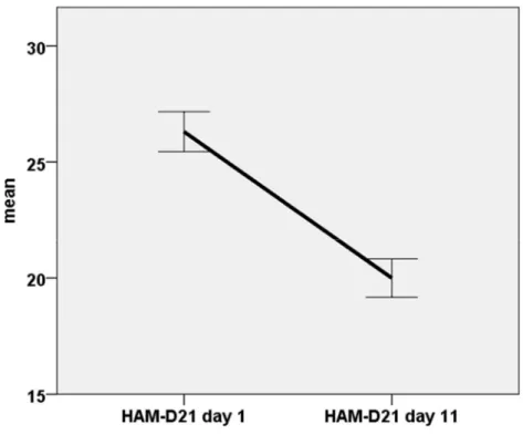
Our goal was to provide a better understanding of the interactions between neural systems during antidepressant treatment, the effect of antidepressants on emotional processing, the relationship between amygdala hyperactivity as well as other brain regions BOLD response to emotional facial expression, and the HPA axis activity in relation to clinical effectiveness of the treatment measured using the 21-item version of the Hamilton Rating Scale for depression (HAM-D21) (Hamilton 1967).
In this study firstly, we considered the evidence for a cognitive neuropsychological model of antidepressant drug action. We used pharmaco-fMRI (3T) to investigate the effect antidepressant treatment on the amygdala (right (R)+left (L)), caudate (R+L), dorsal lateral pre- frontal cortex (DLPFC) (R+L), fusiform gyrus (R+L), hippocampus (R+L) and insula (R+L) BOLD response to facial expression at baseline and after one week of antidepressant treatment (antidepressants or placebo in addition to psychotherapy) in major depressed patients.
Secondly, we investigated the activity of the hypothalamus-pituitary-adrenal (HPA)-axis using the combined dexamethasone suppression / corticotropin releasing hormone (Dex/CRH) stimulation test before and after one week of treatment to elucidate the relationship between BOLD response signal change and HPA-axis activity.
Thirdly, we clinically assessed the patients before, during and after treatment periods using the HAM-D21 (Hamilton 1967) rating scale as the primary outcome criterion.
2.1.2. Study design and drug treatment
Double-blind, placebo-controlled, parallel group study in 33 depressed patients who were randomly assigned to 4 groups. The patients in each group received either 10 mg Escitalopram, 30 mg Mirtazapine, 25 mg Agomelatine or placebo for at least 7 days. The patients receive the medication at 8:00 am in case of escitalopram or at 10:00 pm in case of agomelatine and mirtazapine treatment according to usual clinical procedures. For blinding a double-dummy technique was used. On study day 4 and 11, i.e. the days of pharmaco-fMRI scans, the patients received the medication in the morning at 8:00 am (about 4 hours before the fMRI scan). All four groups then received the fMRI scan before treatment and after the seventh day of the treatment. In addition, on the 2nd and 9th day of the study we investigated the levels of cortisol secretion by using combined Dexamethasone suppression / corticotropine-stimulation (combined Dex/CRH-) tests. Mood and anxiety were assessed primarily using the HAMD-21 scale by trained psychiatrists immediately before starting treatment, on the 7th day of escitalopram, mirtazapine, agomelatine or placebo treatment and then in weekly intervals until discharge of the hospital. After the second fMRI scan in case of partial response (defined as at least 10% reduction in HAMD-21 scale) treatment was continued without change, in case of nonresponse a dose increase or change of the treatment was offered to the patients. The same was true if no reduction of at least 20% was seen after 2 weeks of treatment according usual clinical procedures. On the day of inclusion a laboratory screening and on the days of the fMRI scan blood withdrawals for the estimation of drug plasma levels of the used antidepressants were performed. The measurement of plasma levels was performed after unblinding the medication. For the study flow chart see Table 1.
In case of clinical necessity a concomitand treatment with hypnotics, including lorazepam (up to 3 mg/d), zopiclone (up to 15 mg/d) or zolpidem (up to 20 mg/d) was allowed. On the days before and the days of the pharmaco fMRI scan and the Dex/CRH-test concomitant medication was avoided. After study participation all patients received further treatment according to clinical indications. Each patient advised independently of the study only according to clinical reasons. In case patients responded well to the treatment, it was considered to continue pharmacotherapy. In case of nonresponse other pharmacological and non-pharmacological interventions including augmentation strategies were offered.
Table 1: Study flow chart and investigation plan for the blinded short-term treatment period (MED = medication)
Day
1 Day
2 Day
4 Day
5-8 Day
9 Day
10 Day
11 Day
11+
Study in- clusion HAM- D21
com- bined Dex/CRH
-test 1 fMRI 1 blinded medi- cation or placebo (MED)
MED &
combined Dex/CRH
-test 2 M
ED MED & fMRI 2 HAM-D21 treatment
according to clinical require- ments 2.1.3. Functional magnetic resonance imaging data acquisition
fMRI data collection was done using three different 3 Tesla (3T) scanners due to a replacement of the research scanner of the University Regensburg. From the total of 33 patients, 2 were investigated using a Siemens MAGNETOM Allegra head scanner for 3T brain imaging (Siemens AG, Erlangen, Germany), fitted with a birdcage headcoil, which was located at the department of neuroradiology of the University Regensburg, Center for Clinical Magnetic Resonance Research. Eight of the patients were investigated using a Siemens MAGNETOM Skyra full-body scanner for 3T imaging, located at the Institute of Radiodiagnostics at the University Hospital Regensburg, and 23 of the patients were scanned using a Siemens MAGNETOM Prisma head scanner for 3T brain imaging with the 20-channel headcoil, located at the Bezirksklinikum Regensburg (clinical center of the district Upper Palatinate), Center for Clinical Magnetic Resonance Research. The study participants completed a neuroimaging battery including high-resolution structural, resting state, and functional task scans. Only the data from the facial emotion processing task described in the following was used for the current analysis.
2.1.4. Functional MRI experimental task
Before each fMRI task investigation all patients were screened and asked to disclose any ferromagnetic implants and devices such as cardiac pace makers, before they entered the MRT scanner completely metal free. During the fMRI scanning, participants were asked to process visual stimuli and complete a simple gender discrimination task, involving the rapid presentation of emotional and neutral facial expressions or scrambled pictures. In this task, they viewed male and female emotional faces. As a starting basis the Averaged Karolinska Directed Emotional Faces (AKDEF) were used. This is a set of totally 70 pictures of averaged human facial expressions. The material was developed in 1998 by Daniel Lundqvist and Jan-Eric Litton
at Karolinska Institutet, Department of Clinical Neuroscience, Section of Psychology, Stockholm, Sweden (Lundqvist D 1998). All faces were modified from the original pictures of the KDEF set to provide a similar proportion and distribution of light and dark areas to prevent confounding effects independent from facial expressions. Therefore, we masked all pictures to cover parts of the hair and to present identical black and white pictures to keep the attention only on facial expression. The patients were asked to report the gender of the face via an MRI compatible keypad. Stimuli were presented on a computer using A simple framework (ASF) for behavioral and neuroimaging experiments (Schwarzbach 2011) based on the psychophysics toolbox (Brainard 1997) for the MATLAB software package (MATLAB_R2016b,
Adalperostraße 45, 85737 Ismaning, Germany,
https://de.mathworks.com/products/matlab.html) and on a cloned projection display to patients on an opaque screen located at the head of the scanner bore, which subjects view using angled mirrors. Subject responses were registered via an MRI-compatible keypad. Immediately before scanning, all subjects received training with another set of stimuli to ensure that they fully understood the requirements of the task.
There were 18 blocks of the emotional task that contained 10 images (for a given condition), and each image was presented for 1.6 seconds (s). The task had seventeen 16s blocks of a baseline fixation point, an interleave with eighteen 16s blocks of the facial or scrambled task blocks of afraid, angry, happy, neutral, and sad faces, and of scrambled pictures (figure 1).
There was no masking between the images. They were merely presented back-to-back within a block. Each condition was repeated 3 times per run (i.e., 6 conditions [afraid, angry, happy, neutral, sad, scrambled]*3 repetitions = 18). Each run was repeated 3 times in a given session (i.e., pre or post).
Figure 1: Presented emotional faces during the scan time. The task was to press a button corresponding to the recognized gender in facial pictures or the localization of dark areas in scrambled pictures.
During each emotional block, participants viewed 10 emotional faces (male and female). Each face was presented for 1.6 s and subjects were asked to report the gender of the face via a MRI compatible keypad to ensure patients remaining on focus during the task. They were instructed to press one key with the index finger when they recognized a female face and another key with the middle finger when they recognized a male face. In case the scrambled pictures were presented, they should press one key with their index finger when a picture with more darkness at the bottom was shown and another key with their middle finger in case darker areas are located in the upper parts of the pictures. The patients were asked to lie calmly without moving their head. The total duration of the fMRI procedure was about 60 minutes per session. Each session consisted of 30 min facial task, the results of which are presented here. This was combined with 20 min resting state and 12 min diffusion tensor imaging (DTI) sequences which have been analyzed in another subproject.
2.1.5. Dex/CRH Test
To investigate the activity of the HPA axis, two Dex/CRH-tests in each patient were performed.
The first test was performed before treatment on days 1 and 2, the second after the short-term treatment of one week on day 8 and 9.
The patients received 1.5mg Dexamethasone (Dex) (Fortecortin®, Merck KG, Darmstadt, Germany) orally at 11:00 pm on the day 1 (first day of the test) before the CRH challenge (second day of the test). On the day of the CRH stimulation test at 2:30 pm an intravenous forearm catheter was inserted and the first blood samples were collected. The patients had to