Rat Histamine H 4 Receptor Orthologs: Correlations and Discrepancies between Distal and Proximal Readouts
Uwe Nordemann1, David Wifling1, David Schnell1, Gu¨nther Bernhardt1, Holger Stark2, Roland Seifert3, Armin Buschauer1*
1Institute of Pharmacy, University of Regensburg, Regensburg, Germany,2Institute of Pharmaceutical and Medicinal Chemistry, Heinrich-Heine-Universita¨t Du¨sseldorf, Du¨sseldorf, Germany,3Institute of Pharmacology, Medical School of Hannover, Hannover, Germany
Abstract
The investigation of the (patho)physiological role of the histamine H4receptor (H4R) and its validation as a possible drug target in translational animal models are compromised by distinct species-dependent discrepancies regarding potencies and receptor subtype selectivities of the pharmacological tools. Such differences were extremely pronounced in case of proximal readouts, e. g. [32P]GTPase or [35S]GTPcS binding assays. To improve the predictability of in vitro investigations, the aim of this study was to establish a reporter gene assay for human, murine and rat H4Rs, using bioluminescence as a more distal readout. For this purpose a cAMP responsive element (CRE) controlled luciferase reporter gene assay was established in HEK293T cells, stably expressing the human (h), the mouse (m) or the rat (r) H4R. The potencies and efficacies of 23 selected ligands (agonists, inverse agonists and antagonists) were determined and compared with the results obtained from proximal readouts. The potencies of the examined ligands at the human H4R were consistent with reported data from [32P]GTPase or [35S]GTPcS binding assays, despite a tendency toward increased intrinsic efficacies of partial agonists. The differences in potencies of individual agonists at the three H4R orthologs were generally less pronounced compared to more proximal readouts. In conclusion, the established reporter gene assay is highly sensitive and reliable.
Regarding discrepancies compared to data from functional assays such as [32P]GTPase and [35S]GTPcS binding, the readout may reflect multifactorial causes downstream from G-protein activation, e.g. activation/amplification of or cross-talk between different signaling pathways.
Citation:Nordemann U, Wifling D, Schnell D, Bernhardt G, Stark H, et al. (2013) Luciferase Reporter Gene Assay on Human, Murine and Rat Histamine H4Receptor Orthologs: Correlations and Discrepancies between Distal and Proximal Readouts. PLoS ONE 8(9): e73961. doi:10.1371/journal.pone.0073961
Editor:Vladimir N. Uversky, University of South Florida College of Medicine, United States of America ReceivedJune 18, 2013;AcceptedJuly 24, 2013;PublishedSeptember 2, 2013
Copyright:ß2013 Nordemann et al. This is an open-access article distributed under the terms of the Creative Commons Attribution License, which permits unrestricted use, distribution, and reproduction in any medium, provided the original author and source are credited.
Funding:This work was supported by the Graduate Training Program (Graduiertenkolleg) GRK760, ‘‘Medicinal Chemistry: Molecular Recognition – Ligand–
Receptor Interactions’’ of the German Research Foundation (DFG), by the European Cooperation in Science and Technology, COST Action BM0806, ‘Recent advances in histamine receptor H4R research’ and by the DFG within the DFG funding programme Open Access Publishing. The funders had no role in study design, data collection and analysis, decision to publish, or preparation of the manuscript.
Competing Interests:Roland Seifert serves as an Academic Editor for PLOS ONE. This does not alter the authors’ adherence to all the PLOS ONE policies on sharing data and materials.
* E-mail: armin.buschauer@chemie.uni-regensburg.de
Introduction
The histamine H4receptor (H4R) [1–5] is preferably expressed on cells of hematopoietic origin such as eosinophils and mast cells and supposed to be involved in inflammatory diseases, e.g. asthma, and pruritis [6–10]. To investigate the (patho)physiological role of the H4R translational, animal models for allergic asthma and allergic contact dermatitis in mice [11–15] or rat models for acute inflammation and conjunctivitis [16,17] were used. Most of the studies confirmed the pro-inflammatory role of the H4R by blocking the H4R-mediated response with JNJ 7777120 (1-[(5- chloro-1H-indol-2-yl)carbonyl]-4-methylpiperazine), which is re- ported to be equipotent as an antagonist at the human, mouse and rat H4R orthologs [18].
However, there are also controversial reports. The administra- tion of the H4R agonist 5(4)-methylhistamine was benefical in a murine asthma model [12], and JNJ 7777120 increased the ocular histamine concentration in a rat conjunctivitis model [17] (for a recent review cf. Neumann et al. [19]). Furthermore, the overall
amino acid identities of H4R species orthologs are remarkably low (human versus mouse and rat: ,70%) compared to other histamine receptor subtypes (H1R, H2R and H3R) [20]. Although relatively small differences in the sequence of histamine receptor species orthologs can result in different potencies and efficacies of individual ligands, the discrepancies are exceptionally high in case of the H4R [21]. In various in vitro assay systems the recombinantly expressed mouse and rat H4R revealed substantial species-dependent differences compared to the human receptor concerning affinity, potency and quality of action of pharmaco- logical tools, compromising the predictive value with respect to translational animal models [20–23]. For example, in comparison to the human H4R, UR-PI294 (N1-[3-(1H-imidazol-4-yl)propyl]- N2-propionylguanidine) and UR-PI376 (2-cyano-1-[4-(1H-imida- zol-4-yl)butyl]-3-[(2-phenylthio)ethyl]guanidine) [24,25] displayed considerably lower potencies and efficacies (UR-PI376) in the [32P]GTPase and [35S]GTPcS binding assays on membrane preparations of Sf9 insect cells expressing the mouse or rat H4R [23]. Most strikingly, JNJ 7777120 exhibited stimulatory effects at
the mouse and rat H4R in functional assays on Sf9 cell membranes [23]. Moreover, the use of JNJ 7777120 as standard antagonist in animal models was questioned due to stimulation of G-protein independent b-arrestin recruitment [26]. Biased signaling of the hH4R has also been shown for other H4R ligands [27].
The aforementioned controversial findings underline the necessity to evaluate pharmacological tools at the H4R species orthologs of interest using different assay systems. For this purpose, a cAMP response element (CRE) controlled luciferase reporter gene assay in HEK293T cells, stably expressing the human, the mouse or the rat H4R, was established. The H4R is Gai/o-coupled and reduces forskolin stimulated cyclic adenosine monophosphate (cAMP) formation after agonist binding [2]. The optimal concentration of forskolin used for pre-stimulation depends on the cell type [28] and should correspond to the EC50of forskolin in the assay system [29]. Therefore, the potency of forskolin was determined, and the effect of the phosphodiesterase (PDE) inhibitor isobutylmethylxanthine (IBMX) was evaluated to opti- mize the sensitivity of the procedure. Due to the delayed onset of gene expression, incubation periods of four to six hours are required [30], increasing the risk of agonist mediated receptor desensitization, which can lead to a decrease in agonist potencies
[30]. Therefore, the time course of the luciferase expression was determined to find the minimum incubation period required for appropriate signal strength. For validation, potencies and efficacies of 23 selected H4R ligands, comprising agonists, inverse agonists and antagonists, were determined (Figure 1).
Materials and Methods Ethics Statement
Human embryonal kidney (HEK293T) cells were purchased from the German Collection of Microorganism and Cell Cultures (DSMZ, Braunschweig, Germany).
Histamine Receptor Ligands
Histamine (HA,1) was purchased from Alfa Aesar (Karlsruhe, Germany). (R)-a-methylhistamine (2), (S)-a-methylhistamine (3), Na-methylhistamine (4), 5(4)-methylhistamine (5), immepip (6), immethridine (7), imetit (8), clobenpropit (9), iodophenpropit (10), proxyfan (PRO, 11), ciproxifan (CIP, 12), clozapine (17), VUF 5681 (18), A 943931 (22) and A 987306 (23) were from Tocris Bioscience (Ellisville, MO, USA), UR-PI294 (13), UR-PI376 (14), VUF 8430 (15), ST-1006 (16), JNJ 7777120 (19), thioperamide Figure 1. Chemical structures of the examined H4R ligands.Agonists (1–17), antagonists/inverse agonists (18–23) at the human H4R.
doi:10.1371/journal.pone.0073961.g001
(THIO, 20), and ST-1012 (21) were synthesized in our laboratories. Chemical structures of the ligands are depicted in Figure 1. Except for 14, 16, 17, 21and 23all stock solutions
(10 mM) were prepared in Millipore water. Stock solution of17 and23were made in 20 mM HCl, whereas14,16and21were dissolved in 50% (v/v) dimethyl sulfoxide (DMSO). Stock solutions of17 and23and those ligands dissolved in water were diluted with Dulbecco’s modified Eagle’s medium (DMEM) supplemented with 10% (v/v) fetal calf serum (FCS). The stock solutions of14, 16and21were diluted with DMEM supplemented with 10% (v/
v) FCS and 10% (v/v) DMSO.
Subcloning of FLAG Epitope- and Hexahistidine-tagged mH4R cDNA into the Shuttle Vector pcDNA3.1(+)
The FLAG epitope (F)- and the hexahistidine (His6)-tagged mH4R cDNA cloned in pGEM-3Z [23] was subcloned atHindIII and XbaI restriction sites into pcDNA3.1(+), encoding G418 resistance. Double digestion withHindIII (Fermentas GmbH, St.
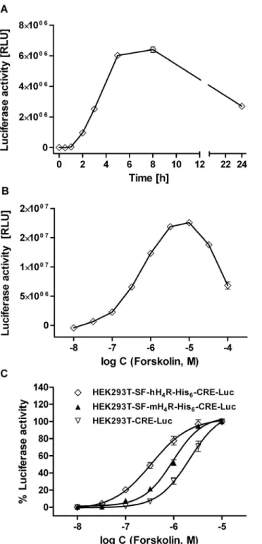
Leon-Rot, Germany) and XbaI (Fermentas) restriction enzymes was performed in reaction buffer Tango (Fermentas) with a two- fold excess ofHindIII at 37uC for 3 h. The DNA bands of the SF- mH4R-His6 (1336 bp) (S stands for the cleavable signal peptide from influenza hemagglutinin, F for flag) insert as well as the linearized pcDNA3.1(+) vector (5352 bp) were extracted from the Figure 2. Stimulation of luciferase activity by forskolin. (A)
Representative time course of the luciferase expression in HEK293T- CRE-Luc cells, stably expressing the CRE-controlled luciferase, upon stimulation with 10mM of forskolin. The luciferase activity was determined after the indicated incubation periods (mean values 6 SEM; n = 9). (B) Representative ‘‘bell-shaped’’ concentration-response curve obtained with HEK293T-SF-hH4R-His6-CRE-Luc cells, stably ex- pressing the hH4R and the CRE-controlled luciferase. (C) Concentration response curves covering the ascending region of the signal obtained with different transfectants.
doi:10.1371/journal.pone.0073961.g002
Figure 3. Inhibition of luciferase activity by histamine in rH4R expressing cells. Gai/o mediated inhibition of forskolin (0.5mM–
5.0mM) stimulated luciferase activities by histamine (HA) in HEK293T- SF-rH4R-His6-CRE-Luc cells, stably expressing the rH4R and the CRE- controlled luciferase. (A) Representative luciferase reporter gene with RLU values as readout. (B) Normalized inhibition of forskolin stimulated luciferase activity (100%) by histamine (HA), with the maximum inhibitory effect of which set at 0%. Data points shown are the mean 6 SEM of at least three independent experiments performed in triplicate.
doi:10.1371/journal.pone.0073961.g003
1% (m/v) agarose (pegGOLD Universal-Agarose, Peqlab, Erlan- gen, Germany) gel using the QIAquick Gel Extraction Kit
(QIAGEN, Hilden, Germany) according to the manufacturer’s protocol. The ligation was performed using T4-DNA-Ligase (6 Weiss U/mL) (New England Biolabs, Ipswich, MA, USA). After the transformation of the ligation product (pcDNA3.1(+)SF- mH4R-His6) into competentE. coli(Top10 strain) cells and plating on agar (Roth, Karlsruhe, Germany) plates containing 100mg/
mL of ampicillin (Sigma, Munich, Germany), one resistant colony was chosen for large scale plasmid DNA preparation using the Qiagen Plasmid Purification kit (Qiagen, Hilden, Germany) according to the manufacturer’s instructions. The restriction analysis withHindIII and XbaI as well as the sequencing of the amplified pcDNA3.1(+)SF-mH4R-His6 vector (performed by Entelechon, Bad Abbach, Germany) confirmed the correct composition of the vector.
Cell Culture and Transfection
HEK293T cells were cultured in Dulbecco’s Modified Eagle Medium (DMEM) (Sigma) containing L-glutamine, 4500 mg/L glucose, 3.7 g/L NaHCO3 (Merck, Darmstadt, Germany), 110 mg/L sodium pyruvate (Serva, Heidelberg, Germany) and 10% (v/v) fetal calf serum (FCS) (Biochrom, Berlin, Germany).
The HEK293T cells, stably expressing the tagged human H4 receptor (HEK293T-SF-hH4R-His6), were cultured in the above- mentioned medium supplemented with 600mg/mL geneticin (G418) (Biochrom). Cells were maintained at 37uC and 5% CO2
in a water-saturated atmosphere in 75-cm2culture flasks (Sarstedt, Nu¨mbrecht, Germany) and diluted (1:10) twice a week with fresh medium.
Figure 4. Effect of histamine and thioperamide on the luciferase activity in hH4R expressing cells. Concentration- response curves of histamine (HA) and thioperamide (THIO) on HEK293T-SF-hH4R-His6-CRE-Luc cells, stably co-expressing the CRE- controlled luciferase and the hH4R. The cells were pre-stimulated with 500 nM of forskolin alone or in combination with IBMX (50mM). The effect of forskolin or that of forskolin plus IBMX was defined as 100%
luciferase activity. Data points shown are the mean 6SEM of two independent experiments performed in triplicate.
doi:10.1371/journal.pone.0073961.g004
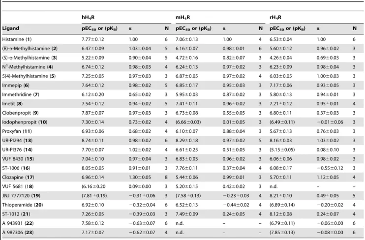
Table 1.Potencies and efficacies of H4R ligands in the luciferase reporter gene assay at the hH4R, the mH4R and the rH4R.
hH4R mH4R rH4R
Ligand pEC50or (pKB) a N pEC50or (pKB) a N pEC50or (pKB) a N
Histamine (1) 7.7760.12 1.00 6 7.0660.13 1.00 4 6.5360.04 1.00 6
(R)-a-Methylhistamine (2) 6.4760.09 1.0360.04 5 6.1660.07 0.9860.01 6 5.6060.12 0.9660.02 3
(S)-a-Methylhistamine (3) 5.2260.09 0.9060.04 5 4.7260.16 0.8260.07 3 4.2660.04 0.6960.03 3
Na-Methylhistamine (4) 6.7460.12 0.9860.03 4 6.2460.13 0.9760.02 3 6.2360.09 0.9860.04 3
5(4)-Methylhistamine (5) 7.2560.05 0.9760.03 3 6.8760.05 0.9760.02 4 6.0360.05 1.0060.03 3
Immepip (6) 7.6460.12 0.9860.02 5 6.8560.17 0.9560.03 3 7.1760.06 0.9360.05 3
Immethridine (7) 6.1260.20 0.6560.02 3 5.9560.03 0.8760.02 3 5.8060.13 0.9460.01 3
Imetit (8) 7.5460.12 0.9460.02 5 7.4160.11 0.9660.02 3 7.2160.12 0.9560.01 4
Clobenpropit (9) 7.8760.07 0.9760.03 3 6.7360.08 0.5560.05 3 6.8060.11 0.3760.03 3
Iodophenpropit (10) 7.3060.14 0.7360.02 4 (6.6660.03) 0.0160.05 3 (6.4960.11) 20.0160.06 3
Proxyfan (11) 6.9360.06 0.6860.02 4 6.1060.07 0.8860.04 3 5.6760.13 0.7660.03 3
UR-PI294 (13) 8.7460.11 0.9860.02 6 8.2960.18 0.9760.02 5 8.1660.03 1.0360.02 3
UR-PI376 (14) 7.7060.07 1.0260.02 4 6.6160.25 0.5160.05 3 (5.1560.05) 0.0860.10 3
VUF 8430 (15) 7.0460.10 0.9760.04 3 6.8360.03 0.9660.02 3 6.0660.06 0.9860.02 3
ST-1006 (16) 8.0560.05 0.9160.01 3 7.7660.11 0.3760.04 4 6.0860.17 20.5560.12 3
Clozapine (17) 6.9660.14 1.3060.05 8 5.4460.06 0.9960.01 3 5.7060.11 1.1260.05 4
VUF 5681 (18) (6.1660.20 0.0960.00 3 5.2060.15 0.4260.02 3 n.d. – –
JNJ 7777120 (19) (7.8160.19) 20.3160.06 3 (7.5860.13) 20.2360.03 4 8.2160.10 0.4960.05 5
Thioperamide (20) 6.9260.10 20.3260.04 6 6.5260.13 20.4460.02 4 (6.8960.14) 20.2060.02 4
ST-1012 (21) 7.2660.05 20.3960.03 3 7.4960.09 0.2460.05 4 8.1260.08 0.2460.07 4
A 943931 (22) 7.5860.12 20.6360.07 6 n.d. – – (6.7960.11) 20.0660.00 6
A 987306 (23) 7.1760.07 20.6260.07 4 n.d. – – (7.8560.13) 20.0860.00 6
Data are represented as mean values6SEM of N independent experiments performed in triplicate.a: intrinsic activity, referred to histamine = 1.00; n.d.: not determined.
doi:10.1371/journal.pone.0073961.t001
HEK293T-SF-hH4R-His6cells were stably co-transfected with pGL4.29[luc2P/CRE/Hygro] (Promega, Mannheim, Germany) encoding hygromycin resistance (Hygro) and the firefly luciferase (luc2P), the transcription of which is controlled by the cAMP responsive element (HEK293T-SF-hH4R-His6-CRE-Luc cells).
HEK293T cells were stably co-transfected with pGL4.29[luc2P/
CRE/Hygro] (HEK293T-CRE-Luc cells) and pcDNA3.1(+)SF- mH4R-His6 (HEK293T-SF-mH4R-His6-CRE-Luc) or pcDNA3.1(+)-SF-rH4R-His6 (HEK293T-SF-rH4R-His6-CRE- Luc cells), respectively. For transfection, the cells were seeded into a 24 well-plate (Becton Dickinson, Heidelberg, Germany), so that they reached 60–70% confluenccy on the next day. The transfection mixture containing 0.5mg of the DNA and either 1mL (4:2 ratio), 1.5mL (6:2 ratio) or 2mL (8:2 ratio) of FuGeneHHD transfection reagent (Roche Diagnostics, Man- nheim, Germany) was prepared according to the manufacturer’s protocol and added to the cells, followed by an incubation period of 36–48 h at 37uC and 5% CO2in a water-saturated atmosphere.
Co-transfected cells were cultured in DMEM supplemented with 10% (v/v) FCS, 600mg/mL of G418 and 200mg/mL of hygromycin B (A.G. Scientific, San Diego, USA).
Luciferase Reporter Gene Assay
Approximately 2?105transfected cells, suspended in DMEM supplemented with 10% (v/v) FCS, were seeded per well into flat- bottomed 96-well plates (Greiner, Frickenhausen, Germany). The cells were allowed to attach for 17 h at 37uC, 5% CO2in a water- saturated atmosphere. A stock solution (10 mM) of forskolin (Sigma) in DMSO was used to prepare feed solutions in DMEM containing 10% (v/v) FCS (final DMSO concentration in the assay was#1%). For experiments in the presence of a PDE inhibitor, the feed solution of forskolin contained 500mM of IBMX (Sigma).
After addition of forskolin (0.4mM for the cells expressing the human H4R and 1mM for the rat and mouse H4R expressing cells) alone (to determine forskolin potency) or in combination with histaminergic ligands, the cells were incubated for 5 h. In antagonist mode, the forskolin solution was supplemented with 0.10, 0.15 or 1.00mM of histamine as the agonist for the human, mouse and rat H4R expressing cells, respectively. Thereafter, the medium was discarded, the cells were washed once with 100mL of phosphate buffered saline (PBS, pH 7.4) (KCl 2.7 mM; KH2PO4
1.5 mM; NaCl 137 mM; Na2HPO45.6 mM; NaH2PO41.1 mM in Millipore water; all chemicals were from Merck, Darmstadt, Table 2.Reference data of H4R ligands determined in the [35S]GTPcS binding assay at the hH4R, the mH4R and the rH4R and reported in literature.
hH4R mH4R rH4R
Ligand pEC50or (pKB) a pEC50or (pKB) a pEC50or (pKB) a
Histamine (1) 7.1–8.2a,e,j,l,m 1.0 5.2–7.5a,d,e,f, l 1.0 4.3–7.1a,d,e,f,l 1.0
(R)-a-Methylhistamine (2) 6.2–7.0e,j 0.8–1.0 6.6e 0.8 6.0e 0.4
(S)-a-Methylhistamine (3) 4.9j 1.0 – – –– –
Na-Methylhistamine (4) 6.1–7.4e,j 0.9–1.0 – – – –
5(4)-Methylhistamine (5) 7.2–7.8d,j,m 0.9–1.0 6.02d 1.0 5.1d 1.1
Immepip (6) 7.7–7.8a,j 0.8–0.9 5.27a 0.7 5.0a 0.7
Immethridine (7) 6.0j 0.5 – – – –
Imetit (8) 7.9–8.5e,j 0.3–0.9 8.1e 0.8 8.1e 0.3
Clobenpropit (9) 7.7–8.3a,j,m 0.5–1.3 6.1a 0.2 (6.3)a 0.0
Iodophenpropit (10) (7.7–8.0)a,j 0.0 (6.4)a 0.0 (6.0)a 0.0
Proxyfan (11) 7.2j 0.5 – – – –
UR-PI294 (13) 8.4–8.5a,d 0.9–1.0 6.1–6.5a,d 1.0 4.6–5.5a,d 1.0–1.6
UR-PI376 (14) 7.5–7.8a,d,m 0.9–1.3 (6.1)a–6.9d 0.0–0.2 (5.5)a–4.5d 0.0–0.4
VUF 8430 (15) 7.3–8.2a,k,m 0.8–1.0 5.1a 0.7 4.5a 0.4
ST-1006 (16) 8.9c 0.2 – – – –
Clozapine (17) 5.8–6.8a,b,j,m 0.7–1.2 ,4a 0.0 ,4a 0.0
VUF 5681 (18) ,5i – – – – –
JNJ 7777120 (19) (7.6)a–7.5d 20.4d 6.1–6.7a,d 0.4–0.6 6.1–6.5a,d 0.2–0.5
Thioperamide (20) 6.4–7.0a,d,j,m –1.0 –21.4a,d (7.1)a 0.0 (6.4)a 0.0
ST-1012 (21) 7.4c 21.1 – – – –
A 943931 (22) (8.2)g–7.3a 21.8a (8.2)g 0.0 (6.2–8.0)a,g 0.0
A 987306 (23) (8.3)h–7.1a 21.5a (8.2)h 0.0 (7.1–8.3)a,h 0.0
Reference data are taken from (unless otherwise noted,avalues referred to histamine = 1.0):
afunctional [35S]GTPcS-binding assay on Sf9 cell membranes co-expressing the hH4R, mH4R or rH4R+Gia2+b1c2;
b,c,d
Steady-state [32P]GTPase assay on Sf9 cell membranes co-expressing: hH4R-RGS19+Gia2+b1c2
b[43], hH4R-GAIP+Gia2+b1c2, rH4R or mH4R+Gia2+b1c2+GAIPd[23], hH4R+Gia2+b1c2c
(avalue of ST-1012 referred to thioperamide =21.0, [39]);
ecalcium mobilization assay in 293-EBNA cells transiently co- expressing the hH4R, mH4R or rH4R with Gqi5[20];
f,g,h
calcium mobilization assay in HEK293 cells stably co-expressing the hH4R, mH4R or rH4R with Gqi5
f[46],g[44],h[45];
i,j,k,l
CRE-b-galactosidase reporter gene assay in SK-N-MC cells stably co-expressing: the hH4R [40–42] or the mH4Rk[57] with the CRE-b-galactosidase reporter gene;
mCRE-luciferase reporter gene assay in HEK293T cells, transiently co-expressing the hH4R with the CRE-controlled luciferase reporter gene [27];
lSRE-luciferase reporter gene assay in HEK293 cells, co-expressing the human, mouse or rat H4R+SRE-luciferase+Gaqichimeric G-protein [57].
doi:10.1371/journal.pone.0073961.t002
Germany) and lysed in 40mL of lysis buffer (pH 7.8) (N-[2- hydroxy-1,1-bis(hydroxymethyl)ethyl]glycine (Tricine) 25 mM (Sigma); glycerol 10% (v/v) (Merck); ethyleneglycoltetraacetic acid (EGTA) 2 mM (Sigma); TritonTM X-100 1% (v/v) (Serva);
MgSO4 ? 7H2O, 5 mM (Merck); dithiotreitol (DTT) 1 mM (Sigma)) for 45–60 min under shaking (180 rpm). For lumines- cence measurement, 20mL of lysate were transferred into a white flat-bottomed 96-well plate (Greiner) and the GENios Pro microplate reader (Tecan, Salzburg, Austria) was primed with the luciferase assay buffer (pH 7.8) (glycyl-glycine (Gly-Gly) 25 mM; MgSO4 ? 7H2O, 15 mM; KH2PO4, 15 mM (Merck);
EGTA, 4 mM; adenosine 59-triphosphate (ATP) disodium salt, 2 mM (Sigma); DTT 2 mM; D-luciferin potassium salt 0.2 mg/
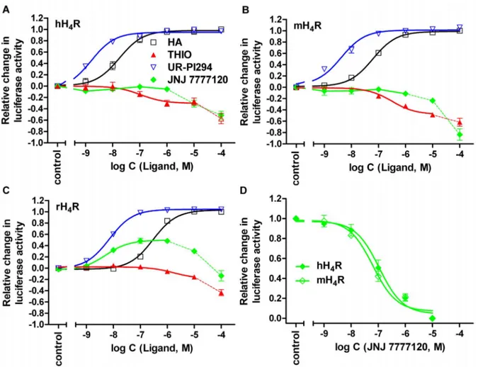
mL (Synchem, Felsberg, Germany)) [31]. Light emission was induced by the injection of 80mL of the luciferase assay buffer into each well. Luminescence, expressed as RLUs (relative light units), was measured for 10 s. The basal luciferase activity was subtracted from each signal. EC50 and IC50 values were analyzed by nonlinear regression and best fitted to sigmoidal concentration- response curves with GraphPad Prism 5.04 (Graph Pad, San Diego (CA), USA). IC50values were converted to KBvalues using the Cheng-Prussoff equation [32]. The intrinsic activity of ligands Figure 5. Effect of selected standard ligands on H4R orthologs.(A) Potencies and efficacies of histamine (HA), thioperamide (THIO), UR-PI294 and JNJ 7777120 at the hH4R, (B) the mH4R and (C) the rH4R (agonist mode). (D) Reversal of the HA (100–150 nM) mediated inhibition of the forskolin- stimulated luciferase activity by JNJ 7777120 at the hH4R and the mH4R (antagonist mode), in the luciferase reporter gene assay in HEK293T cells.
Reaction mixtures contained ligands at the concentrations indicated on the abscissa to achieve saturated concentration response curves. Data points shown are the mean6SEM of at least three independent experiments performed in triplicate. Data points connected by dashed lines reflect H4R- independent increase in luciferase activity at high ligand concentrations. The corresponding values were therefore excluded from non-linear correlations (D).
doi:10.1371/journal.pone.0073961.g005
Figure 6. H4R-independent cellular effects of selected ligand.
Representative H4R-independent increase in the forskolin (1mM) stimulated luciferase activity by ciproxyfan (CIP), proxyfan (PRO), JNJ 7777120 and thioperamide (THIO) in HEK293T-CRE-Luc cells, stably expressing the CRE-controlled luciferase and devoid of the H4R.
doi:10.1371/journal.pone.0073961.g006
was referred to the maximal response to histamine (HA), defined asa= 1 (full agonist). Agonist potencies are given as pEC50values and antagonist activities were calculated as pKBvalues. Measured RLUs were converted to percentual values referred to the span between the maximum effect induced by forskolin and the residual luciferase activity in the presence of histamine at the highest tested concentration. All data are means 6 SEM of N independent experiments, each performed in triplicate. For monitoring the time course of the luciferase expression, transcription was stimulated with 10mM of forskolin, and the cells were lysed after various
incubation periods. For analysis, the respective basal RLUs were subtracted from each value and plotted against the time. For Schild analysis, concentration ratios (r) were obtained by dividing the EC50concentrations of agonist in the presence of JNJ 7777120 (antagonist) by the EC50concentration of agonist in the absence of JNJ 777120. The log (r - 1) values were plotted against the corresponding log [JNJ 7777120] values according to the Schild equation [33] and analyzed by linear regression with GraphPad Prism 5.04. The pA2values were obtained from the intercept of the Schild plot with the x-axis.
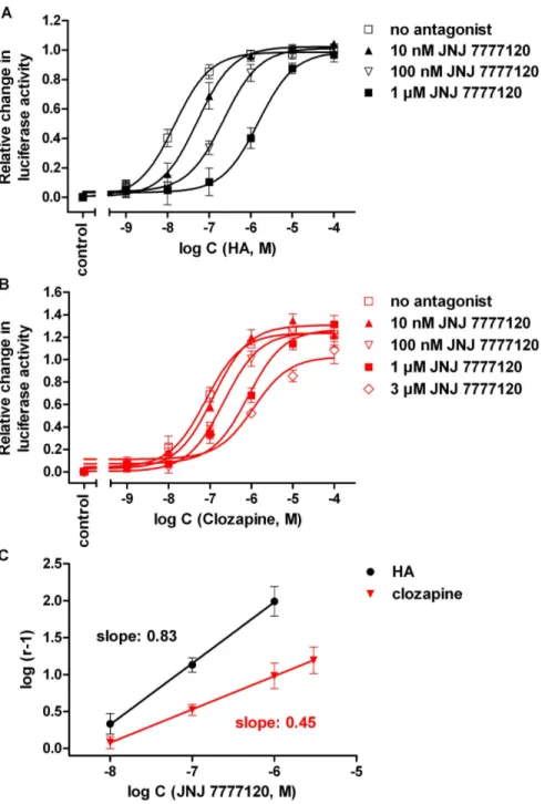
Figure 7. Inhibition of the response to histamine and clozapine by JNJ7777120.Concentration response curves of histamine (A) and clozapine (B) alone and in the presence of JNJ7777120 at increasing concentrations, determined on hH4R expressing HEK293T-CRE-Luc cells in the luciferase reporter gene assay, and corresponding Schild plots (C). The pA2values determined for JNJ 7777120 from Schild regression were 8.39 (slope: 0.8360.02) and 8.17 (slope: 0.4560.01) versus histamine and clozapine, respectively. Data points shown are the mean6SEM of at least three (histamine) or five (clozapine) independent experiments performed in triplicate.
doi:10.1371/journal.pone.0073961.g007
[35S]GTPcS Binding Assay
Cell culture and generation of high-titer recombinant baculo- virus stocks as well as the co-infection of Sf9 cells with high-titer baculovirus stocks encoding Gai2,Gb1c2and the respective H4R were performed as described recently [34,35]. Membrane preparations were performed according to Gether et al. (1995) [36] in the presence of 0.2 mM phenylmethylsulfonyl fluoride, 1 mM ethylenediaminetetraacetic acid (EDTA), 10mg/mL leu-
peptin and 10mg/mL benzamidine as protease inhibitors.
Prepared membranes were resuspended in binding buffer (75 mM Tris/HCl, 12.5 mM MgCl2,1 mM EDTA, pH 7.4) and stored at280uC in 0.5 or 1.0 mL aliquots.
Membranes were thawed, centrifuged for 10 min at 4uC and 13,000 g and carefully resuspended in binding buffer. Experiments were performed in 96-well plates in a total volume of 100mL per well. Each tube contained 8–12mg of protein, 1mM GDP, 100 mM NaCl, 0.05% (w/v) bovine serum albumine (BSA), 20 nCi of [35S]GTPcS ($0.2 nM) and ligand at various concentra- tions. Neutral antagonists were incubated in the presence of histamine at concentrations corresponding to the 10-fold of the EC50 value at the respective receptor. Nonspecific binding was determined in the presence of 10mM unlabeled GTPcS. After incubation under shaking at 200 rpm at room temperature for 2 h, bound [35S]GTPcS was separated from free [35S]GTPcS by filtration through glass microfibre filters using a 96-well Brandel harvester (Brandel Inc., Unterfo¨hring, Germany). The filters were washed three to four times with cold binding buffer (4uC), dried over night and impregnated with meltable scintillation wax prior to counting with a Micro Beta2 1450 scintillation counter (Perkin Elmer, Rodgau, Germany).
Ligands were tested in triplicate. The maximal response to histamine was set to 100% and all other ligands were referenced to histamine.
Results
Optimization of the Assay Conditions
In order to detect a Gai-mediated inhibitory effect on the adenylyl cyclase (AC) activity, the reporter gene assay was performed in the presence of the AC stimulator forskolin. The time course of the luciferase expression upon stimulation with 10mM forskolin is shown in Figure 2A. After a latency period of 0.5–1 h, the enzyme activity steeply increased, and a maximum was reached after 8 h. An incubation period of 5 h was sufficient to obtain 76–94% of the maximum expression. To optimize assay performance, the pEC50value of forskolin in the respective cAMP reporter gene assay system [29] was determined (Figure 2). As the concentration-response curve shows an optimum (Figure 2B), only the ascending part of the curve was considered up to a forskolin concentration of 10mM (Figure 2C). Interestingly, the potency of forskolin was significantly different: pEC50values were 6.4160.05 and 5.9560.04 in the hH4R and mH4R co-transfected cells, respectively, and 5.5060.11 in the HEK293T-CRE-Luc cells (Figure 2C). Forskolin concentration-dependently increased the luciferase expression in HEK293T-SF-rH4R-His6–CRE-Luc cells, which was inhibited by histamine (1) (Figure 3A) with pEC50
values of 6.8160.11, 6.5360.04, 6.2960.07 and 5.9160.04 (Figure 3B) at forskolin concentrations of 0.5, 1.0, 2.5 and 5mM, respectively. Therefore, a concentration of 0.4mM of forskolin was used for pre-stimulating the hH4R expressing cells, whereas 1mM of forskolin was considered optimal for AC stimulation in mH4R and rH4R expressing cells. With respect to comparability of concentration-response curves of H4R ligands at H4R orthologs, the difference between maximum forskolin stimulation in the absence and the presence of the reference agonist histamine (100mM) was set to 100% (Figure 3B).
In the presence of the PDE inhibitor IBMX (50mM) the concentration-response curve of forskolin on HEK293T-SF- hH4R-His6-CRE-Luc-cells was shifted to the left, resulting in an pEC50value of 6.8660.06 (N = 3). Additionally, IBMX increased the receptor-independent luciferase activity by about a factor of four (data not shown). To investigate the effect of IBMX on the Figure 8. Comparison of distal and proximal readouts.
Correlation between agonist potencies in the luciferase reporter gene assay and the [35S]GTPcS assay at the (A) hH4R (slope: 0.9060.20;
r2= 0.80), (B) mH4R (slope: 1.43160.23; r2= 0.95) and (C) rH4R (slope:
1.17160.28, r2= 0.85).
doi:10.1371/journal.pone.0073961.g008
concentration-response curve of the full H4R agonist histamine (1) and the H4R inverse agonist thioperamide (20) HEK293T-hH4R- His6-CRE-Luc cells were pre-stimulated with forskolin (0.5mM) alone or in combination with IBMX (50mM) (cf. Figure 4). The maximum responses to histamine (1) and thioperamide (20), and thus the range of the signals, were reduced in the presence of IBMX. Therefore, further experiments were performed in the absence of IBMX.
Functional Activity of H4R Ligands at the Human, Mouse and Rat H4R
A set of ligands (Figure 1), generally accepted as agonists (1–17), neutral antagonists or inverse agonists (18–23) at the human H4R was selected for functional investigations. The results from the reporter gene assays performed with the H4R species orthologs are summarized in Table 1 and compared to functional data from the [35S]GTPcS binding assay and the literature in Table 2.
hH4R agonists (compounds 1–17). The endogenous ago- nist histamine (1) inhibited forskolin stimulated luciferase activity with pEC50values of 7.77, 7.06 and 6.53 in the hH4R, mH4R and rH4R expressing reporter cells, respectively (Table 1). The methyl- substituted analogs of histamine (2–5) acted, with the exception of 3, as full agonists at the three H4R orthologs. Compared to the hH4R, a trend towards decreased potency was detected at the rodent receptors for compounds1–5(Figure 5A, B, C). Among the enantiomers2and 3, (R)-a-methylhistamine (2) was the eutomer at all species orthologs. Compared to immepip (6), the pyridine analog immethridine (7) showed significantly reduced potency and intrinsic activity at the hH4R. By contrast, immethridine (7) exhibited almost full agonist activity at both, the mouse and rat H4R, with similar moderate potency compared to the hH4R.
Imetit (8) exhibited almost the same potency and efficacy at the three H4R orthologs. In contrast, clobenpropit (9)and iodophen- propit (10), which can be considered as analogs of imetit (8) with an increased distance between the basic moieties and a large lipophilic group in the side chain, displayed a clear decrease in potency and maximal response at the mouse and rat H4R compared to the hH4R. Clobenpropit (9) was a potent full agonist at the hH4R and only a moderate partial agonist at the mouse and rat H4R, whereas iodophenpropit (10) acted as a partial agonist at the hH4R and a neutral antagonist at both, the mouse and the rat H4R. Proxyfan (11) partially activated the three H4R orthologs with significantly lower potencies on the rodent receptors.
Whereas H4R-independent effects of 11 were negligible at concentrations .10mM, the structural analog ciproxifan (12) induced a strong increase (by up to 250%) in luciferase activity at concentrations from 1 to 100mM in HEK293-CRE-Luc cells devoid of H4R expression (Figure 6). Therefore, functional activities of 12 on H4R orthologs were not determined in the luciferase assay. The non-selective acylguanidine-type H3/4R agonist UR-PI294 (13) fully activated the human, mouse and rat H4R (Figure 5A, B, C), being the most potent agonist at all three H4R orthologs (Table 1). In contrast, the selective cyanoguani- dine-type H4R agonist UR-PI376 (14) acted as a potent full hH4R agonist, exhibited only partial agonistic activity at the mH4R and was devoid of agonism at the rH4R (Table 1). VUF 8430 (15) had about the same potency at both, the mH4R and the hH4R, whereas the potency at the rH4R was distinctly lower. At all three H4R species orthologs, VUF8430 (15) was almost as efficacious as histamine (a= 0.96–0.98). The aminopyrimidine-type compound ST-1006 (16) exhibited pronounced differences in the quality of action at the H4R orthologs with nearly full agonism at the hH4R, partial agonism at the mH4R and inverse agonism at the rH4R.
The antipsychotic drug clozapine (17) exhibited only moderate
agonistic potency at the hH4R. However, with anavalue of 1.30, clozapine was even more efficacious than histamine (1). Further- more, clozapine (17) fully activated both, the mouse and the rat H4R, though with low pEC50values (Table 1).
hH4R antagonists and inverse agonists (18–23). Interest- ingly, VUF 5681 (18), with a spacer extended by two carbon atoms compared to the H4R agonist immepip (6), displayed no agonistic activity at the hH4R and only partial agonism at the mH4R. In the antagonist mode at the hH4R, VUF 5681 (18) inhibited the histamine-induced decrease in luciferase activity with a pKBvalue of 6.1660.20. JNJ 7777120 (19) behaved as neutral antagonist at the human and mouse H4R in the luciferase reporter gene assay with comparable pKB values of 7.8160.19 and 7.5860.13, respectively (Figure 5A, B, D). In contrast, at the rH4R JNJ 7777120 (19) acted as a partial agonist (a= 0.4960.05) with a pEC50value of 8.2160.10 (Figure 5C). By analogy with ciproxifan, but much less pronounced, JNJ 7777120 (19) and thioperamide (20) produced receptor-independent increases in luciferase activity at concentrations $10mM in control experi- ments using cells devoid of H4R expression (Figure 6). The corresponding values were therefore omitted in the construction of concentration-response curves of19and20, when studied in the antagonist mode (shown for JNJ 7777120 (19) in Figure 5D).
Thioperamide (20) acted as an inverse agonist, achieving comparable pEC50 values at the human and mouse H4R (Figure 5A, B, Table 1), and revealed moderate antagonistic acitivity at the rH4R with a pKB value of 6.8960.14. The aminopyrimidine ST-1012 (21) acted as an inverse agonist at the hH4R, but revealed partial agonistic activity at the mouse and the rat H4R. The conformationally constrained aminopyrimidines A 943931 (22) and A 987306 (23) were inverse agonists at the hH4R and neutral antagonists at the rH4R.
Discussion and Conclusions Assay Optimization
The pEC50 value of forskolin varied among the different transfectants probably due to different expression levels of the CRE-controlled luciferase. The concentration-response curve revealed a decline at forskolin concentrations higher than 10mM. This decline of the forskolin effect became already obvious at concentrations.3.2mM in the presence of 50mM of the PDE inhibitor IBMX (data not shown), as already described for a CRE-directed luciferase reporter gene assay in Chinese hamster ovary cells (CHO) [37]. By analogy with a report by Kemp et al. [38] an activation of the inducable cAMP early repressor (ICER) may counteract the luciferase expression in HEK293T cells. Gai-protein mediated inhibition of the cAMP synthesis as well as the signal-to-noise ratio was lowered by increasing concentrations of forskolin and IBMX. This was reflected by smaller relative effects and potencies of histamine (1) in the presence of increasing forskolin concentrations (Figure 3) and 50mM of IBMX (Figure 4). Thus, high forskolin concentra- tions should be avoided and the altered potency of forskolin, when used in combination with IBMX, must be considered in this assay.
The co-expression of a CRE-controlled luciferase reporter gene with the human, mouse and rat H4R, respectively, in HEK293T cells enabled the functional analysis of H4R ligands. A set of 23 imidazole and non-imidazole ligands comprising agonists, inverse agonists and antagonists was investigated for ability to effect forskolin stimulated luciferase activity. The obtained pEC50values or pKBvalues were compared with ligand activities from different functional assay systems reported in literature.
Off-target Effects
The luciferase stimulation becoming obvious at concentrations .1mM of JNJ7777120 (19) and thioperamide (20) in cells expressing the H4R orthologs (cf. dashed lines in the concentra- tion-response curves of19and20in Figure 5A-C) suggest inverse agonism. However, the investigation of selected compounds on HEK293T-CRE-Luc cells lacking the H4R (cf. Figure 6) revealed H4R-independent increase in luciferase activity. This effect was most prominent in case of ciproxifan (12), but also pronounced for 19 and 20. Therefore, off-target effects should be taken into account to avoid misinterpretation of biological responses to such compounds at concentrations$10mM.
Activities at the Human H4Receptor
Except for ST-1006 (16) [39], all determined H4R ligand activities at the hH4R were in agreement with results reported in literature [20,23,39–42]. However, a tendency toward elevated intrinsic activities was observed. Contrary to partial agonistic activity of immepip (6) and clobenpropit (9) in the [35S]GTPcS binding assay on membrane preparations of H4R expressing Sf9 cells (a= 0.81and 0.45, respectively) (Table 2), full agonism at the hH4R was determined in the luciferase assay. Iodophenpropit (10), described as a neutral antagonist [40], exerted strong partial agonistic activity at the hH4R in the present study. Partial agonistic activity was also determined for iodophenpropit (10) in a Ca2+mobilization assay in HEK293 cells, co-transfected with the hH4R and the chimeric G-protein Gqi5[5]. ST-1006 (16) had low intrinsic activity in the [32P]GTPase and [35S]GTPcS binding assay at the hH4R [39], but was an almost full agonist in the luciferase assay. The increased intrinsic activity was accompanied with a decrease in potency of about one order of magnitude. In case of clozapine (17), the maximal agonistic response surpassed that of histamine by 30%. In control experiments on HEK293T- CRE-Luc cells devoid of the H4R, clozapine (17) at concentrations as high as 100mM caused an increase in CRE-activity by up to 17% (data not shown). The effect of clozapine on hH4R expressing cells was antagonized by JNJ 7777120 in a concentration- dependent manner, indicating that the (super)agonistic effect was receptor mediated (Figure 7). Using histamine or clozapine as H4R agonists revealed approximately the same pA2 value for JNJ 7777120 (pA2values: 8.39 and 8.17). However, compared to the concentration response curve of histamine in the presence of JNJ 7777120 (Figure 7A), the extent of rightward shift was smaller in case of clozapine (Figure 7B), resulting in different slopes (0.83 compared to 0.45) of the corresponding Schild plots (Figure 7C).
This may be taken as a hint that histamine and clozapine activate the H4R not exactly in the same way. However, due to the pleiotropic character of clozapine (17), effects mediated by targets other than the H4R must be taken into account. Most probably, increased intrinsic activities in the luciferase assay compared to more proximal readouts are caused by amplifications in signaling downstream from G-protein activation [30,37]. For instance, in functional assays on Sf9 cell membranes, ST-1006 (16) [39] and clozapine (17) [43] showed only partial agonism (Table 2).
The constitutive activity of the hH4R, obvious from inverse agonism of thioperamide (20), was rather low compared to functional assays on Sf9 cell membranes [23,34]. In accordance with reported data ST-1012 (21) acted as an inverse hH4R agonist in the [35S]GTPcS assay [39], and JNJ 7777120 (19) behaved as a neutral hH4R antagonist [18,40]. Inverse agonism was also found for A 943931 (22) and A 987306 (23) in the luciferase (Table 1) and the GTPcS assay (Table 2), whereas neutral antagonism was observed in Ca2+(FLIPR) assays [44,45].
Activities at Rodent H4Receptors
Comparing the results from the luciferase assay on mouse and rat H4R with data from other functional assays revealed marked differences. The potencies of histamine (1), 5(4)-methylhistamine (5), immepip (6), UR-PI294 (13), VUF 8430 (15) and clozapine (17) were significantly higher compared to the [32P]GTPase [23]
and [35S]GTPcS binding assay (Table 2). By contrast, the agonist potencies of histamine (1), (R)-a-methylhistamine (2), Na-methyl- histamine (4) and imetit (8) were consistent or lower compared to results from a Ca2+assay using HEK293 cells, co-expressing the mouse or the rat H4R with Gaqi5 [2,46]. For example, in the luciferase assay the pEC50values of histamine (1) were in good agreement with results from the Ca2+assay at the mouse and rat H4R (7.23 and 6.49, respectively) [46], but distinctly higher compared to pEC50values from the [32P]GTPase assay (5.81 and 5.23, respectively) [23]. UR-PI294 (13) achieved pEC50values.8 at the hH4R, mH4R and rH4R in the luciferase assay, whereas the [32P]GTPase assay revealed dramatic differences in pEC50values (8.52, 6.50 and 4.64, respectively) [23]. The potency of imetit (8) was lower compared to the Ca2+assay in HEK293 cells (pEC50
values: 7.4 and 7.2 vs. 8.1 at both receptors) [20]. Whereas being full agonists in the luciferase assay, (R)-a-methylhistamine (2), Na- methylhistamine (4) and imetit (8) only reached 75–80% of the maximal Ca2+response at the mH4R and 30–50% at the rH4R [20].
The pKBvalues of neutral antagonists, such as iodophenpropit (10) at the mouse and rat H4R as well as thioperamide (20) and UR-PI376 (14) at the rH4R were comparable to those determined in the [35S]GTPcS binding assay (Table 2). Mouse and rat H4R- mediated inhibition of forskolin-stimulated luciferase activity in HEK293T-CRE-Luc cells resulted in higher potencies compared to functional assays using Ga-protein activation as readout. This suggests that signal amplification or concomitant activation of different signaling pathways potentiates the inhibition of the luciferase activity. For example, the cAMP pathway may be modulated by a cross-talk with Ca2+signaling elicited by activation of phospholipase C (PLC) [47]. Ca2+is an inhibitor of (forskolin) stimulated and Ca2+sensitive adenylate cyclases type V/VI [48–
50], which are endogenously expressed in HEK293T cells [51]
and interact with the Gaiprotein [52]. Furthermore, the relevance of this crosstalk with regard to the cAMP signaling pathway of G- protein coupled receptors (GPCRs) was demonstrated by the inhibitory effect of the activated Gaqcoupled histamine H1R on the cAMP level in U373 MG cells [53] and, more importantly, by a crosstalk between the Gaicoupled M2mACh receptor and the Gaqcoupled M3mACh receptor. In the latter case the inhibition of forskolin-stimulated cyclic AMP accumulation was facilitated at low agonist concentrations [54]. Further studies on the influence of Ca2+are needed to clarify, whether only the rodent H4Rs are concerned, since agonist potencies at the hH4R were consistent with data from the [32P]GTPase and [35S]GTPcS binding assay (Table 2). Very recently, investigations on human esosinophils revealed a lower Ca2+response to stimulation by histamine (1) and UR-PI376 (14) compared to the chemokine eotaxin via the CCR3 receptor [55]. This may be interpreted as a hint to minor contribution of Ca2+ signaling to the overall H4R mediated response, at least in native human cells. The presence of a range of alternative signaling pathways for the H4R in living cells was underlined recently by the Gaindependent ß-arrestin recruitment of several H4R ligands [26,27].
The results for the standard antagonist JNJ 7777120 (19) at the mouse and rat H4R compared with data reported for other functional assays revealed discrepancies, too. In the luciferase assay JNJ 7777120 (19) acted as a neutral antagonist at the mH4R,
but as a potent partial agonist at the rH4R. Antagonistic activity at both receptors was found in a CRE-drivenb-galactosidase assay in SK-N-MC cells [18] and in a Ca2+assay in HEK293 cells [46], whereas partial agonistic activity was determined at the mouse and rat receptor in the [32P]GTPase [23] and [35S]GTPcS binding assay (Table 2). The pKBvalue at the mH4R in the luciferase assay is consistent with the pKBvalue in the Ca2+assay [46], whereas the agonistic potency at the rH4R is about two orders of magnitude higher compared to the [32P]GTPase assay [23].
Discrepancies between the H4R orthologs in the different assay systems may result from differential equilibria between the active and inactive states of the H4R in the different assay systems as described recently [56]. In the luciferase assay, the constitutive activity, reflected by the inverse agonism of compounds 20–23, was considerably higher for the mH4R than for the rH4R. At the latter JNJ 7777120 shifted the equilibrium toward the active state, becoming obvious as agonistic activity. Inversely, ST1006 (16), a potent agonist a human and mouse H4R, showed considerable inverse agonism at the rH4R. Thus, the outcome of studies in translational animal models cannot be unequivocally predicted by in vitro experiments, but such data may help to interprete conflicting results such as the pro-inflammatory effect of JNJ 7777120 (19) in a rat conjunctivitis model [17].
In case of agonism at the human H4R, the data correlate very well with data provided by more proximal readouts such as GTPase activity or GTPcS binding (Table 1, Table 2, Figure 8A).
This also holds for the rank order of agonists at the mouse and rat H4R (Table 1, Table 2, Figure 8B,C), however, the potencies are up to 100-fold higher in the luciferase assay.
Conclusions
The reporter gene (luciferase) assay in HEK293T cells allows for the quantification of agonistic, inverse agonistic and antago- nistic activity at the H4R species orthologs in a highly sensitive and reliable manner. In view of significantly increased potencies and efficacies of agonists, especially at the rodent H4R orthologs, obviously, there is a positive effect on the readout by activation/
amplification of or cross-talk between different signaling pathways in the luciferase reporter gene assay compared to more proximal functional assays on Sf9 cell membranes. It has now become clear that unequivocal characterization of H4R ligands as agonists, antagonists or inverse agonists in assays using a single readout is impossible. Thus, ligands have to be examined in multiple assays.
But at present, it seems impossible to predict the value of the in vitro data with respect to translational animal studies and their clinical relevance.
Acknowledgments
The authors are grateful to Maria Beer-Kro¨n and Stefanie Dukorn for technical assistance.
Author Contributions
Conceived and designed the experiments: UN GB HS RS AB. Performed the experiments: UN DW DS. Analyzed the data: UN DW DS GB RS AB.
Contributed reagents/materials/analysis tools: HS RS. Wrote the paper:
UN GB RS AB.
References
1. Oda T, Morikawa N, Saito Y, Masuho Y, Matsumoto S (2000) Molecular Cloning and Characterization of a Novel Type of Histamine Receptor Preferentially Expressed in Leukocytes. J Biol Chem 275: 36781–36786.
2. Liu C, Ma X, Jiang X, Wilson SJ, Hofstra CL, et al. (2001) Cloning and pharmacological characterization of a fourth histamine receptor (H(4)) expressed in bone marrow. Mol Pharmacol 59: 420–426.
3. Morse KL, Behan J, Laz TM, West RE Jr, Greenfeder SA, et al. (2001) Cloning and Characterization of a Novel Human Histamine Receptor. J Pharmacol Exp Ther 296: 1058–1066.
4. Nguyen T, Shapiro DA, George SR, Setola V, Lee DK, et al. (2001) Discovery of a Novel Member of the Histamine Receptor Family. Mol Pharmacol 59: 427–
433.
5. Zhu Y, Michalovich D, Wu H-L, Tan KB, Dytko GM, et al. (2001) Cloning, Expression, and Pharmacological Characterization of a Novel Human Histamine Receptor. Mol Pharmacol 59: 434–441.
6. de Esch IJP, Thurmond RL, Jongejan A, Leurs R (2005) The histamine H4
receptor as a new therapeutic target for inflammation. Trends Pharmacol Sci 26:
462–469.
7. Zhang M, Thurmond RL, Dunford PJ (2007) The histamine H(4) receptor: a novel modulator of inflammatory and immune disorders. Pharmacol Ther 113:
594–606.
8. Thurmond RL, Gelfand EW, Dunford PJ (2008) The role of histamine H1 and H4 receptors in allergic inflammation: the search for new antihistamines. Nat Rev Drug Discov 7: 41–53.
9. Leurs R, Chazot PL, Shenton FC, Lim HD, de Esch IJ (2009) Molecular and biochemical pharmacology of the histamine H4 receptor. Br J Pharmacol 157:
14–23.
10. Leurs R, Vischer HF, Wijtmans M, de Esch IJ (2011) En route to new blockbuster anti-histamines: surveying the offspring of the expanding histamine receptor family. Trends Pharmacol Sci 32: 250–257.
11. Dunford PJ, O’Donnell N, Riley JP, Williams KN, Karlsson L, et al. (2006) The histamine H4 receptor mediates allergic airway inflammation by regulating the activation of CD4+T cells. J Immunol 176: 7062–7070.
12. Morgan RK, McAllister B, Cross L, Green DS, Kornfeld H, et al. (2007) Histamine 4 receptor activation induces recruitment of FoxP3+T cells and inhibits allergic asthma in a murine model. J Immunol 178: 8081–8089.
13. Rossbach K, Wendorff S, Sander K, Stark H, Gutzmer R, et al. (2009) Histamine H4 receptor antagonism reduces hapten-induced scratching behav- iour but not inflammation. Exp Dermatol 18: 57–63.
14. Deml K-F, Beermann S, Neumann D, Strasser A, Seifert R (2009) Interactions of Histamine H1-Receptor Agonists and Antagonists with the Human Histamine H4-Receptor. Mol Pharmacol 76: 1019–1030.
15. Beermann S, Glage S, Jonigk D, Seifert R, Neumann D (2012) Opposite effects of mepyramine on JNJ 7777120-induced amelioration of experimentally induced asthma in mice in sensitization and provocation. PLoS One 7: e30285.
16. Coruzzi G, Adami M, Guaita E, de Esch IJP, Leurs R (2007) Antiinflammatory and antinociceptive effects of the selective histamine H4-receptor antagonists JNJ7777120 and VUF6002 in a rat model of carrageenan-induced acute inflammation. Eur J Pharmacol 563: 240–244.
17. Zampeli E, Thurmond RL, Tiligada E (2009) The histamine H4 receptor antagonist JNJ7777120 induces increases in the histamine content of the rat conjunctiva. Inflamm Res 58: 285–291.
18. Thurmond RL, Desai PJ, Dunford PJ, Fung-Leung WP, Hofstra CL, et al.
(2004) A potent and selective histamine H4 receptor antagonist with anti- inflammatory properties. J Pharmacol Exp Ther 309: 404–413.
19. Neumann D, Beermann S, Seifert R (2010) Does the Histamine H(4) Receptor Have a Pro- or Anti-Inflammatory Role in Murine Bronchial Asthma?
Pharmacology 85: 217–223.
20. Liu C, Wilson SJ, Kuei C, Lovenberg TW (2001) Comparison of human, mouse, rat, and guinea pig histamine H4 receptors reveals substantial pharmacological species variation. J Pharmacol Exp Ther 299: 121–130.
21. Strasser A, Wittmann H-J, Buschauer A, Schneider EH, Seifert R (2013) Species-dependent activities of GPCR ligands: lessons from histamine receptor orthologs. Trends Pharmacol Sci 34: 13–32.
22. Lim HD, de Graaf C, Jiang W, Sadek P, McGovern PM, et al. (2010) Molecular determinants of ligand binding to H4R species variants. Mol Pharmacol 77:
734–743.
23. Schnell D, Brunskole I, Ladova K, Schneider EH, Igel P, et al. (2011) Expression and functional properties of canine, rat, and murine histamine H(4) receptors in Sf9 insect cells. Naunyn Schmiedeberg’s Arch Pharmacol 383: 457–470.
24. Igel P, Geyer R, Strasser A, Dove S, Seifert R, et al. (2009) Synthesis and structure-activity relationships of cyanoguanidine-type and structurally related histamine H4 receptor agonists. J Med Chem 52: 6297–6313.
25. Igel P, Schneider E, Schnell D, Elz S, Seifert R, et al. (2009) N(G)-acylated imidazolylpropylguanidines as potent histamine H4 receptor agonists: selectivity by variation of the N(G)-substituent. J Med Chem 52: 2623–2627.
26. Rosethorne EM, Charlton SJ (2011) Agonist-biased signaling at the histamine H4 receptor: JNJ7777120 recruits beta-arrestin without activating G proteins.
Mol Pharmacol 79: 749–757.
27. Nijmeijer S, Vischer HF, Rosethorne EM, Charlton SJ, Leurs R (2012) Analysis of Multiple Histamine H4 Receptor Compound Classes Uncovers Galphai and beta-Arrestin2 Biased Ligands. Mol Pharmacol 82: 1174–1182.
28. Williams C (2004) cAMP detection methods in HTS: selecting the best from the rest. Nat Rev Drug Discov 3: 125–135.