Dimeric histamine H 2 receptor agonists as molecular tools and genetically engineered HEK293T cells as an assay platform to unravel
signaling pathways of hH 1 R and hH 2 R
Dissertation
zur Erlangung des Doktorgrades der Naturwissenschaften (Dr. rer. nat.) an der Fakultät für Chemie und Pharmazie
der Universität Regensburg
vorgelegt von
Nicole Plank geb. Kagermeier aus Straubing
2015
Die vorliegende Arbeit entstand in der Zeit von Mai 2011 bis Dezember 2014 unter der Anleitung von Herrn Prof. Dr. Armin Buschauer am Institut für Pharmazie der Naturwissenschaftlichen Fakultät IV – Chemie und Pharmazie – der Universität Regensburg.
Das Promotionsgesuch wurde eingereicht im April 2015.
Tag der mündlichen Prüfung: 30.04.2015
Prüfungsausschuss: Prof. Dr. A. Jacobi von Wangelin (Vorsitzender) Prof. Dr. A. Buschauer (Erstgutachter)
Prof. Dr. G. Bernhardt (Zweitgutachter) Prof. Dr. J. Heilmann (Drittprüfer)
Für Patrick
I Danksagung
An dieser Stelle möchte ich mich ganz herzlich bedanken bei:
Meinem Doktorvater Herrn Prof. Dr. Armin Buschauer, der mir die Möglichkeit gegeben hat dieses Projekt zu verwirklichen, seine konstruktive Kritik bei der Durchsicht der Arbeit, die Aufnahme ins Graduiertenkolleg, seine Geduld und dass er trotz viel Arbeit stets ein offenes Ohr für mich hatte.
Herrn Professor Dr. Günther Bernhard für seine stetige Unterstützung im Laboralltag, seine fachliche Anleitung, das Durchsehen der Arbeit und natürlich auch für das schöne Sommerfest.
Herrn Professor Dr. Roland Seifert und Frau Kristin Werner für die Durchführung der Monozytenexperimente.
Herrn Professor Dr. Sigurd Elz und Frau PD Dr. Andrea Strasser für das Überlassen der H1R Liganden.
Herrn Dr. Max Keller für die Hilfe bei der Synthese des Radioliganden und seine fachliche Kompetenz.
Herrn Dr. Paul Baumeister für seine Unterstützung bei der Testung und für die immer lustigen Unterhaltungen am Gang.
Frau Dita Fritsch und Frau Elvira Schreiber für die Hilfe der Durchführung vieler Assays.
Herrn Peter Richthammer für seine Hilfsbereitschaft und die netten Gespräche.
Frau Uta Hasselmann, Silvia Heinrich und Karin Reindl für die stets freundliche Unterstützung bei organisatorischen Angelegenheiten.
Dem Graduiertenkolleg 1910 für die Finanzierung interessanter Tagungen.
Meinen Bürokollegen: Frau Frauke Antoni, Frau Stefanie Dukorn und Herrn Andrea Pegoli (grazie buffo puffo).
„14.2.08“: Herrn Dr. Johannes Felixberger für sein enormes Wissen, seine unermüdliche Hilfsbereitschaft und die schöne Zeit auch außerhalb der Arbeit. Herrn Dr. Stefan Huber für die lustigen, aber manchmal auch fachlichen Diskussionen und seine Freundschaft. Herrn Dr. Uwe Nordemann für das Überlassen der HEK293T CRE Luc Zellen und sein unerschütterlich freundliches Gemüt.
Meinen ehemaligen Kolleginnen und Freundinnen Dr. Melanie Kaske, Dr. Carolin Meyer und Dr. Stefanie Rodler für die vielen sportlichen Aktivitäten und den schönen Abenden mit gutem Wein.
Bei Arminia Buschauer, insbesondere: Herrn Dr. Tobias Birnkammer, Herrn Dr. Roland Geyer, Herrn Dr. Tobias Holzammer, Herrn Kilian Kuhn, Herrn Sebastian Lieb, Herrn José Esteban Obreque-Balboa, Frau Dr. Miriam Pluym, Herrn Dr. Nicola Pluym, Herrn Steffen Pockes und Herrn Dr. David Wifling für die gute Zusammenarbeit und die angenehme Atmosphäre.
Zu guter letzt die Menschen, die mehr als Dank verdienen:
Alle meine Freunde außerhalb der Uni, die namentlich zu erwähnen den Rahmen sprengen würde.
Frau Maria Beer-Kroen für die enorme Unterstützung bei der Laborarbeit, die Süßigkeiten im Büro, die stets aufmunternden Worte und ihre nie endend wollende Hilfsbereitschaft.
Meine Schwiegereltern Gisela und Max für Ihre gute Laune und Ihre immerwährende Hilfsbereitschaft.
Meine Geschwister Peter und Ulrike für Ihre Unterstützung in allen Lebenslagen.
Meine Eltern auf die ich mich immer verlassen kann und ohne die es diese Arbeit sicher nicht geben würde.
Mein Mann Patrick für seine Liebe, seine unermessliche Geduld und unser kleines Igelchen.
III
Publications (published results prior to the submission of this thesis):
1. Kagermeier, N.; Werner, K.; Keller, M.; Baumeister, P.; Bernhardt, G.; Seifert, R.;
Buschauer, A. Dimeric carbamoylguanidine-type histamine H2 receptor ligands: A new class of potent and selective agonists. Bioorg. Med. Chem. 2015.
2. Baumeister, P.; Erdmann, D.; Biselli, S.; Kagermeier, N.; Elz, S.; Bernhardt, G.;
Buschauer, A. [3H]UR-DE257: Development of a tritium-labeled squaramide-type selective histamine H2 receptor antagonist. ChemMedChem. 2015.
3. Kagermeier, N.; Nordemann, U.; Bernhardt, G.; König, G.; Buschauer, A. Differential Signaling Pathways of the hH1R and the hH2R. Inflamm. Res. 2014 (Abstract)
4. Birnkammer, T.; Spickenreither, A.; Brunskole, I.; Lopuch, M.; Kagermeier, N.;
Bernhardt, G.; Dove, S.; Seifert, R., Elz, S.; Buschauer, A. The bivalent ligand approach leads to highly potent and selective acylguanidine-type histamine H2
receptor agonists. J. Med. Chem. 2012
Short lecture:
1. Kagermeier, N.: Differential Signaling Pathways of the hH1R and the hH2R. 43th Annual Meeting of the European Histamine Research Society in Lyon; 2014.
Poster presentations:
09/11: 6th Summer School in Medicinal Chemistry, Regensburg.
Kagermeier, N.; Holzammer, T.; Bernhardt, G.; Dove, S.; Buschauer, A.: Thr190 in the human histamine H2 receptor (hH2R) contributes to binding of imidazole-type ligands.
10/13: Annual meeting of the German Pharmaceutical Society (DPhG) in Freiburg.
Kagermeier, N.; Nordemann, U.; Bernhardt, G.; König, G.; Buschauer, A.: Alternative signaling pathways of the histamine H1 (H1R) and the histamine H2 receptor (H2R).
09/14: 13th International Symposium on Medicinal Chemistry (EFMC-ISMC) in Lisbon.
Kagermeier, N.; Keller, M.; Baumeister, P.; Bernhardt, G.; Buschauer, A.: Bivalent carbamoylguanidine-type H2 receptor ligands: Potent and selective agonists, but inappropriate for radio- and fluorescence labeling.
09/14: 7th Summer School in Medicinal Chemistry, Regensburg.
Kagermeier, N.; Nordemann, U.; Felixberger, J.; Bernhardt, G.; König, G.; Buschauer, A.:
Differential signaling pathways of the human histamine H1R (hH1R) and the human histamine H2R (hH2R).
V
Contents
Contents
1 General Introduction ... 1
1.1 G-protein coupled receptors ... 2
1.1.1 Brief history ... 2
1.1.2 GPCRs as drug targets ... 2
1.1.3 GPCR structure and classification ... 2
1.1.4 GPCR activation and signaling ... 3
1.1.4.1 Classical model ... 3
1.1.4.1.1 G-proteins ... 4
1.1.4.1.2 G-protein regulated effectors ... 4
1.2 Histamine and its receptors ... 6
1.2.1 The biogenic amine histamine ... 6
1.2.1.1 Brief history ... 6
1.2.1.2 Biosynthesis, metabolism and function ... 6
1.2.2 The four histamine receptors and their ligands ... 6
1.2.2.1 The histamine H1R ... 6
1.2.2.2 The histamine H2R ... 7
1.2.2.3 The histamine H3R ... 9
1.2.2.4 The histamine H4R ... 10
1.3 GPCR dimerization and bivalent ligands ... 11
1.3.1 GPCR dimerization ... 11
1.3.2 Bivalent ligands ... 12
1.4 Functional selectivity and G-protein independent signaling ... 13
1.5 References ... 15
2 Scope and Objectives ... 25
2.1 Dimeric carbamoylguanidine-type H2 receptor ligands ... 26
2.2 Unraveling signaling pathways of the human histamine receptors H1R and H2R in HEK293T CRE Luc cells ... 26
3 Dimeric carbamoylguanidine-type H
2receptor ligands: A new class of potent and selective agonists ... 27
3.1 Introduction... 28
3.2 Chemistry ... 28
3.2.1 Homobivalent carbamoylguanidine-type H2R ligands ... 28
3.2.2 Toward bivalent fluorescence- and radioligands ... 31
3.3 Results and Discussion ... 33
3.3.1 Stability of carbamoylguanidines compared to acylguanidines ... 33
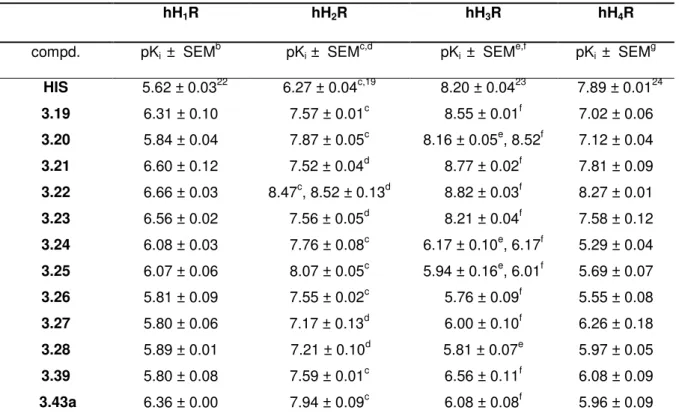
3.3.2 Pharmacological results and discussion ... 34
3.3.2.1 H2R agonism in the GTPγS binding assay ... 34
3.3.2.2 H2R affinities and receptor subtype selectivities ... 35
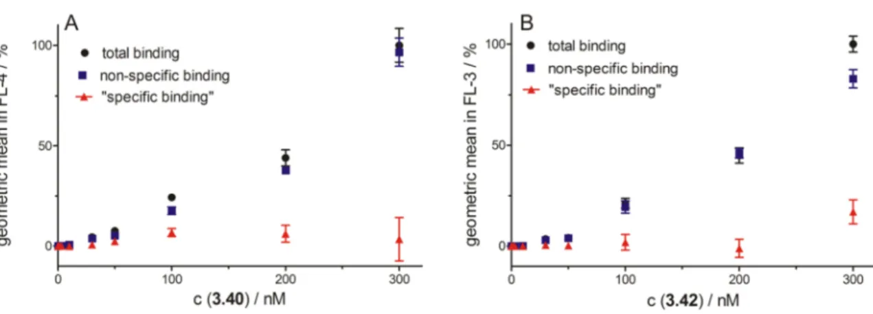
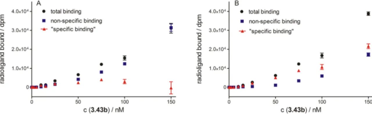
3.3.2.3 Investigation of the fluorescence ligands 3.40-3.42 and the radioligand
3.43b ... 36
3.3.2.4 Investigation of H2R agonists in human monocytesa ... 40
3.3.2.5 Stability of selected carbamoylguanidines in plasma ... 42
3.3.2.6 Plasma protein binding ... 42
3.3.2.6.1 Equilibrium dialysis ... 42
3.3.2.6.2 Ultrafiltration ... 43
3.3.2.7 Cytotoxicity in the crystal violet based chemosensitivity assay ... 44
3.4 Summary and conclusion ... 46
3.5 Experimental section ... 47
3.5.1 General Experimental conditions ... 47
3.5.2 Chemistry: Experimental Protocols and Analytical data ... 47
3.5.3 Pharmacological Protocols ... 61
3.5.3.1 GTPγS binding assay ... 61
3.5.3.2 Competition binding experiments ... 61
3.5.3.3 Studies on human monocytes ... 61
3.5.3.4 EBAO staining ... 62
3.5.3.5 Stability in plasma ... 62
3.5.3.6 Plasma protein binding ... 62
3.5.3.6.1 Recovery after equillibrium dialysis ... 62
3.5.3.6.2 Ultrafiltration ... 63
3.5.3.7 Crystal violet based chemosensitivity assay ... 63
3.6 References ... 64
4 Unraveling signaling pathways of the human histamine receptors H
1R and H
2R in HEK293T CRE Luc cells ... 67
4.1 Introduction... 68
4.2 Materials and Methods ... 69
4.2.1 Cell culture ... 69
4.2.2 Stable transfection of HEK293T CRE Luc cells with the pcDNA3.1(+)- hH1R vector ... 69
4.2.3 Stable transfection of HEK293T CRE Luc cells with the pcDNA3.1(+)- hH2R vector ... 70
4.2.4 Stable transfection of HEK293T CRE Luc hH1R cells with the pcDNA3.1(-)mtAEQ plasmid ... 70
4.2.5 Stable transfection of HEK293T CRE Luc hH2R cells with the pcDNA3.1(-)mtAEQ vector ... 70
4.2.6 Stable transfection of HEK293T CRE Luc-SF-hH4R-His6 with pGL4.29[luc2P/CRE/Hygro] plasmid ... 70
4.2.7 Whole cell radioligand binding assay ... 70
4.2.8 Luciferase reporter gene assay ... 71
4.2.9 Fura-2 Ca2+ assay ... 71
4.2.10 Aequorin assay ... 72
4.2.11 cAMP assay... 72
4.3 Results and Discussion ... 73
4.3.1 Investigation of HEK293T CRE Luc cells expressing the hH1R ... 73
4.3.1.1 Saturation binding assay using HEK293T CRE Luc hH1R cells ... 73
4.3.1.2 Luciferase reporter gene assay using HEK293T CRE Luc hH1R cells ... 73
VII
Contents
4.3.1.2.1 Effect of Gαq inhibition on the H1R dependent luciferase activity ... 74
4.3.1.2.2 Effect of pertussis toxin on the H1R dependent luciferase activity ... 75
4.3.1.2.3 Effect of protein kinase A inhibitors on the H1R dependent luciferase activity ... 77
4.3.1.2.4 Effect of the Gβγ inhibitor gallein on the H1R dependent luciferase activity ... 78
4.3.1.3 Investigation of hH1R expressing HEK293T cells in the fura-2 assay ... 79
4.3.1.4 Investigation of hH1R expressing HEK293T cells in the aequorin assay ... 79
4.3.1.5 Investigation of hH1R expressing HEK293T cells in a cAMP assay... 81
4.3.1.5.1 Optimization of cAMP assay parameters ... 81
4.3.1.5.2 cAMP assay on HEK293T CRE Luc hH1R cells ... 82
4.3.2 Investigation of HEK293T CRE Luc cells expressing the hH2R ... 82
4.3.2.1 Saturation binding assay using HEK293T CRE Luc hH2R cells ... 83
4.3.2.2 Luciferase reporter gene assay using HEK293T CRE Luc hH2R cells ... 83
4.3.2.2.1 Effect of Gαq inhibition on the H2R dependent luciferase activity ... 84
4.3.2.2.2 Effect of protein kinase A inhibitors on the H2R stimulated luciferase activity ... 84
4.3.2.2.3 Effect of pertussis toxin on the H2R stimulated luciferase activity ... 85
4.3.2.2.4 Effect of the Gβγ inhibitor gallein on the H2R dependent luciferase activity ... 86
4.3.2.3 Investigation of hH2R expressing HEK293T cells in the fura-2 assay ... 86
4.3.2.4 Investigation of hH2R expressing HEK293T cells in the aequorin assay ... 86
4.3.2.5 Investigation of hH2R expressing HEK293T cells in a cAMP assay... 87
4.4 Summary and conclusion ... 88
4.5 References ... 90
5 Application of the established luciferase reporter gene assay for the functional investigation of hH
1R ligands ... 93
5.1 Introduction... 94
5.2 Material and Methods ... 94
5.2.1 Cell culture ... 94
5.2.2 Stable transfection of HEK293T CRE Luc cells with the pcDNA3.1(+)- hH1R vector ... 94
5.2.3 Luciferase reporter gene assay ... 94
5.2.4 Crystal violet based chemosensitivity assay ... 94
5.2.5 Selected test compounds ... 94
5.2.5.1 H1R agonists ... 94
5.2.5.2 H1R antagonists ... 95
5.3 Results and Discussion ... 96
5.3.1 Quantification of hH1 receptors per cell ... 96
5.3.2 Optimization of the incubation period ... 96
5.3.3 Effect of the solvent on the luciferase activity... 97
5.3.4 Functional characterization of H1R ligands ... 97
5.3.5 Characterization of histaprodifen derivatives in the crystal violet based chemosensitivity assay ... 100
5.4 Summary and conclusion ... 101
5.5 References ... 102
6 Summary ... 103
7 Appendix ... 107
Chapter 1
General Introduction
2
1.1 G-protein coupled receptors 1.1.1 Brief history
It was in the early 20th century that binding of biologically active molecules to “specific sites on cells” was postulated for the first time by Langley and his student Dale. It is noteworthy that their studies included two of the most important families of receptors, the ion channel receptors and the G-protein coupled receptors (GPCRs).1 Now we know that with about 800 genes, GPCRs, also refered to as seven-transmembrane domaine receptors or heptahelical receptors, represent the largest family of cell surface receptors encoded in the human genome, including approximately 400 chemosensory (olfactory and taste) receptors.2,3
Within the next 50 years the work of Langley and Dale was refined to the concept of classical receptor pharmacology by pioneers like e.g. Black and Furchgott.4 The β2-adrenergic receptor was the first GPCR to be cloned in 1986 and represents the starting point for a molecular understanding of GPCRs.5 Another milestone in the field of GPCR research was achieved in 2000, when Palczewski and coworkers published the first high-resolution X-ray crystal structure of a GPCR, the bovine rhodopsin.6 So far, high-resolution structures are available for 25 GPCRs, including β-adrenoceptors (avian β1-AR7 and human β2-AR8), muscarinic acetylcholine receptors (human M2R9 and rat M3R10), opioid receptors (human nociceptin receptor11, human κ-OR12, mouse µ-OR13 and mouse δ-OR14) and the histamine H1R.15 The importance and significance of GPCRs in biomedical research was underlined in 2012 when Brian Kobilka and Robert Lefkowitz were awarded the Nobel Prize for their work in the GPCR field.
1.1.2 GPCRs as drug targets
Drugs binding to GPCRs show therapeutic benefit in a broad variety of indications, such as pain, bronchial asthma, cardiovascular diseases and neurological disorders. Although addressing only a small number of these membrane proteins,16 more than 30 % of the approximately 500 most important approved drugs bind to GPCRs, making them the most successful therapeutic targets.17 Endogenous ligands were identified for more than 200 GPCRs, including hormones and neurotransmitters.18-20 The endogenous ligands of around 140 GPCRs are yet unidentified.21 These so-called orphan GPCRs represent promising potential new drug targets.
1.1.3 GPCR structure and classification
Characteristic of all GPCRs are seven hydrophobic transmembrane (TM) domains, with an extracellular amino terminus and an intracellular carboxyl terminus (Fig 1.1). GPCRs share the highest homology within the TM domains, whereas the carboxyl terminus, the intracellular loop between TM5 and TM6 and the amino terminus represent the most variable structures.22 The extracellular parts and the transmembrane regions are important for ligand binding, whereas the intracellular regions are crucial for signaling and for feedback modulation of receptor function.23 Under evolutionary aspects, Fredriksson et al. classified GPCRs into five families: Glutamate, Rhodopsin, Adhesion, Frizzled/Taste2 and Secretin (GRAFS).18 The rhodopsin family, by far the largest group, can be subdivided into four main groups: α, β, γ and δ. The histamine receptors belong to the α group. The rhodopsin family shows several characteristic features like the NSxxNPxxY motif in TM7, the DRY motif or
General Introduction
3 D(E)-R-Y(F) at the bottom of TM3, close to IL2.18 Most ligands for the rhodopsin family receptors bind between the TM helices.24
Figure 1.1. Schematic representation of a prototype class A GPCR (IL = intracellular loop; EL = extracellular loop; 1-7 = transmembrane domains)
1.1.4 GPCR activation and signaling
1.1.4.1 Classical modelUpon binding of an agonist, the receptor in its active form is functioning as a nucleotide exchange factor on the α subunit of heterotrimeric G-proteins, triggering the exchange of GDP by GTP. Thereby, the Gα subunit dissociates from the Gβγ subunit and both subunits interact with effector proteins, resulting in an increase or decrease in second messenger concentrations. The G-protein returns to its inactive state upon cleavage of GTP to GDP by the intrinsic GTPase activity of the Gα subunit (Fig. 1.2).23
Figure 1.2. G-Protein cycle upon receptor activation by an agonist (adopted from Seifert et al. 2005).25
The classical model of activation of GPCRs is the ternary complex model.26 This model was extended by the finding that numerous receptors are capable of activating G-proteins in the absence of agonists.27 According to this extended ternary complex model (Fig.1.3A), the receptor exists in an inactive (Ri) and in an active (Ra) conformation. The level of basal
4
receptor activity is given by the equilibrium between the active and the inactive state. Ligands are classified according to their ability to shift the equilibrium. Agonists show higher affinity for Ra and stabilize the active conformation, leading to G-protein activation. Neutral antagonists bind to Ri and Ra with the same affinity and, therefore, they do not alter the equilibrium. In contrast, inverse agonists stabilize the inactive receptor conformation.27,28
Figure 1.3. (A) Extended ternary complex model (L = ligand; Ra = active state of the receptor; Ri = inactive state of the receptor; G = G-protein) (adopted from Rajagopal et al.)29 (B) Effects produced by different ligands in a biological system (adopted from Kenakin).30
1.1.4.1.1 G-proteins
Heterotrimeric G-proteins consist of a 39-52 kDa GTP-binding Gα subunit and a Gβγ subunit.
In the inactive state, the Gβγ dimer is non-covalently bound to the Gα subunit. There are 16 known mammalian Gα subunits and they are grouped into four families (Gαi/o, Gαs, Gαq/11 and Gα12/13).23 Furthermore, there are 5 known Gβ- and 12 known Gγ subunits. In vitro most Gβ and Gγ subunits can form stable heterodimers. In combination with the diversity of the Gα subunits, this provides the potential of numerous combinations to form functional heterotrimeric G-proteins. Still, the role of subunit diversity of G-proteins is not fully understood.31
1.1.4.1.2 G-protein regulated effectors
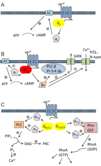
In principle, the interaction between receptor and G-protein is not strictly specific. G-protein mediated signaling can depend on the cellular background 31 and, moreover, some GPCRs are capable of coupling to multiple G-proteins.32,33 An overview of typical signaling patterns of GPCRs is given in Figure 1.4.
Activated G-proteins regulate the function of different effectors. The most widely studied effector are isoforms of adenylyl cyclase (AC). All of the nine ACs are activated by the Gαs
subunit, resulting in an increase in intracellular cAMP. It is noteworthy that some of the ACs can additionally be influenced by Gβγ subunits via calcium-calmodulin.23 The Gαs family includes the ubiquitously expressed Gαs subunit and the olfactory α subunit Gαolf.
The G-proteins of the Gαi/o family are widely expressed, and the main function of the Gαi
subunit is the inhibition of most of the ACs. The role of this family of G-proteins has extensively been studied with pertussis toxin (PTX), resulting in ADP-ribosylation and preventing the coupling of Gαi to the receptor.34 Because of high expression, the activation of
General Introduction
5 Gαi leads to a release of high amounts of βγ subunits. Therefore, Gαi activation is believed to be the major pathway resulting in Gβγ mediated signaling.35
The Gαq and Gα11 subunits are almost ubiquitously expressed, whereas Gα14 and Gα15/16
show a rather limited expression pattern.35 Gαq/11 coupled receptors do not discriminate between the Gαq and the Gα11 subunit.36,37 Both subunits are efficient activators of the isoforms β1, β3 and β4 of PLC, but not of the β2 isoform.38 While the role of Gαq and Gα11 in biological processesis well understood, the function of the other two members of the Gαq/11
family is still unclear.35
The investigation of the function of the G12/13 family is compromised by the lack of selective inhibitors. G12 and G13 are often stimulated by receptors simultaneously activating Gαq/11. Studies revealed that G12/G13 can alter a variety of downstream effectors including phospholipase A2 (PLA2) and the Na+/H+ exchanger.39,40 Moreover, they regulate the formation of actomyosin-based structures and modulate their contractility by increasing the activity of the small GTPase RhoA (Ras homolog gene family, member A).41
Figure 1.4. Typical pattern of GPCRs coupling. (A) Gs mediated signaling (AC = adenylyl cyclase; ATP = adenosine triphosphate; cAMP = cyclic adenosine monophosphate) (B) Gi/o mediated signaling (GIRK = G- protein-regulated inward rectifier potassium channel; PI-3-K = phosphoinositide-3-kinase; PLC-ß = phospholipase C-β (C) Gq/11 and G12/13 mediated signaling (DAG = diacylglycerol; GDP = guanosine diphosphate;
GTP = guanosine triphosphate; IP3 = inositol 1,4,5-trisphosphate; PIP2 = phosphatidylinositol 4,5-bisphosphate;
PKC = protein kinase C; Rho-GEF = Rho-guanine nucleotide exchange factor; RhoA = Ras homolog gene family, member A) (adopted and modified from Wettschureck et al. 2005).35
6
1.2 Histamine and its receptors 1.2.1 The biogenic amine histamine
1.2.1.1 Brief historyMore than 100 years ago, the British physiologist Sir Henry Dale discovered histamine as a constituent of ergot. Subsequently, histamine was identified as endogenous substance in the body (“histos” = “tissue”).42 During the next decade, the pharmacological effects of histamine were extensively studied, and in 1937 the first antihistamines were synthesized by Daniel Bovet and Anne-Marie Staub.43 Five years later, the first H1R antagonists were introduced into therapy.
1.2.1.2 Biosynthesis, metabolism and function
In the body, histamine is formed from the amino acid L-histidine by the action of histidine decarboxylase (HDC).44 After liberation, histamine is rapidly cleaved by histamine N- methyltransferase (HNMT)45 or diamine oxidase (DAO).46 As a neurotransmitter, histamine plays a crucial role in brain functions, including circadian rhythm, nociception and locomotor activity.47 Furthermore, histamine regulates secretion of pituitary hormones, gastrointestinal and circulatory functions, and it is involved in inflammatory reactions and the modulation of the immune response.47 Histamine is mainly stored in the granules of mast cells48 and basophils49 and can be released in large amounts upon degranulation in response to various stimuli. Another depot are the enterochromaffin-like (ECL) cells in the stomach. Histamine released from ECL cells, regulates gastric acid secretion from parietal cells.50 The effects of histamine are mediated by four histamine receptor (HxR) subtypes, termed H1R, H2R, H3R and H4R, all of them belonging to class A GPCRs.
1.2.2 The four histamine receptors and their ligands
1.2.2.1 The histamine H1RThe H1R is mainly expressed in mammalian brain, on smooth muscle and endothelium cells and on lymphocytes.51 On immune cells, the H1R is often co-expressed along with the H2R, complicating detailed studies of both receptors due to the lack of selectivity of some ligands.52 The H1R preferentially couples to the Gαq protein, resulting in an increase in intracellular Ca2+. H1R-mediated biological effects comprise vasodilatation, bronchoconstriction, modulation of endothelial barrier function, as well as pain and itching as a consequence of insect stings.53 The first H1R antagonists, usually referred to as “classical antihistamines” (e.g. mepyramine, diphenhydramine) against allergic diseases, were already established in the 1940s. These 1st generation H1R antagonists are highly lipophilic compounds, able to pass the blood brain barrier and to cause sedation. For that reason, antagonists such as cetirizine and loratadine were developed that cross the blood brain barrier to a much lower degree. This 2nd generation of antihistamines still represents blockbuster drugs for the treatment of allergic disorders (Fig 1.5).54
General Introduction
7
Fig. 1.5. Chemical structures of selected H1R antagonists
The only H1R agonist in clinical use is betahistine (Fig. 1.6), a drug for the treatment of Meniere`s disease.55 Highly potent H1R agonists were developed as pharmacological tools.
Potent agonists, superior to histamine, were obtained by structural optimization of 2- phenylhistamines.56,57 The most successful approach to potent and selective H1R agonists resulted in histaprodifen derivatives, especially suprahistaprodifen.58-61
Figure 1.6. Chemical structures of selected H1R agonists
1.2.2.2 The histamine H2R
Up to the late 1940s, scientists had no explanation why some of the histamine-mediated effects, for example the positive chronotropic response and the stimulation of gastric acid secretion, could not be antagonized by the classical antihistamines.62,63 In 1966, Ash and Schild postulated the existence of a second receptor subtype,64 which was pharmacologically
8
characterized and termed H2R in 1972 by the Nobel laureate Sir James Black.65 The H2R is located in mammalian brain, on parietal cells, on human neutrophils and eosinophils, in cardiac-, airways and uterus tissue.52,66 The H2R canonically couples to the Gαs protein resulting in the activation of PKA and in an increase in cAMP.67 After burimamide,65 the first antagonist used to define the H2R, H2R antagonists with increased affinity, e.g. cimetidine, famotidine and ranitidine, revolutionized the treatment of gastric and duodenal ulcer (Fig.
1.7).68 Nowadays, these drugs have been replaced by proton pump inhibitors. Nevertheless, there is a great interest in CNS permeable H2R ligands to elucidate the role of the H2R in the CNS.63
Figure 1.7. Chemical structures of selected H2R antagonists
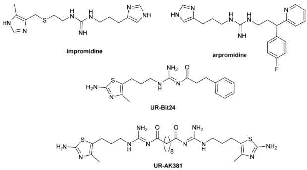
The guanidine derivative impromidine was the first highly potent H2R agonist. A large number of impromidine analogues has been synthesized and characterized with respect to H2R agonism.69 Impromidine was the first H2R agonist clinically tested in patients suffering from severe catecholamine-refractory congestive heart failure.70,71 Structural modification of impromidine led to arpromidine, in which the cimetidine-like moiety was replaced by a more lipophilic pheniramine-like structure. Such compounds showed up to 400 times the potency of histamine.72 With respect to oral bioavailability, the strongly basic guanidine (pKa~13) was replaced by an acylguanidine (pKa~8). Surprisingly, agonistic potency was retained or even increased (e.g. UR-Bit24).73,74 A tremendous increase in potency was achieved according to the bivalent ligand approach. Bivalent H2R agonists (e.g. UR-AK381) were up to 4000 times more potent than histamine and are the most potent H2R agonists known so far (Fig. 1.8).75 Highly selective H2R agonists might be beneficial for patients suffering from acute myeloid leukemia (AML).76,77 In myeloid cells histamine facilitates T-cell mediated killing of tumor cells and is therefore used as an orphan drug in combination with interleukin 2 (IL2) for maintenance treatment of AML.78
General Introduction
9
Figure 1.8. Chemical structures of selected H2R agonists
1.2.2.3 The histamine H3R
In 1983, Schwartz and coworkers suggested the existence of a third histamine receptor.
They discovered that histamine was able to inhibit its own release from cerebral neurons and that this effect was antagonized by burimamide at nanomolar concentrations, which was far below the concentration necessary to block the H2R.79,80 The H3R is predominantly located in neurons where it acts as an autoreceptor regulating the synthesis and release of histamine.
Additionally, the H3R also occurs as a hetero-receptor on non-histaminergic neurons, controlling the release of other neurotransmitters including dopamine, acetylcholine and GABA.63 In the periphery, the receptor is expressed in the gastrointestinal tract, the airways and the cardiovascular system.81 The H3R couples to Gi proteins and has been shown to interfere with various transduction pathways apart from the modulation of the AC activity, for example activation of PLA2 and inhibition of K+-induced Ca2+ activation.82 The first potent H3R antagonists were thioperamide and clobenpropit, which turned out to be additionally highly potent H4R ligands. Moreover, as imidazole-type ligands, these compounds harbor the risk of drug-drug interactions due to interactions with cytochrome P-450 enzymes. With the discovery of high constitutive activity, numerous H3R antagonists had to be re-classified as inverse agonists. To reduce the off-target effects and to improve the drug-like properties, the imidazole ring was replaced mainly by a variety of secondary and tertiary amines, including cyclic amine moieties (e.g. piperazino, pyrrolidino and morpholino).83 Potential therapeutic implications for H3R antagonists (Fig. 1.9) include obesity and numerous CNS disorders such as schizophrenia, epilepsy and Alzheimer`s disease. Very recently, the first H3R inverse agonist pitolisant was introduced as an orphan drug for patients suffering from narcolepsy.84,85
10
Fig 1.9. Chemical structures of selected H3R antagonists/inverse agonists
Modifications of histamine by mono- or dimethyl substitution resulted in more potent and selective H3R agonists (Fig. 1.10), such as (R)-α-methylhistamine. A further improvement regarding selectivity over the H4R was achieved with immethridine86 and methimepip87. All of the H3R agonists represent histamine derivatives, which carry the risk of drug-drug interactions. Bioisosteric replacement of the imidazole moiety in H3R agonists harbors a potential for new drugs, for example, for the treatment of migraine, ischemic arrhythmias, asthma or diabetes mellitus.88,89
Figure 1.10. Chemical structures of selected H3R agonists
1.2.2.4 The histamine H4R
In 1994, Raible and coworkers suggested the existence of a novel histamine receptor on human eosinophils, based on a histamine induced calcium response.90 This hypothesis was corroborated by several workgroups, which independently cloned the H4R.91-95 The H4R was reported to be expressed in bone marrow and immunocytes, such as mast cells, eosinophils and monocytes.52 The identification of the H4R in the CNS96 was not confirmed by several workgroups.97 Additionally, according to a recently published study, there is no evidence for the expression of the H4R in human monocytes.98 Presumably, the erroneous detection of the H4R in brain and monocytes was due to the use of unspecific antibodies and unreliable data on mRNA expression.97-100 The H4R is a Gi coupled receptor and mediates an increase in intracellular Ca2+ concentration. The indole derivative JNJ7777120 was the first selective and potent non-imidazole H4R antagonist described in the literature and has been widely used as a reference compound in vitro and in vivo.101 Surprisingly, JNJ7777120 turned out to be a biased agonist, stimulating H4R mediated β-arrestin recruitment.102 Five H4R antagonists (e.g. UR-63325 [undisclosed structure], PF-3893787103, JNJ39758979104) (Fig.1.11) entered clinical trials for the treatment of allergic rhinitis, allergic asthma, atopic dermatitis and rheumatoid arthritis.105 Despite significantly antipruritic effects in human, the clinical trial of JNJ39758979 was terminated due to two agranulocytosis cases.106,107
General Introduction
11
Figure 1.11. Chemical structures of selected H4R antagonists
In contrast to H4R antagonists, the potential therapeutic value of H4R agonists (Fig 1.12) is still a matter of speculation. Regardless of that, further investigation of the (patho)physiological role of the H4R requires selective and potent H4R agonists as molecular tools. Due to the high sequence homology with the H3R (~ 58 % identity in the TM regions)94 the design of selective H4R proved to be rather challenging. The cyanoguanidine OUP-16 was the first ligand with improved (approximately 30-fold) H4R selectivity. Structural optimization of imidazolylalkylguanidines led to the highly potent and selective H4R agonists, like e.g. UR-PI376.108 One of the most potent H4R agonists known so far was identified among a series of 2-arylbenzimidazoles.109 Potencies and efficacies of numerous H4R agonists are depending on the species considered.110 In this context, oxime-type compounds, structurally derived from JNJ7777120, proved to be of special value. The Z- configured isomers have comparable agonist potencies on human and rodent H4Rs.111
Figure 1.12. Chemical structures of selected H4R agonists
1.3 GPCR dimerization and bivalent ligands 1.3.1 GPCR dimerization
Over a long period of time, it has been assumed that GPCRs exist and function as monomers. However, there is growing evidence that GPCRs are able to form dimers or oligomers. The first receptor shown to form dimers was the GABAB receptor. Only when coexpressed with GABAB2, the GABAB1 receptor is capable of forming a functional GABAB
12
receptor.112-114 Meanwhile, many GPCRs are known to form hetero- and homodimers, respectively (for reviews, cf. Agnati et al. 2003, Hiller et al. 2013).115,116 Receptor dimerization can induce distinct ligand binding properties. For example, it was shown for δOR and µOR coexpressing cells that there was a significant increase in binding of µOR ligands in the presence of δOR antagonists.117 This phenomenon can be explained by positive cooperativity: binding of one ligand to the first receptor molecule increases the interaction of the second ligand to the second receptor protomer (Fig. 1.13A). Receptor dimerization can alter signaling, e.g., induce a switch in G protein coupling. For example, the D1R canonically couples to Gαs, and the D2R couples to Gαi, whereas heterodimers of the two receptors preferentially activate Gαq.118 It is noteworthy that dimerization can also cause a switch from G-protein activation to β-arrestin signaling.119 Furthermore, GPCR dimerization may have an influence on trafficking. For instance, coexpression of the α1D- and the α1B adrenoceptor led to a 10-fold increase in cell surface expression of the α1D-AR, suggesting that heterodimerization enhances the transport of α1D-AR to the cell membrane.120 Drugs selectively targeting receptor dimers might possess higher affinity and selectivity and/or an improved pharmacokinetic profile resulting in reduced side effects.121,122
1.3.2 Bivalent ligands
One possibility to target GPCR dimers is the bivalent ligand approach. Typically, the term
“bivalent ligands” refers to molecules composed of two pharmacophoric moieties linked through a spacer.123 Based on the pioneering work of Porthogese and coworkers, who developed the first bivalent ligands for the investigation of the dimerization of opioid receptors,124-126 the bivalent ligand approach was successfully adopted for many GPCRs, including e.g. serotonin, histamine and dopamine,75,127-131 resulting in compounds with higher affinity and selectivity compared to their monovalent counterparts. The two pharmacophoric entities can either be structurally equal (homobivalent) or different (heterobivalent).
Obviously, the length of the spacer is a key factor for bridging a receptor dimer (Fig. 1.13B).
A very short or a very long spacer decreases the potential to bridge two vicinal receptor protomers.132 Porthogese et al. observed highest agonist potency at a spacer length of 18 atoms (~20 Å).133 However, newer studies revealed that the optimum spacer length for bivalent ligands targerting receptor dimers depends on the dimer interface, the structure of pharmacophores and topicity of the point of attachments.116 Thus, bivalent ligands with clozapine as pharmacophore displayed highest affinity with a spacer length of 16 and 18 atoms.134 Interestingly, the highest potency for bivalent H2R agonists was achieved with compounds bearing an octa- or hexamethylene chain, rather indicating to an additional binding site at the same receptor molecule (Fig. 1.13C).75
Figure 1.13. Possible binding modes of bivalent ligands. (A) Binding to two receptor protomers (B) Bridging of a receptor dimer (C) Simultaneous binding to an orthosteric and allosteric binding site of the same receptor molecule (bitopic or dualsteric binding mode) (adopted and modified from Bonger et al. 2007).135
General Introduction
13 Previously, our workgroup applied the guanidine – acylguanidine bioisosteric approach to imidazolylpropylguanidine-type histamine H2 receptor (H2R) agonists73,136 and explored the bivalent ligand approach, taking into consideration the hypothesis that GPCRs, including histamine receptor subtypes, can form functionally active dimers. Acylation of the guanidine group reduced the basicity by 4-5 orders of magnitude. Surprisingly, agonistic potency was retained or even increased. In combination with a bivalent approach, this strategy resulted in highly potent H2R agonists. The binding mode of these compounds is far from being understood. The optimal spacer length of the most active bivalent H2R agonists is most probably insufficient to enable simultaneous occupation of the recognition sites of putative H2R dimers.75 Probably, the tremendous gain in potency results from interaction with an accessory binding site at the same receptor protomer.
1.4 Functional selectivity and G-protein independent signaling
Besides G-proteins, two additional protein families are known to interact with active state GPCRs: G-protein coupled receptor kinases (GRKs) and β-arrestins.137 Whereas originally, the role of these proteins was simply associated with desensitization, internalization and recycling of GPCRs,138 they are nowadays considered as alternative signal transducers.137,139 Particularly, β-arrestins are known to interact with many proteins and protein kinases, leading to the phosphorylation of numerous targets.140,141
Over the last decades, a growing number of experimental data supported the concept of
“biased signaling” as a consequence of various active states of an individual receptor.
According to the classical model, agonists are considered to have “linear efficacy”, i.e. a pharmacological activity resulting from a sequence beginning with receptor occupancy and G-protein activation.142 However, some ligands show “imbalanced efficacies” regarding the activation of different signaling pathways (Fig. 1.14A).141 Such ligands proved to be antagonists on one pathway, while simultaneously being agonists on another pathway.143,144 To consider such a behavior, the classical two-state model was extended to a multiple-state model (Fig. 1.14B). According to the latter, a given ligand is capable of stabilizing a unique receptor conformation, either triggering the G-protein or the β-arrestin pathway.141,142,145,146
Figure 1.14. (A) Concept of functional selectivity: A given ligand (1-4) stabilizes a unique active receptor conformation (R*1-R*4). The distinct receptor conformations differ in their capability to interact with different G- proteins (G1-G3) and β-arrestins (βArr). As a result, effector systems (E1-E4) are regulated in a ligand specific manner (adopted from Seifert 2013).146 (B) Multiple-state model (L = ligand; Ran = distinct active receptor conformations; Ri = inactive state of the receptor; Tn = distinct transducers) (adopted from Rajagopal et al.
2010)29
14
The ability of a ligand to address either G-protein signaling or β-arrestin signaling is referred to as “functional selectivity”, “biased agonism” or “collateral efficacy” (Fig. 1.15).147,148 By now, functionally selective ligands were discovered for many GPCRs, e.g. for β-adrenergic receptors,149,150 dopamine D2 and D3 receptors151,152 and the histamine H4R.153,154 Such biased agonists harbor a great potential for the development of novel therapeutic agents with reduced adverse effects. Studies already revealed that the pharmacology of some of these functionally selective ligands was different from that of unbiased compounds.148,155,156
Figure 1.15. Functionally selective ligands: (A) Binding of an unbiased ligand leads to G-protein activation and β-arrestin recruitment in a balanced manner. (B) Binding of a G-protein biased ligand. (C) Binding of a β-arrestin biased ligand (βArr = β-arrestin; GRK = G-protein receptor coupled kinase) (adopted from Rajagopal et al.
2010).29
General Introduction
15
1.5 References
1. Lefkowitz, R. J. Historical review: A brief history and personal retrospective of seven- transmembrane receptors. Trends Pharmacol. Sci. 2004, 25, 413-422.
2. Liapakis, G.; Cordomi, A.; Pardo, L. The G-protein coupled receptor family: actors with many faces (vol 18, pg 175, 2012). Curr. Pharm. Des. 2012, 18, 4583-4583.
3. Sharman, J. L.; Benson, H. E.; Pawson, A. J.; Lukito, V.; Mpamhanga, C. P.; Bombail, V.; Davenport, A. P.; Peters, J. A.; Spedding, M.; Harmar, A. J.; Nc-Iuphar. IUPHAR- DB: updated database content and new features. Nucleic Acids Res. 2013, 41, D1083-D1088.
4. Furchgott, R. F. Receptor mechanisms. Annu. Rev. Pharmacol. 1964, 4, 21-50.
5. Dixon, R. A.; Kobilka, B. K.; Strader, D. J.; Benovic, J. L.; Dohlman, H. G.; Frielle, T.;
Bolanowski, M. A.; Bennett, C. D.; Rands, E.; Diehl, R. E.; Mumford, R. A.; Slater, E.
E.; Sigal, I. S.; Caron, M. G.; Lefkowitz, R. J.; Strader, C. D. Cloning of the gene and cDNA for mammalian beta-adrenergic receptor and homology with rhodopsin. Nature 1986, 321, 75-79.
6. Palczewski, K.; Kumasaka, T.; Hori, T.; Behnke, C. A.; Motoshima, H.; Fox, B. A.; Le Trong, I.; Teller, D. C.; Okada, T.; Stenkamp, R. E.; Yamamoto, M.; Miyano, M.
Crystal structure of rhodopsin: A G protein-coupled receptor. Science 2000, 289, 739- 745.
7. Warne, T.; Serrano-Vega, M. J.; Baker, J. G.; Moukhametzianov, R.; Edwards, P. C.;
Henderson, R.; Leslie, A. G.; Tate, C. G.; Schertler, G. F. Structure of a beta1- adrenergic G-protein-coupled receptor. Nature 2008, 454, 486-491.
8. Cherezov, V.; Rosenbaum, D. M.; Hanson, M. A.; Rasmussen, S. G.; Thian, F. S.;
Kobilka, T. S.; Choi, H. J.; Kuhn, P.; Weis, W. I.; Kobilka, B. K.; Stevens, R. C. High- resolution crystal structure of an engineered human beta2-adrenergic G protein- coupled receptor. Science 2007, 318, 1258-1265.
9. Haga, K.; Kruse, A. C.; Asada, H.; Yurugi-Kobayashi, T.; Shiroishi, M.; Zhang, C.;
Weis, W. I.; Okada, T.; Kobilka, B. K.; Haga, T.; Kobayashi, T. Structure of the human M2 muscarinic acetylcholine receptor bound to an antagonist. Nature 2012, 482, 547- 551.
10. Kruse, A. C.; Hu, J.; Pan, A. C.; Arlow, D. H.; Rosenbaum, D. M.; Rosemond, E.;
Green, H. F.; Liu, T.; Chae, P. S.; Dror, R. O.; Shaw, D. E.; Weis, W. I.; Wess, J.;
Kobilka, B. K. Structure and dynamics of the M3 muscarinic acetylcholine receptor.
Nature 2012, 482, 552-556.
11. Thompson, A. A.; Liu, W.; Chun, E.; Katritch, V.; Wu, H.; Vardy, E.; Huang, X. P.;
Trapella, C.; Guerrini, R.; Calo, G.; Roth, B. L.; Cherezov, V.; Stevens, R. C. Structure of the nociceptin/orphanin FQ receptor in complex with a peptide mimetic. Nature 2012, 485, 395-399.
12. Wu, H.; Wacker, D.; Mileni, M.; Katritch, V.; Han, G. W.; Vardy, E.; Liu, W.;
Thompson, A. A.; Huang, X. P.; Carroll, F. I.; Mascarella, S. W.; Westkaemper, R. B.;
Mosier, P. D.; Roth, B. L.; Cherezov, V.; Stevens, R. C. Structure of the human kappa-opioid receptor in complex with JDTic. Nature 2012, 485, 327-332.
13. Manglik, A.; Kruse, A. C.; Kobilka, T. S.; Thian, F. S.; Mathiesen, J. M.; Sunahara, R.
K.; Pardo, L.; Weis, W. I.; Kobilka, B. K.; Granier, S. Crystal structure of the micro- opioid receptor bound to a morphinan antagonist. Nature 2012, 485, 321-326.
14. Granier, S.; Manglik, A.; Kruse, A. C.; Kobilka, T. S.; Thian, F. S.; Weis, W. I.; Kobilka, B. K. Structure of the delta-opioid receptor bound to naltrindole. Nature 2012, 485, 400-404.
15. Shimamura, T.; Shiroishi, M.; Weyand, S.; Tsujimoto, H.; Winter, G.; Katritch, V.;
Abagyan, R.; Cherezov, V.; Liu, W.; Han, G. W.; Kobayashi, T.; Stevens, R. C.; Iwata, S. Structure of the human histamine H1 receptor complex with doxepin. Nature 2011, 475, 65-70.
16. Lappano, R.; Maggiolini, M. G protein-coupled receptors: novel targets for drug discovery in cancer. Nat. Rev. Drug Discov. 2011, 10, 47-60.
16
17. Wise, A.; Jupe, S. C.; Rees, S. The identification of ligands at orphan G-protein coupled receptors. Annu. Rev. Pharmacol. Toxicol. 2004, 44, 43-66.
18. Fredriksson, R.; Lagerstrom, M. C.; Lundin, L. G.; Schioth, H. B. The G-protein- coupled receptors in the human genome form five main families. Phylogenetic analysis, paralogon groups, and fingerprints. Mol. Pharmacol. 2003, 63, 1256-1272.
19. Pierce, K. L.; Premont, R. T.; Lefkowitz, R. J. Seven-transmembrane receptors. Nat Rev Mol Cell Bio 2002, 3, 639-650.
20. Vassilatis, D. K.; Hohmann, J. G.; Zeng, H.; Li, F. S.; Ranchalis, J. E.; Mortrud, M. T.;
Brown, A.; Rodriguez, S. S.; Weller, J. R.; Wright, A. C.; Bergmann, J. E.; Gaitanaris, G. A. The G protein-coupled receptor repertoires of human and mouse. Proc. Natl.
Acad. Sci. U. S. A. 2003, 100, 4903-4908.
21. Tang, X. L.; Wang, Y.; Li, D. L.; Luo, J.; Liu, M. Y. Orphan G protein-coupled receptors (GPCRs): biological functions and potential drug targets. Acta Pharmacol.
Sin. 2012, 33, 363-371.
22. Kobilka, B. K. G protein coupled receptor structure and activation. Biochim. Biophys.
Acta 2007, 1768, 794-807.
23. Luttrell, L. M. Reviews in molecular biology and biotechnology: transmembrane signaling by G protein-coupled receptors. Mol. Biotechnol. 2008, 39, 239-264.
24. Baldwin, J. M. Structure and function of receptors coupled to G proteins. Curr. Opin.
Cell Biol. 1994, 6, 180-190.
25. Seifert, R., Wieland T. G Protein‐Coupled Receptors as Drug Targets: Analysis of Activation and Constitutive Activity. Wiley‐VCH, Weinheim 2005.
26. Delean, A.; Stadel, J. M.; Lefkowitz, R. J. A ternary complex model explaines the agonist-specific binding-properties of the adenylate cyclase-coupled beta-adrenergic- receptor. J. Biol. Chem. 1980, 255, 7108-7117.
27. Samama, P.; Cotecchia, S.; Costa, T.; Lefkowitz, R. J. A mutation-induced activated state of the beta 2-adrenergic receptor. Extending the ternary complex model. J. Biol.
Chem. 1993, 268, 4625-4636.
28. Leff, P. The two-state model of receptor activation. Trends Pharmacol. Sci. 1995, 16, 89-97.
29. Rajagopal, S.; Rajagopal, K.; Lefkowitz, R. J. Teaching old receptors new tricks:
biasing seven-transmembrane receptors. Nature Reviews Drug Discovery 2010, 9, 373-386.
30. Kenakin, T. A Pharmacology Primer. Elsevier 2014, 28.
31. Schmidt, C. J.; Thomas, T. C.; Levine, M. A.; Neer, E. J. Specificity of G-protein-beta subunit and gamma subunit interactions
J. Biol. Chem. 1992, 267, 13807-13810.
32. Eason, M. G.; Kurose, H.; Holt, B. D.; Raymond, J. R.; Liggett, S. B. Simultaneous Coupling of Alpha-2-Adrenergic Receptors to 2 G-Proteins with Opposing Effects - Subtype-Selective Coupling of Alpha-2c10, Alpha-2c4, and Alpha-2c2 Adrenergic- Receptors to G(I) and G(S). J. Biol. Chem. 1992, 267, 15795-15801.
33. Galandrin, S.; Bouvier, M. Distinct signaling profiles of beta(1) and beta(2) adrenergic receptor ligands toward adenylyl cyclase and mitogen-activated protein kinase reveals the pluridimensionality of efficacy. Mol. Pharmacol. 2006, 70, 1575-1584.
34. Mangmool, S.; Kurose, H. G(i/o) protein-dependent and -independent actions of Pertussis Toxin (PTX). Toxins (Basel) 2011, 3, 884-899.
35. Wettschureck, N.; Offermanns, S. Mammalian G proteins and their cell type specific functions. Physiol. Rev. 2005, 85, 1159-1204.
36. Watson, S. P.; Asazuma, N.; Atkinson, B.; Berlanga, O.; Best, D.; Bobe, R.; Jarvis, G.; Marshall, S.; Snell, D.; Stafford, M.; Tulasne, D.; Wilde, J.; Wonerow, P.;
Frampton, J. The role of ITAM- and ITIM-coupled receptors in platelet activation by collagen. Thromb. Haemost. 2001, 86, 276-288.
37. Wu, D. Q.; Katz, A.; Lee, C. H.; Simon, M. I. Activation of Phospholipase-C by Alpha- 1-Adrenergic Receptors Is Mediated by the Alpha-Subunits of Gq Family. J. Biol.
Chem. 1992, 267, 25798-25802.
General Introduction
17 38. Rhee, S. G. Regulation of phosphoinositide-specific phospholipase C. Annu. Rev.
Biochem 2001, 70, 281-312.
39. Dhanasekaran, N.; Dermott, J. M. Signaling by the G(12) class of G proteins. Cell.
Signal. 1996, 8, 235-245.
40. Hooley, R.; Yu, C. Y.; Symons, M.; Barber, D. L. G alpha 13 stimulates Na+-H+
exchange through distinct Cdc42-dependent and RhoA-dependent pathways. J. Biol.
Chem. 1996, 271, 6152-6158.
41. Buhl, A. M.; Johnson, N. L.; Dhanasekaran, N.; Johnson, G. L. G-Alpha(12) and G- Alpha(13) Stimulate Rho-Dependent Stress Fiber Formation and Focal Adhesion Assembly. J. Biol. Chem. 1995, 270, 24631-24634.
42. H. H. Dale, P. P. L. The physiological action of β-Imidazolethylamine. J. Physiol.
1910, 41, 318–344.
43. D. Bovet, A. S. Action protectrice des éthers phenoliques au cours l'intoxication histaminique. C.r. séances Soc. biol. ses. fil. 1937, 124, 547-549.
44. Ackermann, D. Z Physiol Chem 1910, 60, 482-501.
45. Schwartz, J. C.; Arrang, J. M.; Garbarg, M.; Pollard, H.; Ruat, M. Histaminergic transmission in the mammalian brain. Physiol. Rev. 1991, 71, 1-51.
46. Beaven, M. A. Factors Regulating Availability of Histamine at Tissue Receptors Pharmacology of Histamine Receptors 1982, Ganellin, C.R., Parsons, M.E. (Eds.).
Wright PSG, Bristol, London, Boston, 102-145.
47. Dy, M.; Schneider, E. Histamine-cytokine connection in immunity and hematopoiesis.
Cytokine Growth Factor Rev. 2004, 15, 393-410.
48. Riley, J. F.; West, G. B. The presence of histamine in tissue mast cells. J. Physiol.
1953, 120(4), 528–537.
49. Falcone, F. H.; Zillikens, D.; Gibbs, B. F. The 21st century renaissance of the basophil? Current insights into its role in allergic responses and innate immunity. Exp.
Dermatol. 2006, 15, 855-864.
50. Mossner, J.; Caca, K. Developments in the inhibition of gastric acid secretion. Eur. J.
Clin. Invest. 2005, 35, 469-475.
51. Hill, S. J. Distribution, Properties, and Functional-Characteristics of 3 Classes of Histamine-Receptor. Pharmacol. Rev. 1990, 42, 45-83.
52. Seifert, R.; Strasser, A.; Schneider, E. H.; Neumann, D.; Dove, S.; Buschauer, A.
Molecular and cellular analysis of human histamine receptor subtypes. Trends Pharmacol. Sci. 2013, 34, 33-58.
53. Bongers, G.; de Esch, I.; Leurs, R. Molecular Pharmacology of the Four Histamine Receptors. Histamine in Innflammation 2010, 709, 11-19.
54. Simons, F. E. R.; Simons, K. J. Histamine and H-1-antihistamines: Celebrating a century of progress. J. Allergy Clin. Immunol. 2011, 128, 1139-1152.
55. Barak, N. Betahistine: whats new on the agenda? Expert Opin Inv Drug 2008, 17, 795-804.
56. Zingel, V.; Elz, S.; Schunack, W. Histamine Analogs .33. 2-Phenylhistamines with High Histamine H-1-Agonistic Activity. Eur. J. Med. Chem. 1990, 25, 673-680.
57. Leschke, C.; Elz, S.; Garbarg, M.; Schunack, W. Synthesis and Histamine H-1 Receptor Agonist Activity of a Series of 2-Phenylhistamines, 2-Heteroarylhistamines, and Analogs. J. Med. Chem. 1995, 38, 1287-1294.
58. Elz, S.; Kramer, K.; Pertz, H. H.; Detert, H.; ter Laak, A. M.; Kuhne, R.; Schunack, W.
Histaprodifens: Synthesis, pharmacological in vitro evaluation, and molecular modeling of a new class of highly active and selective histamine H-1-receptor agonists. J. Med. Chem. 2000, 43, 1071-1084.
59. Carman-Krzan, M.; Bavec, A.; Zorko, M.; Schunack, W. Differences in the binding characteristic and intracellular effects of specific H-1 agonists-histaprodifens in the cardiovascular tissue. Fundam. Clin. Pharmacol. 2001, 15, 72-72.
60. Strasser, A.; Striegl, B.; Wittmann, H.-J.; Seifert, R. Pharmacological profile of histaprodifens at four recombinant histamine H-1 receptor species isoforms. J.
Pharm. Exp. Ther. 2008, 324, 60-71.
18
61. Menghin, S.; Pertz, H. H.; Kramer, K.; Seifert, R.; Schunack, W.; Elz, S. N-alpha- imidazolylalkyl and pyridylalkyl derivatives of histaprodifen: Synthesis and in vitro evaluation of highly potent histamine H-1-receptor agonists. J. Med. Chem. 2003, 46, 5458-5470.
62. Ashford, C. A.; Heller, H.; Smart, G. A. The action of histamine on hydrochloric acid and pepsin secretion in man. Br. J. Pharmacol. Chemother. 1949, 4(2), 153–156.
63. Parsons, M. E.; Ganellin, C. R. Histamine and its receptors. Br. J. Pharmacol. 2006, 147, S127-S135.
64. Ash, A. S. F.; Schild, H. O. Receptors Mediating Some Actions of Histamine. Br. J.
Pharmacol. Chemother. 1966, 27, 427-&.
65. Black, J. W.; Parsons, E. M.; Durant, C. J.; Duncan, W. A. M.; Ganellin, C. R.
Definition and Antagonism of Histamine H2-Receptors. Nature 1972, 236, 385-390.
66. Hill, S. J.; Ganellin, C. R.; Timmerman, H.; Schwartz, J. C.; Shankley, N. P.; Young, J.
M.; Schunack, W.; Levi, R.; Haas, H. L. International Union of Pharmacology. XIII.
Classification of histamine receptors. Pharmacol. Rev. 1997, 49, 253-278.
67. Johnson, C. L.; Weinstein, H.; Green, J. P. Studies on Histamine H-2 Receptors Coupled to Cardiac Adenylate-Cyclase - Blockade by H-2 and H-1 Receptor Antagonists. Mol. Pharmacol. 1979, 16, 417-428.
68. Cataldi, M.; Borriello, F.; Granata, F.; Annunziato, L.; Marone, G. Histamine receptors and antihistamines: from discovery to clinical applications. Chem. Immunol. Allergy 2014, 100, 214-226.
69. Dove, S.; Elz, S.; Seifert, R.; Buschauer, A. Structure-activity relationships of histamine H-2 receptor ligands. Mini-Rev. Med. Chem. 2004, 4, 941-954.
70. Baumann, G.; Permanetter, B.; Wirtzfeld, A. Possible Value of H-2-Receptor Agonists for Treatment of Catecholamine-Insensitive Congestive Heart-Failure. Pharmacol.
Ther. 1984, 24, 165-177.
71. Baumann, G.; Felix, S. B.; Heidecke, C. D.; Riess, G.; Loher, U.; Ludwig, L.; Blomer, H. Apparent Superiority of H-2-Receptor Stimulation and Simultaneous Beta- Blockade over Conventional Treatment with Beta-Sympathomimetic Drugs in Post- Acute Myocardial-Infarction - Cardiac Effects of Impromidine - a New Specific H-2- Receptor Agonist - in the Surviving Catecholamine-Insensitive Myocardiumic. Agents Actions 1984, 15, 216-228.
72. Buschauer, A. Synthesis and Invitro Pharmacology of Arpromidine and Related Phenyl(Pyridylalkyl)Guanidines, a Potential New Class of Positive Inotropic Drugs. J.
Med. Chem. 1989, 32, 1963-1970.
73. Ghorai, P.; Kraus, A.; Keller, M.; Götte, C.; Igel, P.; Schneider, E.; Schnell, D.;
Bernhardt, G.; Dove, S.; Zabel, M.; Seifert, R.; Buschauer, A. Acylguanidines as bioisosteres of guanidines: N(G)-acylated imidazolylpropylguanidines, a new class of histamine H2 receptor agonists. J. Med. Chem. 2008, 51, 7193-7204.
74. Xie, S. X.; Ghorai, P.; Ye, Q. Z.; Buschauer, A.; Seifert, R. Probing ligand-specific histamine H1- and H2-receptor conformations with NG-acylated Imidazolylpropylguanidines. J. Pharmacol. Exp. Ther. 2006, 317, 139-146.
75. Birnkammer, T.; Spickenreither, A.; Brunskole, I.; Lopuch, M.; Kagermeier, N.;
Bernhardt, G.; Dove, S.; Seifert, R.; Elz, S.; Buschauer, A. The bivalent ligand approach leads to highly potent and selective acylguanidine-type histamine H(2) receptor agonists. J. Med. Chem. 2012, 55, 1147-1160.
76. Aurelius, J.; Martner, A.; Brune, M.; Palmqvist, L.; Hansson, M.; Hellstrand, K.;
Thoren, F. B. Remission maintenance in acute myeloid leukemia: impact of functional histamine H-2 receptors expressed by leukemic cells. Haematol-Hematol J 2012, 97, 1904-1908.
77. Werner, K.; Neumann, D.; Seifert, R. Analysis of the histamine H2-receptor in human monocytes. Biochem. Pharmacol. 2014, 92, 369-379.
78. Romero, A. I.; Thoren, F. B.; Aurelius, J.; Askarieh, G.; Brune, M.; Hellstrand, K. Post- consolidation immunotherapy with histamine dihydrochloride and interleukin-2 in AML.
Scand. J. Immunol. 2009, 70, 194-205.
79. Schwartz, J. C. Histamine as a Transmitter in Brain. Life Sci. 1975, 17, 503-517.


![Figure 3.12. EBAO staining of human monocytes. Monocytes were incubated with 100 µM of compounds 3.20 [B, E, H] and 3.25 [C, F, I] for 2 h](https://thumb-eu.123doks.com/thumbv2/1library_info/5555441.1689175/55.892.105.790.150.305/figure-ebao-staining-human-monocytes-monocytes-incubated-compounds.webp)
