Constitutive activity of the human histamine H 4 receptor: Molecular
modelling, binding and functional studies on wild-type and mutant H 4 R orthologs
Dissertation
zur Erlangung des Doktorgrades der Naturwissenschaften (Dr. rer. nat.) an der Fakultät für Chemie und Pharmazie
der Universität Regensburg
vorgelegt von
David Wifling
aus Bad Griesbach2015
Anleitung von Herrn Prof. Dr. A. Buschauer und Herrn Prof. Dr. S. Dove am Institut für Pharmazie der Fakultät für Chemie und Pharmazie der Universität Regensburg.
Das Promotionsgesuch wurde eingereicht im Februar 2015 Tag der mündlichen Prüfung: 13. März 2015
Prüfungsausschuss:
Prof. Dr. A. Slenczka (Vorsitzender) Prof. Dr. A. Buschauer (Erstgutachter) Prof. Dr. S. Dove (Zweitgutachter) Prof. Dr. S. Elz (Drittprüfer)
Widmung
Für meine liebste Maria
„Phantasie ist wichtiger als Wissen, denn Wissen ist begrenzt.“
Albert Einstein, 1879-1955
An dieser Stelle möchte ich mich bedanken bei:
Herrn Prof. Dr. A. Buschauer, dass ich an einem so interessanten Thema arbeiten durfte, sowie dass ich meine in silico erzeugten Hypothesen experimentell mittels Herstellung von Rezeptormutanten überprüfen durfte. Ganz herzlichen Dank auch dafür, dass ich sehr frei war in der Projektplanung, für die sehr guten Diskussionen sowie die konstruktive Kritik bei der Durchsicht dieser Arbeit und der Publikationen.
Herrn Prof. Dr. S. Dove für seine konstruktive Kritik zu diversen Modelling-Themen, bei der Interpretation der Ergebnisse sowie bei der Durchsicht dieser Arbeit und der Publikationen.
Herrn Prof. Dr. R. Seifert (Institut für Pharmakologie, Medizinische Hochschule Hannover) für seine zielführenden Beiträge unter anderem zu Western Blots und diversen Assays, sowie bei der Durchsicht der im Br. J. Pharmacol. veröffentlichten Publikation.
Herrn Prof. Dr. G. Bernhardt insbesondere für die Unterstützung bei der Bestimmung der Rezeptor- und G-Proteinexpressionslevel verschiedener Rezeptor-Konstrukte, sowie für die Durchsicht der Publikationen.
Frau PD Dr. A. Strasser und Herrn Prof. Dr. S. Dove für die Bereitstellung des auf der Kristallstruktur des inaktiven hH1R (PDB ID: 3RZE) basierenden hH4R Homologiemodells.
Herrn Prof. Dr. S. Elz für die Ermöglichung, ein Tutorium für die Studentinnen und Studenten im ersten Semester Pharmazie zu halten, sowie für die Bereitschaft als Drittprüfer an der mündlichen Prüfung teilzunehmen.
Herrn Prof. Dr. A. Slenczka für die Bereitschaft, dem Prüfungsausschuss vorzustehen.
Frau K. Löffel für die gute Zusammenarbeit sowie die zahlreichen fachlichen Diskussionen während der Anfertigung ihrer Diplomarbeit. Insbesondere danke ich für die Bereitstellung der in [3H]Histamin Sättigungs-, Bindungs- und [35S]GTPγS-Experimenten ermittelten Daten zu den von ihr generierten hH4R-F169V und mH4R-V171F Mutanten.
Herrn Dr. U. Nordemann für die Testung zweier Rezeptormutanten in der [3H]Histamin Sättigung sowie die Durchführung von [3H]Histamin Kompetitions-Bindungs Experimenten mit dem hH4R Wildtyp und fünf hH4R Mutanten. Ferner vielen Dank für die fachlichen Diskussionen.
Frau Dr. I. Brunskole und Frau K. Ladova für die Bereitstellung der Baculoviren für die hH4R-R341S und hH4R-R341E Mutanten.
Herrn Dr. P. Baumeister für die Bereitstellung von Daten zum hH4R bezüglich eines [3H]Histamin Sättigungsexperimentes sowie mehrerer [3H]Histamin Kompetitions-Bindungs Experimente mit Histamin, JNJ7777120 und VUF8430 als Kompetitor.
Herrn Dr. T. Holzammer für die Einführung in das Molecular Modelling sowie für die vielen Gespräche zu den Themen Molecular Modelling, Mutagenese und zu pharmakologischen Fragestellungen.
Herrn Dr. M. Keller für die vielen Diskussionen unter anderem zu pharmakologischen Parametern und deren Auswertung bzw. Interpretation.
meinen derzeitigen und ehemaligen Bürokollegen Herrn Dr. P. Baumeister, Frau Dr. M. Ertel, Herrn Dr. T. Holzammer, Frau Dr. A. Kaczor und Herrn Dr. M. Keller für die angenehme Atmosphäre und die vielen fachlichen Diskussionen.
Herrn Dr. P. Igel für die Bereitstellung der H4R Agonisten UR-PI294 und UR-PI376, Frau Dr.
S. Gobleder für die Bereitstellung des H4R Agonisten Isoloxapin sowie Herrn Dr. P. Baumeister für die Bestellung von [35S]GTPγS und [3H]Histamin.
Frau M. Beer-Krön sowie Frau G. Wilberg für die Kultivierung der Sf9 Zellen sowie Frau M.
Beer-Krön und Frau D. Fritsch für die Durchführung einiger Assays.
Herrn P. Richthammer für die tatkräftige Unterstützung bei technischen Problemen.
Frau U. Hasselmann, Frau S. Heinrich und Frau K. Reindl für ihre freundliche Hilfe bei vielen organisatorischen Fragen.
dem Graduiertenkolleg GRK 760 der DFG für die finanzielle Unterstützung sowie die wissenschaftliche Ausbildung.
allen Kollegen und studentischen Hilfskräften für die Vorbereitung und Durchführung zahlreicher Praktika.
Besonders danke ich meinen Eltern Ingrid und Walter, meinen Geschwistern Judith, Raphael und Manuel für ihre Unterstützung jeglicher Art, für ihr stets offenes Ohr sowie die schöne Zeit zu Hause.
Mein besonderer Dank gilt meiner lieben Freundin Maria für die Unterstützung während der Promotion, für die schönen Unternehmungen, und besonders für die Liebe, die sie mir geschenkt hat.
Contents
1 Introduction ... 1
1.1 G-protein coupled receptors ... 1
1.1.1 GPCR classes ... 2
1.1.2 Structure of G-protein coupled receptors ... 3
1.1.3 Function of G-protein coupled receptors... 4
1.1.4 Models of ligand binding and G-protein coupling ... 5
1.1.4.1 Classical model ... 5
1.1.4.2 Operational model ... 5
1.1.4.3 Ternary complex models ... 6
1.1.5 Constitutive activity of GPCRs ... 7
1.1.6 Signal transduction ... 8
1.1.6.1 Structure of G-proteins and G-protein cycle ... 8
1.1.6.2 Functions of different G-protein subtypes ... 10
1.1.6.3 β-arrestin-mediated GPCR internalization and signalling ... 11
1.2 Histamine and histamine receptors ... 13
1.2.1 Historical perspective of histamine ...13
1.2.2 Histamine: occurrence and metabolism ...14
1.2.3 Histamine receptor subtypes and ligands ...15
1.2.3.1 Homologies between the four histamine receptor subtypes ... 15
1.2.3.2 Key amino acids of the four histamine receptor subtypes ... 16
1.2.3.3 Selectivity profile of histamine receptor ligands ... 17
1.2.3.4 H1 receptor ... 18
1.2.3.5 H2 receptor ... 20
1.2.3.6 H3 receptor ... 21
1.2.3.7 H4 receptor ... 22
1.3 Species differences ... 26
1.3.1 Homologies between H4R species variants ...26
1.3.2 Key amino acids of the H4R species orthologs ...26
1.3.3 Pharmacological characteristics of H4R species orthologs ...28
1.4 References ... 31
2 Scope and Objectives ... 47
2.1 References ... 49
3 Computational Chemistry ... 51
3.1 Summary ... 51
3.2 Introduction ... 52
3.3 Methods ... 53
3.3.1 Available GPCR crystal structures ...53
3.3.2 H3R/H4R homology models based on the active state of the β2AR ...54
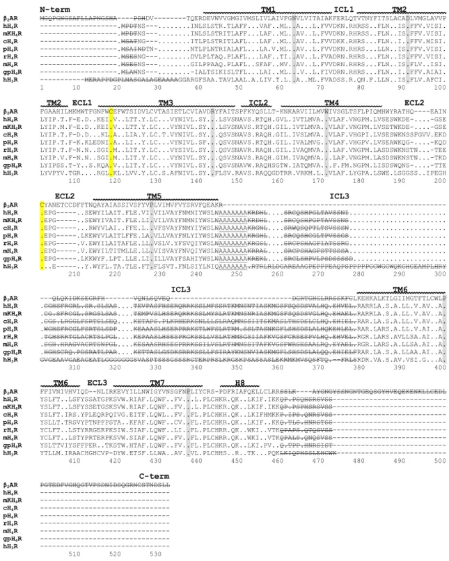
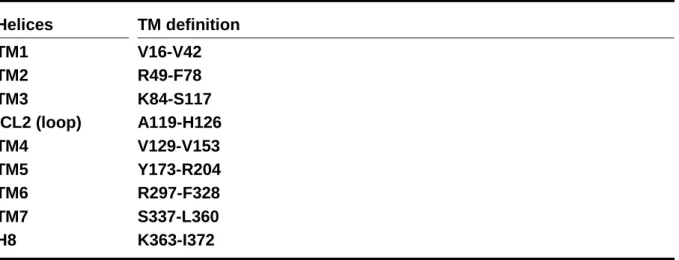
3.3.2.1 Sequence alignment ... 54
3.3.2.2 3D structure generation ... 56
3.3.3 hH4R homology model based on the inactive state of the hH1R ...57
3.3.4 Illustration in PyMOL ...58
II
3.3.5 Docking experiments ...58
3.4 Results and Discussion ... 58
3.4.1 Homology Modelling ...58
3.4.1.1 Template selection ... 58
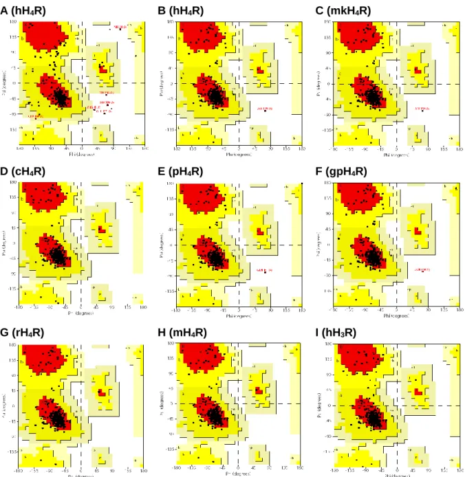
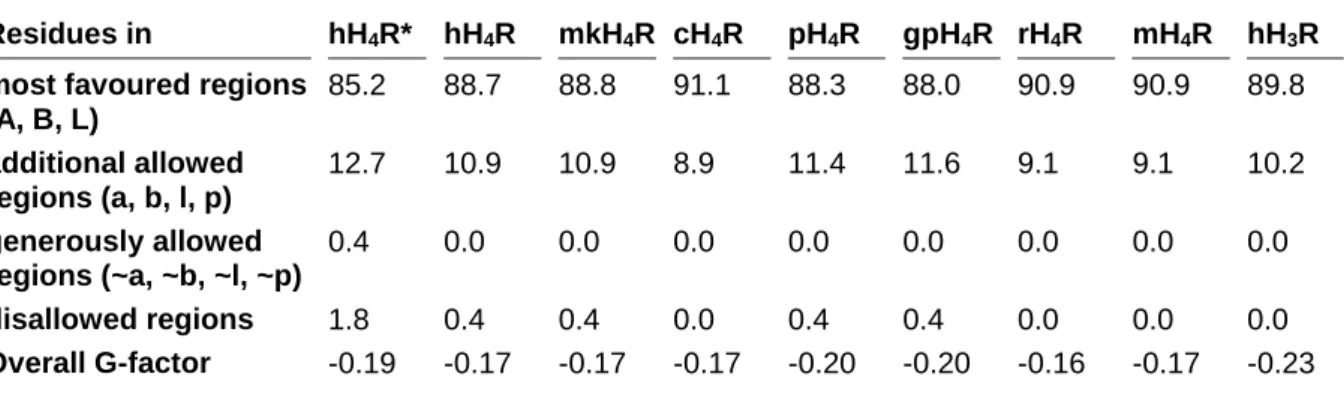
3.4.1.2 3D structure validation ... 59
3.4.2 Species differences between H4R orthologs ...61
3.4.3 Comparison of inactive and active state hH4R models ...62
3.4.4 Analysis of the binding modes of the investigated H4R ligands ...66
3.4.4.1 Ligand-free basal hH4R states ... 66
3.4.4.2 Histamine ... 68
3.4.4.3 UR-PI294 ... 68
3.4.4.4 Thioperamide ... 69
3.4.4.5 JNJ7777120 ... 70
3.4.4.6 VUF8430 ... 72
3.4.4.7 Immepip ... 73
3.4.4.8 Clozapine ... 74
3.4.4.9 Isoloxapine ... 75
3.4.4.10 UR-PI376 ... 76
3.4.4.11 Clobenpropit ... 77
3.5 Conclusions ... 78
3.6 References ... 79
4 Molecular determinants for the high constitutive activity of the human histamine H
4receptor: Functional studies on orthologs and mutants ... 87
4.1 Summary ... 87
4.2 Introduction ... 88
4.3 Methods ... 91
4.3.1 Materials ...91
4.3.2 Construction of the cDNA for hH4R-F169V, hH4R-S179A/M, hH4R-F169V+S179A/M, mH4R-V171F and mH4R-V171F+M181S...92
4.3.3 Cell culture, generation of recombinant baculoviruses, membrane preparation ...93
4.3.4 SDS-PAGE and Coomassie staining ...93
4.3.5 Western blotting ...94
4.3.6 [3H]histamine saturation binding experiments ...94
4.3.7 [3H]histamine competition binding assay ...94
4.3.8 [35S]GTPγS binding assay ...94
4.3.9 Homology model of the hH4R ...95
4.3.10 Miscellaneous ...95
4.4 Results ... 96
4.4.1 Expression of recombinant proteins ...96
4.4.2 [3H]histamine competition binding experiments ...98
4.4.3 Functional analysis of wild-type and mutant H4 receptors in the [35S]GTPγS assay ...99
4.5 Discussion ... 106
4.5.1 Affinities and potencies of the investigated ligands at H4R orthologs and mutants ... 106
4.5.2 Different quality of action of JNJ7777120 ... 109
4.5.3 Maximal effects of agonists at H4R orthologs and mutants ... 109
4.5.4 Constitutive activity ... 110
4.6 Conclusions ... 111
4.7 References ... 112
5 The extracellular loop 2 (ECL2) of the human histamine H
4receptor substantially contributes to ligand binding and constitutive activity ... 117
5.1 Summary ... 117
5.2 Introduction ... 118
5.3 Materials and Methods ... 120
5.3.1 Materials ... 120
5.3.2 Methods ... 121
5.3.2.1 Site-directed mutagenesis of the hH4R ... 121
5.3.2.2 Cell culture, generation of recombinant baculoviruses and membrane preparation ... 121
5.3.2.3 SDS-PAGE and Coomassie staining ... 121
5.3.2.4 [3H]histamine saturation binding experiments ... 122
5.3.2.5 [35S]GTPγS binding assay ... 122
5.3.2.6 Miscellaneous ... 122
5.4 Results ... 123
5.4.1 Receptor expression ... 123
5.4.2 Functional analysis of wild-type and mutant H4 receptors ... 124
5.5 Discussion ... 127
5.5.1 Potencies of ligands at mutated H4 receptors ... 127
5.5.2 Intrinsic activities of ligands and constitutive activity of receptors ... 128
5.6 Conclusions ... 129
5.7 References ... 130
6 Effect of S330R mutation in ECL3 on ligand binding and function of the human H
4R ... 135
6.1 Summary ... 135
6.2 Introduction ... 135
6.3 Materials and Methods ... 137
6.3.1 Materials ... 137
6.3.2 Site-directed mutagenesis of the hH4R ... 137
6.3.3 Cell culture, generation of recombinant baculoviruses and membrane preparation ... 137
6.3.4 SDS-PAGE and Coomassie staining ... 137
6.3.5 [3H]histamine saturation binding experiments ... 137
6.3.6 [3H]histamine competition binding assay ... 137
6.3.7 [35S]GTPγS binding assay ... 137
6.3.8 Miscellaneous ... 137
6.4 Results ... 137
IV
6.4.1 Expression of recombinant proteins ... 137
6.4.2 Competition binding data of H4R ligands at the hH4R-S330R mutant ... 139
6.4.3 Functional analysis of the hH4R-S330R mutant compared to wild-type H4Rs in the [35S]GTPγS assay ... 139
6.5 Discussion ... 142
6.5.1 Affinities and potencies of the investigated ligands at H4R wild-types and the hH4R-S330R mutant ... 142
6.5.2 Maximal agonist effects and constitutive activities determined at H4R orthologs and hH4R-S330R mutant ... 142
6.6 Conclusions ... 143
6.7 References ... 143
7 Investigations on the contribution of R341
7.36to ligand binding and function of the hH
4R ... 145
7.1 Summary ... 145
7.2 Introduction ... 145
7.3 Materials and Methods ... 146
7.3.1 Materials ... 146
7.3.2 Site-directed mutagenesis of the hH4R ... 147
7.3.3 Cell culture, generation of recombinant baculoviruses and membrane preparation ... 147
7.3.4 SDS-PAGE and Coomassie staining ... 147
7.3.5 [3H]histamine saturation binding experiments ... 147
7.3.6 [3H]histamine competition binding assay ... 147
7.3.7 [35S]GTPγS binding assay ... 147
7.3.8 Miscellaneous ... 147
7.4 Results ... 147
7.4.1 Expression of the recombinant proteins hH4R-R341S and hH4R-R341E ... 147
7.4.2 [3H]histamine competition binding on hH4R-R341S and hH4R-R341E ... 149
7.4.3 Functional investigation of wild-type, hH4R-R341S and hH4R-R341E mutant H4 receptors in the [35S]GTPγS assay ... 149
7.5 Discussion ... 153
7.5.1 Affinities and potencies of ligands at H4R wild-types and hH4R-R341S/E mutants ... 153
7.5.2 Maximal agonist effects and constitutive activities determined at H4R orthologs and hH4R-R341S/E mutants ... 154
7.6 Conclusions ... 154
7.7 References ... 155
8 Summary ... 157
9 Appendix ... 159
9.1 Plasmid map of pVL1392-SF-hH
4R-His
6... 160
9.2 Summary of potencies, intrinsic activities and affinities ... 160
9.2.1 Summary of potencies ... 161
9.2.2 Summary of intrinsic activities ... 162
9.2.3 Summary of affinities ... 163
9.3 Comparison of affinities and potencies of H
4R ligands ... 164
9.4 Statistical analysis of wild-type and mutant H
4receptors ... 165
9.4.1 Statistical analysis of H4R ligand potencies ... 165
9.4.2 Statistical analysis of intrinsic activities of H4R ligands ... 166
9.4.3 Statistical analysis of H4 receptor affinities ... 167
9.5 Publications, short lectures, posters and awards ... 168
9.5.1 Publications ... 168
9.5.2 Short lectures ... 168
9.5.3 Poster presentations ... 168
9.5.4 Poster Award ... 169
9.6 Eidesstattliche Erklärung ... 171
VI
Abbreviations
[A] ligand concentration
[R] receptor concentration
3D three-dimensional
5-HT1BR; 5-HT2BR;
5-HT4R
serotonin receptor subtypes
Å Ångström
A2AR adenosine receptor subtype
AC adenylyl cyclase
ATP adenosine 5’-triphosphate
Bmax maximal specific binding of a radioligand determined in saturation binding assays, reflecting the number of receptors
bp base pairs
BSA bovine serum albumin
c canine
cAMP 3’,5’-cyclic adenosine monophosphate
cDNA complementary DNA copied from a messenger RNA
CNS central nervous system
CRE cAMP response element
CREB cAMP response element binding protein
CYP450 Cytochrome P450
D2R; D3R dopamine receptor subtypes
DAG 1,2-diacylglycerol
DAO diamine oxidase
DMSO dimethyl sulfoxide
DNA deoxyribonucleic acid
E. coli Escherichia coli
EC50 molar concentration of an agonist producing 50 % of the maximum response for that agonist
ECL1, ECL2, ECL3 first, second and third extracellular loop of a GPCR EDTA ethylenediaminetetraacetic acid
Emax maximal possible response in a given system ERK extracellular signal regulated kinase
FLAG FLAG-tag, octapeptide epitope (mostly DYKDDDDK) for labelling of proteins
GDP guanosine 5’-diphosphate
gp guinea pig
GPCR G-protein coupled receptor
VIII
G-protein guanine nucleotide binding protein
GPS GPCR proteolytic site
GRK G-protein coupled receptor kinase
GTP guanosine 5’-triphosphate
GTPase guanosine 5’-triphosphate hydrolase GTPγS guanosine 5’-O-[gamma-thio]triphosphate
Gαi/o α subunit of G-proteins that inhibits the adenylyl cyclase Gαq/11 α subunit of G-proteins that stimulates phospholipase C-β Gαs α subunit of G-proteins that stimulates the adenylyl cyclase Gβγ βγ-subunits of a heterotrimeric G-protein
h human or hour(s)
H1R; H2R; H3R; H4R histamine H1, H2, H3 and H4 receptor subtypes
H8 helix 8
H-bond hydrogen bond
HDC histidine decarboxylase
His6 hexahistidine tag, epitope for labelling of proteins
HNMT histamine-N-methyltransferase
IC ion channel
ICL1, ICL2, ICL3 first, second and third intracellular loop of a GPCR IP3 inositol-1,4,5-trisphosphate
IP3R inositol-1,4,5-trisphosphate receptor IUPHAR International Union of Pharmacology
Ka equilibrium association constant for the binding of a ligand to an inactive receptor
KA equilibrium dissociation constant for the binding of a ligand to an inactive receptor
Kb equilibrium dissociation constant of an antagonist-receptor complex in a functional assay; molar concentration occupying 50 % of the receptors at equilibrium
Kd equilibrium dissociation constant for a radioligand-receptor complex
kDa kilodalton
Kg equilibrium association constant of the receptor/G-protein complex KG equilibrium dissociation constant of the receptor/G-protein complex Ki analogue to Kb, but determined in radioligand competition binding
assays; molar concentration of the competing ligand occupying 50 % of the receptors
L allosteric constant for the activation of the receptor
LGIC ligand-gated ion channel
m mouse
M1R; M2R; M3R; M4R;
M5R
muscarinic receptor subtypes
MAO B monoamine oxidase B
MAPK mitogen-activated protein kinase
min minute(s)
mk monkey
n.a. not applicable
n.d. not determined
NHR nuclear hormone receptor
NMS N-methylscopolamine
OCT organic cation transporter
ORF open reading frame
p pig
P2Y1R P2 purinoceptor subtype
PAGE polyacrylamide gel electrophoresis PAM250 Point Accepted Mutation matrix
PCR polymerase chain reaction
PDB Protein Data Bank
pEC50 negative decadic logarithm of the EC50 value
Pi inorganic phosphate
PI3K phosphatidylinositol-3-kinase
PIP2 phosphatidylinositol-4,5-bisphosphate
PKA protein kinase A
pKb negative decadic logarithm of the Kb value
PKC protein kinase C
pKi negative decadic logarithm of the Ki value
PLA2 phospholipase A2
PLC phospholipase C
PLC-β phospholipase C-β
QNB 3-quinuclidinyl benzilate
r rat
Ra active state of the receptor
RGS regulators of G-protein signalling
Ri inactive state of the receptor
rpm revolutions per minute
Rt total number of receptor sites
s second(s)
SDS sodiumdodecylsulfate
X
SEM standard error of the mean
SF signal peptide and FLAG N-terminal tag Sf9 Spodoptera frugiperda insect cell line
t turkey
TM1-TM7 transmembrane domains 1-7 of GPCRs
Tris tris(hydroxymethyl)aminomethane
TSHR thyroid stimulating hormone receptor
VFTM Venus flytrap mechanism
VGIC voltage-gated ion channel
X-ray roentgen radiation (wavelength in the range of 0.01 to 10 nanometres) α intrinsic activity or constant describing the affinity of the ligand for Ra
over Ri (effect of receptor activation on ligand binding) α1BAR adrenergic receptor subtype
β effect of receptor activation on the coupling of G-protein to the receptor (effect of G-protein coupling on receptor activation)
β1AR; β2AR adrenergic receptor subtypes
γ effect of ligand binding on the coupling of G-protein to the receptor (effect of G-protein coupling on the binding of ligand)
δ constant describing the synergism between receptor activation, G-protein coupling or the binding of a ligand
т power of the agonist to produce response
Colour codes and symbols
Wild-type or mutant H4 receptors Ligands
-12 -10 -8 -6 -4 -2
-50 0 50 100
150 hH4R-R341E
hH4R
hH4R-S179M hH4R-S179A hH4R-R341S hH4R-S330R hH4R-F169V
hH4R-F169V+S179M hH4R-F169V+S179A mH4R-V171F+M181S hH4R-F168A
mH4R-V171F mH4R
rH4R
log c
% h is ta m in e e ff e c t
5 6 7 8
5 6 7
8 histamine
UR-PI294 thioperamide JNJ7777120 VUF8430 immepip clozapine isoloxapine UR-PI376 clobenpropit
pK
ip E C
50XII
Chapter 1 1 Introduction
Introduction
1.1 G-protein coupled receptors
There are approximately 20,000–25,000 protein encoding genes in the human genome (Pennisi, 2012). The number of both, disease modifying and “druggable” genes (Rask- Andersen et al., 2014), was estimated to be in the range of 3000 for each group (Hopkins and Groom, 2002), with an overlap, representing potential drug targets (Rask-Andersen et al., 2011), of about 600 to 1500 genes (Hopkins and Groom, 2002). G-protein coupled receptors (GPCRs) represent the most important class of biological targets (Figure 1.1) of the currently approved drugs (~30 %) and are considered as promising targets for the discovery and development of future drugs as well (Jacoby et al., 2006). About 800 GPCRs are encoded in the human genome. As a kind of molecular switches, GPCRs play a major role in signal transduction from the outside into the cell. Signals may be external sensory stimuli or molecules emitted from other cells (cellular communication, e. g., neurotransmission). Various signals such as photons, ions, biogenic amines, purines, lipids, nucleic acid derivatives, peptides and proteins (Jacoby et al., 2006) are recognized by GPCRs which include 388 olfactory receptors and roughly 400 receptors recognizing hormones, neurotransmitters and other endogenous ligands (Pawson et al., 2014).
Figure 1.1: Number of human targets, measured by the number of distinct human UniProt entries:
enzymes, catalytic receptors (enzyme-linked receptors);
transporter, GPCRs (without olfactory receptors), VGICs (voltage-gated ion channels);
LGICs (ligand-gated ion channels); other ICs (other ion channels), NHRs (nuclear hormone receptors) and other proteins. Adapted from the IUPHAR DB (Pawson et al., 2014) on 05.11.2014
1138
240 508
394 141
8447 48 108
Enzymes: 1138 Catalytic receptors: 240 Transporter: 508 GPCRs: 394 VGICs: 141 LGICs: 84 Other ICs: 47 NHRs: 48
Other proteins: 108
2 1.1 G-protein coupled receptors
1.1.1 GPCR classes
GPCRs (Davenport et al., 2013; Foord et al., 2005) are subdivided into families (classes A-F).
All proteins that were proven to bind G-proteins were included and the remaining 7TM receptors were assigned to the O (Other) family (Kolakowski, 1994). The well-known system of the International Union of Pharmacology (IUPHAR) is similar with the exception that the Frizzled class is referred to as a separate family instead of being included in family O (Foord et al., 2005; Kolakowski, 1994), dividing GPCRs into classes A-C, Frizzled GPCRs, Other 7TM proteins and Adhesion GPCRs (Figure 1.2;(Fredriksson et al., 2003; Pawson et al., 2014).
Figure 1.2: Members of different GPCR classes (A (without olfactory receptors), B, C, Frizzled, Other and Adhesion). Numbers in parentheses indicate orphan GPCRs included in the figure.
Adapted from the IUPHAR DB (Pawson et al., 2014) on 05.11.2014.
Class A, also called the Rhodopsin-like family, represents the largest class, comprising more than 75 % of all GPCRs (Figure 1.2). It is a very heterogeneous group of GPCRs with a highly conserved short N-terminus and differently conserved motifs within the TMs, for example the DRY motif in TM3, the FxxCWxP motif in the middle of TM6, the NPxxY motif at the bottom of TM7 and the disulphide bond forming cysteines in TM3 and ECL2. All in all the conservation in TM regions is very low (Lagerström and Schiöth, 2008). Class A of GPCRs is subdivided into four groups (α, β, γ and δ). Receptors of biogenic amines such as adrenaline, dopamine, serotonin or histamine, belong to group α.
Characteristic of the small Secretin family (class B) is an extracellular hormone-binding domain, a disulphide bond between two cysteine residues in ECL1 and ECL2 as well as a relatively long N-terminus with three cysteines (Lagerström and Schiöth, 2008).
The Glutamate family is assigned to class C of GPCRs. Typical for this class is a long N-terminus, which is the region where the endogenous ligand is bound according to the so- called Venus flytrap mechanism (VFTM). Like in most GPCRs the disulphide bond between ECL1 and ECL2 is present (Lagerström and Schiöth, 2008).
Long highly glycosylated and diverse N-termini, a GPCR proteolytic site (GPS) as well as conserved cysteines in ECL1 and ECL2 are characteristic of the Adhesion family (Lagerström and Schiöth, 2008).
371 15
29 11
12 66
Class A: 282 (89) Class B: 15 (0) Class C: 22 (7) Frizzled: 11 (0)
Other 7TM proteins: 6 (6) Adhesion: 33 (33)
The less characterized Frizzled GPCR class consists of the frizzled and smoothened receptors (Lagerström and Schiöth, 2008).
1.1.2 Structure of G-protein coupled receptors
The first high resolution crystal structure of a GPCR, the bovine rhodopsin, was published in 2000 (Palczewski et al., 2000). Bovine rhodopsin was used as a template for homology modelling until the first structure of a biogenic amine GPCR, the β2 adrenoceptor (β2AR) in the inactive state, was published in 2007 (Cherezov et al., 2007; Rasmussen et al., 2007). As the stabilization of the active state proved to be particularly challenging, the resolution of the structure of the β2AR active state in 2011 (Rasmussen et al., 2011a) may be considered as the beginning of a new era of GPCR research.
A GPCR consists of an extracellularilly located N-terminus, seven transmembrane domains, connected by three extracellular (ECL1-3) and three intracellular loops (ICL1-3), and an intracellular C-terminus, including a membrane-associated helical domain (helix 8, H8) (Figure 1.3;(Alexander et al., 2013).
Figure 1.3: Structure of the human histamine H4R in the active state. Homology model was generated based on the nanobody stabilized active state of the β2AR (PDB (Protein Data Bank) ID: 3P0G) as template (Rasmussen et al., 2011a). The surface illustrates the binding pocket region.
hH4R Surface binding pocket
ECL2
TM7
TM6 TM5
TM4
TM3
TM2
TM1 ECL3 ECL1
ICL3
ICL1
ICL2
H8
4 1.1 G-protein coupled receptors
GPCRs are embedded in the membrane lipids and the binding pocket (orthosteric site, in Figure 1.3 illustrated as surface) is formed by extracellular parts of the TMs as well as by ECL2 on top of the binding pocket (Granier and Kobilka, 2012). GPCR function can be modulated by ligand binding to allosteric sites, which are less conserved than orthosteric sites as demonstrated by X-ray structures of muscarinic acetylcholine receptor subtypes (Christopoulos, 2014; Granier and Kobilka, 2012; Haga et al., 2012; Kruse et al., 2012; Kruse et al., 2014; Kruse et al., 2013).
1.1.3 Function of G-protein coupled receptors
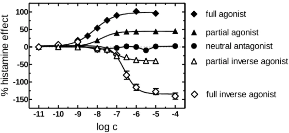
A GPCR toggles between both the inactive and active state (Figure 1.4). An agonist elicits a biological response by stabilizing the receptor in the active state Ra (Figure 1.4 and Figure 1.5).
The intrinsic activity of partial agonists ranges from 0 to 100 %. Partial agonists are ligands not capable of producing the maximal response. This is in agreement with the idea that partial agonists stabilize the active state less effective than full agonists (Kenakin, 2001).
A neutral antagonist (intrinsic activity = 0 %) neither activates nor inhibits the receptor, i. e., the basal equilibrium between both active Ra and inactive Ri states remains unaltered. A neutral antagonist is capable of competing with both agonists and inverse agonists for orthosteric binding.
Considering constitutive activity as a prerequisite, an inverse agonist decreases the elevated level of basally activated receptors by stabilizing the inactive state (Kenakin, 2004).
Differentiation between full and partial inverse agonists is possible, enabling full inverse agonists to stabilize the inactive state Ri more effectively (α ≤ -100 %) than partial inverse agonists (0 > α > -100 %). In terms of intrinsic activity, the responses to GPCR ligands are referenced to the maximal effect produced by the endogenous ligand as the standard agonist (e. g. histamine in case of the H4R, i. a. = 100 %). Therefore, depending on the basal activity of the GPCR of interest in the respective assay, the intrinsic activity of a full inverse agonist can by definition be lower than -100 %. (Seifert and Wieland, 2005).
Figure 1.4: Two state model of a GPCR. The receptor toggles between the inactive state Ri and the active state Ra. Adapted from Seifert and Wenzel-Seifert (2002).
full inverse agonist
partial inverse agonist
neutral
antagonist partial agonist full agonist
R
aR
iFigure 1.5: Effects of ligands with different intrinsic activities shown as concentration-response curves. [35S]GTPγS assays were performed with different H4 receptor species orthologs. Modified from Seifert and Wieland (2005).
1.1.4 Models of ligand binding and G-protein coupling
1.1.4.1 Classical model
Binding of ligand A to a receptor R is described by the Langmuir adsorption isotherm according to Clark (1933) and Clark (1937) (Equation(1.1):
ρ =[AR]
[Rt] = [A]
[A] + KA (1.1)
Herein, AR is referred to as the ligand bound receptor, Rt corresponds to the total number of receptor sites and KA to the equilibrium dissociation constant (Ka to the equilibrium association constant) of the agonist receptor complex. ρ describes the fraction of ligand bound and total receptor concentration (Kenakin, 2009).
Different modifications, introduced by Ariens (1954), Stephenson (1956) and Furchgott (1966) led to the assumption (Equation(1.2):
(1.2) The term ε, referring to the agonist-specific term intrinsic efficacy, and the total concentration of receptors [Rt] were introduced in the binding function (Equation(1.3):
Response = f ( [A]
[A] + KA× ε × [Rt]) (1.3)
1.1.4.2 Operational model
The “operational” model, developed by Black and Leff (1983), improved the model describing the relationship between agonist concentration and response (Kenakin, 2009). The assumption of one ligand binding to one receptor is valid.
-11 -10 -9 -8 -7 -6 -5 -4 -150
-100 -50 0 50
100 full agonist
partial agonist neutral antagonist
full inverse agonist partial inverse agonist
log c
% histamine effect
Stimulus AR
A + R Ka Response
KA
6 1.1 G-protein coupled receptors
The response is dependent on the agonist concentration [A], KA, т (power of the agonist to produce response) and Emax (maximal response of the system). Both Emax and т are receptor and system dependent (Equation(1.4):
response = [A] × τ × Emax
[A] × (τ + 1) + KA (1.4)
High levels of т (e. g. 100-1000) imply a relatively high percentage of activated receptor species over a broad range of т. But if т → 0, the maximal response → 0. Moreover, the smaller KA
(higher affinity), the less agonist is required to attain the same response.
1.1.4.3 Ternary complex models
The ternary complex model – firstly described by De Lean et al. (1980) – takes into account the binding of the activated receptor to membrane proteins such as G-proteins (Equation(1.5;(Kenakin, 2009):
(1.5)
The extended ternary complex model additionally implies the equilibrium between the inactive Ri and the active Ra receptor states (Figure 1.6;(Kenakin, 2009; Samama et al., 1993).
Figure 1.6: Illustration of the extended ternary complex model.
According to Kenakin (2009). KG: equilibrium dissociation constant of the receptor/G-protein complex and Kg: respective equilibrium association constant.
The conversion of Ri to Ra is considered in the allosteric constant L (Equation(1.6):
L = [Ra]
[Ri] (1.6)
Furthermore, the terms α, referring to differences in affinity of the ligand to Ra compared to Ri, and γ, referring to differences in affinity of the ligand bound receptor to the G-proteins compared to the ligand-unbound receptor, were introduced (Kenakin, 2009). For example, an α or a γ value of 10 means, that the ligand has a tenfold higher affinity to Ra compared to Ri or
ARG AR + G
A + R Ka
KG KA
Kg
ARi ARa ARaG
RaG Ra
Ri
Ka
L Kg
G
αγKa γKg
αL
αKa
G
a tenfold higher affinity of the ligand-receptor complex to G-proteins compared to the ligand- free receptor.
The fraction ρ (Equation(1.7) of the two G-protein activated receptor species [RaG] and [ARaG]
and the total receptor population is given as:
ρ =
L × [G]
KG (1 +α × γ × [A]
KA )
[A]
KA (1 + α × L (1 +γ × [G]K
G )) + L (1 +[G]K
G) + 1 (1.7)
The terms α and γ define the intrinsic activity (Kenakin, 2004). If a ligand binds with high affinity to the active state of the receptor (α > 1) and this ligand-bound receptor binds to G-proteins with high affinity (γ > 1), the ligand will be an agonist with positive intrinsic activity. By contrast, α and γ < 1 means that the ligand preferentially stabilizes the inactive state of the receptor with high affinity and the affinity of the ligand-bound receptor to G-proteins decreases. The corresponding quality of action becomes obvious in a functional system with constitutive activity, and the respective ligand is referred to as an inverse agonist.
Further refinements were made with the cubic ternary complex model (Figure 1.7), taking into account the interaction of inactive receptor states (Ri and ARi) with G-proteins (Kenakin, 2009;
Weiss et al., 1996a; Weiss et al., 1996b; c).
Figure 1.7: Cubic ternary complex model, according to Kenakin (2009). β: effect of receptor activation on the coupling of G-protein to the receptor (effect of G-protein coupling on receptor activation); δ: constant describing the synergism between receptor activation, G-protein coupling or the binding of a ligand.
1.1.5 Constitutive activity of GPCRs
Constitutive activity describes the ability of a GPCR to spontaneously produce a cellular response in the absence of a ligand (Lefkowitz et al., 1993). So far, constitutive activity has been observed for more than 60 wild-type GPCRs and several disease-causing mutants (Seifert and Wenzel-Seifert, 2002).
ARi ARa
Ra Ri
Ka
L
βKg αL
αKa
ARiG ARaG
RaG RiG
Kg
βL δαβL
γKg δγβKg
γKa δγαKa
8 1.1 G-protein coupled receptors
The higher the constitutive activity of a GPCR, the more the basal equilibrium between inactive and active state is shifted towards the active state. Therefore, the amplitude of the response elicited by a full agonist is lower at a constitutively active receptor compared to a GPCR devoid of constitutive activity (Kenakin, 2004). Inversely, the maximum effect of an inverse agonist increases with the level of constitutive activity of the receptor of interest. The hallmarks of a constitutively active receptor are a high basal activity, a high intrinsic activity and potency of partial agonists and a high inverse agonistic effect of inverse agonists (Seifert et al., 1998).
The phenomenon of constitutive activity can be derived from the extended ternary complex model (Equation(1.8;(Kenakin, 2004; 2009):
(1.8)
1.1.6 Signal transduction
GPCRs may be considered as molecular switches transferring extracellular signals to intracellular responses. Conformational changes from inactive to active state(s) allow for activation of signal transducers, i. e., effectors such as G-proteins (Chapter 1.1.6.2) or β-arrestins (Chapter 1.1.6.3) (Granier and Kobilka, 2012).
1.1.6.1 Structure of G-proteins and G-protein cycle
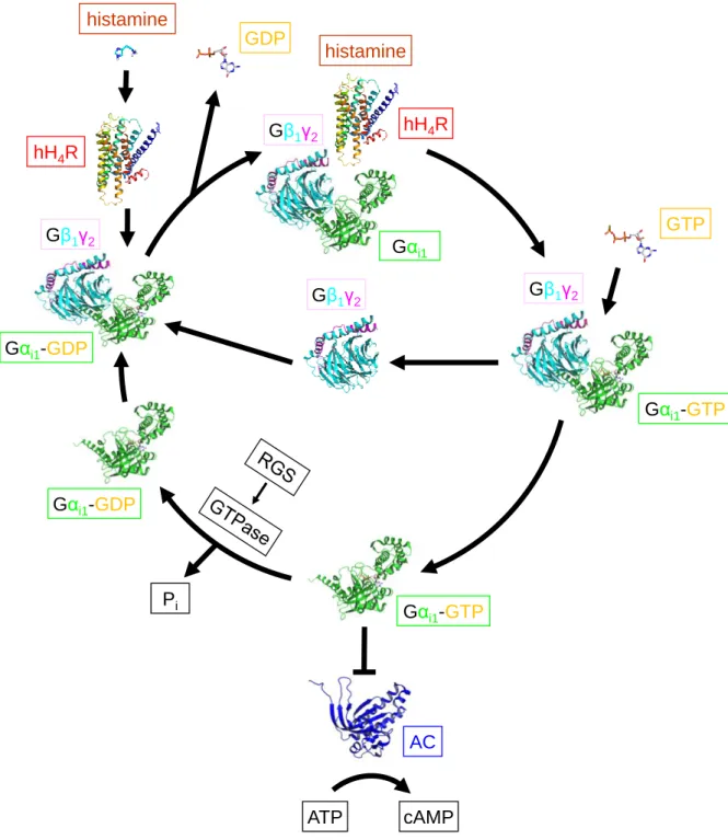
Currently, 16 different Gα G-protein subunits as well as 5 Gβ and 12 Gγ subunits are known (Cabrera-Vera et al., 2003; Downes and Gautam, 1999). In G-proteins not activated by a GPCR, the GDP-bound Gα subunit is combined with the Gβγ-dimer to form a heterotrimeric complex (Figure 1.8 and Figure 1.9;(Hurowitz et al., 2000). Both the Gα as well as the Gβγ subunit are attached to the membrane via lipid anchors (Chen and Manning, 2001; Dupre et al., 2009). Binding of a ligand to a GPCR gives a ternary complex consisting of the agonist- bound active receptor and the nucleotide free Gα and Gβγ subunit (Bünemann et al., 2003;
De Lean et al., 1980; Kling et al., 2013; Ratnala and Kobilka, 2009). Upon Gα activation by a GPCR, GDP is released and replaced by GTP, and Gα (Figure 1.10) dissociates from Gβγ (Rasmussen et al., 2011b). Gα and Gβγ have their own effectors and influence the levels of second messengers (Tuteja, 2009). The active state of the Gα subunit is switched off by the intrinsic GTPase activity to give inactive GDP-bound Gα, which re-associates with Gβγ.
Regulators of G-protein signalling (RGS proteins; GTPase activating proteins, GAPs) can enhance the activity of the GTPase (Neitzel and Hepler, 2006; Wieland et al., 2007; Willars, 2006).
RaG Ra
Ri L
KG G
Figure 1.8: G-protein cycle, turned on by histamine H4 receptor stimulation as an example. Gαi1
is coloured in green, the Gβ1 subunit in turquoise, the Gγ2 subunit in pink and the adenylyl cyclase (AC) in blue. The following crystal structures were used: inactive heterotrimeric complex of Gαi1 and Gβ1γ2
(PDB ID: 1GG2 (Wall et al., 1995)), active Gαi1 (PDB ID: 1GIA (Coleman et al., 1994)), adenylyl cyclase (1CUL (Tesmer et al., 2000)); hH4R was generated as a homology model with the active state of the β2AR (PDB ID: 3P0G (Rasmussen et al., 2011a)) as template. Modified from Gilman (1987) and Rasmussen et al. (2011b).
GDP
Gαi1-GDP Gβ1γ2
Gβ1γ2
Gαi1-GTP GTP
Gβ1γ2
Gαi1-GTP
AC
ATP cAMP
Pi Gαi1-GDP hH4R
histamine
Gαi1 Gβ1γ2 hH4R
histamine
10 1.1 G-protein coupled receptors
Figure 1.9: Crystal structure of heterotrimeric G-proteins Gαi1 and Gβ1γ2. PDB ID:
1GG2 (Wall et al., 1995). Gαi1
is coloured in green, Gβ1 in turquoise and Gγ2 in pink. The bound GDP is illustrated as sticks.
Figure 1.10: Crystal structure of Gαi1 in the active state. The non-hydrolysable GTP analogue GTPγS is bound to the Gαi1 subunit.
PDB ID: 1GIA (Coleman et al., 1994).
1.1.6.2 Functions of different G-protein subtypes
According to the α subunits, G-proteins are usually divided into four families: Gs, Gi/o, Gq/11 and G12 (Downes and Gautam, 1999). Activation of Gαs, for instance mediated by the histamine H2R, leads to an increase in the production of 3’,5’-cyclic adenosine monophosphate (cAMP) by the adenylyl cyclase (AC) (Figure 1.11;(Liu et al., 2003; Neves et al., 2002). By contrast, the stimulation of Gαi/o-coupled receptors such as the H3R and H4R results in an inhibition of the AC activity and a decreasing cAMP level (Figure 1.11;(Neves et al., 2002). cAMP stimulates many kinases, most prominently the protein kinase A (PKA), which is capable of phosphorylating numerous substrates, including the cAMP response element binding protein (CREB) (Birnbaumer, 2007; Hur and Kim, 2002).
Although the term inhibitory G protein (Gi) was initially derived from the inhibitory effect on the AC, members of the Gαi family reveal also signal transmission via the Gβγ-subunit (Khan et al., 2013), e. g., by activation of phospholipase C- (PLC-;(Harden et al., 1987)) (Figure 1.11).
Moreover, K+ channels are activated and inactive Ca2+ channels are stabilized resulting in hyperpolarization and inhibition of excitation. Kinase (Akt) pathways are activated via the stimulation of PI3K (phosphatidylinositol-3-kinase) and formation of the second messenger PIP3 (phosphatidylinositol-3,4,5-trisphosphate) (Hur and Kim, 2002). Activation of PLC-β2 and 3 induces synthesis of the second messengers IP3 (inositol-1,4,5-trisphosphate) and DAG (1,2-diacylglycerol) (Hokin and Hokin, 1955; Hokin and Hokin, 1953). Whereas DAG activates the PKC directly, IP3 increases the intracellular calcium level (Berridge et al., 1983) and therefore indirectly activates the PKC (Birnbaumer, 2007).
Gαq/11-coupled receptors such as the H1R activate PLC-β1 and 4 (Wu et al., 1992) cleaving PIP2 (phosphatidylinositol-4,5-bisphosphate) to give IP3 and DAG (Birnbaumer, 2007; Neves et al., 2002) (Figure 1.11).
Figure 1.11: Signalling pathways of GPCRs, exemplified by the histamine receptor subtypes H1R- H4R. Gα proteins are marked in green, the Gβ-subunit in turquoise and the Gγ-subunit in pink; enzymes are highlighted in blue, except for kinases (orange), and ion channels are marked in yellow ochre.
Modified from Steinhilber et al. (2005) and Aktories et al. (2006).
1.1.6.3 β-arrestin-mediated GPCR internalization and signalling
Out of four different known isoforms of arrestins, two arrestins are expressed in the retina (“visual arrestins”) and two “non-visual” arrestins were identified, namely β-arrestin1 and 2
H1R H2R H3R/H4R
histamine
Gαq/11
γ β GTP
GDP
Gαs
γ β GTP
GDP
Gαi/o
γ β GTP
GDP AC
cAMP ATP
PKA
Raf1 CREB Epac
Rap1 B-Raf MEK ERK1,2
L-Ca2+
channel
K+ channel
Ca2+
channel transmitter release hyper-
polarisation
excitation
PI3K PIP2 PIP3
PDK1 PKB multiple kinases
PLC-β2,3 DAG IP3
IP3R PKC intracellular Ca2+ release
PIP2 PLC-β1,4
PIP2 IP3 DAG
IP3R PKC intracellular Ca2+ release
CRE
12 1.1 G-protein coupled receptors
(Luttrell and Gesty-Palmer, 2010). β-arrestins are capable of both directly influencing diverse signalling pathways (e. g. protein kinases) as well as of modifying the number of active receptors by the internalization and downregulation machinery (Luttrell and Lefkowitz, 2002;
Shenoy and Lefkowitz, 2011). In particular, continuous stimulation of a GPCR by an agonist implies a β-arrestin mediated desensitization (Hanyaloglu and von Zastrow, 2008). In the first step of this process the receptor is phosphorylated by GRKs (G-protein coupled receptor kinases) (Figure 1.12), a class of enzymes comprising seven different subtypes (Luttrell and Lefkowitz, 2002; Ribas et al., 2007; Watari et al., 2014). In detail, GPCRs are preferentially phosphorylated at serine and threonine residues in ICL3 and the C-terminus. Phosphorylation increases the affinity of β-arrestins to the receptor; β-arrestin binding precludes further coupling of G-proteins to the receptor. Unlike the visual arrestins, β-arrestins additionally contain clathrin and β2-adaptin (AP2 complex) binding motifs and are therefore involved in the processes of endocytosis, resensitization and downregulation (Goodman et al., 1996). Inhibiting the function of the GTPase dynamin (Zhang et al., 1996), being responsible for the endocytosis via clathrin- coated pits, prevents the internalization of GPCRs. After endocytosis, the receptor is either recycled or degraded in lysosomes, depending on the duration of β-arrestin binding to the receptor. If β-arrestins dissociate from the receptor upon endocytosis (e. g. β2AR), the receptor is preferentially recycled to the plasma membrane (class A β-arrestin recruitment). However, in case that the receptor (e. g. V2R) remains bound to β-arrestin, the receptor is most probably degraded (class B β-arrestin recruitment) (DeWire et al., 2007; Gurevich and Gurevich, 2006;
Luttrell, 2008).
Apart from the internalization process, β-arrestins can directly modulate several effector proteins such as ERK (extracellular signal-regulated kinase), a MAPK (mitogen-activated protein kinase), and can activate PI3K or inhibit the transcription factor NFκΒ by activation of IκΒ (Figure 1.12;(Reiter et al., 2012; Shenoy and Lefkowitz, 2011).
According to the concept of “functional selectivity” or “biased signalling”, depending on the bound ligand, the receptor should be capable to activate, either G-proteins (G-protein biased ligand) or β-arrestins (β-arrestin biased ligand). Ligands causing the GPCR-mediated activation of both, G-proteins and β-arrestins, are referred to as balanced ligands (Reiter et al., 2012).
Figure 1.12: β-arrestin-mediated desensitization and signalling of GPCRs. On the one hand, agonist binding to a GPCR, e. g., histamine to the hH4R, initiates G-protein dependent signalling. On the other hand, activated receptors are specifically phosphorylated (red) by GRKs (dark green). This phosphorylation increases β-arrestin recruitment (brown) to the receptor and precludes further G-protein activation. β-arrestin links the receptor to the internalization machinery of clathrin (blue) and clathrin adaptor (AP2, pink) leading to a dynamin-dependent (grey) receptor internalization via “clathrin coated pits”. In the endosomes, the receptor is either degraded in lysosomes or agonist dissociation is facilitated by the low pH in the endosome leading to the dissociation of β-arrestin. The receptor is dephosphorylated and recycled to the plasma membrane. Besides, β-arrestins influence many signalling pathways such as ERK, PI3K or NFκΒ. Downregulation modified from Gurevich and Gurevich (2006) and β-arrestin signalling from Reiter et al. (2012).
1.2 Histamine and histamine receptors
1.2.1 Historical perspective of histamine
Histamine was firstly synthesized by Windaus and Vogt from histidine (Windaus and Vogt, 1907). Three years later, Sir Henry Dale and his colleagues isolated histamine from the mould ergot in the Wellcome Laboratories (Dale and Laidlaw, 1910). Subsequently, Dale and Laidlaw performed studies on the physiological effects of histamine. When injected into animals,
15
Gαi/o γ β
histamine
GRK PP P
β-arrestin PP P
β-arrestin PP P
Clathrin AP2
Dynamin
β-arrestin PP P
Clathrin AP2
β-arrestin PP P β-arrestin
P P P
degradation
lysosome endosome
recycling
clathrin coated pit PI3K ERK
NFκΒ IκΒ
protein translation apoptosis
γ β
14 1.2 Histamine and histamine receptors
histamine caused contractions of smooth muscles in the gut and the respiratory tract, vasodepression, increase in cardiac contractility and a shock-like syndrome (Dale and Laidlaw, 1910; 1911; 1919; Parsons and Ganellin, 2006). In 1927, histamine was identified as an endogenous substance in the lung and liver (Best et al., 1927).
The finding that histamine induced anaphylaxis and was involved in allergies inspired the search for compounds antagonizing the pathological effects of histamine. The first antihistamines blocking the action of histamine in an anaphylactic reaction were identified in the 1930s (Bovet and Staub, 1937; Fourneau and Bovet, 1933).
1.2.2 Histamine: occurrence and metabolism
Stored in vesicles (granules), histamine predominates in mast cells (Riley and West, 1952), basophils (Falcone et al., 2006) and thrombocytes. Herein, histamine forms an ionic interaction with the acidic mucopolysaccharid heparin. Besides, histamine is stored in enterochromaffin- like cells in the gastric mucosa (Prinz et al., 2003) and acts as a neurotransmitter of histaminergic neurons (Dy and Schneider, 2004).
Histamine features two basic groups with the amine moiety in the side chain being more basic (pKa2 = 9.4) than the nitrogen in the imidazole ring (pKa1 = 5.8). Under physiological conditions (pH = 7.4), the monocationic form (amine in the side chain protonated) predominates (Figure 1.13). However, the monocation is not a single molecular entity, since the imidazole ring can undergo 1,3-tautomerism. In aqueous solution the Nτ-H-tautomer is preferred compared to the Nπ-H-nitrogen tautomer (Figure 1.13;(Ganellin, 1973).
Figure 1.13: 1,3-Tautomerism of imidazole ring in the histamine monocation.
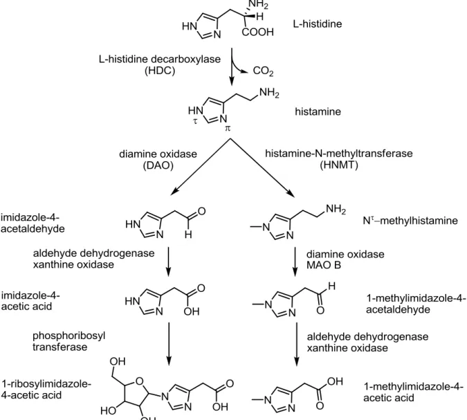
Histamine is a biogenic amine, synthesized from the amino acid L-histidine by the L-histidine- decarboxylase (HDC) (Figure 1.14;(Beall and Vanarsdel, 1961). Whereas special transporters are known for catecholamines or serotonin, histamine re-uptake was reported to be mediated by organic cation transporters (OCTs;(Gründemann et al., 1999; Schneider et al., 2011;
Schneider et al., 2005). The main route of biotransformation and the only one in brain is the Nτ-methylation by histamine-N-methyltransferase (HNMT;(Weinshilboum et al., 1999) prior to oxidation by aldehyde dehydrogenase and xanthine oxidase. The second route of inactivation leads to 1-ribosyl-imidazole-4-acetic acid by oxidation and ribosylation (Beall and Vanarsdel, 1961).
Figure 1.14: Histamine synthesis and metabolism. Modified from Aktories et al. (2006).
1.2.3 Histamine receptor subtypes and ligands
1.2.3.1 Homologies between the four histamine receptor subtypes
Whereas the hH3R is highly related to the hH4R (41 % sequence identity; Table 1.1;(Hough, 2001; Leurs et al., 2009), the first two histamine receptor subtypes share a relatively low sequence homology to the hH3R and hH4R (18-22 % sequence identity; Table 1.1;(Leurs et al., 2009; Lovenberg et al., 1999). For example, the hH1R is more similar to the muscarinic receptors (De Backer et al., 1993), and the hH2R shares a higher level of sequence identity with the 5-HT4R or the D2R-like family than with hH3R and hH4R (Vassilatis et al., 2003).
16 1.2 Histamine and histamine receptors
Table 1.1: Homologies (%) between the four human histamine receptor subtypes hH1R, hH2R, hH3R and hH4R.
Receptor hH1R hH2R hH3R hH4R
hH1R 100
hH2R 25 100
hH3R 21 21 100
hH4R 22 18 41 100
Sequence alignment was performed with Clustal-X 2.1 together with about 80 class A GPCRs. Identities between histamine receptor subtypes hH1R-hH4R were calculated and the number of identical amino acids were divided by the number of amino acids of the respective shorter sequence.
1.2.3.2 Key amino acids of the four histamine receptor subtypes
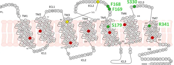
All histamine receptor subtypes feature the highly conserved residues among class A GPCRs (grey;(Mirzadegan et al., 2003), the conserved disulphide bond forming cysteine residues in TM3 and ECL2 (yellow;(Strader et al., 1994) as well as the DRY motif (DRF motif in H3R), the FxxCWxP and the NPxxY motif (green; Figure 1.15). The presence of an acidic residue at the bottom of TM6 (D/E6.30) enables the H1R, H2R and the H3R, unlike the H4R (A2986.30), to form an ionic lock. Besides, the acidic D3.32, a very important residue for ligand binding (Gantz et al., 1992; Ohta et al., 1994; Shin et al., 2002), and Y6.51 are highly conserved. The in ECL2 located FF motif is highly conserved in case of the H3R and H4R, but replaced by FY in the H1R and VQ in the H2R. S5.43, present in case of the H3R and H4R, is replaced by A at the H1R and G at the H2R. Position 5.46 (E in H3R and H4R, N in H1R and T in H2R) has been proven to be very important for ligand binding, in particular in case of agonists (Gantz et al., 1992;
Ohta et al., 1994; Shin et al., 2002; Uveges et al., 2002). In TM7, residues, which are probably also involved in ligand binding and activation, are only poorly conserved: This is, e. g., the case for the basic R3417.36 at the hH4R, which is replaced by an acidic E at the hH3R and neutral residues (M and A) at the hH1R and hH2R, respectively.