Host FcγRs CD16, CD32 and CD64 Reveals a Selective Inhibition through Herpesviral FcγRs
Dissertation
Zur Erlangung des akademischen Grades d o c t o r r e r u m n a t u r a l i u m
(Dr. rer. nat) im Fach Biologie eingereicht an der
Mathematisch-Naturwissenschaftlichen Fakultät I Der Humboldt-Universität zu Berlin
von
Eugenia Corrales-Aguilar
geboren am 22.03.1978 in San José, Costa Rica
Präsident der Humboldt-Universität zu Berlin Prof. Dr. Dr. h.c. Christoph Markschies
Dekan der Mathematisch-Naturwissenschaftlichen Fakultät I
Gutachter: 1. Prof. Dr. Hartmut Hengel 2. Prof. Dr. Richard Lucius 3. Prof. Dr. Günther Schönrich
Gedruckt mit Unterstützung des Deutschen Akademischen Austauschdienstes
“Around here, however, we don’t look backwards for very long. We keep moving forward, opening up new doors and doing new things… and curiosity keeps leading us down new paths.”
Walt Disney
Table of Contents
Summary ... 8
Zusammenfassung... 10
Abbreviations... 12
1 Introduction... 15
1.1 Viruses ... 15
1.1.1 Herpesvirus Family...16
1.1.2 Human Cytomegalovirus (HCMV) ...18
1.1.3 Herpes simplex virus (HSV) ...21
1.2 IgG and IgG-mediated effector functions for viral control ...22
1.3 Host Fcγ Receptors (FcγRs) ...25
1.3.1 CD64 (FcγRI)...27
1.3.2 CD32 (FcRγII)...28
1.3.3 CD16 (FcγRIII)...28
1.3.4 Mouse FcγRs...30
1.4 Immune evasion by herpesviruses ...31
1.4.1 NK immune evasion by CMV...32
1.5 Viral FcγRs ...35
1.5.1 HSV vFcγRs ...35
1.5.2 CMV vFcγRs...38
1.6 Methods for Measuring Antiviral IgG ...40
1.7 Aim of the Thesis...42
2 Results ... 43
2.1 Establishment of a novel assay system for detection and quantification of virus specific IgG antibodies triggering host FcγRs ...43
2.1.1 Stable expression of FcγR-ζ constructs and activation of BWFcγR-ζ transfectants43 2.1.2 IgG dependent activation of the BWFcγR-ζ transfectants ...47
2.1.3 Fc of IgG is required for activation for the BWFcγR-ζ Assay...50
2.1.4 Immune IgG activation of chimeric hCD32-ζ and hCD64-ζ receptors ... 51
2.1.5 Opsonization of MCMV infected target cells by polyclonal virus-specific IgG activates FcγR-ζ transfectants ...54
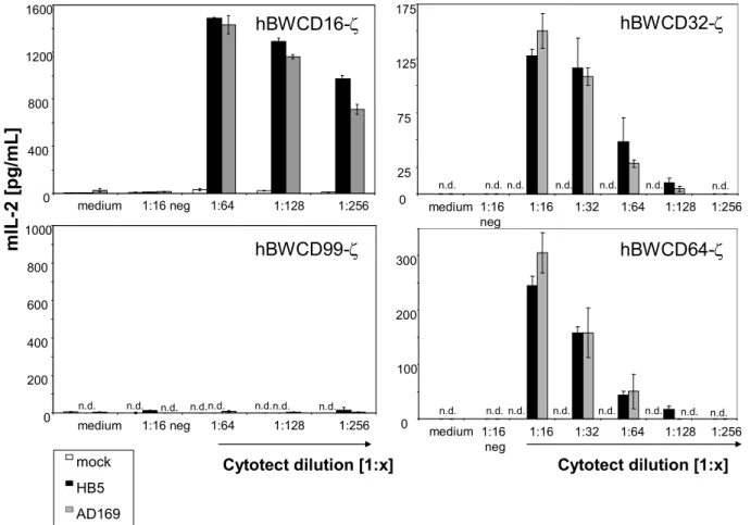
2.1.6 Opsonization of HCMV infected target cells by polyclonal virus-specific IgG activates FcγR-ζ transfectants ...56
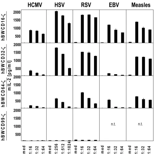
2.1.7 Universal applicability of the assays to detect virus-specific IgG ...58
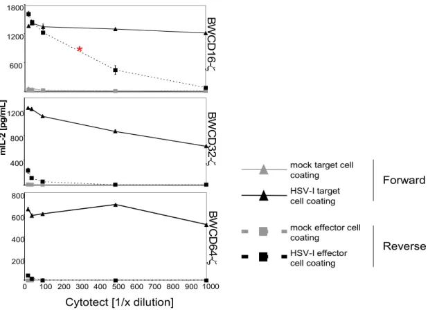
2.1.8 IgG binding to virus-infected target cells is required before activation of FcγR-ζ effector cells ...60
2.1.9 Comparing CD16 activation in the BWCD16-ζ assay vrs CD107a degranulation assay with primary human NK cells ... 65
2.2 Diagnostic Application of the BWFcγR-ζ assay...67
2.2.1 BWFcγR-ζ assay efficiencies for MV-specifc IgG...67
2.2.2 BWFcγR-ζ assay efficiencies and reaction patterns assessed for CMV-IgG in human sera ...71
2.3 Selective inhibition of herpesviral FcγR of IgG mediated effector functions ... 75
2.3.1 Herpesviruses encode vFcγR binding Fcγ on the surface of infected cells ... 75
2.3.5 The HSV vFcγR gE inhibits CD16 and CD32 activation but fails to inhibit CD64.. 82
2.3.6 HCMV vFcγRs interfere with CD16 activation ...84
2.3.7 HCMV vFcγRs interfere with CD16 downstream signalling...86
2.3.8 The HCMV vFcγRs inhibit antibody dependent NK cell degranulation... 88
2.3.9 HCMV vFcγRs interfere with CD32 and CD64 activation ... 90
2.3.10 MCMV fcr-1/m138 diminishes IgG-dependent activation of the murine CD16 .... 94
2.3.11 m138/fcr-1 mediates IgG bipolar bridging ...98
2.3.12 m138/fcr-1 blockade of CD16 activation by W6/32 IgG2a is extremely efficient102 3 Discussion ... 104
3.1 The novel FcγR activation assay opens a window for understanding the role of antiviral antibodies...105
3.1.1 Advantages of the FcγR activation assay...105
3.1.2 Virus-specific IgG responses as a correlate of immunity...106
3.1.3 The BWFcγR-z activating assay’s open questions and future needs for optimization ...108
3.1.4 Applicability of the FcγR activation assay...113
3.2 The Novel Method for Measuring IgG-Dependent Triggering of Host FcγRs CD16, CD32 and CD64 Reveals a Selective Inhibition through Herpesviral FcγRs ... 113
3.2.1 Not all herpesviral FcγRs interfere with neutralization and complement activation114 3.2.2 Herpes simplex virus vFcγR gE interferes with the host FcγRs CD16 and CD32 but not with CD64 activation ...115
3.2.3 Human Cytomegalovirus FcγRs interfere with the host Fc Rs activation...117
3.2.4 Mouse Cytomegalovirus FcγR m138/fcr-1 interferes with the host CD16 activation121 3.2.5 vFcγRs selective interference of Host FcγRs...123
3.3 Herpesviruses and antibodies ...124
3.4 Future Perspectives ...125
4 Materials... 128
4.1 Devices... 128
4.2 Chemicals and biochemicals ...129
4.3 Kits ... 131
4.4 Solutions and Buffers ...132
4.5 Oligonucleotides...136
4.6 Cell lines... 137
4.7 Viruses ... 137
4.8 Antibodies and IVIGs...138
5 Experimental Procedures ...140
5.1 Molecular biology methods...140
5.1.1 Plasmids and working with E. coli ...140
5.1.2 DNA protocols ...141
5.3.8 RSV titration ...151
5.3.9 RSV infection for analysis...151
5.3.10 Measles virus (MV) Stock preparation...151
5.3.11 MV titration ...152
5.3.12 MV infection for analysis...152
5.3.13 accinia virus stock ...152
5.3.14 Titration of VACV stock ...153
5.3.15 VACV infection for analysis ...153
5.3.16 Mouse Cytomegalovirus Stock preparation...153
5.3.17 Mouse Cytomegalovirus titration ...154
5.3.18 MCMV infection for analysis ...154
5.3.19 HCMV and MV plaque reduction neutralization (PRNT) ...155
5.3.20 Production of MCMV Latent serum ...156
5.4 Immunological/Protein Methods ...156
5.4.1 Generation of protein lysates for expression control western blots ... 156
5.4.2 Generation of protein lysates for determination of phosphorylated proteins or for the bipolar bridging proofing...157
5.4.3 SDS-PAGE (Polyacrylamide –Gel electrophoresis) ... 157
5.4.4 Western blot ...158
5.4.5 Immunoprecipitation (IP) ...159
5.4.6 FACS...160
5.5 Novel Assay System for the Detection and Quantification of Virus-specific IgG Antibodies Triggering FcγReceptors (BWFcγR-ζ Assay) ...161
5.5.1 Interleukin-2 (IL-2) ELISA ...163
5.5.2 Crosslinking experiments for determining responsiveness of the newly established FcγR-ζ transfectants...164
5.5.3 Fab Fragments preparation from human IgG ...164
5.5.4 Virus specific IgG and IgM ELISA ...164
5.5.5 Validation of the newly established assay for measuring antibodies that trigger the host FcγRs...165
5.5.6 Statistical Analysis for the novel FcγR-ζ Assay ...166
5.6 Working with human NK cells...167
5.6.1 Isolation and culture of PBMCs ...167
5.6.2 CD107a NK cells degranulation assay ...168
6 List of References ... 170
7 Acknowledgments ...185
8 Publications ... 186
9 Curriculum Vitae... 187
10 Suplementary Data...192
11ERKLÄRUNG...197
Summary
To study the possible interference of the herpesviral vFcγRs with the host FcγRs and IgG- mediated effector functions, a new methodological approach to detect FcγR activating antibodies had to be developed. The novel assay comprises the co-cultivation of virus infected cells upon opsonization with immune IgG antibodies and the stably transfected FcγR-ζ BW5147 transfectants as responder cells. The transfectants express chimeric receptors bearing the extracellular domain of the host FcγRs (i.e. human CD16, human CD32, human CD64, mouse CD16 and mouse FcRIV) fused to the transmembrane and tail domains of the murine CD3ζ chain. Triggering the CD3ζ chain is sufficient to elicit IL-2 secretion in a dose dependent manner which is measured in an ELISA. The setup of the new assay provides a defined effector cell population bearing one Fcγ receptor on the surface, which becomes activated in the presence of immune IgG antibodies bound to the native viral antigens displayed on the surface of infected cells. The assay system allows us to detect and quantify Fcγ receptor-activating immune IgG in an FcγR-specific way, which is thought to have an important biological function in antiviral defense.
Several α- and β- herpesviruses express on the surface of infected cells virally encoded Fc binding glycoproteins. The assay described above was applied to determine if the viral
found that HCMV FcγR gp68 affects activation and downstream signaling of CD16 > CD32 = CD64, while gp34 attenuates CD16 > CD64 > CD32. In clear contrast, HSV gE impairs CD16 activation and weakly CD32, but has no effect on CD64. Furthemore, MCMV m138/fcr-1 diminishes activation of mouse CD16.
Taken together, this data uncover herpesviral FcγRs as hierarchical and redundant antagonists precluding host FcγRs from triggering immune responses.
Zusammenfassung
Um die Wirkung herpesviral-kodierter FcγRezeptoren (vFcγR) auf wirtskodierte zelluläre FcγRezeptoren (FcγR) und IgG-vermittelten Effektorfunktionen untersuchen zu können, war es notwendig einen methodisch neuen Ansatz zu entwickeln, der die Detektion FcγR- aktivierender Antikörper ermöglicht. Dieses neuartige Assay beinhaltet die Kokultivierung virusinfizierter Zellen, die mit virusspezifischen IgG-Antikörpern opsoniert sind, mit FcγR- ζ BW5147-Transfektanten als Reporterzellen. Diese stabilen Transfektanten exprimieren
chimäre Rezeptoren, die aus der extrazellulären Domäne der zellulären FcγRezeptoren (d.h.
entweder humanem CD16, humanem CD32, humanem CD64, murinem CD16 oder murinem FcRIV) bestehen, welche mit der Transmembrandomäne und intrazellulären Domäne der murinen CD3ζ-Kette fusioniert wurden. Die Aktivierung der CD3ζ-Kette führt zu einer IgG- dosisabhängigen mIL-2 Sekretion, die im ELISA gemessen werden kann. Der Versuchsaufbau dieses neuartigen Assays gewährleistet eine definierte Effektorzellpopulation, die dadurch gekennzeichnet ist, nur einen bestimmten FcγRezeptor auf der Oberfläche zu tragen. Dieser chimäre FcγR wird durch virusspezifisches IgG, das an native virale Antigene auf der Zelloberfläche infizierter Zellen gebunden ist, aktiviert. Dieses Versuchssystem ermöglicht uns die Detektion und Quantifikation von FcγR-aktivierenden
oben beschriebene Assay angewandt. In einem systematischen Ansatz wurde die Wirkung der einzelnen vFcγR auf jeden wirtskodierten zellulären FcγRezeptor überprüft. Durch Anwendung einer „loss of function“ (Deletionsmutanten der viralen FcγR)- und einer „gain of function“ (Expression der vFcγR durch rekombinante Vakziniaviren)-Strategie konnte eine selektive Inhibition der wirtskodierten zellulären FcγRezeptoren durch die einzelnen herpesvirale FcγRezeptoren mittels antiviraler IgG-Bindung ermittelt werden.
Es wurde festgestellt, dass der HCMV-kodierte FcγR gp68 die Aktivierung und die nachfolgende Signalkaskade von CD16 > CD32 = CD64 inhibiert, während der HCMV- kodierte FcγR gp34die Aktivierung von CD16>CD64>CD32 inhibiert. In klarem Kontrast dazu wirkt der HSV-kodierte FcγR gE, der CD16 Aktivierung vermindert, CD32 hingegen nur sehr schwach und CD64 gar nicht beeinflußt. Der MCMV-kodierte FcγR m138/fcr-1 vermindert die Aktivierung des murinen CD16.
Zusammenfassend betrachtet zeigen die ermittelten Daten, dass es sich bei den herpesviral- kodierten FcγRezeptoren um hierarchische und redundante Antagonisten der wirtskodierten zellulären FcγRezeptoren handelt. Herpesviral-kodierte FcγRezeptoren wirken somit der Aktivierung des Immunsystems entgegen.
Abbreviations
A Ampere
aa amino acid
Ab Antibody
ADCC Antibody dependent cell cytolysis AIDS Acquired Immunodeficiency Syndrome ATCC American type culture collection BAC bacterial artificial chromosome
bp Base pairs
°C Celcius degree
C1q Complement 1 q component CD Cluster of differentiation
CMV Cytomegalovirus
CPE Cytopathic Effect d days
DAF Decay accelerating factor
DC dendritic cell
DMEM Dulbeccos Modified Eagle Medium DMSO Dimethylsulfoxide
DNA 2-Desoxyribonucleic acid
dNTPs all four 2-Desoxy-Nucleotides (A, T, C, G) dpi days post infection
ds Double strand
DTT 1,4- Dithiothreitol E early
EBV Epstein-Barr Virus
EDTA Ethylenediaminetetraacetic adic (+Na2) ELISA Enzyme-Linked ImmunoSorbent Assay Fab Fragment antigen binding
FACS Fluorescent activated cell sorting Fc Fragment crystallisable Fcγ Fragment crystallisable from IgG
FcR Fc Receptor
FcRn Fc Receptor neonatal FcαR Fc alpha Receptor FcδR Fc delta Receptor FcεR Fc epsilon Receptor FcγR Fc gamma Receptor
FcμR Fc mu Receptor
IE immediatly early
IFN Interferon
IgA Immunoglobulin A
IgD Immunoglobulin D
IgE Immunoglobulin E
IgG Immunoglobulin G
IgM Immunoglobulin
IgSF Immunoglobulin superfamily
IL Interleukin
IP Immunoprecipitation IRL internal repeats long IRS internal repeats short
ITAM immunoreceptor tyrosine-based activation motif ITIM immunoreceptor tyrosine-based inhibitory motif
k Kilo (1000)
kDA Kilo-Dalton
l Liter L late
m Milli (1/1000)
µ Mikro (1/106)
mAb monoclonal Antibody
MCMV Mouse Cytomegalovirus MCP Membrane Cofactor Protein MEF mouse embryonic fibroblast MEM Minimal Essential Medium MHC major histocompatibility complex
MIC A or B MHC-class-I-chain-related molecules A or B
min Minute
MOI multiplicity of infection (pfu/cell)
mRNA messengerRNA
NA Neutrophil antigen
n.d not detectable
NK Natural Killer cells
NKG2A, C, or D natural-killer group 2, member A, C or D
nm nanometer
NP-40 Nonidet P-40 Detergent
n.t. not tested
ORF open reading frame
PAGE Polyacrylamide –Gel electrophoresis PBMC Peripherial blood mononuclear cells PBS phosphate buffered saline
PCR Polymerase chain reaction PEC Peritoneal exudates cells pfu plaque forming units pH potentia hydrogenii
PMN Polymorphonuclear leukocytes PMSF Phenylmethan-Sulfonylflouride
pp Phosphoprotein
PRNT Plaque reduction neutralization test RAE-1 Retinoic acid early inducible 1 Rag recombinase activating gene
SDS Sodium dodecylsulfate
sec Second
SHIV simian human immunodeficiency virus
ss Single Strand
TBE Buffer: Tris, Borat and EDTA in Water TRL Terminal repeats long
TRS Terminal repeats shrot
UL unique long
ULBP UL16-binding protein
US unique short
VACV Vaccinia Virus
vFcγR viral Fc gamma Receptor VZV Varizella Zoster Virus WHO World Health Organization
1 Introduction
1.1 Viruses
The Russian researcher Dimitrii Ivanowsky was the first to describe a pathogenic agent smaller than bacteria in 1892 (Ivanowsky, 1892). He observed after a series of filtration procedures, that the causative agent of the tobacco mosaic disease was not retained. Six years later, Martinus Beijerinck made the same observation independently. He accomplished a conceptual leap primordial for the history of virology: he draw the conclusion that the agent causing the tobacco mosaic disease must belong to a distinctive class of agents so small that it could even circumvent the traditional filtering procedures used for trapping bacteria (Beijerinck, 1898). This same year, Friedrich Loeffler and Paul Frosch determined that the agent causing foot-and-mouth disease was also not filterable (Loffler and Frosch, 1898).
After further experiments, Beijerinck termed the submicroscopic agent contagium vivium fluidum emphasizing its characteristics and its infectious nature. This term eventually evolved into what we know nowadays as virus, from the latin poison (Hughes, 2001).
The primordial characteristic of viruses is their absolute dependence on a living host for reproduction, thus constituting obligate intracellular parasites. They are very small and infectious. The virus genome comprises DNA or RNA, encased in a protective protein structure called capsule or nucleocapsid. Many viruses are surrounded by a lipid bilayer which they acquire when the nucleocapsid buds through the cell membranes. This lipid bilayer is known as virus envelope and contains not only host encoded proteins, but virus encoded proteins needed for infectivity as well. Within an adequate host cell, they are able to replicate their genome and the virion (free virus particle) components are synthesized by the host cell machinery. This progeny or virion particle assembled during the infectious cycle is the vehicle of transmission of the viral genome to the next host cell or organism. There it
1.1.1 Herpesvirus Family
The word “Herpes” comes from the Greek word root herpein, meaning "creeping thing," or serpent. The given name is appropriate for describing the way that herpetic lesions “creep”
and erupt in blisters in a serpent-like pattern. Herpesviruses have a high prevalence worldwide. They are highly disseminated in nature. There is at least one herpersvirus yielded from most animal species. To date, there are 8 human herpesviruses described:
• herpes simplex virus type 1 and 2 (HSV-1, HSV-2) and varizella zoster virus (VZV) belong to the α-subfamily,
• human cytomegalovirus (HCMV) and human herpesvirus 6 and 7 (HHV-6, -7) belong to the β-subfamily
• Epstein Barr-virus (EBV) and human herpesvirus 8 (HHV-8) belong to the γ1- γ2−subfamily, respectively.
All herpesviruses have a similar virion structure consisting of a core containing a large linear double-stranded DNA; an icosahedral capsid from approximately 125 nm surrounding the double stranded DNA; the tegument, an amorphous-appearing protein-filled space surrounding the nucleocapsid; and the envelope, which encloses the virus particle and contains viral glycoproteins on its surface (Figure 1.1).
capsid envelope
capsid envelope
A. B.
All Herpesviruses share 4 major characteristics:
1. They encode a large array of enzymes involved in nucleic acid metabolism (like thymidine kinases, ribonucleotide reductase, etc), DNA synthesis (DNA polymerase, helicase, etc) and processing of proteins (protein kinases)
2. The synthesis of DNA and the assembly of the capsid takes place in the nucleus 3. Production of virions is accompanied by a lytic infection, e.g. destruction of the
infected cell, in the majority of cases
4. Herpesviruses remain invariably latent in their natural hosts. Latent genomes retain the capacity to replicate and cause disease after reactivation. The molecular mechanisms causing the reactivation of the productive replication cycle are not yet fully understood.
Members of this family have also some differences in respect of molecular events leading to their reactivation and as well in their biological features. Some are strictly species specific and replicate relatively slow, as CMV, and others have a wide host cell range in culture and multiply fast as HSV-1. The target cells or tissues where every member of the family remains latent also differ between individual members of this family. For example CMV remains latent in endothelial cells and monocyte-derived Macrophages (Jarvis and Nelson, 2002) and HSV-1 in sensory neurons (Cook et al., 1991). The disease manifestations of every herpesvirus are also very different.
Herpesviruses are well adapted to their hosts. In the natural immunocompetent host, fatal infections are rare.
Many of the herpesviral gene products are essential for virus replication in cell culture, but many others are considered non-essential, meaning, not needed for growing. Since herpesviruses have evolved over a long period of time (80 million years) with their hosts, there has been a lot of time for the host immune response to evolve as well. But, the high coding capacity of this virus family, with so much possible antigenic proteins to be targeted by the immune system positions them masters of immune evasion and molecular piracy (Hengel et al., 1998c). Immune evasion refers itself to virus mediated mechanisms that
the end, a kind of equilibrium is reached by the immunocompetent host, to maintain the virus under control and to allow the virus the establishment of a lifelong latency with episodically reactivations (Sissons et al., 2002).
1.1.2 Human Cytomegalovirus (HCMV)
CMV is a prototypical member of the β-subgroup of herpesviruses. It is an ubiquitous virus infection with worldwide distribution in humans. The virion of CMV has a typical herpesvirus structure, but it is larger (200-300 nm in diameter). CMV has an icosahedral nucleocapsid composed of five core proteins embedded in a tegument containing at least 27 viral proteins and a sampling of cellular and viral RNA (Baldick and Shenk, 1996). Many tegument proteins are phosphorylated and are thought to play a role in initial replication phases, in virion maturation or in immune evasion. The tegument is then enclosed by a lipid bilayer of at least 20 different virally coded glycoproteins (Britt and Mach, 1996). In the envelope we can find a series of glycoproteins which are essential for virus replication, like gB, the gH:gL complex, and the gM:gN complex (See Figure 1.1). These glycoproteins have been shown to be potential targets for antiviral neutralizing antibodies antibodies.
HCMV has the largest genome of the herpesviruses (Chee et al., 1990). The AD169 laboratory strain of HCMV has been completely sequenced and annotated. It has a genome of 230 kbp in length. It has an unique long (UL) region and an unique short (US) region flanked by terminal (TRL or TRS) and internal repeats (IRL or IRS) long and short, respectively. Inversion of these two genome components by recombination during replication can give rise to 4 different isomers (Figure 1.2). An estimation of the coding capacity of CMV
lacking specific genes of CMV. The expression of HCMV genes is tightly coordinated and can be divided into immediate early (IE), early (E) and late (L) expression kinetics.
Figure 1.2 The 4 isomers of the HCMV genome with the UL region and US region flanked by terminal and internal repeats. Figure taken from Fields Virology, 2001
Human Cytomegalovirus (HCMV) causes opportunistic disease. HCMV infection leads to clinical complications in immunocompromised patients, like AIDS and allograft transplantation patients or when the immune system is not yet matured as in foetuses or newborns. Infection in an immunocompetent host usually follows an asymptomatic course.
The primary infection is effectively controlled by a competent immune system, but HCMV persists in the host for life in a latent state with periodic reactivations. Factors causing a recurrent infection are not well understood. Intimate contact is necessary for HCMV transmission due to excretion of HCMV particles into saliva, genital fluids and breast milk.
Vertical infection from mother to child by breastfeeding is a common mode of infection (Hamprecht et al., 2001). HCMV is the only reported herpesvirus that shows natural transplacental transmission (Stagno et al., 1986). The reported incidence of HCMV congenital infection is 0,5-2,0 % (Peckham, 1991) and is a leading cause of mental retardation and deafness in live-born infants. HCMV seroprevalence in adultcan reach almost 100% in Asia and Africa. In Germany, seroprevalence lies approximately at 50% (Enders et al., 2003).
supernatants. Animal models are essential for studying CMV biology in vivo, especially for understanding the immune responses and virus spread. Therefore, Mouse cytomegalovirus (MCMV) is frequently used for this purpose. Mouse and Human CMV show a range of similarities concerning pathogenesis, immunomodulation and latency, but it seems that they achieved these ends via evolutionary divergent mechanisms.
Primary cytomegalovirus infection is controlled by a combination of coordinated innate and adaptive immune responses. The immune response starts with the innate response comprising interferons (IFN) and activation of NK cells. The efficient protective adaptive response appears to be T-cell mediated (Reddehase et al., 1987; Polic et al., 1998), with antibody playing a role in recurrent infection (Jonjic et al., 1994d). Nevertheless, experimental and clinical evidence suggests that humoral immunity to CMV also plays a key role in protecting the host from disease. In the mouse system, the adoptive transfer of immune sera to naïve Rag-/- mice was sufficient for effective clearance of the virus after challenge (Klenovsek et al., 2007d). Antibodies against numerous immunogenic HCMV proteins can be detected in sera from seropositive humans, but they vary in their reactivity against these antigens. Nearly all seropositive sera contain antibodies against gB, gH, the tegument protein pp150 and to a nonstructural DNA binding phosphoprotein pp52 (Greijer et al., 1999; Schoppel et al., 1997). Other targets are matrix antigens like pp71 and pp65. In a traditional view, the role of antibodies is mainly to limit the severity of disease, since the antibody response is too late in comparison with the T cell response to control infection.
Nevertheless, antibody response against CMV is important in reactivation or reinfection settings (Jonjic et al., 1994c).
attenuated viruses and recombinant proteins have been made. Unfortunately, these approaches have not resulted in protection against viral infection (Adler et al., 1995).
Currently, 4 antivirals are approved to be used in immunocompromised patients: Ganciclovir, Valganciclovir, Foscarnet and Cidofovir. All of them have the downside that they may also produce some toxic side effects. Furthermore, chronic administration of these antivirals can select resistant viruses. Therefore, patient treatment with hyperimmunoglobulin preparations can be an option. These preparations consist of an enriched pool of IgG from more than 10.000 patients. Preparations like Cytogam® or Cytotect® are indicated for prophylaxis of HCMV disease associated with transplantation of solid organs (Boeckh et al., 2004; Sulowicz et al., 1998). Unfortunately, most of the studies performed with these preparations had demonstrated only a moderate reduction in CMV associated morbidity (Pakkala et al., 1992).
1.1.3 Herpes simplex virus (HSV)
Herpes simplex viruses were the first of the human herpesviruses to be discovered. They constitute the prototype virus of the α-subfamily. Two types of human herpes simplex viruses have been described. HSV-1 causes usually oropharyngeal blisters and HSV-2 causes mainly genital infections. HSV-1 virus can also produce severe life-threatening encephalitis.
Transmission of HSV requires intimate personal contact. Both viruses produce latency and frequent episodes of reactivation. In most countries, the seroprevalence for HSV-1 is almost of 95-100%. In Germany, the seroprevalence for HSV-1 is approximately 88, 5% in adults, and for HSV-2 approx. 20% (Hellenbrand et al., 2005).
HSV has a genome of ca. 150 kbp. Expression of HSV genes is tightly coordinated and occurs in a sequential manner with immediate early, early and late kinetics. The laboratory strain HSV-1F encodes for approximately 90 unique transcriptional units. At least 84 encode proteins (Roizman, 1999). In some individuals, reactivation of HSV is followed by clinical
The virion of HSV has the typical herpesvirus morphology. The envelope of the HSV-1 virion contains at least 11 different viral glycoproteins (Roizman and Knipe, 2001) which are gB, gC, gD, gE, gG, gH, gI, gL and gM. All these glycoproteins could be potential targets for antiviral antibodies directed against the virion or against infected cells.
HSV infection is controlled by innate and adaptive immune defense mechanisms. Innate control begins with the interferon (IFN) system and NK cells (Nash and Cambouropoulos, 1993). Afterwards, the adaptive immune branch plays an important role reducing viral replication by CD4 and CD8 positive T cells (Nash et al., 1987). Antiviral antibodies are produced, but reactivation and re-infection occurs even in their presence, hence, they are not entirely protective (Kahlon and Whitley, 1988) .
Several protective strategies that limit the severity of pathogenesis and impede infection by herpes simplex viruses have been developed. One approach is the antiviral chemotherapy using Acyclovir, Penciclovir, Famciclovir and Valacyclovir, among others. But, the antiviral chemotherapy can give rise to resistant virus mutants and unresponsiveness to treatment.
The other approach is the prevention method: safe sex practices like condoms and vaccination. A successful vaccination strategy has not yet been accomplished.
1.2 IgG and IgG-mediated effector functions for viral control
Viruses and viral proteins are recognized as foreign by the host immune system. Therefore, following infection through viruses, specific antibody responses against many viral proteins are produced.
Antibodies exist as five different classes: Immunoglobulin A (IgA), Immunoglobulin E (IgE),
Figure 1.3 Monomeric IgG1: the Fab part or antigen binding part that has the paratope recognizing the epitopes expressed by pathogens, eg viruses;
and the Fc part or fragment crystallisable which is important for mediating the different effector functions against pathogens
In orange: Heavy chain In yellow: Light chain Picture from: Woof and Burton, 2004
IgG accounts for almost 75% of the circulating immunoglobulin, constituting the main antibody class in serum. It has a crucial protective capacity against bacteria and viruses.
There are 4 subclasses: IgG1, IgG2, IgG3 and IgG4 in the human and IgG1, IgG2a, IgG2b and IgG3 in the mouse.
IgG antibodies can be divided in two parts: the Fab part or fragment antigen binding that contains the paratope recognizing the epitopes expressed by pathogens, eg viruses; and the Fc part or fragment crystallisable which is important for mediating different effector functions against pathogens. Therefore, antibodies are crucial molecules that function as adaptors linking pathogens with appropriate pathogen elimination mechanisms.
The IgG-mediated effector functions can be divided in two major groups (Parren and Burton, 2001):
1. One targeting directly the integrity of the virus particle, thus impeding infection of target cells:
a. Neutralization: Inhibition of the virus binding to entry receptors and therefore, infection of the target cells.
b. Complement-mediated virolysis: Activation of the classical pathway of the complement cascade which terminates in lysis of the envelope which surrounds the virion or capsid, therefore finally destroying the virus before infection.
c. Virus aggregation: No binding to entry receptors or inability to loose the envelope.
d. Fc-mediated phagocytosis of immune complexes or pathogens: after binding of the antibody to the virion particle, host cells bearing FcγRs (see below) are recruited and phagocytosis occurs to improve antigen processing and presentation.
2. One targeting the virus-infected cells, thus impeding virus replication, spread and re-infection
a. Complement mediated-cell lysis: activation of the classical pathway of the complement cascade resulting in lysis of the infected cell.
b. Antibody dependent cellular cytolysis (ADCC) upon binding of the cellular FcγR: degranulation of NK cells, Macrophages, Neutrophils or Dendritic cells resulting of lysis of infected cells.
c. Induction of apoptosis of infected cells upon binding to host FcγRs: death receptors become activated and apoptosis mediators are released.
antibodies inhibit the virus binding to entry receptors and impede infection of the target cells.
However, the majority of antibodies produced after infection specific against viral antigens, do not have neutralizing activity. They are predominantly directed against defined viral epitopes for which antibody binding does not inhibit viral attachment and/or entry, thus, they are non-neutralizing antibodies. The protective function of such antibodies is widely discussed. Yet, many of these antibodies can elicit immune functions and control certain virus infections through activation of complement, increasing phagocytosis and or eliciting antibody dependent cellular cytotoxicity (ADCC). For example, Hessell AJ et al, reported that there was a dramatic decrease in the ability of a broadly neutralizing antibody to protect macaques against SHIV challenge when Fc receptor and complement-binding activities are engineered out of the antibody. Furthermore, they showed that no loss of antibody protective activity is associated with the elimination of complement binding alone. The results in vivo were consistent with in vitro assays indicating that interaction of Fc-receptor-bearing effector cells with antibody-complexed infected cells is important in reducing virus yield from infected cells (Hessell et al., 2007b).
1.3 Host Fc γ Receptors (Fc γ Rs)
The elimination of pathogens, including viruses, and the successful host immune control require close collaboration between the humoral and cellular components of the immune system. Fc-Binding Receptors (FcRs) function as an important link between these two branches of immunity. The FcRs can be divided in two major groups: those which mediate transport of immunoglobulin across the epithelium or the endothelium like the FcRneonatal (FcRn) (Van Vugt and Van den Winkel, 2001) and those expressed in the surface of leukocytes which are specific receptors for each of the 5 different immunoglobulin classes and trigger diverse effector functions.
FcεR (CD23) has a high affinity to IgE and plays an important role in mast cells as trigger for releasing immune mediators. For the FcμR and FcδR, for IgM and IgD respectively, little is known but it has been described that they help in the activation of B cells and in antibody production.
The last type of FcR is the receptor for the Fc-domain of IgGs (FcγR). Upon IgG binding, FcγRs trigger a diversity of effector mechanisms such as antibody dependent cellular cytotoxicity (ADCC), phagocytosis, endocytosis of immune complexes, cytokine production, antibody production and facilitation of antigen presentation. FcγRs are subdivided into FcγRI (CD64), FcγRII (CD32) and FcγRIII (CD16) differing in cell distribution, affinities for the IgG isotypes and effector functions elicited upon activation (Ravetch and Kinet, 1991;
Nimmerjahn and Ravetch, 2007a). (Figure 1.4)
Most leukocyte FcRs exist as heterodimers with unique ligand binding α chains and promiscuous accessory subunits, like ζ, γ or β chains (Van Vugt and Van den Winkel, 2001).
They can be activating receptors, which bear in the cytoplasmic region an ITAM motif (immunoreceptor tyrosine-based activation motif) or function as downmodulatory receptors because of ITIM motifs (immunoreceptor tyrosine-based inhibitory motif) (Gessner et al., 1998). Usually, after crosslinking of the different FcRs, a range of biological functions can be initiated varying from activating to inhibitory outcomes. Co-engagement of both activating and inhibitory signalling pathways is mostly the rule, setting thresholds for, and ultimately determining the magnitude of the cell response elicited (Ravetch and Bolland, 2001;
Nimmerjahn and Ravetch, 2005a).
Figure 1.4 Fc receptors for IgG. Human Fc receptors for IgG (FcγRs) can be distinguished by their affinity for the antibody Fc-fragment and by the signalling pathways they induce. Humans have one high-affinity receptor, FcγRI; all other FcRs have low to medium affinity. With respect to the type of signals triggered by FcR crosslinking, there is one single-chain inhibitory receptor, FcγRIIB, which contains an ITIM in its cytoplasmic domain. An activating FcR usually consists of a ligand-binding α−chain and a signal-transducing γ-chain dimer, which carries ITAMs. In addition, humans have a glycosylphosphatidylinositol (GPI)-linked receptor that is exclusively expressed by neutrophils, called FcγRIIIB, which exists in two allelic variants NA1 and NA2. Moreover, a variety of human FcγR alleles with altered functionality exist. Thus, FcγRIIA131H and the FcγRIIIA158V have a higher affinity for certain IgG subclasses compared to their allelic counterparts. (from Nimmerjahn and Ravetch, 2007).
1.3.1 CD64 (FcγRI)
CD64 is mainly found on monocytes and macrophages, but cytokine dependent expression can be induced in neutrophils, eosinophils and subpopulations of dendritic cells. CD64 represents the sole leukocyte FcγR capable of and preferentially binding monomeric IgG with high affinity (Hulett and Hogarth, 1994; Ravetch and Kinet, 1991). This receptor shows specificity for the subclasses IgG3, IgG1 and IgG4, in decreasing affinity. It has an extracellular region with three Ig-like domains, a transmembrane domain and a cytoplasmic tail. The physiological importance of this receptor has not yet been completely elucidated.
The high affinity for the IgG can result in a ligand-dependent saturation under serum conditions (Ravetch, 1997). Even though this may be the case, a role in antigen presentation
phagocytosis, cytokine production and ADCC (antibody dependent cellular cytolysis) (Van Vugt et al., 1998).
1.3.2 CD32 (FcRγII)
In contrast, CD32 has a low/medium affinity for monomeric IgG but responds to aggregated IgG (Hulett and Hogarth, 1994). This receptor exists in three isoforms transducing activating or inhibitory signals. These isoforms are conserved in their extracellular domain and transmembrane regions but differ in their cytoplasmic domains. The isoforms are expressed on most types of leukocytes: the FcγRIIa (activating) is expressed on myeloid cells, while the FcγRIIb isoforms (inhibitory) are found in mast cells, basophils, B cells and dendritic cells (Van den Herik-Oudjik et al., 1996). The FcγRIIc (activating) has been found on NK cells (Metes et al., 1998). Crosslinking of CD32 may result in activation or downmodulation of immune responses, depending on which isoform is engaged. FcγRIIa crossllinking results in phagocytosis, cytokine production, superoxide generation, antigen presentation and ADCC.
This isoform is unique, since it has a noncanonical ITAM directly in its cytoplasmic region, making any accessory chain dispensable. On the other hand, the FcγRIIb isoform is also unique, since it has an ITIM directly in their cytoplasmic tails (Van Vugt and Van den Winkel, 2001). This isoform has been recently shown to play an important function in B plasma cells regulation (Xiang et al., 2007; Ono et al., 1996) and in macrophage function (Bruhns et al., 2003). The IgG subclass binding hierarchy follows IgG3>IgG1. IgG2 binding specificity resides on each individual’s FcγRIIA genotype (Hulett and Hogarth, 1994). Namely, a
dependent upon association with the FcR γ chain in monocytes and macrophages or with the γ or δ chains on NK cells. There are two subclasses: the FcγRIIIa and the FcγRIIIb. The
FcγRIIIa is mainly found in macrophages and NK cells and has two allotypes which differ by a single amino acid substitution at position 158 (valine or phenylalanine). The allotypes show differential capacities to bind IgG. The FcγRIIIa-158 valine has a higher affinity for IgG1 and IgG3 and it is able to bind IgG4 in contrast to FcγRIIIa-158 phenylalanine (van der Pol and van de Winkel, 1998). The FcγRIIIb subclass exhibits low affinity for IgG and it is expressed on PMNs (Polymorphonuclear leukocytes) exclusively. As a whole, the FcγRIII has a variety of biological functions, ranging from phagocytosis, superoxide generation, degranulation and ADCC (Van Vugt and Van den Winkel, 2001) (Figure 1.5).
Figure 1.5 Antibody Dependent Cellular Cytolysis. After binding of an IgG antibody to the viral antigen expressed on the surface of infected cells, an effector cell bearing an FcγR (e.g. NK cells) becomes activated and kills the target cell.
Since more than 90% of NK cells are positive for this FcγR, CD16, in combination with CD56,
transmembrane protein present on prototypical NK cells, arguing for a prominent role in NK biology (Mandelboim et al., 1999a).
1.3.4 Mouse FcγRs
Concerning the mouse FcγRs, there are some remarkable differences in IgG specificity, genetic locuses encoding the expression of the receptors and in some functions (Nimmerjahn and Ravetch, 2007a). Murine CD16 is a low-affinity receptor for complexed IgG1, IgG2a and IgG2b but not to IgG3. Furthermore, a recently described mouse FcγR, FcRIV, exerts additional functions by binding to IgG2a and IgG2b and triggering very efficiently ADCC in the mouse (Nimmerjahn et al., 2005a).
The availability of knock-out mice lacking single, double, or triple FcγR, or even the Fcγ chain (Clynes and Ravetch, 1995) will elucidate better the mechanisms involved against viruses in ADCC in in vivo infection models.
Taken together, the cellular phenotypes associated with the FcγR activation in mice and humans include, among many: degranulation, ADCC and release of cytokines. In general, these phenotypes are indicative for a primordial role mediating inflammatory responses by cytotoxic IgGs or IgG immune complexes and virus control. Little is known about the actual role of FcγRs in ADCC in the context of antiviral responses. Despite the supposition that engagement of FcγRs by IgG is decisive for the generation of immune responses and the outcome of many diseases, there exists relatively little methodology to measure these
1.4 Immune evasion by herpesviruses
Herpesviruses achieve permanent infections in their host. For this purpose, they have developed several strategies to ensure co-existence. First, they are able to limit the expression of viral genes to a minimum at the latent phase of infection. Second, they use as target tissue of replication immuno-privileged sites (e.g tissues with less stringent immune surveillance). Third, they are able to compromise the antiviral activities of the host by means of expression of immune evasive proteins. They are masters of immune evasion and molecular piracy (Hengel et al., 1998a). Almost every branch, innate or adaptive, of the immune system has been blocked by the herpesviral genes (Mocarski, 2002). Distinct herpesviral gene products have been characterized for inhibition in induction of type I interferons, inhibition of interferon γ effect, inhibition of antigen presentation, inhibition of T, B and NK cell function, inhibition of complement activation, inhibition of chemokine and growth factors effect, inhibition of apoptosis and, last but not least, putative inhibition of antibody function. New publications show also the inhibiton of CD1 antigen presentation to NKT cells (Raftery et al., 2008). (Figure 1.6)
CD4+
NK
US2, US3, US6, US11, UL83
CD8+
US2
UL16, UL18, UL40, m144, m138
IFNγ
IFNαβ IRS/TRS1, M27,UL83, M43, IE1, IE2
?
CMV infected cell
UL119-118, IRL/TRL11, m138/fcr-1
1.4.1 NK immune evasion by CMV
The importance of NK cells in HCMV infection was highlighted by the fact that one patient with a complete and selective NK cell deficiency suffered from an unusual severe HCMV infection (Biron et al., 1989). Also, NK depletion experiments in mice indicated that NK cells reduce viral replication in the first days post-infection (Tay et al., 1998). For this goal, CMV has developed several mechanisms to evade NK cell recognition and activation (Rajagopalan and Long, 2005a). Such mechanisms help cytomegalovirus to persist in its host. These include expression of viral homologs of MHC I, which block NK cell-mediated killing; selective modulation of MHC I expression to increase inhibition of NK cells;
interference with cytokine or chemokine pathways involved in NK-cell activation; and antagonism of NK-cell activation receptors and their ligands on target cells (See table below).
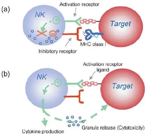
Natural killer (NK) cell activation is controlled by the integration of signals from activation and inhibitory receptors (Figure 1.7). Inhibitory NK cell receptors recognize self MHC class I and restrain NK cell activation (Figure 1.7a). When not impeded by the inhibitory receptors signals, binding of NK cell activation receptors to their ligands on target cells results in NK cell stimulation (Figure 1.7b). In the absence or downregulation of self MHC class I on the target cells, these stimulatory signals are no longer suppressed, resulting in NK cell responses including cytokine production and granule release leading to cytotoxicity. NK cells do not kill by default; that is, when MHC-I inhibition is absent; the NK cell must still be stimulated through activatory receptors. Moreover, whether or not an individual NK cell is activated by a target is determined by this complex balance of receptors with opposing function and expression of the corresponding ligands. In general, however, inhibition
Figure 1.7 Natural killer (NK) cell activation is controlled by the integration of signals from activation and inhibitory receptors. (a) Inhibitory NK cell receptors recognize self MHC class I and restrain NK cell activation. (b) When not impeded by the inhibitory receptors, binding of NK cell activation receptors to their ligands on target cells results in NK cell stimulation, resulting in cytokine production or granule release and killing. Picture taken from French and Yokoyama, 2004
Indeed, recent publications had revealed an increasing number of NK cell subversive mechanisms by CMV which affect this equilibrium (Table 1.1).
Table 1.1 Subversive CMV genes that control NK cell activation
HUMAN CYTOMEGALOVIRUS GENE PRODUCTS THAT SUBVERT NK ACTIVATION
ORF Function References
UL83 (pp65) Inhibition through NKp30 (Arnon et al., 2005) UL16 Sequesters MICB, ULBP1, ULBP2 (NKG2D
ligands) (Dunn et al., 2003)
UL18 MHC-I homologue (decoy for inhibitory NK Receptors)
(Cosman et al., 2001) (Prod'homme et al., 2007)
UL40 Enhances surface expression of HLA-E (Ligand for CD94/NKG2A Inhibitory NK receptors)
(Tomasec et al., 2000) (Ulbrecht et al., 2000) (Wang et al., 2002) UL112 Virally encoded microRNA that downregulates
MICB expression
(Stern-Ginossar et al., 2007)
UL141 Blocks surface expression of CD155, a ligand for
activatory NK receptors (Tomasec et al., 2005) UL142 Inhibition of NK cell-mediated lysis (Wills et al., 2005) MURINE CYTOMEGALOVIRUS GENE PRODUCTS THAT SUBVERT NK ACTIVATION
ORF Function References
m157 Inhibition by Ly49I (Arase et al., 2002)
m152 Downregulation of RAE1 and H60 (Ligands to
NKG2D) (Lodoen et al., 2003)
m155 Downregulation of H60 (Hasan et al., 2005;
Krmpotic et al., 2002) m145 Downregulation of MULT-1 (Ligand to NKG2D) (Krmpotic et al., 2005) m138 Downregulation of H60 and MULT-1 (Lenac et al., 2006a) m04 Counteracting MHC-I retention in ER (Kleijnen et al., 1997)
The majority of these subversive mechanisms are directed against direct activation of NK cells by downregulating or sequestering inhibitory ligands, like MICB or H60. Human NK cells are potent mediators of ADCC due to the expression of the FcγRIII CD16. In the presence of immune IgG and an appropriate target, NK cells become engaged and activated, secreting
1.5 Viral Fc γ Rs
While antibodies can be effective and protective against some viral infections as it is in the case for polio virus (Hovi, 2001) or measles virus (Cutts et al., 1991), in the case of herpesviruses, their potency seems to be restricted (Budt et al., 2004). Herpesviruses are able to re-infect a host or reactivate even in the presence of antibodies. The uneffectiveness of the antiviral antibodies against herpesviruses can be caused by the viral encoded FcγRs.
Two human Cytomegalovirus (HCMV) glycoproteins, gp68 and gp34, encoded by UL119-118 and IRL11/TRL11, respectively, were identified to exhibit Fc binding properties on the surface of HCMV-infected cells, thus constituting viral FcγR (vFcγRs) (Atalay et al., 2002b) (Lilley et al., 2001b) Likewise, Herpes simplex virus (HSV) forms a heterodimeric vFcγR, the gE/gI complex (Frank and Friedman, 1989e). Various pathogens also code for proteins that bind IgG in order to evade the host immune response. Bacteria as Staphylococcus aureus (Langone, 1982) and Streptococcus (Christensen et al., 1976), protozoa as schistosomes (Torpier et al., 1979) and viruses like coronaviruses (Oleszak and Leibowitz, 1990) and hepatitis C virus (Maillard et al., 2004) also express in their surface proteins capable of binding the Fc part of IgG.
1.5.1 HSV vFcγRs
The ability of HSV infected cells to bind to IgG was first demonstrated in 1979 (Baucke and Spear, 1979). A heterogenic complex of the two viral glycoproteins gE and gI has been identified as the HSV vFcγR (Nagashunmugam et al., 1998a). gE and gI are encoded by the genes US7 and US8, respectively. gE alone is able to bind aggregated IgG with low affinity.
When gE is in complex with gI, the affinity for IgG is augmented and the binding of monomeric IgG can be performed. This heterodimer binds IgG with 1:1 stoichiometry (Chapman et al., 1999); (Sprague et al., 2004; Sprague et al., 2006a). The IgG subclass
The IgG binding of the gE:gI complex arises many immune evasion possibilities to the virus.
In antibody- and complement-dependent virus neutralization assays, the gE:gI complex was shown to be of biological relevance. The HSV-1 strain resisted neutralization only slightly but resisted complement-mediated virolysis to a higher extent as a mutant virus lacking the vFcγR (Frank and Friedman, 1989d). Furthermore, this heterodimer protects infected cells from antibody dependent cellular cytotoxicity (Dubin et al., 1991e). The observed phenomena have been explained by a model called antibody bipolar bridging (Figure 1.8).
This model states that an IgG molecule recognising an epitope on the surface of an infected cell or virion via its Fab part is simultaneously bound to the vFcγR through the Fc part.
Therefore, the IgG Fc part is not free to recruit the effector mechanisms, i.e, C1q (the initial component of the classical complement cascade) (Lubinski et al., 1998a) or host cytotoxic FcγR bearing cells. Experiments in vivo further supported the notion that the HSV vFcγR enables the virus to evade antibody attack (Nagashunmugam et al., 1998d). Recently, Sprague ER et al resolved the crystal structure indicating that the ternary organization of the gE-gI/Fc complex is compatible with antibody bipolar bridging (Sprague et al., 2006b).
Antibody bipolar bridging: binding of a single immunoglobulin G (IgG) molecule by its Fab end to its antigenic
target and by its Fc end to an Fc receptor (FcR)
I Frank and HS Friedman. J Virol. 1989 November, 63(11):
4479–4488 Virus infected cell
Ag Ag
IgG vFcγR
cFcγR-ζ
IL-2
Effector cell
Figure 1.8 Antibody bipolar bridging: binding of a single immunoglobulin G (IgG) molecule by its Fab end to its antigenic target and by its Fc end to a viral Fcγ receptor (FcγR)
Experiments with animals indicate that ADCC is a component of the host's immune response to HSV infection. In immunocompromised mice, survival after HSV infection was improved following adoptive transfer of leukocytes and antiviral IgG (Ragerzisman and Allison, 1976).
In separate studies, passive immunization with intact antiviral IgG, but not with F(ab')2 fragments (which do not mediate ADCC), prevented HSV infection (Mckendall, 1985). In humans, high levels of ADCC antibody correlate with less severe neonatal HSV infections (Kohl, 1992; Kohl, 1991). Despite the success of ADCC in controlling HSV infection, previous experiments demonstrate that this response is blunted by vFcγRs (Dubin et al., 1991d), but
Furthermore, IgG Fc independent functions have been described for gE:gI. This glycoprotein is important also for virus cell-to-cell spread in cultured epithelial cells, as well as in epithelial and neuronal tissues (Polcicova et al., 2005). gE:gI promotes cell-to-cell spread by the direction of the assembly of virions into intracellular compartments, so that the virus progeny is ultimately delivered to the cell junctions and not released to the supernatant.
1.5.2 CMV vFcγRs
IgG binding activity of HCMV infected cells was detected on their plasma membrane, at intracellular sites and also in association with virions (Stannard and Hardie, 1991). Three viral genes encoding for two different glycoproteins which bind the Fc part of IgG were identified within the HCMV genome. Namely the two glycoproteins, gp68 and gp34, to be encoded by UL119-118 and IRL11/TRL11, respectively were characterized as the responsible molecules of this function, thus constituting vFcγRs (Atalay et al., 2002c; Lilley et al., 2001a). These three genes are dispensable for replication. The structural composition of both consists of a single chain type I transmembrane protein with low homology to the host FcγR and they are predicted to form IgSF-like domains (Atalay et al., 2002d). Both HCMV FcγRs are highly N-glycosylated and the gp68 has been shown to be also highly o- glycosylated (Manuela Fiedler, unpub data)
gp68 and gp34 are expressed during the early and late phase of HCMV replication. In contrast to the gE:gI complex, the HCMV vFcγRs are able to bind all human IgG subclasses.
Structural information revealing the binding and contact sites for gp68 were recently revealed
and releases it at the acidic pH of lysosomes, consistent with a role in facilitating the degradation of antiviral antibodies (Sprague et al., 2008).
Unlike the gE:gI complex of HSV, the HCMV vFcγR do not inhibit the neutralising function of IgG. Surprisingly, they seem not to affect the neutralizing potency of human IgG (Henrike Reinhard, unpub data). In complement mediated virolysis, the gp68 and gp34 do not show significant interference. Furthermore, in complement mediated cell lysis, they may not play a significant role, since the host encoded complement regulators: CD46 (MCP) and CD55 (DAF) were reported to be upregulated in HCMV infected cells (Spiller et al., 1996a), thus inhibiting activation of the complement cascade.
MCMV-infected cells can also bind mouse IgG-Fc fragment both intracellularly and on the surface of infected cells (Thale et al., 1994e). A single gene coding for the mouse FcγR was identified as m138. It codes for a highly glycosylated type I transmembrane protein of a calculated mass of 105 kDa. It is expressed in the early and late phases of MCMV replication. Alignment of the m138 sequence with mouse host FcγR shows a significant homology and predicts a composition of 3 IgSF-like domains (Matthias Budt, unpub data).
The subclass specificity of the m138 protein lies exclusively by IgG2a and IgG2b, the two most important cytotoxic antibodies in the mouse.
Surprisingly, in B cell deficient mice, the primary infection of MCMV takes the same course in the absence and presence of antibodies (Jonjic et al., 1994b). Recently, antibody independent functions for the m138/fcr-1 were identified. Namely, it was shown that it promotes a rapid down-regulation of NKG2D ligands murine UL16-binding protein like transcript (MULT)-1 and H60 from the cell surface (Lenac et al., 2006b). Deletion of the m138/fcr-1gene from the MCMV genome showed attenuation of viral replication to natural killer (NK) cell response in an NKG2D-dependent manner in vivo.
Murayama T et al reported already in 1987 that the presence of HCMV infected cells inhibits
cells (Murayama et al., 1987). ADCC seems to play furthermore an important role in control of MCMV since passive transfer of antibody of pre-immunized mice is able to control infection, but the neutralizing capacity of these antibodies was demonstrated to be very weak (Klenovsek et al., 2007c). Therefore, we pursued to test if the vFcγRs can constitute important immunosubversive genes inhibiting ADCC with a novel FcγR activation assay.
Given the fact that all the herpesviral FcγRs are expressed on the surface of infected cells and that herpesviruses infect host cells that constitutively express host FcγR, like monocytes, macrophages and dendritic cells, vFcγRs could act as antagonists or inhibitors of the host FcγR function. Thus, the assessment if herpesviral downregulation of IgG-mediated immune control targets the inhibition of host FcγR activation must be investigated.
1.6 Methods for Measuring Antiviral IgG
Antiviral antibodies are detected in the laboratory mainly with three prototypic assays (Hangartner et al., 2006a). These assays comprise the basic protocol of ELISA (Enzyme- linked immunosorbent assay), neutralization assays and the so called in vivo protection assays using appropriate animals. ELISA is used to detect antibodies that bind to a viral antigen, which is immobilized on a plastic surface. Samples to be tested are diluted serially and added to this pre-coated plastic surface. Bound antibodies are thereafter detected by a colorimetric reaction mediated by an enzyme. The intensity of this colour can be afterwards quantified. The conformation of the antigens detected is mostly denatured due to the binding procedures to the ELISA plate under highly basic pH conditions. Even though ELISA analysis
particles are recognized. The disadvantages of this method are mainly its unsuitability as a high throughput procedure and the inability to measure other antibodies which may have an important antiviral biological function besides neutralization.
In in vivo protection assays, an actively or passively immunized animal is challenged with the same pathogen. Virus titres are then determined in blood or organs and are compared with control animals which lack antiviral antibodies. The information gained after such an assay is a direct assessment of protection against the virus. The disadvantages of type of assay are the need of in vivo facilities, the requirement of an animal model for comparison with human diseases or the animals must be permissive for the virus infection and mount a comparable immune response as the one in humans.
The true biological function of antibodies can not easily be tested and therefore, until now, neutralization assays and in vivo protection assays are the only methods available to define these functions. An important biological antiviral function is the one elicited after engagement of the host Fcγ receptors by antibodies bound to target antigens. A process referred as antibody dependent cellular cytotoxicity or ADCC. This method is classically a radioactive approach with 51chromium release assays, difficult to reproduce and variability inherent of the effector cells can alter the results obtained. Newly methods to measure ADCC have been developed in the past couple of years. Examples of these new approaches are the novel EGFP-CEM-NKr flow cytometric method (Kantakamalakul et al., 2006), the usage of genetically modified antigen-specific human T lymphocytes as effector cells (Clemenceau et al., 2006), the rapid fluorometric method using two different double-staining target cells with a membrane dye (PKH-26) and a viability dye (CFSE) prior to the addition of antibody and effector cells called RFADCC (Gomez-Roman et al., 2006), and the substitution of radioactive Cr51 with a non-radioactive dye as CalceinAM (Lichtenfels et al., 1994). All these methods have obvious disadvantages: some require radioactive facilities, FACS (Fluorescence-activated cell sorting) facilites, the limiting factor of having an undefined mixed
defined population. Furthermore, viruses can encode for inhibitors of activation of the effector cells, like CMV for NK cells (see above). Therefore, the possibility to define clearly the role of specific (cytotoxic) antibodies that trigger a single host FcγR is still difficult.
1.7 Aim of the Thesis
Herpesviruses express on the surface of infected cells virally encoded FcγRs. To determine if viral FcγRs circumvent IgG mediated effector functions, IgG- and complement mediated cytotoxicity of herpesviruses infected cells had to be analyzed. A crucial IgG mediated immune control mechanism involves the activation of the host FcγRs through antibody-bound antigens. The activation of the host FcγRs elicits several effector mechanisms, e.g.
degranulation and killing of target cells by NK cells, cytokine secretion, phagocytosis and modulation of dendritic cell functions. Despite the fact that engagement of FcγRs by IgG is decisive for the generation of immune responses and the outcome of many diseases, there exists relatively little methodology to measure these immune responses in vitro. This limitation has been attributed to the lack of a simple, reliable and standardized assay.
Therefore, a method for detection of IgG antibodies able to trigger host FcγRs had to be established to study the possible interference of the herpesviral FcγRs with the host FcγRs.
2 Results
2.1 Establishment of a novel assay system for detection and
quantification of virus specific IgG antibodies triggering host Fc γ Rs
2.1.1 Stable expression of FcγR-ζ constructs and activation of BWFcγR-ζ transfectants
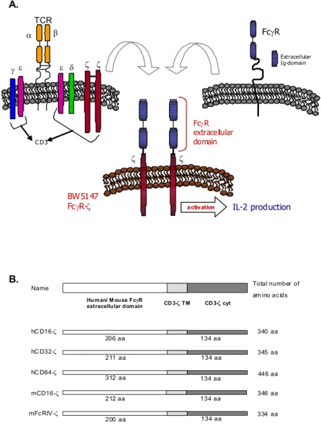
To quantify antiviral IgG antibodies able to trigger defined FcγRs individually, a novel set of chimeric FcγR/CD3-ζ response genes was constructed and stably transfected into BW5147 T cell hybridomas. In order to express the FcγR-ζ constructs, BW5147 thymoma cells lacking the TCR α, β and ζ chains were transfected. These constructs encode for chimeric FcγR proteins in which the extracellular portion of human CD16 (FcγRIIIa, the high responder 158V isoform), human CD32 (FcγRIIa), human CD64 (FcγRIa), mouse CD16 (FcγRIIIa) and mouse FcRIV were fused to the transmembrane and tail domains of the CD3-ζ chain (Figure 2.1), allowing the combination of IgG binding characteristics of the extracellular FcR domain with a well described and easy measurable signalling module as the T cell receptor complex. The CD3-ζ chain is sufficient for triggering activation (Irving and Weiss, 1991c) and secretion of mIL-2, a surrogate marker easily measured by ELISA.
γ ε ε δ
α β
ζ ζ
C D3
TCR
FcγR
Extracellular Ig-domain
IL-2 production ζ
FcγRextracellular
domain ζ
a ctiv ation
BW5147 FcγR-ζ
340 aa hC D16-ζ
hC D32-ζ hC D64-ζ mCD16-ζ mFc RIV-ζ
206 aa 134 aa
211 aa
312 aa
212 aa
200 aa
134 aa
134 aa
134 aa
134 aa CD 3-ζ cyt CD 3-ζ TM
Human/ M ouse FcγR extracellular domain
Name Total number of
am ino ac ids
345 aa
446 aa
346 aa
334 aa
B.
A.
Figure 2.1 (A) Schematic representation of the BWFcγR-ζ effector cells after
cell surface of BW cells (Figure 2.2A). The expression of FcRIV-ζ was difficult to assess, since there are no commercially available antibodies. Nevertheless, mouse Fc-IgG staining on the surface of half of the transfectants was detected, suggesting the presence of a considerable subfraction of cells to be positive for the construct (Figure 2.2A).
To prove intact signal transduction through the hBWCD16-ζ, hBWCD32-ζ, hBWCD64-ζ and the mouseBWCD16-ζ receptors, mAbs directed against each of the FcγR ectodomains were used in crosslinking experiments. All transfectants triggered mIL-2 production upon crosslinking with specific mAbs. An isotype control mAb directed against CD99 did not induce mIL-2 in chimeric FcγR transfectants. In some further experiments, hBWCD99-ζ transfectants (obtained from O. Mandelboim, Israel) were included as a negative control.
CD99 is, like the FcγRs, a member of the immunoglobulin superfamily (IgSF), but it is not an FcγR, thus, it does not bind IgG antibodies. In conclusion, all established BW transfectants responded when crosslinked by specific mAbs directed to the extracellular domain of the FcγR chimeras. (Figure 2.2B)
Thus, human BWCD16-ζ, BWCD32-ζ, BWCD64-ζ, mouse BWCD16-ζ and mouse BWFcRIV-ζ can be further tested for IgG mediated activation.
A. B.
coated in 96-well cell culture plates at a concentration of 2 µg/ml in binding buffer (0.1 M Na2HPO4 pH 9.0). After blocking and washing, mouse mAb specific for human CD16-A/B, human CD32, human CD64 and rat anti- mouse CD16/CD32 were added. As a negative control, mAb anti-human CD99 was used. After removal of unbound antibodies, 200.000 cells per well of the BWFcγR-ζ transfectants were added. mIL-2 measurement was performed after 16 hours of incubation at 37°C with the ELISA.
Summary
1. Stable transfection of chimeric receptors bearing the extracellular domain of host FcγRs and transmembrane and cytoplasmic tails of mouse CD3-ζ chain into BW5147 mouse cells was achieved.
2. Correct expression on the surface of the BWFcγR-ζ transfectants was observed with FACS analysis.
3. Intact signalling (efficient mIL-2 release) was detected from the BWFcγR-ζ transfectants after crosslinking with mAb directed against the ectodomain of the respective FcγR.
2.1.2 IgG dependent activation of the BWFcγR-ζ transfectants
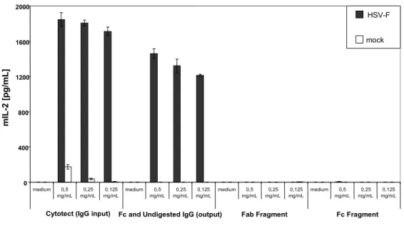
To assess if the BWFcγR-ζ transfectants cells are activated by IgG, human IgG alone, human IgM alone or IgG-depleted human sera were coated to a plate. To assess if the BWFcγR-ζ transfectants cells are activated by immune complexes, HCMV- or MCMV-purified virion preparations alone were directly coated to ELISA plates. Afterwards, a human polyclonal preparation Cytotect® was used to opsonize this plate (which was previously coated with HCMV- and MCMV-virions) in order to elicit immune complex formation. The antigens recognized in this experimental setting are denatured due to the coating procedures. IgG or immune complexes (HCMV virion-IgG) mediate a dose dependent activation of the BWCD16-ζ effector cells (Figure 2.3A-B). To test whether the BWFcγR-ζ transfectants respond to IgG recognizing a native viral surface antigen, a cocultivation assay
single defined surface epitope of HSV-1 gD, was used to opsonize HSV-1 infected MRC-5 fibroblasts. A dose dependent activation of the mouse BWCD16-ζ effector cell was observed (Figure 2.3C). To test if polyclonal antibodies against viral targets also activate the BWFcγR- ζ effector cells, HSV-1 infected cells were opsonized with Cytotect® (Figure 2.3D). Dose
dependent triggering of the BWFcγR-ζ effector cells was observed, confirming IgG dependent activation after opsonization of an antigen. Mock cells did not induce any mIL-2 release by the effector cells. A schematic diagram of the different experimental settings for analysing IgG-dependent activation is depicted in Figure 2.3.
Furthermore, to test if cell bound HD-1 triggers mIL-2 release in a dose dependent manner from FcRIV-ζ cells, a co-cultivation assay with HSV-1 infected fibroblasts was performed.
Comparison between the amount of activation within mCD16-ζ and FcRIV-ζ transfectants showed that the IgG dependent response of the FcRIV-ζ cells was less efficient (Figure 2.3E), which can be explained by lower expression of the chimeric receptor on the surface of the BW transfectants.