Ubiquitin-Independent Proteolytic Targeting of Ornithine Decarboxylase
Inaugural-Dissertation Zur
Erlangung des Doktorgrades
der Mathematisch-Naturwissenschaftlichen Fakultät der Universität zu Köln
vorgelegt von
Roshini Beenukumar Renukadevi aus Trivandrum, Kerala, Indien
Köln, 2015
Berichterstatter
Prof. Dr. Jürgen Dohmen Prof. Dr. Kay Hofmann
Tag der mündlichen Prüfung: 29 June 2015
“Science is the rational way of revealing the existing realities of nature”
My Grandfather
Abstract
Protein degradation mediated by the 26S proteasome is fundamental for cell survival in eukaryotes. There are two known routes for substrate presentation to the 26S proteasome- the ubiquitin-dependent route and the ubiquitin-independent route. Ornithine decarboxylase (ODC) is one of the most well-known ubiquitin-independent substrates of the proteasome. It is a homodimeric protein functioning as a rate-limiting enzyme in polyamine biosynthesis.
Polyamines regulate ODC levels by a feedback mechanism mediated by the ODC regulator called antizyme. Higher cellular polyamine levels promote translation of antizyme mRNA and inhibit ubiquitin-dependent proteasomal degradation of the antizyme protein. Antizyme binds ODC monomers and targets them to the proteasome without ubiquitylation. The mechanism of this ubiquitin-independent proteasomal degradation is poorly understood. Therefore, the major aim of this study was to investigate the mechanism of ubiquitin-independent degradation of the ODC by the 26S proteasome. We show that polyamines, besides their role in regulating antizyme synthesis and stability, directly enhance antizyme-mediated ODC degradation by the 26S proteasome. Polyamines specifically enhanced the degradation of ODC by the proteasome both in vivo in yeast cells and in a reconstituted in vitro system.
ODC is shown to be targeted in a manner quite distinct from ubiquitin-dependent substrates as its degradation was enhanced in a mutant lacking multiple ubiquitin receptors. These and other findings indicate, however, that there is a convergence point for the two routes of degradation because ubiquitin-dependent substrates compete with ODC for degradation.
Using an in vitro assay, it could be shown that the unstructured N-terminal degron, ODS, is essential for binding of ODC to the proteasome. In vivo studies using proteasomal ATPase mutants, in which tyrosine residues in so-called pore loops were mutated to alanine (Y-A), further showed that the pore loops of Rpt4 and Rpt5 are of critical importance for ODC degradation and suggested that ODS might be recognized by these ATPase subunits.
Additional experiments revealed that antizyme promotes ODC degradation most likely by providing an additional binding site. An ODS-antizyme-Ura3 fusion protein was degraded faster in a ubiquitin-independent but proteasome-dependent manner than ODS-Ura3.
Furthermore, a ubiquitin-dependent mode of ODC degradation is also reported. Upon overexpression under the PCUP1 promoter, efficient degradation of ODC involved a ubiquitin- dependent mechanism. This degradation of ODC was independent of ODS and antizyme.
Together, the findings described in this thesis provide novel insights into the mechanism of proteolytic regulation of ODC. With ODC being a validated target for cancer therapy, a detailed understanding of this mechanism may contribute to the discovery of new therapies targeting the polyamine pathway.
Zusammenfassung
Proteinabbau durch das 26S-Proteasom ist von fundamentaler Bedeutung für das Überleben eukaryotischer Zellen. Substrate können dem Proteasom auf zwei Arten präsentiert warden, entweder Ubiquitin-abhängig oder Ubiquitin-unabhängig. Ornithine decarboxylase (ODC) ist das bekannteste Ubiquitin-unabhängige Substrat des Proteasoms. Es ist ein homodimeres Protein mit einer geschwindigkeitsbestimmenden Funktion in der Biosynthese von Polyaminen. Polyamine regulieren die ODC-Konzentration durch einen Feedback- Mechanismus, der durch das ODC-Regulatorprotein Antizym vermittelt wird. Höhere zelluläre Polyamin-Konzentrationen stimulieren die Translation von Antizym-mRNA und hemmen den Ubiquitin-abhängigen Abbau des Antizym-Proteins. Antizym bindet an ODC- Monomere und vermittelt deren Ubiquitin-unabhängigen Abbau durch das Proteasom. Der Mechanismus des Ubiquitin-unabhängigen Proteinabbaus durch das Proteasom ist noch nicht gut verstanden. Das Hauptziel dieser Arbeit war es daher, den Mechanismus des Abbaus der ODC näher zu untersuchen. Es konnte gezeigt warden, dass Polyamine, neben ihrer Rolle in der Regulation der Synthese und Stabilität von Antizyme, einen direkt verstärkenden Effekt auf den Antizym-vermittelten Abbau der ODC durch das 26S- Proteasom sowohl in vivo in Hefezellen als auch in einem rekonstituierten In vitro-System hat. ODC wird auf eine andere Art und Weise vom Proteasom erkannt als Ubiquitin- abhängige Substratproteine, wie der verstärkte Abbau der ODC in Hefemutanten mit fehlenden Ubiquitin-Rezeptoren zeigte. Diese und andere Ergebnisse deuteten an, dass ODC und Ubiquitin-abhängige Substrate aber auch an einem bestimmten Punkt zusammenkommen, da diese Substrate um den Abbau durch das Proteasom konkurrieren.
Durch In vitro-Bindungsstudien konnte gezeigt werden, dass das unstrukturierte N-terminale Abbaussignal (ODS) für die Bindung der ODC an das Proteasom essentiell ist. In vivo- Experimente mit Hefemutanten, in denen kritische Tyrosinreste in den so genannten Pore Loops der ATPase-Untereinheiten zu Alanin mutiert sind, zeigten das diese Loops der Untereinheiten Rpt4 und Rpt5 für den Abbau der ODC von kritischer Bedeutung sind, vermutlich indem sie das ODS erkennen. Weitere Experimente erbrachten Hinweise darauf, dass Antizym wahrscheinlich den Abbau der ODC fördert, indem es eine zusätzliche Bindestelle für das Proteasom beisteuert. So wurde beobachtet, dass ein ODS-Antizym- Ura3-Fusionsprotein schneller Ubiquitin-unabhängig abgebaut wurde als ODS-Ura3. Neben dem zuvor genannten Mechanismus wurde auch ein Ubiquitin-vermittelter Abbau der ODC beobachtet, wenn diese in stärkerem Maße in den Zellen synthetisiert wurde. Dieser Abbau erwies sich als unabhängig von ODS und Antizym. Die Ergebnisse dieser Arbeit eröffnene neue Einblicke in die Mechanismen der proteolytischen Kontrolle der ODC. Da ODC bereits als Zielstruktur von Krebstherapien validiert ist, kann ein detaillierteres Verständnis dieser
regulatorischen Mechanismen zur Entwicklung neuer Therapien beitragen, die einer häufig mit der Zellentartung einhergehenden Erhöhung der Polyaminsynthese entgegen wirken.
Table of Contents
1. Introduction ... 1
1.1. Proteolysis in eukaryotes ... 1
1.1.1. The 26S proteasome ... 1
1.1.2. Ubiquitin-dependent proteasomal targeting ... 4
1.1.3. Ubiquitin-independent proteasomal targeting ... 7
1.2. Feedback regulation of polyamines in eukaryotes ... 8
1.2.1. Polyamine types and their biosynthesis ... 8
1.2.2. Regulation of ODC by antizyme ... 10
1.2.3. ODC degradation: the story so far ... 12
1.3. Other ubiquitin-independent substrates ... 13
2. Aim of current study ... 15
3. Materials and Methods ... 16
3.1. Materials ... 16
3.1.1. Saccharomyces cerevisiae strains ... 16
3.1.2. Escherichia coli strains ... 17
3.1.3. Plasmids ... 17
3.1.4. Oligonucleotides ... 18
3.1.5. Enzymes ... 19
3.1.6. Antibodies ... 19
3.1.7. Chemicals ... 20
3.1.8. Instruments ... 22
3.2. Methods ... 23
3.2.1. Molecular biology and genetic techniques ... 23
3.2.1.1. Isolation of plasmid DNA from E. coli ... 23
3.2.1.2. Estimation of DNA concentration ... 23
3.2.1.3. Isolation of genomic DNA from yeast ... 23
3.2.1.4. PCR amplification ... 23
3.2.1.5. Agarose gel electrophoresis ... 25
3.2.1.6. Extraction of DNA from agarose gels ... 25
3.2.1.7. Restriction digestion of DNA ... 25
3.2.1.8. Ligation of DNA fragments ... 25
3.2.1.9. Preparation of chemically competent E.coli cells ... 26
3.2.1.10. Transformation of chemically competent E. coli cells ... 26
3.2.1.11. Cultivation of yeast cells ... 26
3.2.1.12. Yeast phenotypic analysis by spot tests ... 27
3.2.1.13. Yeast mating type testing and crossing of haploids ... 28
3.2.1.14. Sporulation and tetrad dissection ... 28
3.2.1.15. High efficiency yeast transformation (Gietz and Woods, 2006) ... 28
3.2.2. Biochemical and immunological methods ... 29
3.2.2.1. Yeast cell lysis with glass beads (Dohmen et al., 1995) ... 29
3.2.2.2. Yeast cell lysis by boiling ... 29
3.2.2.3. Yeast cell lysis by grinding ... 30
3.2.2.4. Estimation of protein concentration ... 30
3.2.2.5. SDS polyacrylamide gel electrophoresis (SDS-PAGE) (Laemmli, 1970) ... 30
3.2.2.6. Western blot analysis ... 31
3.2.2.7. Reprobing of western blot membranes ... 32
3.2.2.8. Analysis of protein stability by cycloheximide chase ... 32
3.2.2.9. ODC-Oaz1 interaction analysis ... 32
3.2.2.10. Purification of proteasomes from yeast (Ha et al., 2012) ... 32
3.2.2.11. Purification of Oaz1, ODC and ODC/Oaz1 heterodimer ... 33
3.2.2.12. Analysis of proteasomes by Native-PAGE (Elsasser et al., 2005) ... 34
3.2.2.13. Proteasomal peptidase activity assay (Dohmen et al., 2005) ... 35
3.2.2.14. In vitro proteasomal degradation assay (Ha et al., 2012) ... 35
3.2.2.15. Polyamine binding assay (Palanimurugan and Dohmen, 2012) ... 35
3.2.2.16. Native-PAGE analysis of proteasomal binding ... 36
4. Results ... 37
4.1. Characterization of the direct role of polyamines in ODC targeting ... 37
4.1.1. In vitro recapitulation of proteasomal degradation of ODC ... 37
4.1.2. Polyamines directly promote proteasomal degradation of ODC in vitro ... 41
4.1.3. Polyamines do not enhance ubiquitin-dependent proteasomal degradation .... 42
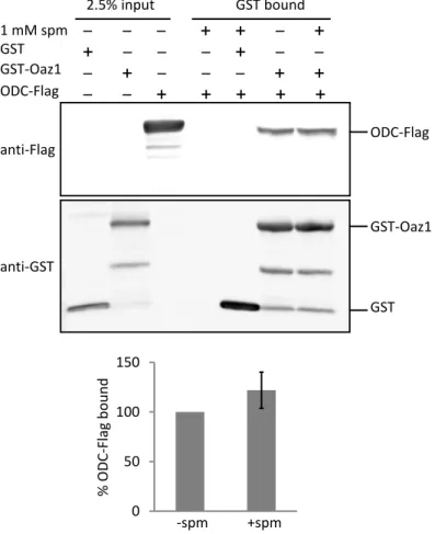
4.1.4. Polyamines do not alter the affinity of ODC/antizyme interaction ... 44
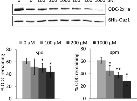
4.1.5. Both spermidine and spermine promote antizyme and ODC degradation in vivo ... ………45
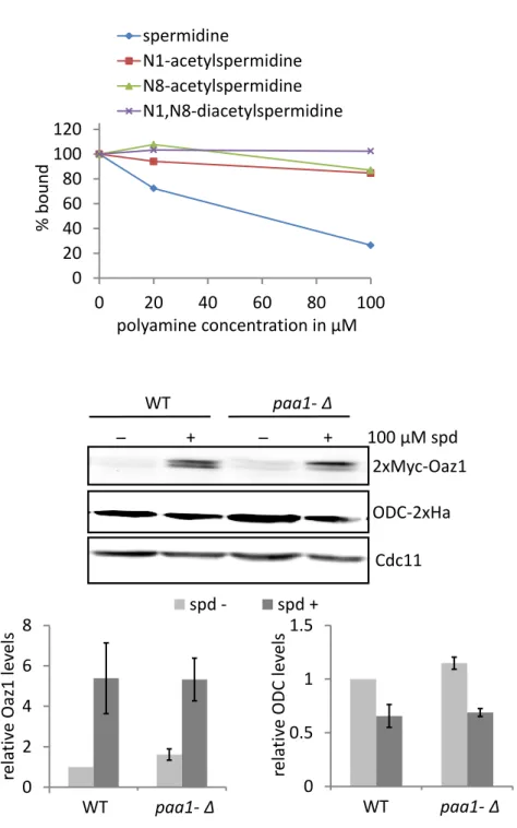
4.1.6. Acetylation of polyamines inhibit their binding to antizyme ... 47
4.2. Characterization of ubiquitin-independent substrate recognition by the proteasome.. ... 49
4.2.1. ODS is essential for proteasomal binding of ODC ... 49
4.2.2. Proteasomal lid is dispensable for in vitro degradation of ODC by the
proteasome ... 52
4.2.3. ODC is not recognized by the canonical ubiquitin receptors ... 53
4.2.4. Human p21 is degraded by the yeast proteasome and competes with ODC for degradation ... 54
4.2.5. Specificity of Rpt4 and Rpt5 Ar-Φ loop in substrate engagement and targeting of ODC……….57
4.3. Characterization of the role of antizyme in ODC targeting ... 60
4.3.1. ODS fused to stable antizyme is degraded in a ubiquitin-independent manner………60
4.3.2. Antizyme might have a binding site on the proteasome ... 63
4.3.3. A genetic screen for the isolation of ODC stabilizing mutants ... 65
4.4. Ubiquitin-dependent degradation of ODC ... 67
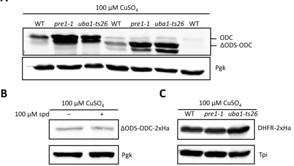
4.4.1. ODC is degraded by the proteasome in a ubiquitin-dependent manner upon overexpression ... 67
4.4.2. Ubiquitin-dependent ODC degradation is independent of its unstructured domain……….68
4.4.3. Ubiquitin-dependent ODC degradation is independent of antizyme ... 70
4.4.4. Protein quality control pathway might not influence ubiquitin-dependent ODC degradation ... 71
5.Discussion ... 73
5.1. Role of polyamines in feedback regulation of ODC ... 73
5.1.1. An additional role of polyamines in ODC degradation ... 73
5.1.2. Spermidine and spermine play similar roles in ODC regulation in yeast ... 75
5.1.3. Role of acetylated polyamines in ODC degradation ... 76
5.2. Ubiquitin-independent substrate targeting to the proteasome ... 77
5.2.1. Factors involved in ODC/Oaz1 targeting to the proteasome ... 77
5.2.2. Human p21 is degraded by the yeast proteasome in a ubiquitin-independent manner………78
5.2.3. Rpt4 and Rpt5 Ar-Φ pore loops are involved in ODC recognition at the proteasome ... 78
5.3. Antizyme might provide an additional binding site to the proteasome ... 80
5.4. Ubiquitin-dependent ODC degradation ... 82
5.5. Conclusions and outlook ... 84
6. References ... 85
List of Abbreviations... 94
Acknowledgement ... 96
Eidesstattliche Erklärung ... 98 Lebenslauf ... 99
1
1. Introduction
1.1. Proteolysis in eukaryotes
About 70 years ago, it was widely accepted that proteins were stable constituents in living cells (Ciechanover, 2012). After years of pioneering research we now know that protein degradation plays a pivotal role in the maintenance of all cells. Abnormal and unwanted proteins are timely eliminated by the cellular machinery. Proteins are broken down into its constituent amino acids which are then utilized for new protein synthesis. Proteolysis in eukaryotes is carried out via two known systems– lysosome- dependent macroautophagy (autophagy) and ubiquitin-proteasome system (UPS).
Lysosomes are organelles that contain an array of enzymes which degrade proteins as well as cellular organelles through the mechanism of autophagy. Alternatively, proteasomal degradation of proteins is achieved by conjugation of target proteins with a post-translational polyubiquitin modification and their subsequent degradation by a barrel shaped complex called the 26S proteasome (Lilienbaum, 2013). Until recently, the two mechanisms were thought to be independent of each other.
However, recent studies suggest a cross-talk between them. Impairment of UPS has been shown to induce autophagy (Pandey et al., 2007) and the inhibition of autophagy led to an induction of proteasome activity by up-regulation of proteasomal subunits (Wang et al., 2013). The principles of proteasome-mediated degradation are described in sections below.
1.1.1. The 26S proteasome
During the late 70s, Hershko, Ciechanover and Rose characterized a non-lysosomal energy requiring proteolytic system now known as the ubiquitin-proteasome system (Ciechanover, 2012). A decade later, Hough et al. partially purified the protease responsible for such an ATP-dependent degradation of ubiquitin-conjugated proteins which later came to be known as the 26S proteasome (Hough et al., 1986). Since then, decades of intensive research have increased our understanding of the structure and function of the proteasome. The proteasome is now a validated target for cancer therapies (Almond and Cohen, 2002). In May 2003, Bortezomib, a proteasome inhibitor was approved by the US FDA as a treatment for multiple myeloma.
The 26S proteasome is a ~2.5 MDa multi-subunit degradation machinery which selectively degrades 80-90% of cellular proteins (Lilienbaum, 2013). It consists of two major subcomplexes, the 20S core particle (20S or CP) and the 19S regulatory particle (19S or RP) (Fig. 1A). The crystal structure of the yeast 20S proteasome
2 revealed that it is composed of four stacked heptametrical rings arranged as an (α1- α7, β1- β7)2 complex. The α-ring consists of seven subunits which are predominantly structural components of the 20S whereas the β-ring houses the catalytic domains of the proteasome. Out of the seven subunits of the β-ring, the β1, β2 and β5 subunits harbor the proteolytic active sites (Tomko and Hochstrasser, 2013).
Fig.1: Structure of the 26S proteasome. (A) Surface representation of the 26S proteasome structure with the 20S core particle capped on both sides by the 19S regulatory particles (RP). (B) Surface representation of the 19S RP structure showing the relative positions of the subunits of the lid and base subcomplexes. (C) Surface representation of the lid subcomplex. (D) Surface representation of the base subcomplex. All structural representations were obtained using the PDB structures 4CR and 1RYP and viewed using the 3D molecular visualization software PyMOLTM.
The 19S RP (Fig. 1B) caps the CP, thereby regulating substrate entry into the CP. It does so by harbouring receptors for ubiquitin binding, detaching ubiquitin tags, opening the 20S CP gate, as well as by unfolding and translocating the substrate into the CP. Under certain in vitro conditions, the RP was shown to dissociate into two subcomplexes, the base and the lid (Michael H. Glickman, 1999). The base consists of a heterohexameric AAA+ ATPase ring (consisting of the subunits, Rpt1-Rpt6) and three non-ATPase subunits, namely Rpn1, Rpn2 and Rpn13 (Fig. 1B and 1D). The major functions of the base are 20S gate opening and substrate unfolding and
19S
19S 20S
26S proteasome
19S
Lid
Base Rpn13
Rpn1 Rpn2
Rpn11
Rpt ring
A B C
D
Rpn3, 5-9, 12, Sem1
Rpn10
3 translocation into the 20S. The unfolded substrates are then threaded through the narrow 20S pore by using the chemical energy from ATP hydrolysis. The Rpn1 and 2 are the largest subunits of the proteasome. Rpn1 serves as the docking sites of extrinsic ubiquitin receptors such as Rad23, Dsk2 and Ddi1. Rpn1 and 2 are hypothesised to serve as a loading platform for incoming substrates. Apart from these, the Rpn13 functions as a ubiquitin receptor. The RP lid consists of 9 Rpn subunits-Rpn3, 5-9, 11, 12 and Sem1 (Fig. 1B and 1C). The lid is essential for the degradation of ubiquitylated substrates. Rpn11 functions as the deubiquitylating enzyme (DUB). Rpn10 is an intrinsic ubiquitin receptor (Forster et al., 2009; Nickell et al., 2009; Tomko and Hochstrasser, 2013; Walz et al., 1998).
It took more than a decade after the solving of the crystal structure of the 20S CP to resolve the structure of the 19S RP. An atomic structure of the latter by crystallography could not be achieved mainly because of its dynamic nature. In 2012, two laboratories independently published subnanometer CryoEM structures of the yeast 19S RP (Beck et al., 2012; Lander et al., 2012). This was achieved using various techniques combined with CryoEM, including a novel approach for heterologous co-expression in E.coli, antibody and GST-fusion labelling and the use of deletion mutants. Interestingly, the lid subcomplex was found to be attached to the side of the 19S RP, which contrasted with previous ideas (Fig. 1B and 1C). The Rpt subunits of the base (marked as Rpt ring in Fig. 1B) were shown to be arranged in a spiral staircase, and the pore of the Rpt ring does not align with the pore of the 20S.
The ubiquitin receptors, Rpn10 and Rpn13, are flexibly attached to the periphery of the RP. The Rpn11 deubiquitylase subunit is positioned directly above the entrance of the pore of the Rpt ring. Rpn1 is very closely associated with the ATPase ring, whereas Rpn2 is placed distally along the long axis of the proteasome and a part of it is positioned above the pore of the Rpt ring (Fig. 1B and 1D).
In a recent review, Inobe and Matouschek have described three different modes of substrate recognition by the proteasome: (1) ubiquitin-dependent, (2) adaptor- mediated, and (3) ubiquitin-independent. The first two modes depend on ubiquitin- tagging of the substrate for proteasome recognition whereas the third one is independent of ubiquitin-tagging. In all three modes of proteasome recognition, a common principle is the engagement of an unstructured domain in the substrate by the ATPase ring to initiate degradation (Inobe and Matouschek, 2014). The modes of proteasomal targeting are detailed in the subsections below.
4
1.1.2. Ubiquitin-dependent proteasomal targeting
There are a growing number of proteins whose cellular function is regulated by their timely elimination by the ubiquitin-proteasome system (UPS). The UPS is known to be involved in many vital cellular processes ranging from DNA repair, cell cycle regulation, and cell migration to immune responses (Melvin et al., 2013). On the clinical side, therapies targeting the UPS are underway for several diseases. Two drugs, Bortezomib and Carfilzomib, both proteasome inhibitors, are already in the clinic as a treatment for multiple myeloma (Melvin et al., 2013). It is therefore critical to further understand the details of the mechanisms of protein degradation by the UPS.
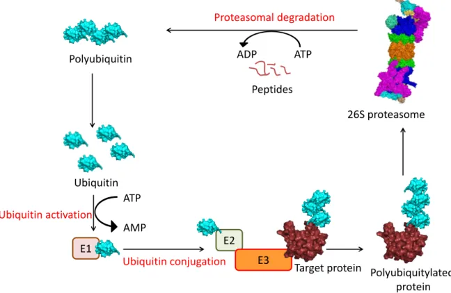
The ubiquitin-dependent substrate targeting and recognition is the more extensively studied route of proteasome targeting. It involves a series of enzymatic reactions wherein ubiquitin, a 8.5 KDa protein modifier, is conjugated to a target protein as detailed in Fig. 2. In Saccharomyces cerevisiae, there is only one known E1 enzyme encoded by UBA1, 11 known E2s and 42 different E3s. (Lee et al., 2008; McGrath et al., 1991). In most cases, ubiquitin conjugation takes place via a peptide bond formed between the Gly76 of ubiquitin and a lysine residue in the substrate. Polyubiquitin chains are formed by attaching another ubiquitin to a lysine residue (e.g. Lys48) of the preceding ubiquitin (Glickman and Ciechanover, 2002). This process is reversible as cells also contain deubiquitylating enzymes (DUBs) which remove the ubiquitin chains from substrates (Komander et al., 2009).
5
Fig. 2: The ubiquitin-proteasome system. Shown are the steps involved in the ubiquitin-dependent degradation of a target protein. Individual ubiquitin moieties are activated in an ATP dependent manner by the E1 ubiquitin-activating enzyme. The ubiquitin is then transferred to an E2, ubiquitin- conjugating enzyme. Subsequently, the activated ubiquitin-loaded E2 interacts with a specific E3, which are protein ligases directly in contact with a substrate. Ubiquitin is then covalently attached to one or more lysines within the target protein. Polyubiquitylation is achieved by attaching additional ubiquitins to the initial ubiquitin via one of its seven lysines. The polyubiquitylated substrate is then recognised by the shuttle factors or intrinsic ubiquitin receptors in the 19S RP. The ubiquitin chain is cleaved off from the substrate by the deubiquitylase, Rpn11 and recycled. The substrate is unfolded by the Rpt1-6 ATPases and translocated into the CP for degradation.
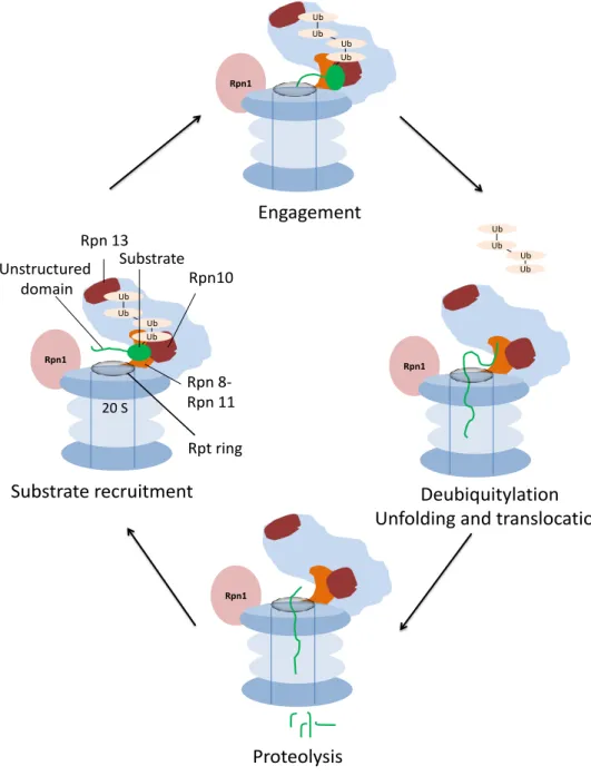
Recently, a more detailed understanding of the ubiquitin-dependent substrate targeting to the proteasome has emerged. The various steps involved in this process are detailed in Fig. 3. Apart from the ubiquitin tag, an unstructured region in the substrate is required for efficient proteasomal degradation (Prakash et al., 2004).
Recent CryoEM structure of an actively translocating 26S proteasome shows that this unstructured initiation region makes contact with the N-ring (the ring formed by the N-terminal domains of the ATPases) once the substrate is tethered to the proteasome via a ubiquitin receptor. Furthermore, the active site of the Rpn11 deubiquitylase is masked to prevent premature deubiquitylation. Upon substrate
E1 E2
E3 Polyubiquitin
Ubiquitin
26S proteasome ADP ATP
ATP AMP
Target protein Polyubiquitylated protein Proteasomal degradation
Peptides
Ubiquitin activation
Ubiquitin conjugation
6 engagement by the Rpt ring, the proteasome undergoes structural changes. As a result, a continuous central channel to the 20S core is formed for substrate degradation. The Rpn11 active site is unmasked as it shifts to a position directly above the N-ring thereby scanning and removing ubiquitin chains from the translocating polypeptide.
Fig. 3: Proteasomal degradation of a ubiquitylated substrate. Upon substrate recognition through polyubiquitin binding to the receptors Rpn13 and Rpn10, the Rpt ring of the proteasome initiates degradation at an unstructured region in the substrate. Structural rearrangements upon successful engagement lead to unfolding and translocation of the substrate to the 20S CP as well as cleavage of the ubiquitin tag. The substrate is completely unfolded and cleaved into peptides. Adapted from (Bhattacharyya et al., 2014).
Rpn1
20 S
Rpn1
Rpn1 Ub
Ub Ub
Ub
Ub Ub
Ub Ub
Ub Ub Ub
Ub
Rpn1
Rpn 13
Rpn10 Substrate
Rpn 8- Rpn 11 Rpt ring Unstructured
domain
Substrate recruitment
Engagement
Deubiquitylation Unfolding and translocation
Proteolysis
7
1.1.3. Ubiquitin-independent proteasomal targeting
Most of the known proteasomal substrates require ubiquitylation for their degradation. However, there are a significant number of proteins the degradation of which does not require ubiquitin conjugation. This mode of degradation is hypothesised to be a remnant of the ubiquitin-free degradation observed in the archaea and bacteria (Erales and Coffino, 2013; Inobe and Matouschek, 2014). Such substrates are characterized by observing their proteasomal degradation when ubiquitylation is impaired either by inactivating the ubiquitin-activating enzymes or by mutating all receptor lysines on the protein (Jariel-Encontre et al., 2008). The mechanism of such a ubiquitin-independent proteasomal targeting still remains unclear. However, it has been speculated that presence of an unstructured domain in these proteins is sufficient for proteasome association (Inobe and Matouschek, 2014). Based on biochemical analyses of mammalian lysates, Baugh et al. have reported that more than 20% of cellular proteins are regulated by degradation in an ubiquitin-independent manner by both the 20S and 26S proteasomal species (Baugh et al., 2009). Using in vitro experiments, it was shown that oxidatively damaged proteins can be degraded by the 20S proteasome, independent of ubiquitin (Davies, 2001). Ornithine decarboxylase (ODC), Rpn4 and thymidylate synthase are examples of ubiquitin-independent substrates, degradation of which is strictly ATP- dependent and therefore requires the 26S proteasome (Erales and Coffino, 2013).
Taken together, these observations bring us to two different modes of ubiquitin- independent proteasomal degradation, one that is ATP-dependent mediated solely by the 26S proteasome and another ATP-independent one mediated mainly by the 20S proteasome. This thesis deals with the former mode of ubiquitin-independent proteasomal degradation in particular with the ubiquitin-independent degradation of ornithine decarboxylase (ODC) in yeast.
ODC is the best-studied ubiquitin-independent substrate. It is conserved from yeast to humans (Palanimurugan et al., 2014). In 1989, Bercovich et al. observed that the degradation of ODC occurs in a ubiquitin-independent but ATP-dependent manner in reticulocyte lysates (Bercovich et al., 1989) which was later shown to occur in vivo in mammalian cells as well (Rosenberghasson et al., 1989). Since then several laboratories have tried to further understand this mechanism of ubiquitin-independent degradation of ODC. However, this mechanism is still not fully understand and is therefore a major focus of this thesis.
8
1.2. Feedback regulation of polyamines in eukaryotes
ODC is the rate-limiting enzyme in the biosynthesis of a class of molecules called polyamines. The current understanding of the feedback regulation of polyamines involving ODC and other players is described in subsections below.
1.2.1. Polyamine types and their biosynthesis
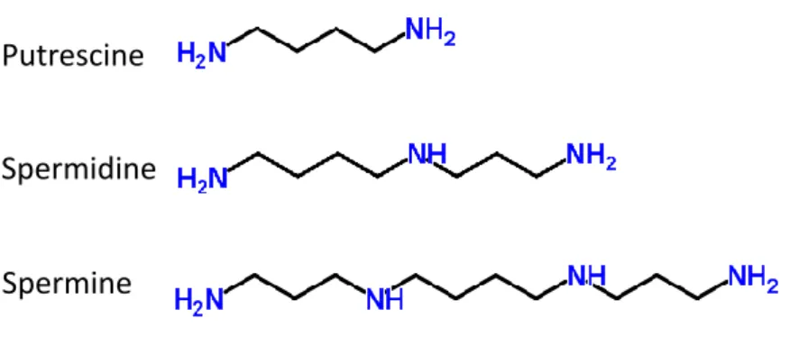
Polyamines are ubiquitous polycations essential for cell survival. Polyamines play multiple roles in the cell and are involved in almost all cellular processes. These include DNA replication, apoptosis, transcription, translation and membrane stability (Palanimurugan et al., 2014). Spermidine and spermine derived from the diamine precursor putrescine are the major polyamines in the cell (Fig. 4). Spermidine is formed from putrescine and spermine from spermidine by the addition of an aminopropyl group (Wallace, 2009).
Fig. 4: Polyamine types and their structure.
Biosynthesis of polyamines in S. cerevisiae is outlined in Fig. 5. The polyamine biosynthetic pathway and it regulation is highly conserved from yeast to humans.
Therefore, S. cerevisiae serves as a useful model organism to delve deeper into the regulatory mechanisms of this pathway.
Polyamines are regulated not only at the level of their biosynthesis but also at their catabolism and transport. Acetylated polyamines are either exported from the cell or subjected to oxidation by polyamine oxidase (Fms1 in yeast; shown in Fig. 5). In mammals, oxidation of N-acetylspermine and N-acetylspermidine produces spermidine or putrescine respectively, along with 3-aceto-aminopropanal and hydrogen peroxide (H2O2) (Palanimurugan et al., 2014). In mammals, the spermidine/spermine N1-acetyltransferase (SSAT) is a well-characterized enzyme in polyamine catabolism (Casero and Pegg, 1993). Liu et al. have recently
Putrescine
Spermidine
Spermine
9 characterized a yeast gene that encodes a polyamine acetyltransferase called Paa1.
All polyamine types were shown to be acetylated by Paa1 in vitro (Liu et al., 2005).
The polyamine biosynthetic pathway is an established chemopreventive and chemotherapeutic target (Nowotarski et al., 2013). The major reasons as summarized by Wallace are (1) polyamines are essential for cell growth (2) elevated polyamine levels are observed in cancer cells (3) ODC is designated as an oncogene as its levels are also elevated in cancer cells (4) inhibition of polyamine biosynthesis inhibits cell growth (Wallace, 2009).
Numerous tumour types have been associated with altered polyamine levels. These include breast, colon, prostrate and skin cancers. The ODC inhibitor 2- difluoromethylornithine (DFMO)/eflornithine was once a promising candidate for chemotherapy although later the clinical trials did not validate its effectiveness (Nowotarski et al., 2013). However in recent clinical trials, DFMO showed promise as a chemopreventive agent. For example, recent phase II clinical trials for prostate cancer have shown that the ODC inhibitor difluoromethylornithine (DFMO) reduced prostate polyamine levels in patients at risk for invasive prostate cancer (Meyskens et al., 2014). AdoMetDC and polyamine oxidases are also potential targets for therapy. Methylglyoxal bis(guanylhydrazone) (MGBG) and 4-Amidoinoindan-1-one- 2’-amidinhydrazone (SAM486A) are inhibitors of AdoMetDC. N,N1-Bis(2,3- butadienyl)-1,4-butanediamine (MDL 72527) is an inhibitor of polyamine oxidases (Nowotarski et al., 2013). An alternative approach for inhibiting the polyamine pathway is by using polyamine analogues (Porter and Bergeron, 1988). Polyamine analogues like BENSpm can be easily taken in by the cell using the polyamine transport pathway thereby inhibiting polyamine biosynthesis and increasing polyamine catabolism. For example BENSpm downregulates ODC and AdoMetDC while inducing SSAT and SMO (spermine oxidase) (Nowotarski et al., 2013).
10 Fig. 5: Biosynthesis of polyamines in yeast. Ornithine, the precursor for polyamine biosynthesis is decarboxylated to the diamine putrescine by Spe1/ODC (Ornithine decarboxylase). Putrescine is then converted to the triamine spermidine by Spe3 (Spermidine synthase) and subsequently to the tetraamine spermine by Spe4 (Spermine synthase). The aminopropyl moieties for spermidine and spermine synthesis come from decarboxylated S-adenosylmethionine (dc-SAM) upon decarboxylation of s-adenosylmethionine (SAM) by Spe2 (SAM decarboxylase). Spermine can be converted back to spermidine by Fms1 (Polyamine oxidase). The polyamines can be converted to several acetylated forms by the enzyme Paa1 (Polyamine acetyltransferase). Adapted from (Rato et al., 2011).
1.2.2. Regulation of ODC by antizyme
Due to their myriad roles in the cell, polyamines are subjected to tight regulation.
Polyamines regulate their biosynthetic enzymes through feedback control. This is mainly accomplished by controlling ODC levels in the cell (Igarashi and Kashiwagi, 2010). In mammals, ODC levels are regulated at the level of transcription, translation as well as degradation. Odc gene promoter contains elements responsive to hormones and growth factors. ODC is also a target of the oncogene c-myc. ODC mRNA has a long 5´-untranslated region (UTR) which contributes to its translational regulation. High leveIs of the translation initiation factor, eIF-4E, therefore enhances the translation of ODC mRNA, which is suggested to be involved in malignant transformation. The ODC 5´-UTR also contains an upstream open reading frame (uORF) that has been shown to regulate ODC translation in vitro (Pegg, 2006; Perez- Leal and Merali, 2012).
ODC is one of the most short-lived enzymes in eukaryotic cells with a half-life estimated between 10 and 60 min in mammals (Persson, 2009). This rapid turnover is mediated by the 26S proteasome in a ubiquitin-independent manner. Fig. 6 depicts
Fms1 Ornithine
Putrescine
Spermidine
Spermine Spe1
Spe3
Paa1
S-adenosylmethionine (SAM)
Spe4
Antizyme
Paa1
Paa1
Acetyl putrescine
Acetyl spermidine
Acetyl spermine 3-amino-propanol dcSAM
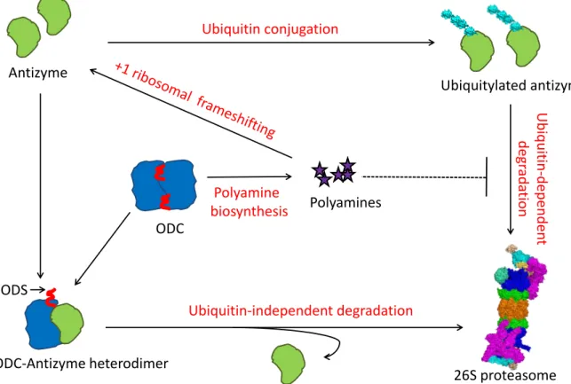
Spe2
11 an overview of the post-translational regulation of ODC by polyamines. ODC degradation is regulated through the synthesis of a regulatory protein called ODC antizyme (Kahana, 2009). In mammals four different ODC inhibiting antizyme isoforms are known. Among these, the most predominant is antizyme-1 which has a wide tissue distribution. Though expressed at lower levels, antizyme-2 is similar to antizyme-1 and promotes ODC degradation in vivo. Antizyme-3 is a testis specific protein, which is restricted to a late stage in spermatogenesis. It however does not target ODC for degradation. There is also an antizyme-4 but it is not very well characterized (Olsen and Zetter, 2011). In yeast, however, only one isoform is known that is encoded by OAZ1 (Palanimurugan et al., 2004). Antizyme levels are also strictly regulated by cellular polyamines. It occurs at the level of translation of antizyme mRNA as well as at its degradation, which is inhibited in response to increased cellular polyamine levels (Palanimurugan et al., 2004). The translational control of antizyme takes place via a conserved mechanism of +1 ribosomal frameshifting (Matsufuji et al., 1995; Palanimurugan et al., 2004). Antizyme mRNA is unique as it has a stop codon in its reading frame. For synthesis of full length protein, the ribosome has to skip the inner stop codon and continue till it reaches the stop codon at the end of the mRNA. This process is regulated by cellular polyamine levels, the mechanism for which remained elusive for a long time. Recent studies from our laboratory have shown that polyamine binding to a PRE (polyamine responsive element) on the nascent antizyme polypeptide is the key that regulates antizyme translation. At low cellular polyamine concentrations, ribosomes that undergo +1 ribosomal frameshifting within a polysome on antizyme mRNA, stall close to the end of the coding sequence thereby preventing completion of translation.
When polyamine levels rise, the binding of polyamines to the PRE deregulates the inhibition resulting in the release of full length antizyme polypeptide. Although this study was carried out in yeast, there is an indication that this is a conserved phenomenon as they also showed that polyamines bind human antizyme in vitro (Kurian et al., 2011). The second level of antizyme regulation is by its ubiquitin- dependent proteasomal degradation. High polyamine levels stabilize antizyme by preventing its degradation, the mechanism of which is not yet fully understood (Palanimurugan et al., 2004).
In mammals, antizyme is also regulated by a ODC-like protein called antizyme inhibitor. Unlike ODC, under physiological conditions antizyme inhibitor is a monomer and therefore binds antizyme with an affinity greater than ODC. This interaction inactivates antizyme thereby resulting in higher cellular polyamine concentrations by
12 synthesis and uptake. Antizyme inhibitor is rapidly degraded in a ubiquitin- independent manner. To date, two different isoforms of antizyme inhibitor are known -antizyme inhibitor-1 and antizyme inhibitor-2 (Kahana, 2009; Olsen and Zetter, 2011).
Fig. 6: Feedback regulation of polyamines in yeast. Shown here is an overview of the feedback regulation of cellular polyamines via ODC and antizyme. Following high cellular polyamine levels, antizyme synthesis is augmented via a unique +1 ribosomal frameshifting of antizyme mRNA.
Antizyme forms heterodimers with ODC monomers resulting in the exposition of an N-terminal unstructured domain in ODC termed ODS (ODC degradation signal). ODC is subsequently targeted to the 26S proteasome in a ubiquitin-independent manner whereas the antizyme is recycled.
Moreover, antizyme levels are controlled posttranslationally by its ubiquitiylation followed by proteasomal degradation. Polyamines inhibit this ubiquitin-dependent degradation of antizyme.
1.2.3. ODC degradation: the story so far
ODC is a 52 KDa protein functional only in its homodimeric form. The ODC monomer exists in equilibrium with the homodimer (Coleman et al., 1994). Antizyme binds to ODC monomers and facilitates their ubiquitin-independent degradation by the 26S proteasome. A 37 amino acid C-terminal region of mouse ODC (termed cODC) was found to be essential for its degradation. cODC was later confirmed as the degron by sequence comparison of mODC with ODC from Trypanosoma brucei (TbODC) which
26S proteasome Ubiquitin conjugation
Ubiquitin-independent degradation
Ubiquitin-dependent degradation
ODC-Antizyme heterodimer Antizyme
Polyamines Polyamine
biosynthesis
Ubiquitylated antizyme
ODS
ODC
13 lacks cODC and is therefore a stable protein in mammalian cells. cODC functioned as a transplantable degron as it mediated ubiquitin-independent degradation of TbODC once transplanted. Antizyme is not essential for the turnover of mODC but it greatly enhances it. Antizyme binding is thought to expose the cODC which is otherwise buried in the ODC homodimer. However, it remains unclear whether antizyme plays a further role in mODC degradation (Erales and Coffino, 2013). In S.
cerevisiae, however, antizyme is essential for the degradation of ODC (Palanimurugan et al., 2004). The yODC (yeast ODC) degron is a ~45 residue N- terminal unstructured domain called ODC Degradation Signal (ODS) which is exposed upon antizyme binding. This degron is both transplantable and replaceable.
However, the transplantable nature of the degron depends on the structural context of the receptor protein. An alpha helical domain succeeding the unstructured domain was found to be a contributing factor in degradation (Godderz et al., 2011; Li and Coffino, 1993).
1.3. Other ubiquitin-independent substrates
There are only a handful of well-characterized ubiquitin-independent substrates.
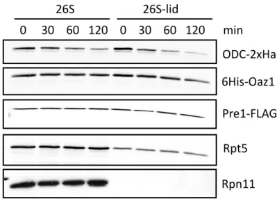
Apart from ODC, yeast Rpn4, a transcriptional regulator of proteasome genes, and mammalian thymidylate synthase (TS), an enzyme involved in the synthesis of DNA precursors are the other known substrates. Presence of an unstructured domain containing degron is the common feature among these substrates (Erales and Coffino, 2013). Rpn4 distinguishes itself from the other two as it is degraded via the ubiquitin-dependent as well as ubiquitin-independent modes (Ju and Xie, 2004). Like yeast ODC, the ubiquitin-independent degron of Rpn4 is at its N-terminus consisting of the first 80 residues. Recently, Ha et al. have reported that the N-terminal degron of Rpn4 interacts with the proteasomal subunits Rpn2, Rpn5 and Rpt1 by using a cross-linking label transfer technique (Ha et al., 2012). On the other hand, in vivo data from our laboratory showed that the proteasome lid is dispensable for ODC degradation (Godderz et al., 2011). Therefore, the mode of proteasomal reception of ubiquitin-independent substrates still remains to be elucidated. Similar to ODC and Rpn4, the degron of TS consists of an N-terminal unstructured domain spanning residues 1-28. Besides this degron, the presence of an N-terminal proline and the residues 9-15 was also critical for TS degradation. Another common feature with ODC was the α-helix following the degron which contributed to the efficiency of degradation (Pena et al., 2009).
14 Apart from these, there are substrates that are ubiquitylated but also degraded in a ubiquitin-independent manner. The mode of their degradation is still under debate.
These comprise p21/Cip1, the TCRα subunit of the T cell receptor, IκBα, c-Jun and calmodulin (Hoyt and Coffino, 2004). p21 is a loosely folded protein that is ubiquitylated in vivo. However, a lysine-less variant of p21 is still unstable showing that its degradation does not completely rely on ubiquitylation (Sheaff et al., 2000).
Later, p21 was shown to be polyubiquitylated at the free amino group of its N- terminal methionine which was shown to be sufficient for its degradation (Bloom et al., 2003). Therefore, the exact mode of degradation of p21 in mammalian cells still remains uncertain. It is possible that both pathways might be involved.
15
2. Aim of current study
Recent structural studies of the 19S regulatory particle have shed light on the mechanistic details of ubiquitin recognition and subsequent engagement of substrates by the proteasome. This model was based on the relative positions of ubiquitin receptors, the deubiquitylase Rpn11 and the ring formed by the 6 Rpts.
(Beck et al., 2012; Lander et al., 2012) However, the ubiquitin-independent recognition of substrates is still poorly understood. This study is aimed at elucidating the mechanism of reception and engagement of ubiquitin-independent substrates by the proteasome. ODC is the primary ubiquitin-independent substrate studied in this thesis although other substrates such as p21 and artificial fusion proteins are also employed in comparative approaches. The major questions addressed in this thesis can be summarized as follows.
1. Do polyamines directly influence ODC degradation?
2. Does ODC bind the proteasome through its unstructured domain, ODS?
3. What are the receptors in the proteasome for ODS and other unstructured domains?
4. Does antizyme have a binding site on the proteasome?
5. Is there a ubiquitin-dependent component for ODC degradation?
16
3. Materials and Methods
3.1. Materials
3.1.1.
Saccharomyces cerevisiae strainsStrain Genotype Lab stock Source
JD47-
13C MATa his3∆200 leu2-3,112 lys2-801 trp1∆63 ura3-52 Sc. 188 (Ramos et al., 1998) BY4741 MATa his3Δ1 leu2Δ0 met15Δ0 ura3Δ0 Sc. 1195 Euroscarf MO24 MATa his3∆200 leu2-3,112 lys2-801 trp1∆63 ura3-52
pre1::PRE1-FLAG-6xHIS Sc. 3534 (Kock et al.,
2015) DG10 MATa his3Δ1 leu2Δ0 met15Δ0 ura3Δ0 oaz1∆::Kan-MX5 Sc. 2583 Euroscarf spe4-∆ MATa his3Δ1 leu2Δ0 met15Δ0 ura3Δ0 spe4∆::Kan-MX5 Sc. 1202 Euroscarf paa1-∆ MATa his3Δ1 leu2Δ0 met15Δ0 ura3Δ0 paa1∆::Kan-MX5 Sc. 3188 Euroscarf YGA40 MATa his3∆200 leu2-3,112 lys2-801 trp1∆63 ura3-52
PGAL1 HSP82::Nat hsc82Δ::Kan-MX6 pdr5Δ::Hph Sc. 3112 (Kandasamy, 2014)
YGA95
MATα his3∆200 leu2-3,112 lys2-801 trp1∆63 ura3-52 PGAL1-HSP82::Nat hsc82Δ::Kan-MX6, pdr5Δ::Hph rpn10- UIM::Phe rpn13-KKD::Trp1 rad23Δ::His dsk2 Δ::Kan
Sc. 3335 (Kandasamy, 2014)
YHI29/1 MATα pre1-1 his3-11,15 leu2-3 ura3 Cans Sc. 324 Lab collection JD77-
ts26
MATa uba1Δ::HIS3 pRSts26-1(uba1-ts-26) leu2-3,112 lys2-
801 ura3-52 Sc. 440 Lab
collection JD59 MATa ump1∆::HIS3 his3∆200 leu2-3,112 lys2-801 trp1∆63
ura3-52 Sc. 227 (Ramos et
al., 1998) YAH96 MATα his3-∆200 leu2-3,112 lys2-801 trp1-1 ura3-52
rpn11::RPN11-3xFLAG-HIS Sc. 3936 (Erales et al.,
2012) JE03 MATα his3-∆200 leu2-3,112 lys2-801 trp1-1 ura3-52
rpn11::RPN11-3xFLAG-HIS Rpt1::rpt1 (Y283A) Sc. 3937 (Erales et al., 2012) MHY292 MATα his3-∆200 leu2-3,112 lys2-801 trp1-1 ura3-52
rpn11::RPN11-3xFLAG-HIS Rpt2::rpt2 (Y256A) Sc. 3938 (Erales et al., 2012) RB18 MATα his3-∆200 leu2-3,112 lys2-801 trp1-1 ura3-52
rpn11::RPN11-3xFLAG-HIS Rpt3::rpt3 (Y246A) Sc. 3968 This study MHY294 MATα his3-∆200 leu2-3,112 lys2-801 trp1-1 ura3-52
rpn11::RPN11-3xFLAG-HIS Rpt4::rpt4 (Y255A) Sc. 3940 (Erales et al., 2012) MHY295 MATα his3-∆200 leu2-3,112 lys2-801 trp1-1 ura3-52
rpn11::RPN11-3xFLAG-HIS Rpt5::rpt5 (Y255A) Sc. 3941 (Erales et al., 2012) RB19 MATα his3-∆200 leu2-3,112 lys2-801 trp1-1 ura3-52
rpn11::RPN11-3xFLAG-HIS Rpt6::rpt6 (Y222A) Sc. 3969 This study
17 AM33 MATa pdr5∆::KanMX5 his3∆200 leu2-3,112 lys2-801
trp1∆63 ura3-52 Sc. 1954 Lab
collection RB4 MATα his3∆200 trp1∆63 met15::Nat ura3∆::Kan
Leu2::PODC-ODC-LEU2::Hph Can::POAZ1-OAZ1-TOAZ1
Sc.3518 This Study
JN54 MATa his3-11,15 leu2-3,112 lys2 trp1-∆1 ura3-52 Sc. 1032 (Nelson et al., 1992) YMF15 MATa ssa1-45 ssa2∆::LEU2 ssa3∆::URA3 ssa4::LYS2 Sc. 1203 (Fröhlich,
2005)
3.1.2.
Escherichia coli strainsStrain Genotype Source
XL1Blue F- φ80 lacZΔM15 Δ(lacZYA-argF)U169 deoR recA1 endA1 hsdR17(rk-, mk+) phoAsupE44 thi-1 gyrA96 relA1 λ-
Lab collection MC1061 hsdR2 hsdM+ hsdS+ araD139 Δ(ara-leu)7697Δ(lac)X74 galE15
galK16 rpsL (Strr) mcrA mcrB1
Lab collection Rosetta™ 2(DE3)
pLysS F- ompT hsdSB(rB- mB-) gal dcm (DE3) pLysSRARE2 (CamR) Lab collection BL21 codon F- ompT gal dcm lon hsdSB(rB
- mB
-) λ(DE3 [lacI lacUV5-T7gene 1 ind1 sam7 nin5])
Lab collection
3.1.3. Plasmids
Name Details Lab stock Source
YCplac33 CEN/URA3 Ec. 201 (Gietz and Sugino,
1988)
YCplac111 CEN/LEU2 Ec. 202 (Gietz and Sugino,
1988)
YCplac22 CEN/TRP1 Ec. 200 (Gietz and Sugino,
1988)
pPM323 PCUP1-2xMyc-OAZ1-if-TCYC1, CEN/URA3 Ec. 3842 (Kurian et al., 2011) pDG240 pET11a-6His-OAZ1(codon optimised for E.coli) Ec. 2770 (Kurian et al., 2011) pDG246 pET11a-6His-OAZ1L245A,L246A,K247A,W251A (codon
optimised for E.coli) Ec. 2776 Lab collection
pRB11 pET11a-6His-OAZ1(codon optimised for E.coli)-
pQE-ODC-2xHa Ec. 3038 This study
pRB12 pET11a-6His-OAZ1(codon optimised for E.coli)-
pQE-ΔN47-ODC-2xHa Ec. 3039 This study
pPM97 PODC-ODC-2xHa-TCYC1, CEN/LEU2 Ec. 3089 (Godderz et al., 2011)
pMAF17 PCUP1-Ub-R-Ha-eK-URA3-TCYC1, CEN/LEU2 Ec. 2380 Lab collection
18 pMAF18 PCUP1-Ub-V76-Ha-eK-URA3-TCYC1, CEN/LEU2 Ec. 3665 Lab collection
pGEX-4T-2 GST Ec. 2445 GE Healthcare
pDG241 pGEX4T-2-GST-OAZ1(codon optimized for
E.coli) Ec. 2771 Lab collection
pDG273 pET11a-ODC-FLAG Ec. 2803 Lab collection
pRB24 pET11a-ODC-2xHa-6His Ec. 3339 This study
pDG269 pCUP1-hp21-2xHa-TCYC1, CEN/URA3 Ec. 2799 Lab collection pDG258 PODC-ODC1-42-URA3-2xHa-TCYC1, CEN/LEU2 Ec. 2788 (Godderz et al.,
2011)
pDG268 PODC-URA3-2xHa-TCYC1, CEN/LEU2 Ec. 2798 (Godderz et al., 2011)
pFS1 PODC-URA3-2xHa-TCYC1, CEN/LEU2 Ec. 3551 (Stadelmayer, 2014) pFS2 PODC- OAZ1-if L245A,L246A,K247A,W251A URA3-2xHa-
TCYC1, CEN/LEU2 Ec. 3552 (Stadelmayer, 2014)
pRB40 PODC- ODC1-47OAZ1-if L245A,L246A,K247A,W251A-TCYC1,
CEN/LEU2 Ec. 3553 This study
pRB41 PODC- ODC1-47OAZ1-if L245A,L246A,K247A,W251A-
URA3-2xHa-TCYC1, CEN/LEU2 Ec. 3577 This study pRB45 PODC- ODC1-47OAZ1-if L245A,L246A,K247A,W251A-
URA3-2xHa-TCYC1, CEN/TRP1 Ec. 3649 This study pJDRZ1 pGAL1-Ub-R-LacZ, 2µ/HIS3 Ec. 3257 Lab collection pPM96 pCUP1- ODC-2xHa-TCYC1, CEN/LEU2 Ec. 3088 (Rangasamy, 2005) pPM106 pCUP1-ΔN47-ODC-2xHa-TCYC1, CEN/LEU2 Ec. 3096 (Rangasamy, 2005) pRB14 pCUP1- ODC-TCYC1, CEN/LEU2 Ec. 3193 This study
pRB15 pCUP1-ΔN47-ODC-TCYC1, CEN/LEU2 Ec. 3194 This study pMAF59 PCUP1-Ub-R-eK-DHFR-2xHa-TCYC1, CEN/LEU2 Ec. 3451 Lab collection
3.1.4. Oligonucleotides
Name Sequence Description
RB4115 GAGGATCCATTAAAGAGGAGAAATTAACTATGTCTAGTACTC AAGTA
BamH1-SD (PQE)- ODC-FP
RB4116 CTGGATCCTACTAGTTGAGCTCTCTAGACTGCATAGTCAGG TACG
Ha-stop-Sac1-Spe1- BamH1-RP
RB4117 GAGGATCCATTAAAGAGGAGAAATTAACTATGAACCAAGATT TGGAA
BamH1-SD (PQE)- ΔODS-ODC-FP
RB4118 GGTTGGTGGCAAACTGAT pRB11-sequencing-FP
RB4119 GATATAGTTCCTCCTTTCAGC pRB11-sequencing-RP
RB4133 ACGAATTCATGTCTAGTACTCAAGTA EcoR1-ODC-FP
RB4134 ACGAATTCATGAACCAAGATTTGGAA EcoR1-dODS-ODC-FP
19
RB4135 TTGGATCCTCAATCGAGTTCAGAGTCTAT ODC-stop-BamH1-RP
RB4343 GACGAGCTCATCGAGTTCAGAGTCTATGT ODC-nostop-Sac1-RP
RB4623 CGCCTCGAGGCATTCAAACTCTAAAATAACAAAG Xho1-nostop-OAZ1-RP
RB4688 GAAGAAAAGCCTGACGTTACTTA RPT1-int-FP
RB4689 TCAATTATATTGCATATAACGCGA RPT1-stop-RP
RB4690 GGTTTCGGTCATGAAAATGGATA RPT2-int-FP
RB4691 TCACAAGTATAAACCTTCTAAATT RPT2-stop-RP
RB4692 TGACGTCACTTATGCAGATGTTG RPT3-int-FP
RB4693 TCATTTGTAGAAGTCGAATTTATC RPT3-stop-RP
RB4694 GTATAATATGACCAGTTTTGAAC RPT4-int-FP
RB4695 TCATAATTTTTGGTATTCTATAGT RPT4-stop-RP
RB4696 GAATTTGATTCTCGTGTAAAAGC RPT5-int-FP
RB4697 TTATGCATAAAAGGATACCGATTT RPT5-stop-RP
RB4698 GACCCACTAGTTTCGTTGATGAT RPT6-int-FP
RB4699 TCACTTGAACAGCTTGGCGACAGA RPT6-stop-RP
3.1.5. Enzymes
Enzyme Supplier
Alkaline Phosphatase NEB
DnaseI Roche
DreamTaq DNA Polymerase Thermo Scientific
Lysozyme Sigma
Phusion® High-Fidelity DNA Polymerase NEB
Restriction Endonucleases Thermo Scientific
T4-DNA Ligase NEB
β-glucoronidase Roche
3.1.6. Antibodies
Antibody Derived from Supplier
Anti-βeta2 Rabbit Lab collection
Anti-Cdc11 Rabbit Santa Cruz Biotechnology
Anti-GST Rabbit Santa Cruz Biotechnology
Anti-HA (16B12 clone) Mouse HISS Diagnostic
Anti-HA (3F10 clone) Rat Roche
Anti-HSP90 Rabbit Lab collection
Anti-FLAG (M2 clone) Mouse Sigma ANTI-FLAG® M2 Affinity Gel Mouse Sigma
Anti-MYC (9B11 clone) Mouse Cell Signaling Technology
Anti-Mouse, HRP Goat Sigma
20
Anti-Mouse, 680 Goat Invitrogen
Anti-Oaz1 Rabbit Lab collection
Anti-ODC Rabbit Lab collection
Anti-PGK Mouse Invitrogen
Anti-Rabbit, HRP Donkey GE Healthcare, UK
Anti-Rabbit, 800 Goat Rockland
Anti-Rat, HRP Goat Abcam
Anti-Rpn5 Rabbit Lab collection
Anti-Rpn11 Rabbit Santa Cruz Biotechnology
Anti-RPT5 Rabbit Abcam
Anti-TPI Rabbit Lab collection
3.1.7. Chemicals
Chemical Supplier
Acetic Acid Merck
N1-Acetylspermidine Wako
N8-Acetylspermidine Wako
Acrylamide Roth
Adenine Applichem
Adenosine triphosphate Apllichem
Agarose Sigma
Ammonium persulfate Sigma
Ampicillin Sigma
L-Arginine Roth
Agar MP Biomedicals
β-Mercaptoethanol Sigma
Boc-LLR-AMC Bachem
Bradford-Reagent Bio-Rad
Bromophenol Blue Serva
Calcium chloride (CaCl2) Acros
Chloramphenicol Sigma
Complete protease inhibitors EDTA free Roche
Coomassie Biliant Blue R-250 Merck
Copper (II) sulfate Acros
Deoxyribonucleotides triphosphate (dNTPs) Sigma
N1, N8-Diacetylspermidine Wako
di-Sodiumhydrogenphosphate (Na2HPO4) Roth
Dimethylformamid (DMSO) Roth
Dithiothreitol (DTT) AppliChem
21
Ethanol VWR
Epoxomycin Enzo life sciences
FLAG peptide Sigma
5-Fluoroorotic acid (5-FOA) Sigma
Formaldehyde Riedel-de Haën
D(+) Galactose VWR International
Geneticin disulfate (G418) Sigma
Glas beads (E. coli) 0.10- 0.11mm Satorius Stedium
Glass beads (yeast) 0.4- 0.6mm Satorius Stedium
D(+) Glucose Roth
Glutathione sepharose 4B GE Healthcare
Glycerol AppliChem
Glycine Merck
L-Histidine AppliChem
4-(2-hydroxyethyl)-1-piperazineethanesulfonic acid (HEPES) Serva
Imidazole Sigma
L-Isoleucine Merck
Isopropanol VWR
Isopropyl-β-D-thiogalactoside (IPTG) Formedium
L-Leucine AppliChem
Lithium acetate Alfa Aesar
L-Lysine Roth
LumiLight Western Blot Substrate Roche
Magnesium chloride (MgCl2) Roth
m-Cresol purple sodium salt Sigma
Methanol Roth
L-Methionine Merck
MG132 Sigma
Milk powder Roth
Nourseothricin sulfate Jena bioscience
Ni-sepharose GE Healthcare
Polyethylene Glycol (PEG) [3350] Sigma
Peptone Formedium
L-Phenylalanine Roth
Pierce ECL Plus Western blotting substrate Thermo Scientific
Ponceau S solution Sigma
Potassium acetate Merck
Potassium chloride Acros
Potassium dihydrogen phosphate Roth
22
Sodium dodecyl sulfate (SDS) VWR
Sodium azide Sigma
Sodium carbonate Sigma
Sodium chloride AppliChem
Sodium hydroxide Roth
Di-sodium hidroxyphosphate Fluka
Sodium phosphate Merck
Sodium thiosulphate Sigma-Aldrich
Spermidine Sigma
Spermine Sigma
[3H]-Spermidine PerkinElmer
Suc-LLVY-AMC Bachem
N, N, N’, N’-tetramethylethylenediamine (TEMED) AppliChem
L-Threonine Roth
Tris Roth
Triton X-100 Sigma
L-Tryptophan AppliChem
Tryptone Formedium
TWEEN 20 Sigma
Uracil Sigma
Urea Usb
Yeast extract powder Formedium
Yeast Nitrogen Base Formedium
3.1.8. Instruments
Major Instruments Source
Centrifuge Avanti J-20 XP, Optima TLX Ultracentrifuge 120,000-rpm,
Allegra X-22R, Scintillation counter LS5000 TD Beckman Coulter
Curix 60-System developer machine Agfa
FLUOstar Galaxy Microplate Reader BMG Labtech
Incubators New Brunswick
Mini-gel gel electrophoresis, Blotting chamber Bio-Rad
Mixer Mill MM400 Retsch
Odyssey Infrared imaging system LI-COR biosciences, USA LI-COR
Thermocycler Biometra
Refrigerated centrifuge 5417R, centrifuge 5415D, Thermomixer
compact, BioSpectromoter Eppendorf
23
3.2. Methods
3.2.1. Molecular biology and genetic techniques 3.2.1.1. Isolation of plasmid DNA from E. coli
E. coli cells harbouring the plasmid of interest were grown overnight (or a minimum of 7 hours) at 37°C with constant shaking in LB medium supplemented with the appropriate antibiotics (ampicillin at 70 µg/ml and chloramphenicol at 34 µg/ml). Cells were collected by centrifugation and plasmid DNA was isolated using E.Z.N.A.® Plasmid Mini Kit I (Omega Bio-Tek) according to manufacturer’s instructions.
LB media
Tryptone 1 %
Yeast extract 0.5 %
NaCl 1 %
Agar (for plates) 2 %
3.2.1.2. Estimation of DNA concentration
DNA concentration was measured using the preprogrammed method and Eppendorf µCuvette™ in an Eppendorf BioSpectrometer®.
3.2.1.3. Isolation of genomic DNA from yeast
Yeast cells were grown overnight at 30°C with constant shaking in 5 ml YPD medium. Cells were collected by centrifugation and genomic DNA was isolated using E.Z.N.A.® Yeast DNA Kit (Omega Bio-Tek) according to manufacturer’s instructions.
3.2.1.4. PCR amplification
Polymerase chain reaction (PCR) amplification of DNA fragments for cloning was performed using the Phusion® High-Fidelity DNA Polymerase (NEB) according to manufacturer’s instructions.