Comparative Analysis
of the Ubiquitin-proteasome system
in Homo sapiens and Saccharomyces cerevisiae
Inaugural-Dissertation zur Erlangung des Doktorgrades
der Mathematisch-Naturwissenschaftlichen Fakultät der Universität zu Köln
vorgelegt von Hartmut Scheel
aus Rheinbach
Köln, 2005
Berichterstatter:
Prof. Dr. R. Jürgen Dohmen Prof. Dr. Thomas Langer Dr. Kay Hofmann
Tag der mündlichen Prüfung: 18.07.2005
Natura non saltat
(Linné)
Zusammenfassung I
Zusammenfassung
Das Ubiquitin-Proteasom System (UPS) stellt den wichtigsten Abbauweg für intrazelluläre Proteine in eukaryotischen Zellen dar. Das abzubauende Protein wird zunächst über eine Enzym-Kaskade mit einer kovalent gebundenen Ubiquitinkette markiert.
Anschließend wird das konjugierte Substrat vom Proteasom erkannt und proteolytisch gespalten. Ubiquitin besitzt eine Reihe von Homologen, die ebenfalls posttranslational an Proteine gekoppelt werden können, wie z.B. SUMO und NEDD8. Die hierbei verwendeten Aktivierungs- und Konjugations-Kaskaden sind vollständig analog zu der des Ubiquitin- Systems. Es ist charakteristisch für das UPS, daß sich die Vielzahl der daran beteiligten Proteine aus nur wenigen Proteinfamilien rekrutiert, die durch gemeinsame, funktionale Homologiedomänen gekennzeichnet sind. Einige dieser funktionalen Domänen sind auch in den Modifikations-Systemen der Ubiquitin-Homologen zu finden, jedoch verfügen diese Systeme zusätzlich über spezifische Domänentypen.
Homologiedomänen lassen sich als mathematische Modelle in Form von Domänen- deskriptoren (Profile) beschreiben. Diese Deskriptoren können wiederum dazu verwendet werden, mit Hilfe geeigneter Verfahren eine gegebene Proteinsequenz auf das Vorliegen von entsprechenden Homologiedomänen zu untersuchen. Da die im UPS involvierten Homologie- domänen fast ausschließlich auf dieses System und seine Analoga beschränkt sind, können domänen-spezifische Profile zur Katalogisierung der UPS-relevanten Proteine einer Spezies verwendet werden. Auf dieser Basis können dann die entsprechenden UPS-Repertoires verschiedener Spezies miteinander verglichen werden.
In dieser Arbeit wurden basierend auf UPS-relevanten Homologiedomänen und unter Verwendung der Profilmethode solche Kataloge für den Menschen und die Hefe Saccharomyces cerevisiae erstellt. In Kombination mit phylogenetischen Methoden wurden die evolutionären Beziehungen zwischen den UPS-Komponenten dieser beiden Organismen untersucht und in geeigneten Fällen eine Orthologiebeziehung abgeleitet. Durch die Verwendung der hoch-sensitiven Profiltechnik und die Einbeziehung von genomischen Datenbanken wurden im Rahmen dieser Arbeit eine Reihe von Proteinen identifiziert, die bisher nicht mit dem UPS assoziiert worden waren. Zusätzlich konnten einige unerwartete Verwandtschaftsbeziehungen zwischen Proteinen des UPS abgeleitet werden. So konnte z.B.
das lange gesuchte Hefe-Ortholog des 'Antizyms' der Ornithin- Decarboxylase identifiziert werden - eine wichtige Voraussetzung zur experimentellen Untersuchung des Ubiquitin-
Zusammenfassung II
unabhängigen Proteinabbaus durch das Proteasom. In einem weiteren Beispiel konnte gezeigt werden, daß Ataxin-3 aus Mensch eine Homologiedomäne mit funktioneller Ähnlichkeit zu den deubiquitylierenden Enzymen besitzt. Da Ataxin-3 bei Patienten mit spinocerebellarer Ataxie 3 (SCA3) mutiert ist, kann diese Entdeckung zur Aufklärung des Krankheitsmechanismus von SCA3 beitragen. In einer dritten exemplarischen Anwendung konnten weitreichende Vorhersagen für den strukturellen Aufbau des 19S-Proteasoms getroffen werden, insbesondere mit Bezug auf dessen 'lid' Subkomplex.
Ein Vergleich der UPS-relevanten Proteinrepertoires der Hefe und des Menschen erlaubte Schlüsse über den evolutionären Ursprung einiger Komponenten des UPS.
Insbesondere bei Proteinfamilien mit einer etablierten oder angenommenen Rolle in der Substraterkennung und -ubiquitylierung oder im reversen Prozess der Deubiquitylierung findet man beim Menschen eine starke Diversifizierung der Proteinfamilien, während die elementaren Funktionen des UPS durch annähernd vergleichbare Proteinsets ausgeführt werden. Trotz der teilweise erheblich größeren Proteinfamilien im Menschen, konnten nicht allen UPS- assoziierten Proteinen der Hefe humane Orthologe zugeordnet werden, was auf spezifische Prozesse innerhalb des UPS von S. cerevisiae hindeutet. Ingesamt überwiegen jedoch die Ähnlichkeiten der beiden Systeme und unterstreichen die Rolle von S. cerevisiae als Modellorganismus zur Aufklärung des UPS.
Abstract III
Abstract
The UPS (ubiquitin-proteasome system) is the most important degradation pathway for intracellular proteins in the eukaryotic cell. In a first step, the protein to degrade (substrate) is tagged covalently with a Ubiquitin chain via an enzyme cascade. Subsequently, the Ubiquitin chain is recognized by the proteasome and the substrate is proteolytically cleaved. Ubiquitin has several homologues, which can be conjugated to proteins posttranslationally, e.g. SUMO or NEDD8. The enzymes used for activation and conjugation of the Ubiquitin homologues are completely analogous to the ones used in the UPS. A hallmark of the UPS is that most proteins involved belong to only a few protein families, which are characterized by common functional homology domains. Several of these homology domains are found in the modification systems of Ubiquitin homologues, but these systems appear to have specific homology domains on their own as well.
Homology domains may be described as mathematical models in terms of domain descriptors (profile). These profiles together with appropriate search algorithms can be applied to screen a given protein sequence for the occurrence of the corresponding homology domains.
As the homology domains involved in the UPS are almost exclusively found in proteins of this and analogous systems, profiles corresponding to these homology domains seem to be an appropriate means to catalogue proteins of the UPS of a given species.
In this work catalogues of proteins with a known or putative role in the UPS or analogous systems were set up for human and Saccharomyces cerevisiae (here referred to as
‘yeast’) based on relevant homology domains and their corresponding profiles. In combination with phylogenetic methods the evolutionary relationships between the UPS components of these two organisms were analyzed and, if possible, orthologous relationships were derived. Using the highly sensitive profile technique and including genomic databases, several new proteins were identified that have not been associated with the UPS so far. Additionally, several unexpected relationships were revealed between proteins of the UPS. For example, the postulated yeast orthologue of the antizyme of the ornithine decarboxylase could be revealed, which may be important for the experimental analysis of Ubiquitin independent protein degradation by the proteasome. Another example is human ataxin-3, in which a homology domain was found with similarity to the catalytic site of deubiquitylating enzymes. As ataxin-3 is mutated in patients with a spinocerebellar ataxia 3 (SCA3), this discovery might have implications for the elucidation of the SCA3 disease mechanism. Furthermore, predictions on the structure of the
‘lid’ of the 19S regulatory particle could be formulated.
Abstract IV
A comparison of the UPS-relevant protein repertoires of yeast and human allowed conclusions on the evolutionary origin of UPS components. Especially protein families with an established or putative role in substrate recognition/ubiquitylation or in the reverse process of deubiquitylation exhibited a strong diversification in human. Simultaneously, elementary functions of the UPS are carried out by almost identical protein sets in both yeast and human.
Despite the extensively expanded protein families in human, not all yeast proteins associated with the UPS could be assigned to human orthologues. This finding might indicate specific processes within the yeast UPS. To summarize, the similarities of both yeast and human UPS are significant and underline the role of S. Cerevisiae as a model organism used in analyzing the UPS.
Abbreviations V
Abbreviations
AAA ATPases associated with a variety of
cellular activities MPN Mpr1, Pad1 N-terminal
APC anaphase-promoting complex
(cyclosome) MSA multiple sequence alignment
BAG Bcl2-associated athanogene domain NEDD neural precursor expressed, developmentally downregulated BIRC6 baculoviral IAP repeat-containing
protein 6 OAZ ODC antizyme
BLOSUM blocks substitution matrix ODC ornithine decarboxylase
bp base pair ORF open reading frame
BTB Bric-a-brac (bab), Tramtrack (ttk), and
Broad-Complex (BR-C) PAM per cent accepted mutation
cDNA complementary DNA PAZ poly-Ub associated Zn-finger
CHIP carboxy terminus of hsp70-interacting
protein PCI proteasome, COP9, initiation factor 3
Clp caseinolytic protease PCNA proliferating cell nuclear antigen CP core particle (20S proteasome) PIAS protein inhibitor of activated STAT
CSN COP9 signalosome POMP proteasomal maturation protein
CUE coupling of Ub conjugation to ER
degradation RING really interesting new gene
Cvt Cytoplasm-to-vacuole targeting RP regulatory particle (19S proteasome subcomplex)
DNA desoxyribonucleic acid RPN regulatory particle non-ATPase
DUB deubiquitylating enzyme RPT regulatory particle triple A ATPase
E1 Ub activating enzyme SC, sc Saccharomyces cerevisiae
E2 Ub conjugating enzyme SCF Skp1, cullin, F-box (E3 complex)
E3 Ub ligating enzyme SGD Saccharomyces Genome Database
E4 Ub chain elongation factor SUMO small Ub-like modifier
eIF3 eukaryotic translation initiation factor
3 TrEMBL translated EMBL nucleotide sequence
data library ENTH Epsin N-terminal homology domain Ub Ubiquitin
ER endoplasmatic reticulum UBA Ubiquitin-pathway associated domain
ERAD ER-associated protein degradation UBC Ub conjugating
EST expressed sequence tag UBL Ub-like modifier
GAT GGA and TOM (target of myb) UBP/USP Ub-specific protease HAUSP herpes-associated ubiquitin-specific
protease UBX Ub-like motif, sometimes referred to
as UX domain HECT Homologous to the E6-AP Carboxyl
Terminus UCH Ub C-terminal hydrolase
HMM hidden Markov model UEV Ub conjugating enyzme variant
HS, hs Homo sapiens UFD Ub fusion degradation pathway
JAMM JAB1/MPN/Mov34 metalloenzyme UIM Ub interacting motif
kDa kilodalton UPS Ub proteasome system
Contents VI
Contents
ZUSAMMENFASSUNG I
ABSTRACT III
ABBREVIATIONS V
CONTENTS V
1 INTRODUCTION 1
1.1 Comparative sequence analysis 1
1.1.1 Functional classification of protein sequences 1
1.1.2 The modular architecture of proteins 2
1.1.3 Homology domains and their impact on protein classification 3
1.2 The ubiquitin-proteasome system 4
1.2.1 Ubiquitin and its relatives 6
1.2.2 Ub-activating enzymes (E1) 8
1.2.3 Ub-conjugating enzymes (E2) 9
1.2.4 Ub-ligases (E3) 10
1.2.5 Ub-hydrolases (DUB) 12
1.2.6 Ub-binding proteins 13
1.2.7 Proteasome 16
1.3 Detection of homologues and protein family analysis 18
1.3.1 Sequence comparison methods 18
1.3.2 Multiple sequence alignments (MSA) 18
1.3.3 Profile searching 19
1.3.4 Substitution matrices 21
1.3.5 Dendrogram analysis of proteins and genes 22
1.4 Data sources and functional prediction 23
1.4.1 Protein protein interactions and their prediction 23
Contents VII
1.4.2 Model systems as source for biological data 23
1.5 Aim of the study 25
2 METHODS 26
2.1 Protein and nucleotide sequences for database searches 26
2.2 Databases for homology domain descriptors 27
2.3 Constructing, refining and application of profiles 27
2.3.1 Obtaining an initial alignment 28
2.3.2 Construction of the profile 29
2.3.3 Scaling the profile and database search 29
2.3.4 Iterative improvement 30
2.3.5 Determination of complete sets of protein families 32
2.4 Other tools 32
2.4.1 Dendrogram analysis 32
2.4.2 BOXSHADE 33
2.4.3 Secondary structure prediction 33
2.4.4 18S rRNA tree 33
3 RESULTS 34
3.1 Ubiquitin and its relatives 34
3.1.1 Type I Ubiquitin- like modifiers 34
3.1.1.1 Ubiquitin in the yeast and human genome 34
3.1.1.2 Type I Ubiquitin- like modifiers 36
3.1.2 Type II Ub- like proteins 37
3.1.2.1 Ub-like domains detected by the Ub-profile 37
3.1.2.2 Remotely Ub- like domains in Ub-activating enzymes 40 3.2 Activating enzymes for Ub and related modifiers (E1) 42
3.2.1 E1 protein sets for yeast and human 42
3.2.2 Specific genes and homology domains in the E1 family 43 3.2.3 Repetitive motifs in some E1s contain the active site 45
3.2.4 Other domain arrangements in E1 45
Contents VIII
3.3 Ub-conjugating enzymes (E2) 46
3.3.1 E2 protein sets for yeast and human 46
3.4 Ligases for Ub and related modifiers 48
3.4.1 Finding RING finger proteins 48
3.4.1.1 Construction of a pure RING finger profile 48
3.4.1.2 Role of zinc-coordinating residues for subfamily determination 49 3.4.1.3 Overview of the RING superfamily including U-Box proteins 50
3.4.2 Comparing the RING finger and its variants 51
3.4.2.1 The Parkin finger triad 51
3.4.2.2 PIAS-type RING finger 52
3.4.2.3 Highly degenerated RING- finger 52
3.4.2.4 No zinc binding capabilities, but still E3 activity: U-box 53 3.4.3 Assignments of orthologues for the RING-type ligases 54
3.4.3.1 Dma1/Dma2 vs. CHFR/RNF8 54
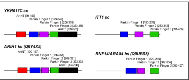
3.4.3.2 YKR017C vs. ARIH1/UBCH7BP 55
3.4.4 No evidence for PHD fingers as Ubiquitin ligases 56
3.4.5 RING-cullin based E3s 57
3.4.5.1 Few scaffolds for RING-cullin based E3s 57
3.4.5.2 Defining the substrate-binding subunits 58
3.4.5.3 Large families of potential substrate binding subunits 59
3.4.5.4 Assignment of orthologues 60
3.4.5.5 Five novel BTB proteins in yeast 60
3.4.6 HECT type Ub- ligases 62
3.4.7 A20-zinc-finger-type Ub-ligases 63
3.4.8 Non-RING based SUMO- ligases 64
3.5 Ub-hydrolases (DUB) and desumoylating enzymes 65
3.5.1 UCH family 65
3.5.2 USP family 65
3.5.2.1 USP-type DUBs are highly diversified 65
3.5.2.2 The catalytic domain consists of six major boxes 68 3.5.2.3 Large insertions between the individual boxes 71 3.5.2.4 A potential zinc finger is inserted into the catalytic domain 72
3.5.3 Other families 74
Contents IX
3.5.3.1 A cysteine-protease motif in ataxin-3 74
3.5.3.2 OTU family 78
3.5.3.3 MPN family 78
3.5.3.4 Desumoylating enzymes 79
3.6 Ub- and SUMO-binding proteins 80
3.6.1 The UBA domain and its relatives 80
3.6.1.1 The UBA domain family comprises several subfamilies 80 3.6.1.2 The CUE domain is related to the UBA domain and may be subdivided 82
3.6.1.3 Additional UBA-related domains 84
3.6.1.4 Human orthologues of yeast Snf1 lack the UBA domain 86
3.6.2 The Ub- interacting motif (UIM) 87
3.6.3 The UEV domain 89
3.6.4 The NZF domain 90
3.6.5 The PAZ domain 91
3.6.6 The GAT domain 92
3.6.6.1 Few GAT members in yeast and human 92
3.6.6.2 The GAT domain and the UIM appear exchangeable 92
3.6.7 Identification of a SUMO interaction motif 93
3.6.7.1 Working hypothesis 93
3.6.7.2 Working scheme for SIM identification 93
3.6.7.3 Integration of multiple criteria to evaluate putative SIMs 94 3.6.7.4 Identification of a putative SUMO interaction motif 95 3.6.7.5 The putative SIM is present in numerous SUMO interactors 96 3.6.7.6 The SIM is orthologous to a previously defined SUMO binding motif in PIAS2 97
3.7 Proteasome 99
3.7.1 The 20S proteasome 99
3.7.1.1 The immunosubunits are paralogues of the catalytic 20S proteasome subunits 99
3.7.1.2 Human PSMA7/α4 has recently been duplicated 99
3.7.2 Subunits of the 19S regulatory particle 101
3.7.3 Comprehensive analysis of the PCI subunits of the proteasomal lid, the CSN and the
eIF3 101
3.7.3.1 Determining subunits of the PCI complexes 101
3.7.3.2 TPR- like he lical repeats in PCI proteins 102
Contents X
3.7.3.3 A previously unrecognised PCI domain in eIF3k 104 3.7.3.4 A structural model for the canonical PCI domain 107
3.7.4 Other activators of the proteasome 109
3.7.4.1 PA28αβ, PA28γ and PA200 109
3.7.5 Proteins involved in subunit synthesis and assembly of the 26S proteasome 109
3.7.5.1 Rpn4 109
3.7.5.2 Ump1/hUMP1 110
3.7.5.3 Hsp90 110
3.7.6 Proteins involved in substrate delivery to the proteasome 110
3.7.6.1 Ub-like/UBA-adaptor proteins 110
3.7.6.2 Ub-recognition components of the proteasome 111
3.7.6.3 Hsp90/Hsp70/BAG1/CHIP 111
3.7.6.4 Cdc48/VCP/p97 112
3.7.6.5 Antizyme: a model for Ub- independent proteasomal targeting 113 3.7.6.6 Ubr1/ClpS: a common domain in the N-end-rule-pathway of eukaryotes and
bacteria 120
3.7.7 Physiological proteasome inhibitors 124
3.7.7.1 PI31 124
4 DISCUSSION 125
4.1 Cataloguing the UPS with the profile technique 125
4.1.1 Functional domains help structuring the UPS 125
4.1.2 The UPS and related systems resemble intracellular signal transduction pathways126
4.1.3 Benefits from cataloguing the UPS 126
4.1.4 Limitations of existing sequence profile collections 127
4.2 Revised and novel homology domains 128
4.2.1 Novel Ub-like domains in E1s and their functional role 128
4.2.2 Subfamilies of the RING superfamily 128
4.2.3 Extension of the UBA family 130
4.2.4 A functional role for the USP zinc-finger 131
4.2.5 Prediction of a common structural scaffold for proteasome lid, COP9-signalosome and
eIF3 complexes 131
4.2.5.1 The bipartite structure of the PCI domain 131
XI
4.2.5.2 The nature of the N-terminal helical repeat extension 132 4.2.5.3 A structural scaffold for three multi-protein complexes 134 4.2.6 Ubr1 might use its ClpS domain for proteasome binding 135
4.3 Comparing the yeast and human UPS 136
4.3.1 Ub family, E1 and E2 enzymes 136
4.3.2 Simple and complex E3 enzymes 138
4.3.3 Deubiquitylating enzymes 143
4.3.4 Ub-binding proteins 144
4.3.5 Comparison of PCI complexes regarding their PCI/MPN architecture 145
4.4 Conclusions and future directions 148
5 REFERENCES 1
6 APPENDIX 164
7 ACKNOWLEDGMENTS 170
8 EIDESSTATTLICHE ERKLÄRUNG 171
9 LEBENSLAUF 172
1
1 Introduction
1.1 Comparative sequence analysis
1.1.1 Functional classification of protein sequences
Comparative sequence analysis of proteomes from distinct species is a generally applied approach to derive knowledge on phylogenetic relationships, evolution and function of any new protein sequence. Transfer of available functional information from already characterized proteins to novel ones is often performed based on sequence homology. Homologous protein sequences are sequences that share a common evolutionary ancestor. Homology is often inferred from sequence similarity measurements, although in a few cases sequence similarity seems to have arisen by convergence. Sequences of homologous proteins can diverge greatly over evolutionary time, but function or structure may be maintained anyway. Thus, if sufficient sequence similarity is detected between a well studied and an uncharacterised protein, available information can be transferred along the homologous relationship between the two proteins.
Homologues can be divided into orthologues and paralogues. Orthologues have diverged from each other by a speciation event, i.e. the evolution of new biological species from a common ancestor, while paralogues have diverged from each other by gene duplication events (Fitch, 2000). Unlike orthologues and paralogues, a xenologue represents a homologue that has entered the genome of a species by interspecies gene transfer (horizontal gene transfer). While paralogues often evolve new functions, even if related to the original one, orthologues typically occupy the same functional niche, which remains the same even in phylogenetically distant species. Therefore the identification of orthologues is more reliable for functional inference than comparing two paralogous sequences, which are similar without necessarily fulfilling the same biological role. Besides estimating sequence similarity for the purpose of functional transfer, the actual phylogenetic relationship between sequences is important as well. In addition to detecting sequence similarity and phylogenetic relationships, the possibility of convergence has to be accounted for in the homology approach.
Homologues of a given protein are normally found in a protein or DNA database by specialized tools, for example BLAST (Altschul, 1997) or FASTA (Pearson, 1988). As a simplistic approach, the function of the best scoring hit returned by such a search is transferred to the query sequence. This method can be refined by examining a larger number of hits that exhibit a certain degree of sequence similarity. Consideration of many hits in turn may include sequences, which only show regional similarity to the query. When trying to use those 'partial
Chapter 1 Introduction 2
homologies' for functional inference, it is a prerequisite that the information meant to be transferred from the hit to the query really resides within the matching region. By deducing a consensus from the classification of multiple reliable and preferably global hits, the query sequence can be assigned a specific function or a more general classification such as the involvement in a certain biological process. In this respect the usage of a unified functional vocabulary greatly facilitates the determination of a consensus classification (Ashburner, 2000).
1.1.2 The modular architecture of proteins
In the best case of a sequence-to-sequence comparison involving both the query and a clearly similar sequence, the region of similarity spans the complete length of the query. More frequently, the direct comparison reveals only a partial match between the two sequences.
Strictly speaking, this situation allows only a functional classification of the particular stretch of the query sequence that was responsible for the reported database hit. One possible explanation might be a higher divergence of the sequences in the dissimilar region, but many of these constellations are caused by the modular architecture of proteins involved.
From the analysis of 3D protein structures it is known that a large portion of proteins contain multiple folding units rather than one monolithic fold. A folding unit, generally termed a domain, is a compact structure that folds independently from other parts of the protein. Typical domains have a hydrophobic core and consist of secondary structure elements such as β-strands or α-helices, which in turn can arrange themselves into sheets or a-hairpins. The exterior of a domain is usually hydrophilic due to its exposure to the solvent, but may also exhibit hydrophobic patches in order to fulfil certain functions, e.g. acting as a binding site. Within some multi-domain proteins, such binding sites are used to minimize the intramolecular repulsion of the domains. Others have their domains connected via flexible linkers. Another interpretation of a domain is that of an intra-protein subunit in analogy to the formation of a quaternary structure by separate proteins. As structure defines function, domains are generally associated with particular functions like enzymatic activity or ligand recognition.
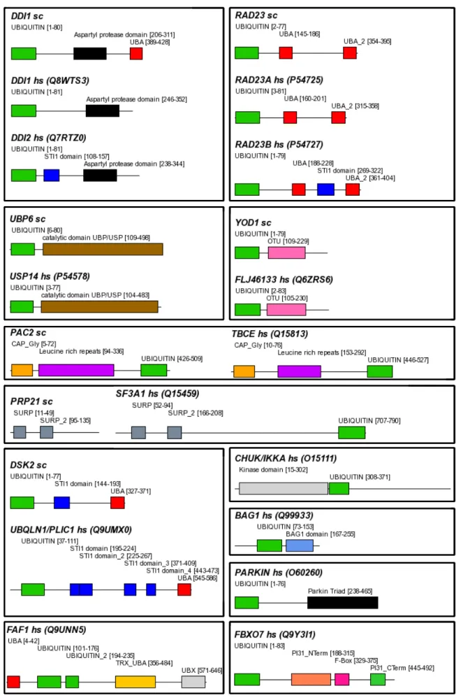
In the course of evolution, the autonomous folding capabilities of domains seem to have made them suitable evolutionary units that could be arranged in different domain contexts without disturbing their structural integrity and therefore their function. An example for a domain, which exists as a monolithic protein as well as part of multi-domain proteins with different structures, is the ubiquitin-like domain (Figure 1-1). The underlying evolutionary events that lead to the formation of new domain organisations are mainly exon shuffling, duplication and fusion of whole genes or just gene regions (Li, 1997). Duplication of gene
Chapter 1 Introduction 3
regions may lead to a repetitive domain structure, whereas fusion or insertion can generate 'mosaic proteins' composed of domains originating from genes with different evolutionary histories.
Figure 1-1 Domain topologies of multi-domain proteins with a Ubiquitin-like domain. In proteins like Ubiquitin, SUMO and Rub1/NEDD8 (shown here), this type of fold is able to form monolithic proteins. ‘sc’ following the protein name indicates S. cerevisiae.
1.1.3 Homology domains and their impact on protein classification
Common domains of otherwise unrelated protein sequences may be used for functional classification. It has to be kept in mind that this type of classification is restricted to the domains under consideration. Common domains from different proteins often share congruent boundaries and a homology relationship and are thus called ‘homology domains’. In many cases these homology domains correspond to structural domains, which are thought to exhibit folding independence. Homologous regions shorter than approximately 20 residues are too small to form an independent hydrophobic core and thus should not be considered true 'domains'.
Nevertheless, those small conserved regions can be carrier of important functional information, e.g. by being a recognition target for other proteins. In the following, short conserved regions are referred to as 'motifs' instead of 'domains'.
As many proteins are multi-domain proteins, the protein classification based on homology domains inevitably leads to more than one function for such proteins, because each homology domain can have its own characteristic function. Therefore these proteins belong to more than one protein family. As a consequence, the most accurate approaches to protein classification rely on domain-to-domain rather than on complete protein-to-protein comparisons.
Another reason to compare domains individually comes from the observation that within mosaic proteins with the same set of domains, the domain organization may be shuffled.
Chapter 1 Introduction 4
Once a homology domain is identified as a sequence stretch conserved across various proteins and attributed to a certain function, it can serve as a template for the classification of novel sequence data. To that end, several techniques have been developed that aim at the extraction of the essential features of a homology domain, and store them in terms of motif descriptors ('profiles') (Bucher, 1996). This concept will be introduced in the following, as sequence profiles can be applied as a very sensitive method to find distant homologous members of a protein family and therefore are a central technique of this work.
1.2 The ubiquitin-proteasome system
Most of the homology domains used by proteins of the ubiquitin-proteasome system (UPS) described below are present exclusively in this pathway. Thus, novel proteins containing one of these UPS-specific domains may be considered as new components of the UPS with a high reliability. Indeed, in the recent past the mining of sequence databases for proteins with domains relevant to the UPS has been a valuable source for new components and regulators of this system (Bai, 1996, Hofmann, 1996, Hofmann, 1998, Hofmann, 2001).
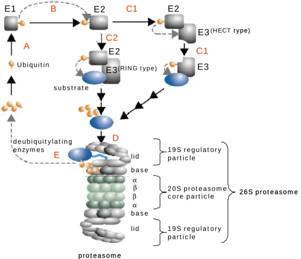
Common to all eukaryotic cells is their capability to degrade proteins and peptides, and for this purpose two major proteolytic system, the 26S proteasome and the lysosome are present within the cells. While the lysosome is responsible for the non-specific degradation of endocytosed proteins such as receptors, the proteasome bears the main load of intracellular proteins to be degraded. The proteasome is a multi-subunit protease that combines substrate recognition, unfolding and hydrolytic cleavage (see Figure 1-2 for a rough overview of the UPS). Prior to proteasomal digestion, substrates are usually tagged with a poly-ubiquitin chain via a covalent isopeptide bond that links the free C-terminus of a ubiquitin (Ub) and the ε-amino group of a lysine. This multi-step enzymatic reaction is generally known as ubiquitylation.
Until Ub is linked to a substrate protein in a covalent manner, it passes through several enzymatic reactions. First, Ub precursors have to be processed to allow activation by Ub- activating enzymes (E1) (see chapter 1.2.1 and 1.2.2).
Secondly, the activated Ub is transferred from the E1 to a so-called Ub-conjugating enzyme (E2) (see chapter 1.2.3) (Hershko, 1983).
Then a Ub-ligase (E3) catalyses the transfer of the Ub moiety to a substrate via one of two major types of transfer mechanisms (Huang, 2004). E3 enzymes form a heterogeneous group of proteins belonging to different protein families and will be described in more detail in chapter 1.2.4. Enzymes that catalyse the elongation of Ub chains by ligating Ub to existing poly-Ub chains are often referred to as E4 enzymes (Koegl, 1999). They can be considered as
Chapter 1 Introduction 5
specialized E3s, and correspondingly share the same sequence motifs as the other E3s (Pickart, 2004).
Ubiquitylation is a reversible process, and eukaryotic genomes harbour a set of deubiquitylating enzymes of various evolutionary background. These enzymes use different homology domains to cleave both isopeptide bonds between Ub moieties in poly-Ub chains and protein-Ub conjugates and are described in chapter 1.2.5.
The proper attachment of Ub to a substrate requires a lysine-based ubiquitylation site and specific surface patches that are recognized by the substrate-binding site of an E3. The sequence features involved are diverse and hardly amenable to sequence analysis (Peters, 2002).
In contrast, Ub recognition motifs are readily recognizable in multiple Ub binding proteins and seem to be widely applied throughout the UPS (see chapter 1.2.6) (Hofmann et al., 1996, Hofmann et al., 2001).
The multi-subunit proteasome itself is also characterized by recurring homology domains. For example, the cylindrical, proteolytic core particle of the proteasome (20S) consists of 28 homologous subunits. Moreover, the two subcomplexes of the 19S regulatory particle, base and lid, contain particular homology domains (Ferrell, 2000). In this respect, the base complex harbours six AAA-ATPases and the 'lid' consists of eight subunits stemming from two different protein families (Hofmann et al., 1998, Maytal-Kivity, 2002). Each protein family contributing to the 26S proteasome structure will be described in chapter 1.2.7.
Chapter 1 Introduction 6
E1 E2 E2
E3
E3(HECT type)
E2
substrate
deubiquitylating enzymes
26S proteasome Ubiquitin
E3(RING type)
αβ α β base lid lid base
20S proteasome core particle
proteasome
19S regulatory particle
19S regulatory particle
B C1
C2
C1
D E
A
E1 E2 E2
E3
E3(HECT type)
E2
substrate
deubiquitylating enzymes
26S proteasome Ubiquitin
E3(RING type)
αβ α β base lid lid base
20S proteasome core particle
proteasome
19S regulatory particle
19S regulatory particle
B C1
C2
C1
D E
A
Figure 1-2 Simplified overview of the ubiquitin-proteasome pathway. A, activation of Ub by an E1; B, transfer of activated Ub to an E2; C1/C2, recognition of a substrate molecule by an E3 and biosynthesis of a substrate-linked poly-Ub chain; D, binding of the ubiquitylated substrate to the proteasome and substrate degradation; E, recycling of Ub by deubiquitylating enzymes for subsequent rounds of substrate ubiquitylation. More details will be given in the text. Figure adapted from Kloetzel, 2004.
1.2.1 Ubiquitin and its relatives Ubiquitin
Ubiquitin (Ub) is a small protein of 76 residues and is ubiquitously found in all eukaryotic species. Its primary sequence is extremely well conserved and can easily be detected in quite different organisms. Ub is usually translated as a precursor, which consists of multiple in-frame fused Ub copies or of Ub fused to other highly expressed proteins like ribosomal subunits (Redman, 1994). Prior to their use in the UPS, Ub precursors have to be processed by Ub specific hydrolases (Amerik, 2000, Finley, 1989).
The primary role of ubiquitin as a degradation signal for proteins is achieved by its attachment to the substrate via a covalent isopeptide bond. The substrate may be Ub as well, leading to poly-Ub chains. So far, three lysine residues of Ub have been demonstrated as possible ubiquitylation sites (K29, K48 and K63) and different types of linkage seem to be associated
Chapter 1 Introduction 7
with different functions within the cell (Pickart, 2000). Even monoubiquitylation serves a specific role and was reported to be utilized as a signal for internalisation of receptors (Di Fiore, 2003, Terrell, 1998).
Type I Ub-like proteins
Ubiquitin has a multiplicity of homologous proteins that also can be attached to other proteins posttranslationally, e.g. SUMO family proteins (Melchior, 2003, Muller, 2001), NEDD8/Rub1 (Ohh, 2002), Urm1 (Goehring, 2003), FUBI/FAU (Michiels, 1993), Hub1 (Dittmar, 2002), ISG15 (Kim, 2003) and Fat10 (Raasi, 2001). The latter two consist of two fused Ub-like domains and have only been found in vertebrates so far. Besides the obvious Ub homologues, there are several analogous protein modifiers, e.g. Atg8 (Mizushima, 2003), Atg12 (Wang, 2003) and Ufm1 (Komatsu, 2004). Their relationship to the Ub family is not yet fully understood. Like Ub, at least some of these modifiers use cascades of activating and conjugating enzymes, as well as proteins recognizing and removing the modification of a substrate. Ub and proteins that can act as modifiers are generally referred to as type I Ub-like proteins or Ub-like modifiers.
Except for Hub1, Ub and its homologous type I Ub-like proteins end with a "GG" motif. As the
"GG" motif has been discussed as a prerequisite for conjugation to substrates (Jentsch, 2000, Rudolph, 2001), Hub1 probably requires mechanisms different from that of the "GG" motif containing type I Ub-like proteins. Whether Hub1 is covalently attached to other proteins is still controversial (Luders, 2003). Despite the described similarities, type I Ub-like proteins typically do not mark their substrates for proteasomal degradation. For example, SUMO-conjugation targets cytosolic RanGAP1 to the nuclear pore complex (Matunis, 1996) and SUMOylation of p53 leads to its activation (Gostissa, 1999). NEDD8/Rub1 is conjugated to the Cullin subunits of SCF complexes in order to regulate their activity (Lammer, 1998, Ohh et al., 2002) and was found to modify p53 (Xirodimas, 2004). Fat10 was reported to be conjugated to so far unknown proteins and to stimulate apoptosis (Raasi et al., 2001). Unlike Ub but similar to Nedd8, Fat10 is a substrate of the proteasome (Hipp, 2004).
Type II Ub-like proteins
Type II Ub-like proteins contain a Ub-like homology domain, but are not conjugated to substrates (Jentsch et al., 2000). The Ub-like domain lacks the C-terminal "GG" motif, which is a hallmark of type I Ub-like proteins and a likely prerequisite for attachment to other proteins.
Proteins with a Ub-like domain often are mosaic proteins containing other, UPS associated homology domains; some examples are shown in Figure 1-1. Prominent examples for type II Ub-like proteins are yeast Rad23 and its human orthologues Rad23A and Rad23B, which in
Chapter 1 Introduction 8
addition to the Ub-like domain contain two Ub-binding UBA domains. These proteins play an important role in nucleotide excision repair of DNA, and the Ub-like domain is necessary for this function (Prakash, 2000, Watkins, 1993). Furthermore, the Ub-like domain of Rad23B was shown to associate with the S5a subunit of the human 26S proteasome (Hiyama, 1999).
Another widespread Ub-like domain is the UBX domain, which shares the same fold with Ub and plays a role in the UPS as well (Buchberger, 2001). For example, fission yeast Ubx2 and Ubx3 both contain a UBA and a UBX domain with the UBX domain mediating interaction with the hexameric p47/VCP/Cdc48 complex (Hartmann-Petersen, 2004).
1.2.2 Ub-activating enzymes (E1)
Before free Ub enters the ubiquitylation machinery, it is activated by enzymes termed E1 (Ub activating enzymes). The activation is ATP-dependent and is subdivided into two steps, both catalysed by an E1. First, the C-terminus of the free Ub or Ub-like protein becomes adenylated. In the second step, the activated C-terminus is transferred to the catalytic cysteine residue of the E1 yielding a highly energetic thioester bond (Pickart et al., 2004). Besides activation, E1s bind to Ub conjugating enzymes (E2), which are downstream components of the ubiquitylation process, and transfer the Ub moiety to the catalytic cysteine of these enzymes afterwards.
All E1s known so far share a common homology domain, which contains a NAD binding site. This domain assumes a fold found in many NAD binding proteins and can be traced back even to bacteria. Here, it is detected in proteins of thiamine and molybdopterin biosynthesis pathways, ThiF and MoeB, respectively (Begley, 1999, Unkles, 1999). These proteins catalyse the adenylation of the C-termini of ThiS and MoaD, two proteins with structural similarity to Ub (Lake, 2001, Rudolph et al., 2001), but do not transfer them to proteins. Rather, activation of ThiS and MoaD serves for sulphur transfer in the corresponding biosynthetic pathways (Pitterle, 1993, Taylor, 1998).
Interestingly, E1s harbouring two NAD binding domains like yeast Uba1, human UBE1 or human UBE1L are active as monomers, while those with a single copy fulfil their function only in complex with a protein that have a second NAD-binding domain (Huang et al., 2004).
This may be a copy of the same protein as it is the case for Atg7, or a distinct homologue, as found in the pairings of yeast Aos1/Uba2, human Aos1/Uba2 and human APPBP1/Uba3 (Johnson, 1997, Komatsu, 2001, Walden, 2003).
E1s exhibit a modular architecture as seen in several solved E1 structures (Lois, 2005, Walden et al., 2003). Besides the common NAD binding domain, in some E1s a Rhodanese
Chapter 1 Introduction 9
domain is present, e.g. in Uba4/MOCS3. This observation is of particular interest, as the Rhodanese is also found in bacterial ThiI, a protein involved in the thiamine biosynthesis pathway (Palenchar, 2000). The Rhodanese domain is known to have sulphur transferase activity, but its role in some E1s is so far unknown. Another example for the modular architecture of E1s is the Ub-like domain recently reported in the human SUMO-E1 Sae2 (Lois et al., 2005). A detailed analysis of E1 domain structures will be given in chapter 3.
1.2.3 Ub-conjugating enzymes (E2)
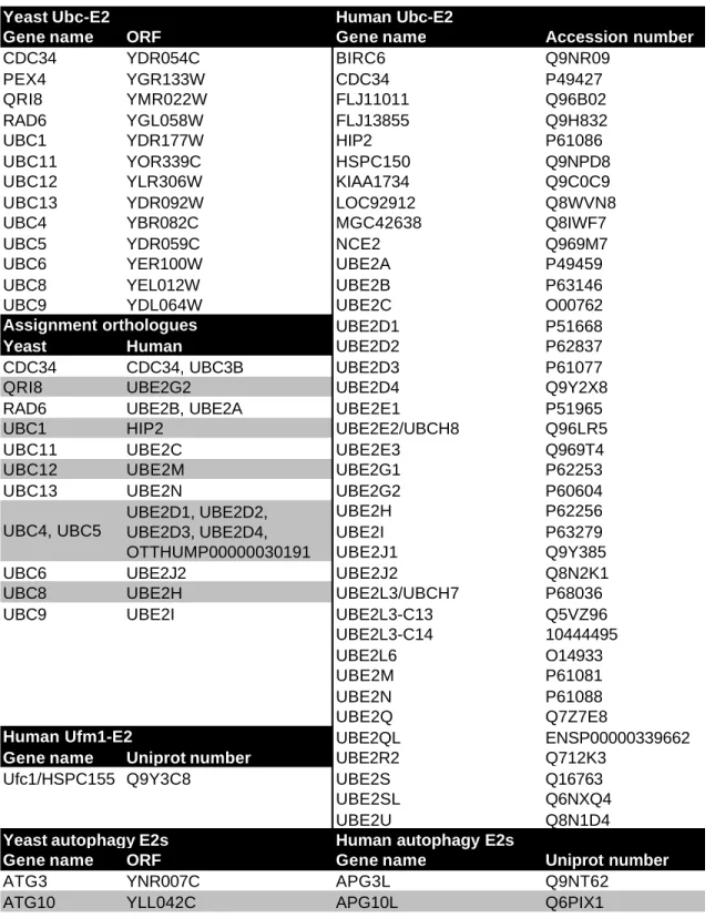
After a thioester bond is established between Ub and an E1, the latter recruits a second class of enzymes important for ubiquitylation, dubbed E2s or Ub-conjugating enzymes (UBC).
Once recruited, the E2 itself becomes ubiquitylated itself in a transthiolation reaction (Pickart, 1985). Interestingly, there are multiple Ub-specific E2s known (11 in yeast) (Pickart et al., 2004), while for the enzymatic cascades of the modifiers SUMO/Smt3 or NEDD8/Rub1 only one E2 has been discovered so far, which is Ubc9 or Ubc12, respectively (Hershko et al., 1983, Johnson, 1997, Pickart et al., 1985, Schwarz, 1998). The function of an E2 is not necessarily restricted to one particular modifier, as seen in the case of human UBE2E2/UCH8. The latter was recently reported to conjugate Ub as well as the linear di-Ub-like ISG15, indicating at least in this case overlapping pathways of Ub and a Ub-like modifier (Zhao, 2004).
Independent of the modifier conjugated, all E2s have a conserved homology domain termed UBC (Ub conjugating) in common, which is ~150 residues in length and harbours the catalytic cysteine (VanDemark, 2002). Besides this domain, some E2s have large sequence extensions up- and downstream of this domain, which in some cases play a role in E3 recognition (Mathias, 1998). A striking example for an E2 bearing much primary sequence outside the common homology domain is the ~5000 aa BRUCE/BIRC6 (Hauser, 1998).
E2s act on Ub function in distinct biological processes. One very specific and important function is performed by the yeast E2 Cdc34, which is responsible for the degradation of cyclin- G1 and Sic1, two key regulators of the cell cycle (Blondel, 1996). Yeast Rad6 has been shown to be essential for degradation of N-end-rule pathway substrates as well as for modification of histones and the polymerase processing factor PCNA (Dohmen, 1991, Dover, 2002, Hoege, 2002). Human UBEL1 and UBE2E2 are interferon inducible E2s important for ISG15 conjugation and therefore play a role in immune response (Kim, 2004, Zhao et al., 2004). More E2s and their specific function are reviewed by Haas and Siepmann (Haas, 1997).
In rare cases, the E2 can directly transfer the modifier to some substrates, but most often a specificity factor for substrate recognition is required. Another mode of modifier transfer
Chapter 1 Introduction 10
involves an intermediate step, in which the modifier is transferred to the specificity factor before final conjugation to the substrate. The specificity factor is in both cases termed E3 or Ub-ligase.
1.2.4 Ub-ligases (E3)
Ub-ligases (E3) have the function of recognizing a substrate and mediating the transfer of Ub to the substrate. Organisms generally possess a large number of E3s, each responsible for a limited set of substrates. Thus, E3s provide specificity in substrate ubiquitylation. In the case of Ub, E3s are a prerequisite for substrate recognition, while SUMO can be transferred to some substrates in the absence of an appropriate E3 (Hershko, 1998, Seeler, 2003). There is evidence that the E2 for SUMO/Smt3, Ubc9, directly interacts with the substrate RanGAP1 via binding to a sumoylation consensus site hKx(D|E) (Bernier-Villamor, 2002). In this regard, Ubc9 acts as a combined E2/E3 enzyme taking over the activated SUMO from its E1 and transferring it to the substrate. Nonetheless, several SUMO-specific E3s have been reported, which act as bridging factors like Ub-E3s bringing both E2 and substrate into a sterically favourable arrangement (Dohmen, 2004).
So far, no universal recognition motif for ubiquitylation comparable to the sumoylation consensus site is known and this observation is likely associated with the large number of Ub- specific E3s in the UPS. One exception might be the 'N-end-rule-pathway' that is responsible for the proteasome dependent degradation of proteins with a destabilizing N-terminal residue (F, H, I, K, L, R, T, W), which may be regarded as a degradation signal ('degron'). Based on sequence analysis, the E3 components for the ligation of Ub can be subdivided into three major classes (HECT, RING and U-Box), but a fourth protein family characterized by a particular homology domain (A20 zinc finger) has recently joined the ranks of ubiquitin ligases (Deshaies, 1999, Jiang, 2001, Scheffner, 1990, Wertz, 2004).
HECT based Ub-ligases
Proteins of the HECT family share a C-terminal homology domain of approximately 350 residues. The first HECT family member discovered to have ligase activity was E6AP.
Therefore, the homology domain was named after this protein, 'homologous to E6AP carboxyl terminus' (Scheffner, 1995). E6AP has been shown to bind and ubiquitylate the tumour suppressor p53 in cells infected with the human papilloma virus, leading to proteasomal degradation of p53. One of the natural targets of E6AP is human Rad23A (Kumar, 1999).
The HECT domain binds the E2 enzyme and contains the catalytic cysteine that gets linked to Ub in a transthiolation reaction between a ubiquitylated E2 and a HECT E3 (Pickart, 2001, Scheffner et al., 1995). HECT type E3s are thought to transfer a poly-Ub chain onto the
Chapter 1 Introduction 11
substrate at once, with the poly-Ub chain being assembled first on the HECT E3 (Pickart et al., 2004).
RING-based Ub-ligases
The second class of E3s is the so-called RING-finger family (for 'really interesting new gene'). RING finger proteins share a globular domain, whose structure depends on complexing two zinc ions. Unlike the HECT proteins, RING finger proteins do not get covalently linked to Ub, but rather function as adaptors between the substrate and the E2. Another characteristic feature of the RING family is that some members are subunits of large multi-subunit E3 complexes like the SCF-complex or the APC (Deshaies, 1999, Peters, 2002). Other RING finger proteins, like p53-ubiquitinating Mdm2, work without auxiliary proteins (Li, 2003).
Some Ub ligases use a U-box for E2 recruitment. The U-box represents a highly divergent variant of the original RING finger motif (Aravind, 2000, Pringa, 2001). A well known U-box protein is CHIP, which associates with chaperones like Hsc70 or Hsp90 in order to recognize the substrates to ubiquitylate, e.g. the glucocorticoid receptor (Connell, 2001, Cyr, 2002).
The SCF and other RING-cullin-based Ub-ligase complexes
The SCF complex (for 'Skp1, Cullin, F-box') is a multisubunit Ub-ligase whose core consists of the RING finger protein Hrt1/Roc1/Rbx1, the cullin Cdc53 and Skp1. Analogous to the monomeric RING finger E3s, the RING finger domain of Hrt1 is utilized for E2 recruitment.
Cdc53 serves as a scaffold binding the Hrt1 subunit and Skp1 simultaneously (see Figure 1-3 B). Skp1 in turn ties the real substrate binding protein, which contains two major domains, an F- box domain utilized for Skp1 binding and a further protein interaction domain. For example, the yeast F-box protein Cdc4 contains a repetitive WD40 region that adopts a β-propeller fold suitable for binding the substrate, Sic1 (Deshaies, 1999). The F-box subunit may be regarded as an exchangeable substrate specificity factor, thereby allowing the SCF to ubiquitylate different targets while the E3 core remains unchanged. From the structure of the SCF a more general model was developed valid for similar types of complex E3 ligases (see Figure 1-3 A). The main features that differ between the SCF complex and related complexes is the usage of completely different substrate specificity adaptors like SOCS-box or BTB proteins (see Figure 1-3 C,D) (Willems, 2004). The APC ('anaphase promoting complex') also belongs to the family of complex SCF-type E3 ligases, but uses a distinct RING finger protein, Apc11, and contains markedly more subunits (Zachariae, 1998).
Chapter 1 Introduction 12
A B
C D
A B
C D
Figure 1-3 Cullin-RING-based E3 complexes (SCF-like complexes); A, general composition of a cullin-RING- based E3 complex; B-D, specific examples together with an example adaptor and its corresponding substrate are shown. See 1.2.4 for more details. Figure adapted from Willems (Willems et al., 2004).
1.2.5 Ub-hydrolases (DUB)
Deubiquitylating enzymes (DUBs) form a heterogeneous enzyme group, whose members cleave ubiquitin-linked proteins after Gly76, the terminal ubiquitin residue (Lam, 1997). These enzymes can participate in two different cellular processes, biosynthesis of free Ub and deubiquitylation of Ub-protein conjugates.
A family of DUB enzymes that preferably cleaves Ub monomers from Ub precursors consists of small thiol-proteases (~ 25 kDa) and is often referred to as UCH family (Ub C-terminal hydrolases). They cleave regular peptide bonds at the C-terminus of Ub in poly-Ub precursors or in fusion proteins consisting of ribosomal proteins and Ub. However, there are examples of UCH-type DUBs that act both on precursors and conjugates (Kwon, 2004).
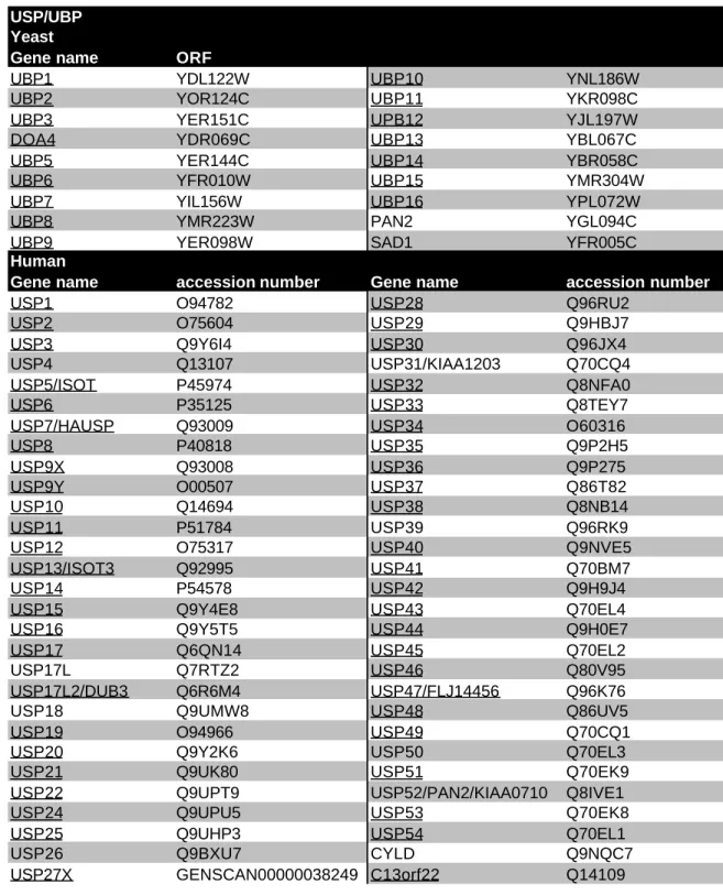
Unlike the processing of precursors, the deubiquitylation of Ub-protein conjugates requires hydrolysis of an isopeptide bond. The family of Ub specific proteases (USP in human, UBP in yeast), which is larger than the UCH family, and whose members are larger in size (60- 300 kDa), prefers the cleavage of such isopeptide bonds. Only for a few USPs functions are known, e.g. USP11 has been shown to deubiquitylate RanBPM (Ideguchi, 2002) and UBP3 has been implicated in gene silencing (Moazed, 1996). There seems to be a broad diversity
Chapter 1 Introduction 13
concerning their function, their dependence on ATP and their localization within the cell. Some USPs occur freely, while others are associated with large complexes, such as the proteasomal lid, the CSN or the SAGA complex (Daniel, 2004, Leggett, 2002, Zhou, 2003). The proteasome-associated USPs are thought to be responsible for protecting Ub from proteasomal degradation. Other USPs like yeast Ubp14 and human UCHL5/UCH37 probably play a role in editing poly-Ub chains on target proteins and for Ub recycling by cleaving free poly-Ub chains instead (Amerik, 1997, Lam, 1997, Lam et al., 1997).
Ataxin-3, the protein mutated in Machado Joseph Disease (SCA3), belongs to a novel group of cysteine-proteases and is active against ubiquitin chains (Burnett, 2003, Scheel, 2003).
Like ataxin-3, the OTU (ovarian tumour) proteases display a structural similarity to the USP protein family in their catalytic core (Makarova, 2000) and a deubiquitylating activity was shown for several OTU proteins (Evans, 2003, Soares, 2004).
Besides these four DUB classes of cysteine proteases, a deubiquitylating activity was found in the MPN subunit Rpn11 of the proteasomal lid (Maytal-Kivity et al., 2002, Verma, 2002, Yao, 2002). Rpn11 generates free poly-Ub chains by hydrolysing the bond that connects the target protein and the proximal Ub of the poly-Ub chain. Interestingly, a deneddylating activity, i.e. the cleavage of Lys-linked Nedd8 conjugates is intrinsic to the MPN protein Csn5, the CSN subunit analogous to Rpn11 (Cope, 2002, Maytal-Kivity et al., 2002). In contrast to UCH and UBP proteases, MPN proteins are metalloproteases coordinating Zn2+ in their active site (Tran, 2003, Verma et al., 2002, Yao et al., 2002).
1.2.6 Ub-binding proteins
Typical intracellular signal transduction pathways are characterized by a modular architecture of the proteins involved in the three fundamental steps of signal generation, signal recognition and signal removal. In protein phosphorylation, the archetype of such transduction systems, the three roles are filled by kinases, phosphatases, and phosphopeptide recognition domains (SH2, PTB, FHA etc), respectively. The components of the UPS obviously form an analogous system. Here, the E1-E2-E3 cascade corresponds to the signal generation, where Ub conjugated to a substrate constitutes the signal itself. Ub-binding proteins serve for recognizing the signal, while DUBs quench it. Similarly, the analogy to signal transduction pathways seems to be valid for most Ub-like modifiers and their associated apparatus.
Within the UPS, there exist many recognition systems, which can recognize the different ubiquitylation states including different types of Ub-to-Ub linkages and various chain lengths.
Ub-recognizing proteins are normally classified according to the homology domains involved and play a crucial role in the UPS.
Chapter 1 Introduction 14
UBA domain
The Ub associated domain (UBA) occurs in many different proteins of the UPS, including E3s, DUBs, Ub conjugases and adaptors (Hofmann et al., 1996). The universal character of the UBA as a Ub binding domain can be seen from many reported interactions between UBA containing proteins and Ub (Bertolaet, 2001, Rao, 2002, Wilkinson, 2001).
The UBA domain is a small domain of only ~40 residues with a three-helix bundle fold (Mueller, 2002). It has a preference for tetra-Ub chains, which is of two orders of magnitude higher than to mono-Ub (Wilkinson et al., 2001). There are contradicting reports on the linkage preference of UBA domains. Both a binding to Lys-48 linked chains and to Lys-29 linked chains have been described (Raasi, 2003, Rao et al., 2002).
While the uncertainty on linkage preference of UBA domains remains, more information exists on the part of Ub that is recognized by UBA domains. By NMR-based methods, Ryu et al. have identified the Ile-44 surface patch of Ub, and a homologous region in the Ub-like domain of human Rad23B, as interacting with UBA domains (Ryu, 2003). These experiments also demonstrate the ability of UBA domains to interact with type II ubiquitin-like proteins.
CUE domain
The CUE domain is another ubiquitin-binding homology domain (Ponting, 2000), which has been suggested to be distantly related to the UBA domain (Shih, 2003). This relationship was recently confirmed by Kang et al., who have solved the Cue2 structure in complex with Ub (Kang, 2003). In this CUE/Ub complex, the CUE domain is bound to Ub's Ile-44 patch, similar to the binding of the human Rad23B-UBA domain to Ub (Ryu et al., 2003). Additional evidence for the CUE domain as a Ub binding domain comes from Donaldson et al. and Shih et al., who have reported the CUE domain of yeast Vps9, Cue2, Cue3, Cue5 and human Tollip to directly bind mono-Ub (Donaldson, 2003, Shih et al., 2003). The preference for mono-Ub is probably valid for all CUE domain proteins and makes it different from the UBA domain, which prefers poly-Ub (Shih et al., 2003). Another CUE family member, Cue1, has been assigned a role in the ER associated degradation pathway (ERAD), which relies on Ub signals (Biederer, 1997).
However, its affinity to ubiquitin is significantly reduced compared to Vps9 and Cue2 (Shih et al., 2003).
UIM
The UIM (Ub interacting motif) was first described in 2001 by Hofmann et al. based on a motif in Rpn10/S5a (Hofmann et al., 2001), a proteasomal subunit, which had been shown to bind ubiquitin (Young, 1998). Other proteins associated with the UPS contain this motif as well, e.g. several DUBs, the yeast F-Box protein Ufo1 and some E3s. Besides the UPS, the UIM
Chapter 1 Introduction 15
appears in proteins that regulate Ub-dependent events of endocytosis. Mono-ubiquitylation of target proteins serves as an internalisation signal, and the Ub-recognizing element in this process has been narrowed down to the UIM in eps15 and Hrs (Di Fiore et al., 2003, Polo, 2002). Interestingly, mono-ubiquitylation of endocytosis components such as ligand-bound receptors in the plasma membrane depends on a functional UIM domain in the same protein (Klapisz, 2002, Polo et al., 2002) and the UIM also keeps the ubiquitylation status of a target on mono-ubiquitylation (Di Fiore et al., 2003).
The UIM is a very short motif of ~20 residues consisting of an α-helix with the conserved residues located on one side of the helix (Shekhtman, 2002). Like the UBA domain, the UIM binds to the Ile-44 patch of Ub. This interaction of a UIM and Ub does not involve the Lys-48 of Ub, which would allow the UIM to differentiate between poly-Ub and mono-Ub.
Nevertheless, within the UPS, the UIM obviously prefers poly-Ub as a binding partner (Perez, 2003, Polo et al., 2002, Shekhtman et al., 2002, Thrower, 2000).
GAT domain
The GAT domain (GGA and Tom1) was initially found in proteins regulating clathrin- mediated trafficking of vesicles (Dell'Angelica, 2000). Recent findings have shown the GAT domain to bind to Ub (Katoh, 2004, Shiba, 2004). The structures of several GAT domains have been solved, presenting the GAT domain as a three-helix bundle with elongated and almost parallel helices, an arrangement quite different from the helix bundle of the UBA structure (Shiba et al., 2004). Like the UBA domain, the GAT domain is thought to interact with the Ile- 44 patch of Ub (Shiba et al., 2004).
UEV domain
The UEV (Ub E2 variant) domain is related to the domain responsible for the catalytic E2 activity, but is devoid of the cysteine important for Ub conjugation (Ponting, 1997). A well known member of this inactive subfamily of E2 enzymes is the tumour susceptibility gene 101 protein (TSG101/Vps23), which plays a role in Ub-dependent protein sorting and is mutated in certain types of breast cancer (Bishop, 2002, Pornillos, 2002, Pornillos, 2002). Budding yeast Mms2, another UEV protein, forms a complex with Ubc13 (functional E2) and is required for Rad6/Rad18 dependent postreplicative DNA repair (Broomfield, 1998, Hofmann, 1999).
Available structural information on TSG101 in complex with ubiquitin demonstrates the ability of the UEV domain to bind Ub (Sundquist, 2004). At the same time, other UEV proteins may differ in their Ub binding modes (Sundquist et al., 2004).
Chapter 1 Introduction 16
NZF domain
The NZF domain (Npl4 Zn-finger) is a C4-type Zn-finger that coordinates a zinc ion via four cysteines, which makes this domain very different from the Ub-binding modules described so far (Wang, 2003). This Zn-finger is found in human Npl4, a VCP/Cdc48/p97 adaptor protein, and in yeast Vps36. Experiments with both proteins revealed Ub-binding properties (Alam, 2004, Meyer, 2002).
1.2.7 Proteasome 20S proteasome
The 20S proteasome is a subcomplex of the 26S proteasome and after binding of two copies of the 19S regulatory particle yields the 26S proteasome. The barrel-shaped 20S proteasome consists of 28 subunits arranged in four stacked rings with seven subunits each. All subunits share a common evolutionary ancestor and can be further subdivided into the α- subunits forming the outer rings and the β-subunits, which are found in the two inner rings. This α7β7β7α7 structure of four rings harbours three major chambers in its centre. All chambers are connected with each other and the surrounding solvent. The largest and centrally located chamber bears the six catalytically active sites, which are located on distinct subunits of the beta-rings. The active subunits are termed β1, β2, and β5, each of which occurs with two copies in the 20S proteasome. The sequestration of the protease activity to the shielded chamber allows the proteasome to limit degradation to the correct substrates. Before a substrate can be degraded within the central chamber, it has to be recognized as a correct substrate at one of the entry pores and unfolded in a subsequent step (Baumeister, 1998).
Archeae and several bacteria also possess proteasome-like proteases, which typically only consist of one or two subunit types. During evolution, the number of distinct subunits has multiplied in higher organisms, i.e. the yeast genome encodes seven different α-subunits and seven β-subunits (Gille, 2003). The situation in mammals is even more diverse with ten distinct β-subtypes, three of which are interferon-γ inducible ('immunosubunits') and only found in so- called 'immunoproteasomes' (Kloetzel, 2004). The three immunosubunits β1i, β2i and β5i occupy the positions of β1, β2 and β5 positions, respectively, of newly synthesized 20S immunoproteasomes upon interferon-γ induction. As a consequence of the altered catalytic subunit layout, immunoproteasomes generate peptides suitable as antigen precursors. The latter normally have to be trimmed to the correct length by cytosolic or ER-based peptidases (Kloetzel, 2004). Suitable antigens are finally presented by MHC class I proteins. Interferon-γ
Chapter 1 Introduction 17 also stimulates the biosynthesis of PA28α and PA28β proteins, which assemble into the heptameric PA28 regulatory complex able to cap the immunoproteasome.
All α-subtypes are non-catalytic in nature and fulfil a regulatory function instead. Their N-terminal extensions, especially that of the α3 subunit, lock the pores of the 20S proteasome.
Only upon binding to the 19S regulatory particle (PA700) or the PA28 complex in the case of the immunoproteasome, the α-subunits' N-termini become delocalised and open up the pores (Groll, 2000, Kloetzel, 2004).
19S regulatory particle
The 19S regulatory particle is essential for proteasomal activity and consists of two subcomplexes. The one binding the 20S proteasome is the 'base', which is a hexameric ring of AAA-ATPases with chaperone activity (Braun, 1999) and three additional subunits, Rpn1, Rpn2 and Rpn10. Base subunit Rpt5 and Rpn10 have been shown to bind to poly-Ub chains and therefore may function as receptors of ubiquitylated substrates (Deveraux, 1994, Lam, 2002). A more indirect role in substrate delivery to the proteasome has been mapped to Rpn1, as this protein associates with the adaptor protein Rad23, which in turn is responsible for the recognition of many ubiquitylated substrates (Elsasser, 2002).
The other subcomplex called the 'lid' has a more complex structure based on eight core subunits (Glickman, 1998). The lid is composed of multiple subunits harbouring the PCI domain, named after the three similar complexes ((i) proteasome lid, (ii) COP9 signalosome or CSN complex, (iii) eukaryotic translation initiation factor eIF3) that contain this domain (Hofmann et al., 1998). These complexes in turn are termed PCI complexes. Other subunits of the lid are characterized by a second homology domain called MPN (Mpr1-Pad1 N-terminal) (Aravind, 1998, Kapelari, 2000). The lid complex contains an intrinsic deubiquitylating activity, which is encoded by the MPN subunit Rpn11 that has the hallmarks of a metalloprotease as described in chapter 1.6.5 (Maytal-Kivity et al., 2002, Verma et al., 2002, Yao et al., 2002).
Interestingly, Csn5, an MPN-bearing subunit of another PCI complex, the signalosome, also encodes a metalloprotease that can cleave Ub from proteins (Groisman, 2003). In addition, Csn5 is needed for the cleavage of the Ub-like protein Nedd8 from cullins (Cope et al., 2002). No specific function has been described for the PCI subunits of the lid so far.
Chapter 1 Introduction 18
1.3 Detection of homologues and protein family analysis
1.3.1 Sequence comparison methods
As homology serves as a vector along which information can be transferred from one known sequence to a new sequence, methods have developed that help deciding if two sequences are sufficiently similar to infer a relationship. The basic concept is the alignment, in which two or more sequences are arranged along each other, so that evolutionary or structurally equivalent residues are opposed. Multiple sequences can be aligned simultaneously as well, leading to multiple sequence alignments ('MSA'). MSAs are not only helpful to illustrate evolutionary events within a protein family, but also constitute the basis for phylogenetic tree construction, secondary structure prediction, homology modelling and, of special interest, the identification of conserved domains. With regard to the latter, homology domains and motifs often appear as columns with obvious conservation, while the adjacent primary sequence has a higher level of variation. When looking for further proteins with a given domain, information on amino acid frequency at each position within the domain was shown to be useful for so called profile-based techniques (Bucher et al., 1996, Gribskov, 1987). A more detailed description on profile construction as well as database search methods will be given in chapter 2.
For calculating a mathematically optimal alignment of two or more sequences, dynamic programming algorithms are typically used, for example the Smith-Waterman algorithm that looks for the best alignment between two subsequences ('local alignment') (Smith, 1981). These algorithms aim at maximizing the alignment score under an additive scoring scheme by incorporating as many positively scoring residue pairs as possible into the alignment. At the same time, negatively scoring pairs from unconserved residues and special penalties for gaps have to be minimized. Finally, a single optimal alignment and its corresponding score are reported. Efforts to increase the sensitivity of this type of comparison aim at scoring alternative alignments as well and weighting them by a probability value (Bucher et al., 1996).
1.3.2 Multiple sequence alignments (MSA)
MSAs are simultaneous alignments of more than two sequences (see Figure 1-4). As mentioned above, MSAs provide information on amino acid composition at each individual position within the alignment. Unfortunately, the calculation of a mathematically optimal MSA is computationally too expensive to be feasible (Wang, 1994). Current MSA generating programs rely on heuristics and none of them guarantees to report the fully optimised MSA. The programs used in this work, ClustalW and T-Coffee (Chenna, 2003, Notredame, 2000), belong