LIQUID-LIQUID PHASE SEPARATION:
MOLECULAR MECHANISMS AND INFLUENCE ON THE mRNA DECAPPING MACHINERY
DISSERTATION ZUR ERLANGUNG DES
DOKTORGRADES DER NATURWISSENSCHAFTEN (DR. RER. NAT.) DER FAKULTÄT FÜR BIOLOGIE UND VORKLINISCHE MEDIZIN
DER UNIVERSITÄT REGENSBURG
vorgelegt von
STEFAN SCHÜTZ
aus QUEDLINBURG
im Jahr
2019
- 2 - Das Promotionsgesuch wurde eingereicht am:
14. Juni 2019
Die Arbeit wurde angeleitet von:
PROF. DR. REMCO SPRANGERS
Unterschrift:
Stefan Schütz
- 3 -
- 4 -
List of Publications and Manuscripts
Published
1. Schütz, S.; Nöldeke, E. R.; Sprangers, R. A Synergistic Network of Interactions Promotes the Formation of in Vitro Processing Bodies and Protects mRNA against Decapping. Nucleic Acids Res. 2017, 45 (11), 6911–6922. https://doi.org/10.1093/nar/gkx353.
2. Damman R; Schütz, S; Luo, Y; Weingarth, M; Sprangers, R; Baldus, M. Atomic-level insight into the maturated state of mRNA processing bodies by combining solid and solution-state NMR spectroscopy. Nat Commun. 2019, 10 (1), 4536. https://doi.org/10.1038/s41467-019- 12402-3.
Accepted
3. Schütz, S.; Sprangers, R. Methyl TROSY spectroscopy: A versatile NMR approach to study challenging biological systems. Prog. Nucl. Magn. Reson. Spectrosc., 2019, in press, https://doi.org/10.1016/j.pnmrs.2019.09.004
In preparation
4. Schütz, S; Sprangers, R. Deciphering the contributions of molecular interactions that lead to liquid-liquid phase separation of the conserved DEAD-box protein Dhh1.
The contributions of the individual authors to the listed publications and manuscripts are indicated at
the beginning of the respective chapters.
- 5 -
TABLE OF CONTENT
SUMMARY ... - 8 -
SELECTED ABBREVIATIONS ... - 9 -
CHAPTER 1 General Introduction ... - 10 -
1.1 A SHORT VIEW ON THE LIFE OF AN mRNA ... - 10 -
1.2 mRNA DEGRADATION IN EUKARYOTES ... - 12 -
1.2.1 3´-5´ decay ... - 14 -
1.2.2 5´-3´ decay and the mRNA degradation machinery ... - 14 -
1.3 LIQUID-LIQUID PHASE SEPARATION ... - 15 -
1.4 NMR SPECTROSCOPY ... - 18 -
1.4.1 The TROSY experiment ... - 18 -
1.4.2 Methyl TROSY ... - 19 -
1.4.3 Methyl labeling ... - 20 -
1.4.4 Methyl resonance assignment ... - 22 -
1.4.5 CSP experiments ... - 27 -
1.4.6 Methionine scanning ... - 29 -
1.5 AIMS OF THIS THESIS ... - 32 -
CHAPTER 2 A synergistic network of interactions promotes the formation of in vitro processing bodies and protects mRNA against decapping ... - 33 -
2.1 INTRODUCTION ... - 33 -
2.2 MATERIALS AND METHODS ... - 35 -
2.2.1 Protein expression and purification ... - 35 -
2.2.2 Protein fluorescence labeling ... - 37 -
2.2.3 RNA in vitro transcription, purification and capping ... - 37 -
2.2.4 Liquid-liquid phase separation experiments ... - 39 -
2.2.5 Microscopy ... - 39 -
2.2.6 NMR ... - 39 -
2.2.7 Decapping assays ... - 40 -
2.2.8 HPLC analysis ... - 40 -
2.2.9 Analysis of degradation data ... - 40 -
2.2.10 RNase A protection assays ... - 40 -
2.3 RESULTS ... - 41 -
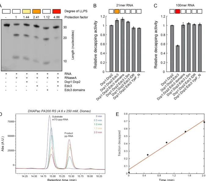
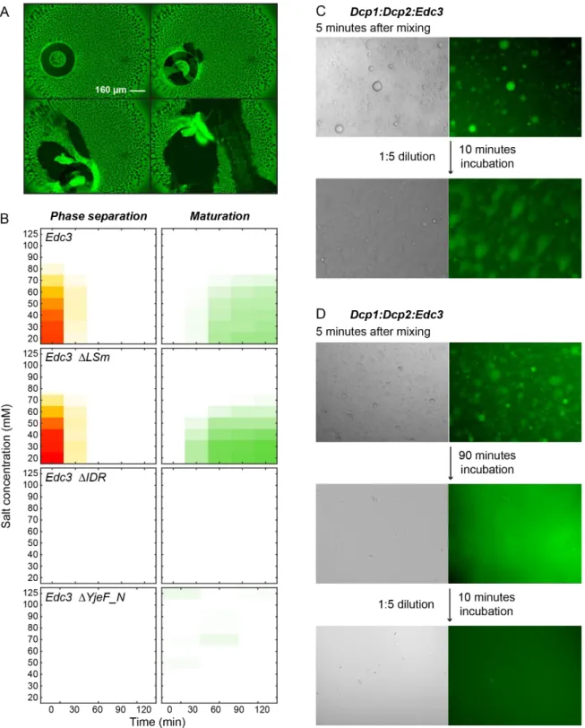
2.3.1 High throughput approach for LLPS determination ... - 41 -
2.3.2 Pdc1 stimulates LLPS ... - 43 -
- 6 -
2.3.3 RNA strongly stimulates LLPS ... - 44 -
2.3.4 RNA is protected against degradation by LLPS ... - 46 -
2.3.5 LLPS reduces the catalytic activity of Dcp2 ... - 46 -
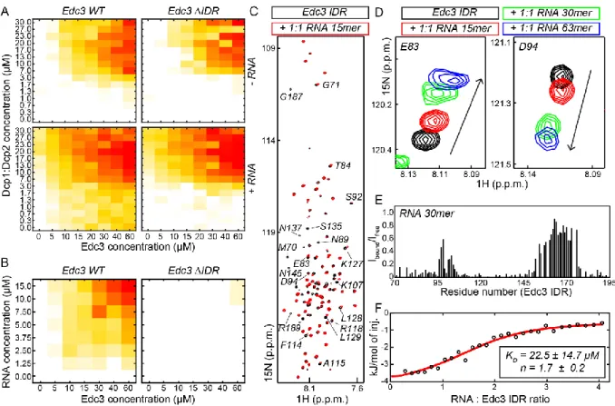
2.3.6 The IDR in Edc3 specifically interacts with RNA ... - 48 -
2.3.7 Interactions of the Edc3 IDR and the RNA-helicase Dhh1 are conserved from yeast to human ... - 51 -
2.3.8 Maturation of processing bodies ... - 51 -
2.4 DISCUSSION ... - 54 -
CHAPTER 3 Atomic level insight into the maturated state of mRNA processing bodies by combining solid- and solution-state NMR spectroscopy ... - 57 -
3.1 INTRODUCTION ... - 57 -
3.2 MATERIALS AND METHODS ... - 59 -
3.2.1 Protein expression and purification ... - 59 -
3.2.2 RNA in vitro transcription and purification ... - 61 -
3.2.3 Liquid-liquid phase separation assays ... - 62 -
3.2.4 Solution-state NMR experiments ... - 62 -
3.2.5 Solid-state NMR experiments ... - 62 -
3.2.6 Molecular Dynamics Simulations ... - 63 -
3.3 RESULTS ... - 63 -
3.3.1 The LSm domain of Edc3 is mobile in the matured state ... - 63 -
3.3.2 The YjeF_N domain forms a rigid core in the matured state ... - 65 -
3.3.3 Interactions between the IDR and the Yjef_N domain are important for phase separation of Edc3 ... - 67 -
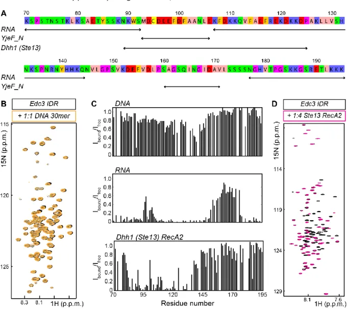
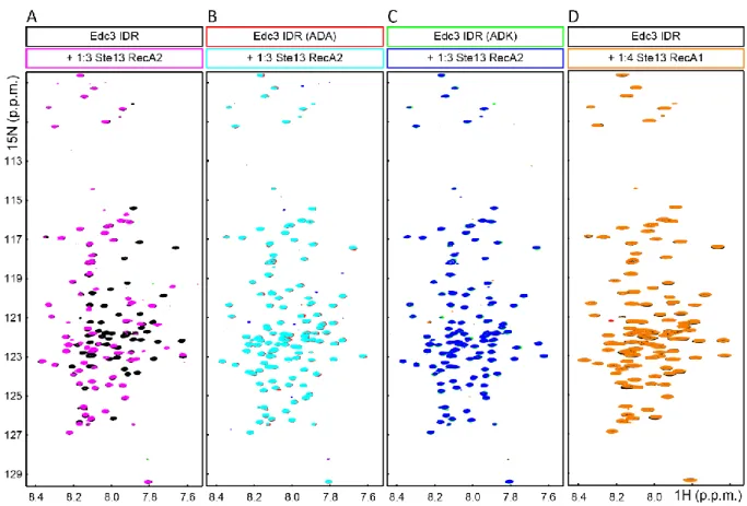
3.3.4 Details of the interactions between the IDR and the Yjef_N domain ... - 69 -
3.3.5 RNA interacts with the IDR and thereby increases rigidity ... - 72 -
3.4 DISCUSSION ... - 74 -
CHAPTER 4 Deciphering the contributions of molecular interactions that lead to liquid-liquid phase separation of the conserved DEAD-box protein Dhh1 ... - 76 -
4.1 INTRODUCTION ... - 76 -
4.2 MATERIALS AND METHODS ... - 80 -
4.2.1 Protein expression and purification ... - 80 -
4.2.2 Complex reconstitution ... - 81 -
4.2.3 Protein labeling for NMR spectroscopy... - 82 -
4.2.4 NMR spectroscopy... - 82 -
4.2.5 RNA in vitro transcription and purification ... - 82 -
4.2.6 Liquid-liquid phase separation experiments ... - 83 -
- 7 -
4.3 RESULTS ... - 83 -
4.3.1 Dhh1 undergoes phase separation in the absence of RNA ... - 83 -
4.3.2 Residues at the C-terminus of the RecA2 domain are crucial for LLPS ... - 85 -
4.3.3 The unstructured extensions enhance LLPS of the Dhh1 helicase core ... - 89 -
4.3.1 ATP and RNA strongly enhance LLPS of full-length Dhh1... - 89 -
4.3.2 Binding of Edc3 or Pat1 disrupts phase separation of the Dhh1 core... - 91 -
4.3.3 In solution, the two RecA domains of Dhh1 tumble independently ... - 93 -
4.3.4 Assignment of the Dhh1 methyl groups ... - 94 -
4.3.5 Residues in the RecA-like domains sense the phase-separated state ... - 99 -
4.4 DISCUSSION ... - 99 -
CHAPTER 5 Conclusion ... - 103 -
FUNDING ... - 104 -
ACKNOWLEDGEMENTS ... - 104 -
REFERENCES ... - 106 -
- 8 -
SUMMARY
Cellular liquid-liquid phase separation (LLPS) results in the formation of dynamic membrane-less granules that play an important role in many biological processes. On a molecular level, the clustering of proteins into a confined space results from an indefinite network of intra- and intermolecular interactions.
Here, we introduce and exploit a novel high-throughput bottom-up approach to study how the interactions between RNA, the Dcp1:Dcp2 mRNA decapping complex and the scaffolding proteins Edc3 and Pdc1 result in LLPS and the formation of processing bodies (P-bodies). We find that the LLPS boundaries are close to physiological concentrations upon inclusion of multiple proteins and RNA. Within in vitro P-bodies the RNA is protected against endonucleolytic cleavage and the mRNA decapping activity is reduced, which argues for a role of P-bodies in temporary mRNA storage.
Interestingly, the intrinsically disordered region (IDR) in the Edc3 protein emerges as a central hub for interactions with both mRNA and mRNA decapping factors. In addition, the Edc3 IDR plays a role in the formation of irreversible protein aggregates that are potentially detrimental for cellular homeostasis.
Until now, a detailed structural characterization of the intrinsically heterogeneous LLPS process has been challenging. Here, we combine solid- and solution-state NMR spectroscopy to obtain atomic-level insights into the assembly and maturation of in vitro P-bodies. Our results reveal that Edc3 domains exhibit diverse levels of structural organization and dynamics after LLPS. In addition, we find that interactions between the different Edc3 domains and between Edc3 and mRNA in solution are largely preserved in the condensed protein state, allowing P-bodies to rapidly form and dissociate upon small alterations in the cellular environment.
Additionally, we aim at unraveling the role of the conserved helicase Dhh1 in the formation of (in vitro) P-bodies. We found that the LLPS process of Dhh1 contains contributions from the RNA, the IDRs at the N- and C-terminal regions and the folded helicase core domains. Based on mutants of the enzyme, we identified residues in the C-terminal part of the second helicase core domain to be crucial for LLPS of Dhh1. In addition, we found that ATP enhances Dhh1 phase separation, even in the absence of RNA. Our results will allow us to conclude to what degree the above interactions contribute in a constructive manner to LLPS and, by employing NMR spectroscopic methods, which residues are involved in the phase separation process.
In summary, our work sheds light on both the molecular mechanisms that underlie
liquid-liquid phase separation and provides clues about how this influences cellular processes.
- 9 -
SELECTED ABBREVIATIONS
ADP/ATP Adenosine di/triphosphate CSA Chemical shift anisotropy CSP Chemical shift perturbation
CV Column volume
DNA Deoxyribonucleic acid DTT Dithiothreitol
Dcp(S) (Scavenger) Decapping protein DDX DEAD-box protein
Dhh DEAD-box helicase homolog Edc Enhancer of decapping eIF eukaryotic Initiation Factor FUS Fused in Sarcoma
GDP/GMP Guanosine di/monophosphate GST Glutathion-S-transferase HEPES 2-(4-(2-Hydroxyethyl)-1-
piperazinyl)- ethanesulfonic acid HLM Helical leucine-rich motif
HMQC Heteronuclear multiple quantum coherence
hn Heterogeneous nuclear HSQC Heteronuclear single quantum
coherence
IDR Intrinsically disordered region IPTG Isopropyl β-D-1-
thiogalactopyranoside
ITC Isothermal titration calorimetry LLPS Liquid-liquid phase separation LSm Like Smith
MAGIC Methyl assignment by graphing inference construct
MBP Maltose binding protein
MES 2-(N-morpholino)-ethanesulfonic acid
(ss)NMR (solid-state) Nuclear magnetic resonance
NOE(SY) Nuclear Overhauser effect (spectroscopy)
PAGE Polyacrylamide gel electrophoresis
PABP Poly(A) binding protein Pat Protein associated with
topoisomerase II P-body Processing body RecA Recombinase A
(m)RNA (messenger) Ribonucleic acid RNP Ribonucleoprotein
Scd Suppressor of clathrin deficiency SDS Sodium dodecylsulfate
SEC Size exclusion chromatography
SH Src-homology
SUMO Small ubiquitin-related modifier TEV Tobacco etch virus
TROSY Transverse relaxation optimized spectroscopy
Xrn Exoribonuclease
Amino acids are abbreviated by their one- or three-letter code. Nucleobases are
abbreviated by their one-letter code.
- 10 -
CHAPTER 1 General Introduction
1.1 A SHORT VIEW ON THE LIFE OF AN mRNA
Eukaryotic messenger RNA (mRNA) is transcribed from its DNA template in the 5’ to 3’
direction by RNA polymerase II (Pol II) in the nucleus
1. Immediately after the first nucleotides emerge from the polymerase, the nascent transcript is protected at its 5’ end by a cap structure
2–4(Figure 1.2 A). Binding of the capping enzyme to the C-terminal domain of Pol II ensures that only Pol II transcripts are capped
5–7.
The simplest eukaryotic 5’ cap structure, the so-called cap 0, consists of an N7-methylated guanosine (m
7G) that is linked to the first transcribed nucleotide via a 5’-5’ triphosphate bridge (Figure 1.1)
2. This unusual linkage results in a free 3’ OH group at the m
7G, which confers stability of the nascent transcript against 5’-3’ exonucleases
8,9. In higher eukaryotes, additional methylation at the 2’-O ribose position of the first and second transcribed nucleotide result in cap 1 and cap 2 structures, respectively
10. Higher methylated cap structures exist for minor RNA species and trypanosomal mRNAs
10. Notably, Pol II has been shown to accept nicotinamide adenine dinucleotide (NAD) and 3’-dephospho coenzyme A as non-canonical nucleotides for transcription initiation in vitro
11. Recent evidence confirms the existence of NAD caps for a subset of yeast mRNAs and underscores the potential that also other adenine-containing nucleotide caps could exist
12.
Figure 1.1: Cap structures protect the mRNA from premature 5’-3’ exonucleolytic degradation. The cap 0 is characterized by an N7-methylated guanosine that is linked via a triphosphate to the first transcribed nucleotide, while the cap 1 has an additional methyl group at the 2’-O ribose position of the first transcribed nucleotide. Higher order cap structures exist for minor RNA species and in higher eukaryotes. The cleavage sites of the decapping enzymes DcpS and Dcp2 are indicated.
Notably, DcpS is inhibited by the Dcp2 decapping product m7GDP.
- 11 -
The capped, premature mRNA is further processed by splicing events that remove introns
13,14and by protecting the 3’ end by polyadenylation (Figure 1.2 A)
15. The length of the polyadenosine (poly(A)) tail differs among species: in yeast, the poly(A) tails reach lengths of around 50-80 nucleotides while in mammals they can be up to 250 nucleotides long
16–18. Thereby, long poly(A) tails are usually associated with high mRNA stability
19. Notably, mRNAs with (not too) short poly(A)-tails can have higher translation rates than long-tailed mRNAs
20. Replication-dependent histone mRNA is the only eukaryotic mRNA species that lacks a poly(A) tail; instead, these histone mRNAs contain a protective 3’ stemloop structure
21.
The cap and the poly(A) tail are parts of the 5’ and 3’ untranslated regions (UTRs), respectively, that flank the protein coding region of a mature mRNA (Figure 1.2 A)
22. The UTRs function in the control of mRNA maturation, localization, stability, translation efficiency and also plays a role in various disease
23–27. Stable secondary structures that are found in the UTRs can interfere with translation by preventing the ribosome from scanning for the start codon
28. Additionally, they provide internal ribosome entry sites (IRES)
29,30or serve as binding sites for regulatory proteins
31,32. In higher eukaryotes, small RNA species like short interfering RNAs (siRNAs) or micro RNAs (miRNAs) can bind to the 3’ UTR, which provides an additional level to regulate gene expression
33–35.
Figure 1.2: Characteristics of mRNA. (A) Structural features of a mature mRNA. The transcript is protected at its 5’ end by an N7-methyl guanosine cap that is linked via a triphosphate to the first transcribed nucleotide. The 3’ end of an mRNA is protected by a polyadenosine (poly(A)) tail. The coding region starts with the initiation codon AUG and terminates with one of three possible stop codons. The 5’ and 3’ untranslated regions (UTRs) can contain highly structured segments and regulate mRNA maturation, translation and degradation. (B)-(D) Schematic representation of closed-loop messenger ribonucleo- protein (mRNP) complexes during translation initiation (B), decapping by Dcp2 (C) and 5’-3’ degradation by Xrn1 (D). These three processes are assumed to be enhanced by bridging the 5’ and 3’ end of the mRNA, which is shown in a simplified manner compared to A. Closed-loop structures of translational repressed mRNPs are not shown.
- 12 -
The 5’ cap and the 3’ poly(A) tail mediate nuclear export of the mature mRNA
36,37. In the cytosol, the eukaryotic translation initiation factor (eIF) 4E binds to the m
7G cap
38, while the poly(A) tail is bound by the poly(A) binding protein (PABP1 in human and Pab1 in yeast)
39. Both proteins are bridged by eIF4G, which leads to a closed-loop structure of the messenger ribonucleoprotein (mRNP) complex (Figure 1.2 B)
40. The circular closed-loop is associated with efficient translation
41–43and protection of the mRNA against decapping and subsequent degradation
44. Further, the circularization serves as a quality control mechanism to ensure that only properly transcribed and processed mRNAs are translated, as a missing cap or poly(A) tail would prohibit circularization
30. Notably, closed-loop structures are supposed to exist not only for actively translated mRNAs
45, but also for repressed mRNAs
46,47or during mRNA degradation (Figure 1.2 C and D)
48,49.
1.2 mRNA DEGRADATION IN EUKARYOTES
Cellular mRNA levels depend on the equilibrium between transcription and mRNA degradation. The amount of actively translated mRNA in a cell must be tightly regulated in a spatiotemporal manner to allow for adaption to environmental changes and to different stages in cell cycle or development. Thus, some mRNA species are turned over rapidly, while others are kept for longer periods
50. The half-life of mRNAs varies considerably among different species, between minutes and a few hours in yeast
51and up to several days in mammals
52,53.
Degradation is the final step in the life of an RNA and provides the last possibility for a cell to control gene expression on the RNA level. mRNA degradation not only serves the purpose of routine mRNA turnover but also of differential gene expression. Additionally, aberrant transcripts must be removed from the cell to prevent their potentially dangerous accumulation. Dedicated quality control pathways exist for the degradation of mRNAs that contain premature stop codons (nonsense- mediated decay, NMD)
54, that lack a stop codon (non-stop decay, NSD)
55,56or that are trapped in stalled ribosomes (no-go decay, NGD)
57,58. Besides these minor surveillance mechanisms two major mRNA decay pathways exist, a 5’ to 3’ and a 3’ to 5’ degradation pathway (Figure 1.3)
59,60.
Both pathways rely on the shortening of the poly(A) tail by deadenylation complexes
61,62,
whereby deadenylation is the rate-limiting step in mRNA turnover
61. Deadenylation occurs in a biphasic
manner
62, where the Pan2/Pan3 complex trims initially very long poly(A) tails of mature mRNAs
63–65,
while further deadenylation by the CCR4-NOT complex leaves only a few adenines on the mRNA
(Figure 1.3, top)
65–67. In many eukaryotes, a stretch of uridine nucleotides is attached to the oligo(A)
remnant, which finally marks the mRNA for degradation
68–70.
- 13 -
Interestingly, the poly(A)-binding protein PABP plays an ambivalent role by not only promoting translation, but also by recruiting the deadenylation machinery that finally displaces PABP from the mRNA when the poly(A) tail is shortened below a critical length
71,72. It was found that poly(A) tails with a high occupancy of PABP are deadenylated slowly by Ccr4
72.
On the contrary, poly(A) tails free of PABP are rapidly deadenylated by Ccr4 and Caf1, another deadenylase of the CCR4-NOT complex
72. Remarkably, low PAPB occupancy on the poly(A) tails was found to correlate with a high degree of sub-optimal codon usage in the coding region of the mRNA
72. Moreover, mRNAs with poor codon optimality were reported to be occupied also with Dhh1, a DEAD-box RNA helicase involved in translational repression and mRNA degradation
73–75. Thus, PABP and Dhh1 link codon optimality to mRNA turnover: efficiently translated mRNAs are protected against deadenylation and degradation, while slow translation triggers mRNA decay
76.
Figure 1.3: Schematic representation of the two major eukaryotic mRNA degradation pathways. Most mRNAs are turned over in a deadenylation-dependent manner. The poly(A) tail is removed in a biphasic process by the Pan2/Pan3 and CCR4- NOT deadenylation complexes. Subsequently, the deadenylated mRNA is subjected to one of two different decay pathways:
in 5’-3’ decay, irreversible decapping is followed by exoribonucleolytic degradation, while in 3’-5’ decay the mRNA is first degraded from its 3’ end before the short remnants are decapped. The decapping enzymes Dcp2 and DcpS produce m7GDP and m7GMP respectively. The cellular fate of the methylated nucleotides is unknown. The monophosphorylated nucleosides produced by Xrn1 and exosome activity can be recycled in the cell to transcribe new RNA molecules.
- 14 - 1.2.1 3´-5´ decay
In 3’-5’ decay (Figure 1.3, bottom right), the cytosolic exosome complex degrades deadenylated mRNA in a processive, hydrolytic manner into monophosphorylated nucleosides (NMPs)
77–80. In the cytosol, the exosome is accompanied by the Ski-complex that assists in mRNA recruitment and possesses helicase activity
80,81. The short mRNA remnants of exosome activity are subsequently decapped by the scavenger decapping protein DcpS
82–84. Thereby, hydrolysis of the triphosphate linkage between the cap structure and the first transcribed nucleotide releases N7-methyl GMP (m
7GMP) as a product (Figure 1.1)
85.
1.2.2 5´-3´ decay and the mRNA degradation machinery
In 5’-3’ mRNA decay (Figure 1.3, bottom left), deadenylation-dependent decapping by the Dcp1/Dcp2 complex precedes exonucleolytic degradation
86. Removal of the cap interferes with translation initiation, which usually requires the recognition of the m
7G-cap by the eukaryotic initiation factor 4E (eIF4E)
87,88. As decapping is irreversible, Dcp2 activity inevitably leads to complete degradation of the mRNA. It is thus crucial for a cell to tightly regulate decapping factors to prevent premature mRNA degradation. Dcp2 hydrolyses the cap structure to release 5’ monophosphorylated mRNA and m
7GDP
89,90, in contrast to m
7GMP that is produced by DcpS in 3’-5’ decay (Figure 1.1).
Subsequently, the decapped mRNA is hydrolyzed to NMPs in a processive manner by the conserved exoribonuclease Xrn1
91–93. Notably, DcpS is inhibited by the Dcp2 decapping product m
7GDP, which provides a means to down-regulate the 3’-5’ decay pathway if degradation in the 5’-3’ direction is highly active
94.
Dcp2 is part of a larger mRNA degradation machinery, whose components increase the low intrinsic decapping activity of Dcp2
95,96. This degradation machinery arises from a plethora of protein:RNA and protein:protein interactions. Although individual components of the mRNA degradation machinery and their specific interactions are not strictly conserved among different species, the basic principles of mRNA decapping and degradation are found to be similar from yeast to humans.
The Dcp1 protein is the main decapping activator and forms a tight complex with Dcp2 in
yeast
97,98. Dcp1 recruits other decapping factors such as the enhancer of decapping 1 (Edc1), Dhh1
(human DDX6) and Pat1 as well as Xrn1
99,100. Further, it could be shown that Dcp2 directly interacts
with the decapping activators Edc3 and Scd6 (human LSm14)
101–104. In turn, Edc3 and Scd6 as well as
Pat1 are bound via conserved peptide motifs by the DEAD-box helicase Dhh1
105–108.
- 15 -
In yeast, the 5’ cap-recognizing Dcp2 enzyme binds directly to the scaffolding protein Pat1
49. In turn, Pat1 interacts strongly with the LSm1-7 complex that binds at the 3’ end of the mRNA with a strong preference for oligo(A) over poly(A) sequences
109,110. Thus, the Pat-LSm complex specifically recognizes deadenylated mRNA and links deadenylation to decapping
48.
The Dcp2:Pat1:LSm1-7 interaction bridges the 5’ and the 3’ end of the mRNA to form a closed- loop structure, which is thought to further enhance decapping (Figure 1.2 C)
59,111. As Pat1 also binds the exoribonuclease Xrn1, Dcp2 can be replaced by Xrn1 after decapping succeeded, thereby maintaining the closed-loop structure to facilitate 5’-3’ degradation (Figure 1.2 D)
49. In human, the scaffolding protein Edc4 adopts the role of yeast Pat1 by mediating the contact between Dcp2 and Xrn1
112, but leaving the closed-loop mechanism untouched
49.
1.3 LIQUID-LIQUID PHASE SEPARATION
Given the many interactions between mRNA degradation factors, it is not surprising that they were found to co-localize in the cytosol. These foci that are enriched in mRNA degradation factors were named processing bodies (P-bodies) and appeared to be membrane-less compartments
113,114. P-bodies or other cytosolic and nuclear foci are thought to arise from a process referred to as liquid- liquid phase separation (LLPS)
115–117. Over the recent years, LLPS evolved into a widely accepted mechanism for subcellular compartmentalization
118. Concepts from polymer physics have been used and extended to provide the theoretical framework to describe cellular and reconstituted phase separation processes involving very heterogeneous biological polymers such as proteins and RNA
119.
The physical properties of phase-separated cellular bodies have first been described for P granules, germ line-specific RNPs in Caenorhabditis elegans
117. P granules exhibit properties of liquid droplets. As such, they are spherical in shape, they fuse, and deform under shear stress
117. Fluorescence recovery within seconds revealed highly dynamic granule components and a viscosity similar to that of glycerol
117. Additionally, the surface tension between the P granules and the cytoplasm was found to be quite small, which facilitates rapid and reversible dissolution and condensation of P granules that is required for proper C. elegans embryo development
117. In the last years, liquid-like behavior has been demonstrated for a variety of phase separated droplets
103,120–124.
RNP containing cellular foci can be grouped into nuclear and cytosolic granules
118,125. The first
group comprises for example nucleoli
126, Cajal bodies
127, Para speckles
128, Histone locus bodies
129, PML
bodies
130and nuclear pore complexes
131, while P-bodies
113, stress granules
132, germ (P) granules
117and
Balbiani bodies
133are in the cytosol. Additionally, signaling complexes
134–136and biosynthetic clusters
such as purinosomes
137can also form by phase separation processes.
- 16 -
Cellular phase transitions are a result of supersaturation of proteins and nucleic acids
119. In a cell, this can be achieved for example by regulating gene expression or the charge state of proteins.
Indeed, methylation, acetylation and phosphorylation interfere with LLPS
120,138–141. Changes in temperature do not only influence gene expression but also have direct effects on phase separation, as some RNPs undergo phase separation at elevated temperatures while others phase separate in the cold
142. Additionally, a cell reacts to environmental stress factors such as osmotic or pH shocks with phase transitions that result in compartmentalization of specific proteins and RNAs. Besides that, in vitro phase separations are influenced directly by changes in salt or proton concentration (pH).
Three main driving forces for liquid-liquid phase transitions have been determined for proteins: (1) interactions within low complexity regions, (2) multivalent interactions involving folded domains and (3) protein:RNA interactions
118. Thereby, different interaction modes can act simultaneously in phase separated droplets to give rise to the high redundancy observed for many LLPS processes
103,143.
Low complexity regions are unfolded protein segments with limited compositional diversity
that are often enriched in glycine, polar, aromatic or charged residues. These intrinsically disordered
regions (IDRs) are found frequently in proteins undergoing LLPS. Thereby, the IDRs mediate
intermolecular contacts via charge-charge, cation-π, dipole-dipole and π-π interactions: For example,
the P granule protein LAF-1 undergoes homotypic phase separation due to interacting clusters of
positive and negative charges. For the DEAD-box RNA helicase DDX4, phase separation is dependent
on an overrepresentation of aromatic FG/GF repeats within clusters of positive charge
120. Dipole-dipole
interactions dominate phase separation of prion-like IDRs in LSm4, huntingtin, Whi3 and a set of mRNA
degradation factors that harbor stretches of poly-glutamine or -asparagine
122,144–146. Lastly, proteins
related to amyotrophic lateral sclerosis (ALS) such as FUS, hnRNPA1 and hnRNPA2 form amyloid-like
fibrils that are stabilized by ladders of aromatic side chains
123,147–149. Along these lines, phenylalanine-
to-serine mutations within the FG-repeat containing nuclear pore protein Nsp1p interfere with the
formation of hydrogel-like assemblies, highlighting the importance of π-π interactions for cellular
phase transitions
131. Notably, phase separations that involve ionic interactions rely on the clustering of
charge, while a more equal charge distribution was found to disfavor intermolecular interactions
120,143.
Multivalency is an instrumental aspect of cellular and in vitro reconstituted liquid-liquid phase
separation. Many phase separations rely on weak but multivalent interactions between the involved
binding partners. For example, the tripartite system of nephrin, NCK and N-WASP associates via a set
of multivalent interactions that manifests in phase separation. First, nephrin contains three phospho-
tyrosine sites, which are recognized by the NCK SH2 domain. And second, the three SH3 domains in
NCK can be bridged by N-WASP that contains six proline-rich motifs (PRMs).
- 17 -
The importance of multivalency has also been demonstrated for artificial two-component LLPS systems. One such system consists of multiple SH3 and PRM repeats on two separate polypeptide chains, where the degree of in vitro phase separation is directly dependent on the number of SH3 and PRM modules within the two proteins
134. Additionally, (SH3)
5and (PRM)
5proteins were found to co- localize in liquid-like compartments in living cells, indicating that multivalent interactions are sufficient to induce cellular phase transitions
134. In another multivalent two-component LLPS system, that has been engineered from multiple copies of SUMO and SUMO-interaction motifs (SIMs), phase separation and the strength of interaction also scaled with the number of compatible modules
150. Other multivalent interactions important for physiological LLPS processes are found between the Edc3 LSm domain and several helical-leucine rich motifs (HLMs) in Dcp2
103or between multiple RNA-recognition motifs (RRMs) in the polypyrimidine tract binding protein (PTB) and UCUCU repeats in RNA
134.
RNA is a key component of many cellular granules
125. Interactions between RNA and IDRs of several proteins such as FUS, hnRNPA1 or LSm4 have been shown to promote LLPS in vitro
151. Analogously, mRNA binding to a folded RRM enhances poly(Q)-driven phase separation of recombinant Whi3
122. In hnRNPA1, RNA-binding to the two RRMs induces phase separation even in the absence of the low complexity region
123and in the case of PTB, binding of pyrimidine-rich clusters to the four RRMs is essential for LLPS
134. Thus, disruption of RNA-binding can result in decreased phase separation and cellular foci formation, as has been shown exemplarily for Pat1, where phosphorylation of the C-terminus interferes with RNA-binding in vitro and P-body formation in vivo
152,153.
It has been observed frequently that liquid-liquid phase separated proteins and RNPs can undergo a second phase transition to a more solid- or gel-like state
145,154–156. This second transition, also referred to as maturation, can result in the formation of dissolution- and salt-resistant structures with non-spherical morphology
122,151,157. In some cases, droplet maturation was found to be driven by the formation of amyloid-like fibrils that are associated with neurodegenerative disorders such as frontotemporal dementia (FTD) or amyotrophic lateral sclerosis (ALS) and disease-related mutants often show enhanced fiber formation
121,123,148. Notably, in vivo maturation processes can also lead to functional instead of pathological states. For example, Balbiani bodies in Xenopus leavis oocytes, yeast stress granules or nuclear pore complexes behave more like solids or hydrogels than like liquids
116,133,158.
Remarkably, high concentrations of RNA were found to prevent fibrillization and to slow down
phase transitions in some cases
121,122,159, although lower RNA concentrations frequently promote LLPS
(see above). The RNA-dependent reduction in fiber formation has been linked to the charge screening
properties of the polyanionic RNA
122.
- 18 -
Similarly, sub-physiological ATP levels facilitate phase separation of FUS, while physiological ATP concentrations between 5 and 10 mM result in droplet dissolution and frequently in protein solubilization and stabilization
160–162. The effect of ATP on LLPS has been attributed to its hydrotropic properties
160. These results hint at cellular mechanisms beyond posttranslational modifications (see above), autophagy-mediated clearance
163or ATP-dependent chaperone and Dhh1 activity
12,164to control LLPS and RNP homeostasis and to prevent pathological fibrilization.
In this thesis, I used the conserved decapping factors and P-body components Edc3 (CHAPTER 2 and CHAPTER 3) and Dhh1 (CHAPTER 4) to study LLPS mechanisms at an atomic level.
1.4 NMR SPECTROSCOPY
This chapter contains parts written for a review that I co-authored which has been accepted by
“Progress in Nuclear Magnetic Resonance Spectroscopy” for publication and which is currently in press.
Until recently, biomolecular NMR spectroscopy studies of proteins with a molecular weight over 40 kDa were challenging and rare. For these systems rapid spin relaxation rates prevented the routine recording of high-quality NMR spectra
165. Currently, this molecular weight limit of solution- state NMR spectroscopy has been shifted significantly and numerous reports demonstrated that complexes that are (far) over 100 kDa in size are amenable to detailed NMR studies. These advances can be ascribed to two important technological advances. On the one hand, sample preparation and isotope labeling methods have been established, where partial or complete deuteration has resulted in significant decreases in transverse relaxation rates by eliminating
1H-
1H dipole-dipole coupling
166–173. On the other hand, the exploitation of transverse relaxation optimized spectroscopy (TROSY) effects
174–177has resulted in additional and significant sensitivity gains in protein NMR spectroscopy
178,179. These TROSY approaches were initially introduced for
1H,
15N-labeled proteins, and later adapted to aromatic
1H-
13C spin systems
180and
13CH
3-labeled methyl groups
181.
1.4.1 The TROSY experiment
The amide
1H-
15N spin system possesses four different energy levels arising from the combination of α and β spin states of the
1H and
15N spins. The four energy levels can be described as magnetization terms (coherences), which are created by the pulse sequence of an NMR experiment.
The four coherences each have an individual relaxation rate, whereof some coherences relax significantly slower than others due to the destructive interference of different relaxation mechanisms.
For the amide
1H-
15N spin system, transverse relaxation receives strong contributions from dipole-
dipole coupling (DD) and chemical shift anisotropy (CSA).
- 19 -
1
H-
15N DD and
15N CSA where found to interfere destructively for one of the four
1H-
15N coherences in an heteronuclear single quantum correlation (HSQC) experiment, which leads to slow relaxation of this magnetization term
176.
In a traditional
1H-
15N HSQC experiment
182, all four magnetization terms are mixed, which results in an averaged relaxation rate and a relatively broad resonance of medium intensity. Contrarily, NMR experiments that exploit the TROSY effect select the slowly relaxing coherence and keep it separated from fast relaxing terms throughout the pulse sequence
176. Due to its slow relaxation, this magnetization term leads to a sharp and intense signal in the spectrum. As only one fourth of the equilibrium magnetization is finally used to record the NMR spectrum, TROSY-type experiments are preferably applied to large proteins and complexes, where relaxation is a severe issue. For small proteins with slow relaxation rates the sensitivity gains due to the TROSY effect do not compensate for the loss of three quarters of the initial magnetization.
In contrast to DD, the CSA for
1H and
15N nuclei in amide groups is dependent on the strength of the external magnetic field. For amide groups, optimal cancellation of DD and CSA and thus the most efficient TROSY effect was found to occur at field strengths of about 21 T, corresponding to a proton Larmor frequency of 900 MHz
176,183. Due to the different CSA of
13C and
15N, aromatic CH-groups show an optimal TROSY effect at about 14 T (600 MHz proton frequency)
180. The largest possible peak heights, in contrast to slowest transverse relaxation rates, are obtained with spectrometers operating at even higher proton frequencies of 900 MHz for
13C-detected TROSY on aromatic CH-moieties and, theoretically, 1.5 GHz for
1H-detected TROSY on amide groups
183. Notably, the most powerful NMR spectrometers that are currently being developed will operate at a proton frequency of “only” 1.2 GHz.
1.4.2 Methyl TROSY
It has turned out that the combination of specialized methyl group labeling schemes and application of the methyl TROSY technique is one of the most successful approaches to make solution- state NMR spectroscopy amenable to assemblies that are far over 200 kDa
184. Notably, amide TROSY and methyl TROSY rely on different principles as the former is effective on AX spin systems such as amide
15N-
1H moieties (or aromatic CH-groups), while the latter requires an AX
3spin system as found in
13CH
3-labeled methyl groups. In contrast to the amide
15N or aromatic
13C chemical shift anisotropy, the methyl
13C CSA is very small and can thus not interfere with the large dipole-dipole couplings.
Instead, proton-carbon and proton-proton dipolar interactions interfere destructively in the isolated
1
H-
13C spin system of methyl groups in high molecular weight proteins. As transverse relaxation in
methyl groups is dominated solely by dipolar interactions, the methyl TROSY effect is independent on
the magnetic field strength.
- 20 -
In methyl groups, the carbon atom is connected to three protons. As each
1H and
13C spin either adopts an α or a β spin state, the combination of all possible spin states results in 16 different energy levels. The energy levels are connected by 28 fast and slowly relaxing single-quantum proton, single- quantum carbon and heteronuclear double-/zero-quantum transitions. The group of Lewis Kay could show that the fast and slowly relaxing coherences never interconvert in a
1H-
13C heteronuclear multiple quantum correlation (HMQC) experiment, which thus is an intrinsic TROSY experiment for methyl groups in high molecular weight proteins
181.
The
1H-
13C HMQC experiment was found to be up to three-times more sensitive for methyl groups than the standard
1H-
13C HSQC experiment
181. In the
1H-
13C HSQC pulse sequence, several 90°
1
H pulses interconvert fast and slowly relaxing methyl coherences multiple times, which results in broader and weaker signals for large proteins. Contrarily, the application of only a single 90°
1H pulse in the
1H-
13C HMQC pulse sequence prevents mixing of the differentially relaxing methyl coherences, which is essential for the gain in sensitivity. As a result, methyl resonances of large proteins are comparably sharp and intense, especially relative to amide resonances of the same protein.
1.4.3 Methyl labeling
Methyl groups occur in around one third of the proteinogenic amino acids (alanine, threonine, valine, leucine, isoleucine and methionine) and are thus abundant probes to study protein structure, function and dynamics. Routine experiments that exploit the methyl TROSY effect are most efficient on fully protonated and
13C-labeled methyl groups (
13CH
3) that are embedded in an otherwise uniformly deuterated background
181. Deuteration eliminates dipolar interactions with non-methyl protons that would lead to additional relaxation mechanisms. However, as
13C has a natural abundance of only 1.1 %, methyl TROSY experiments require the enrichment with NMR-active nuclei by either providing appropriate isotope sources during protein expression (see below) or by posttranslational modifications with isotope-labeled tags
185,186.
During cell-based protein expression in Escherichia coli (E. coli), natural metabolic pathways of
the expression host are utilized to selectively channel
13CH
3-labeled methyl groups into specific
residues. To that end, isotope labeled amino acids or amino acid precursors are added to the growth
medium of the cells. To ensure that these labeled compounds only end up in the target sites and are
not “scrambled” into other amino acids, it might be required to add additional unlabeled
metabolites
187or to genetically modify the expression host
188,189.
- 21 -
Most of the commonly applied labeling schemes work well in growth media that are based on D
2O as a solvent and glucose as the main carbon source. In case other carbon sources such as glycerol are used, the dominant cellular metabolic pathways change, which can result in a situation where specific precursors are no longer solely used in the corresponding amino acid synthesis pathway.
Methyl labeling strategies have been developed for Ala-β
190,191, Ile-γ2
192,193, Ile-δ1
165,181,194–196, Met-ε
197–200and Thr-γ2
188,201–203methyl groups as well as for the simultaneous labeling of the Leu-δ and Val-γ positions
194,204–207. To reduce spectral overlap in the crowded region of Leu and Val methyl resonances, Leu
208or Val
188,189,209can be labeled separately or stereo-specific labeling of the pro-(S) (Leu-δ2 and Val-γ2)
210,211or pro-(R) (Leu-δ1 and Val-γ1)
211methyl groups can be applied.
Figure 1.4: Schematic biosynthetic pathways of the methyl-bearing amino acids (Ala, Ile, Leu, Met, Thr and Val) in E. coli.
Amino acids and key metabolites, that can be used as precursors for methyl labeling, are depicted with their structural formulas. Full arrows indicate one-step reactions, while dashed arrows resemble multiple reactions, double-headed arrows indicate reversible reactions. The scrambling pathways of the Ala-β methyl group (red) into leucine, valine and isoleucine-γ2 and of the Thr-γ2 methyl group (blue) into isoleucine-δ1 are indicated. The methionine methyl group (green) does not scramble. The enzymes or enzyme complexes that catalyze the biosynthetic reactions are abbreviated with their EC number and gene names. EC 1.1.1.85: 3-isopropylmalate dehydrogenase, EC 1.1.1.86: ketol-acid reductoisomerase (KARI), EC 2.2.1.6:
aceto-hydroxy-acid synthase (AHAS), EC 2.3.3.13: 2-isopropylmalate synthase, EC 2.6.1.1: aspartate aminotransferase
,
EC 2.6.1.2: glutamate-pyruvate aminotransferase, EC 2.6.1.42: branched-chain amino acid aminotransferase (BCAT), EC 2.6.1.57: aromatic-amino-acid transaminase, EC 2.6.1.66: alanine-valine transaminase, EC 2.7.1.39: homoserine kinase,EC 4.2.1.9: dihydroxyacid dehydratase, EC 4.2.1.35: 3-isopropylmalate dehydratase, EC 4.2.3.1: threonine synthase, EC 4.3.1.19: threonine deaminase. Further information on the biosynthetic pathways can be found online:
https://www.genome.jp/kegg/
- 22 -
Usually, a combination of methyl groups is labeled
184,196,200,206,212. Choosing the correct precursors and supplements, all possible combinations of methyl-labeling can be achieved. Although not done frequently, methyl labeling of all methyl-bearing amino acids (ILVMAT) has been shown
203,213. In our group, we exploit advanced labeling schemes such as IM-
214,215, ILVM- (Damman, Schütz, et al., under revision) or ILVMA-labeling
96(Schütz et al., in preparation). We also label subsets of amino acids such as IA and IV, if residue-type specific assignments are required, for example as a prerequisite for automated assignment algorithms. In the case of IA- and IV-labeling, label scrambling to Leu/Val and Leu methyl groups is suppressed by supplementing unlabeled α-ketoisovalerate and α-ketoisocaproate, respectively. Whenever feasible, we make use of D
2O with a deuteration level of less than 100% and of protonated supplements for suppression of label scrambling, without compromising the information content of our experiments. This strategy works well for proteins and complexes with a molecular weight of up to 100 kDa. However, we frequently observe that methyl- methyl NOE and protein dynamics experiments such as relaxation dispersion require the highest possible level of deuteration.
In this thesis, I applied ILVM-labeling to the Edc3 YjeF domain (see CHAPTER 3 ) and ILVMA- labeling to the helicase core or isolated RecA-like domains of Dhh1 (see CHAPTER 4). For residue-type specific assignments of the Dhh1 RecA-like domains, I also used IA-, IV-, IMV- and IMA-labeling (see CHAPTER 4).
1.4.4 Methyl resonance assignment
The assignment of methyl resonances to specific methyl groups in the protein is a prerequisite for the analysis of methyl TROSY NMR data. For methyl groups in large proteins and complexes, this process can be time-consuming and challenging, especially when traditional methyl resonance assignment strategies that rely on assigned backbone resonances fail.
Methyl assignment via through-bond correlations
For small proteins, it is usually possible to assign the backbone resonances through traditional
methods
216. In case the spectral quality allows, the methyl resonances can subsequently be assigned
by correlating the methyl chemical shifts with assigned backbone and/or side chain resonances based
on total correlated spectroscopy (TOCSY) transfer methods. Due to fast signal relaxation processes,
this assignment approach will fail for larger proteins. We observe that proteins and complexes with a
molecular weight exceeding 25 kDa require full deuteration and special methyl labeling schemes to
assign methyl groups in this manner
217,218.
- 23 -
Significant magnetization losses, that occur during the TOSCY transfer times, can be prevented by using a series of correlated spectroscopy (COSY)-type magnetization transfer steps as these ensure that magnetization from the methyl groups is solely transferred to one or a few specific backbone nuclei
219. The complete magnetization transfer via COSY is, however, insensitive due to the large number of transfer steps. Thus, more sensitive methyl-detected “out-and-back” experiments have been introduced.
In those experiments, the magnetization is transferred from the methyl groups to side chain or carbonyl carbons with known chemical shifts and subsequently back to the methyl protons for detection
206,220,221.
The assignment of methionine methyl resonances by spectroscopic methods is challenging as the methyl group is an isolated spin system that is separated from the other side chain atoms by an NMR-inactive sulfur atom. Nevertheless, for low molecular weight proteins, small
13C-
13C and
1H-
13C long-range J-couplings have been exploited to link the methyl group to the rest of the side chain
222. For large proteins (> 20 kDa), this strategy will most likely not be of the required efficiency to provide any assignment information.
In this thesis, I initially aimed for an assignment of methyl resonances of the Dhh1 RecA-like domains based on through- bond correlations with backbone amide and side chain Cα and Cβ chemical shifts. However, the triple-resonance experiments required for the assignment of backbone resonances were of insufficient quality to assign methyl groups with this strategy.
I thus turned to alternative assignment strategies that are discussed below.
Divide-and-conquer
For most proteins that have a molecular weight over 50 kDa, the assignment of the backbone becomes challenging
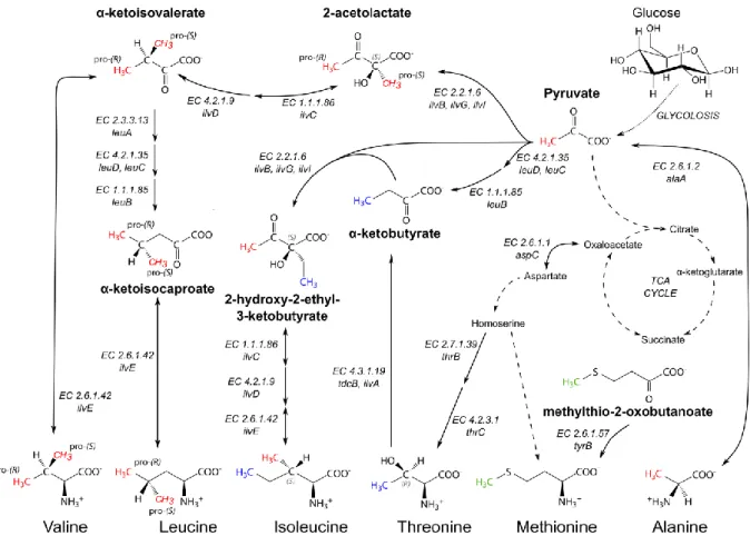
223. This often prevents the assignment of methyl group resonances based on the backbone assignment. In the divide-and-conquer approach, a large complex or multi-domain protein is dissected into smaller building blocks. In case the fold of the building blocks is preserved in isolation, it is possible to transfer the traditionally obtained assignments from the small part onto the larger assembly (Figure 1.6).
Figure 1.5: Methyl resonance assignment strategy based on J-couplings. Chemical shifts of methyl proton and carbon atoms are correlated with assigned backbone or side chain chemical shifts. Blue arrows indicate magnetization transfer pathways from the methyl groups to the backbone amide protons for detection. Red arrows indicate the flow of magnetization in “out- and-back” experiments, where magneti- zation is transferred from the methyl protons via the methyl carbon atoms to side chain and backbone carbons with known chemical shifts and back to the methyl protons for detection.
- 24 -
This approach has turned out to be useful for symmetric multi-subunit assemblies, in case the individual subunits can be prepared in a monomeric form
184. Changes in the chemical shifts between the monomer and the fully assembled complex are often limited, especially in the core of the protein building block. Hence, a straightforward transfer of the assignments from the subunit to the complex is possible. In addition, the divide-and-conquer approach has been successfully applied to complexes that contain more than one unique subunit
224and to large multi-domain proteins
200,214,225,226.
After the transfer of the resonance assignments from the building block to the large assembly, it is required to validate that these are indeed transferred correctly. To that end, additional information is required that can, for example, be derived from the comparison of the chemical shifts of additional side chain carbon atoms through “out-and-back” J-based experiments (see above), or from NOE based experiments (see below).
In this thesis, the divide-and-conquer approach has been applied to assign the Edc3 YjeF methyl resonances in the Edc3 ΔLSm construct, that comprises the IDR and the YjeF-domain (see CHAPTER 3), and to assign the ILVMA-methyl resonances of Dhh1 based on methyl resonance assignments of the isolated RecA1 and RecA2 domains (see CHAPTER 4).
Figure 1.6: The divide-and-conquer approach is used to assign multi-domain proteins or multi-subunit complexes.
(A) Schematic 1H-13C correlation spectrum of a hypothetical protein (black) and of one of its domains in isolation (red).
Assignments from the isolated domain are easily transferred to the full-length protein due to limited chemical shift perturbations. Resonances corresponding to residues that experience a similar chemical environment in the isolated domain and in the full-length protein do not show chemical shift perturbations (see for example the resonance of Ile12 in A; Ile12 is remote from the domain interface in the full-length protein (B)). However, residues that experience a different chemical environment in the isolated domain compared to the full-length protein give rise to resonances that exhibit (small) chemical shift perturbations (see for example the Ile234 resonance in A; Ile234 is in the interface of the two domains in the full-length protein (B)). (B) Schematic representation of an isolated domain (red), whose assignments are transferred to the hypothetical full-length protein (black).
- 25 - Methyl assignment via site-directed mutagenesis
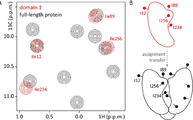
For very large or challenging protein complexes the assignment strategies mentioned above might not be applicable. In addition, in many cases, a full methyl group assignment might not be required to address the question at hand. In those cases, a limited or full methyl group assignment can be obtained through a mutagenesis approach. In this approach, a methyl-bearing residue of interest is mutated into a (closely related) other amino acid, without distorting the fold of the protein. Methyl TROSY spectra are subsequently recorded for the wild-type and for the mutant protein
227. In the ideal case, both spectra are identical apart from one (alanine, methionine, threonine, isoleucine) or two (valine, leucine) resonances that are absent in the spectrum of the mutated protein. These resonance(s) then correspond to the methyl group(s) of the mutated residue (Figure 1.7).
In several cases, the mutagenesis approach has proven to be successful for obtaining methyl group assignments of large complexes
214,215,228,229. In addition, the mutagenesis approach complements other assignment strategies, for example those that are based on the divide-and-conquer approach or the NOE-based approach for residues with other methyl groups in spatial proximity (see below). In practice, this assignment procedure can be complicated in case the introduced mutation results in severe chemical shift perturbations (CSPs) of other resonances so that the peak reporting on the mutation can no longer be unambiguously identified
184. In that case, it might be necessary to include a large number of mutations to be able to distinguish between primary and secondary CSPs
228.
Figure 1.7: Methyl group assignment based on a mutagenesis approach. (A) Schematic 1H-13C correlation spectra of the hypothetical protein (black) and of a mutant (green), where one Ile residue (Ile345) has been mutated into another amino acid X. The Ile345 resonance is thus missing in the spectrum of the mutant protein, while the resonances of all other Ile methyl groups superpose well in the wild-type protein (black) and in the mutant (green). An exemption is the Ile234 resonance, which experiences a small chemical shift perturbation. This is due to the proximity of Ile234 to the mutation site at position 345 that results in a different chemical environment for Ile234 in the wild-type compared to the mutant protein. (B) Schematic representation of the hypothetical protein (black), where Ile345 is mutated to another amino acid X (green).
- 26 -
Here, I utilized the mutagenesis-driven assignment approach to assign several methyl groups in the Edc3 YjeF domain as a prerequisite to apply the methionine scanning methodology (see 1.4.6 and CHAPTER 3).
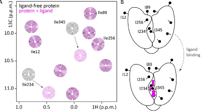
Assignments based on NOEs
Even for very large complexes, it is possible to obtain methyl-methyl NOE contacts with high sensitivity
184. Methyl-methyl NOE spectra reveal methyl resonances that are derived from methyl groups close in space. This data can be used to validate assignments obtained by the divide-and- conquer approach through comparison of NOE patterns (Figure 1.8). In case high resolution structural information of the complex is available, it is possible to directly compare experimental NOE cross-peak patterns with expected, back-calculated NOE patterns. Based on that, assignments of residues that are close in space to already assigned residues can be accomplished.
Experimentally, inter-methyl NOEs are readily obtained with the use of 3D HMQC-NOESY
230or 4D HMQC-NOESY-HMQC
231,232experiments. Due to the lower dispersion of the proton chemical shifts in methyl groups, H-C-H correlations are usually less informative than C-C-H correlations.
Figure 1.8: Methyl group assignment based on NOEs. (A) Schematic 2D methyl TROSY spectrum of a hypothetical protein labeled at the Ile-δ1 methyl groups (left) and three exemplary NOESY “strips” from a C-C-H experiment (right). Dashed lines indicate matching resonances in the 2D spectrum and the NOESY strip of Ile234. The NOESY strips show one intense peak for the respective Ile-δ1 methyl group and less intense cross-peaks for each Ile-δ1 methyl group that is close in space. Notably, the cross-peak intensity decreases with the distance between the methyl groups (r-6 dependence). For example, Ile234 (red, left strip) is in close proximity of Ile345 (green; see B and C) and the Ile345 cross-peak is thus quite intense. Compared to Ile345, Ile256 (blue) is more distant to Ile234 and its cross-peak is weaker. Above an inter-methyl distance of approximately 7 Å, NOE cross-peaks are no longer detectable using routine NOESY-experiments (see also panel B). For example, Ile345 shows an NOE to Ile234, but not to Ile256 (see A, right NOESY strip). In practice, the NOE transfer between two methyl groups can be more efficient in one than in the other direction, giving rise to cross-peaks of different intensity. For example, the cross- peak of Ile234 to Ile256 is more intense than the cross-peak of Ile256 to Ile234 (compare left and middle NOESY strip in A).
(B) Schematic drawing of the three Ile side chains discussed above. Black dashed lines indicate observable NOEs, while a gray dashed line indicates an inter-methyl distance that is too large to detect NOEs. (C) Schematic representation of the hypothetical protein, where the assignment for Ile345, that has been obtained by a mutagenesis approach (see above), can be confirmed by an NOE to the proximal Ile234. The dashed ellipse indicates the area that is enlarged in B.