Research Collection
Doctoral Thesis
Phase Separation in Model Bilayer Membranes
Author(s):
Scheidegger, Laura Publication Date:
2021
Permanent Link:
https://doi.org/10.3929/ethz-b-000479137
Rights / License:
In Copyright - Non-Commercial Use Permitted
This page was generated automatically upon download from the ETH Zurich Research Collection. For more information please consult the Terms of use.
ETH Library
Phase Separation in Model Bilayer Membranes
Laura Scheidegger DISS. ETH NO. 27498
DISS. ETH NO. 27498
Phase Separation in Model Bilayer Membranes
A thesis submitted to attain the degree of DOCTOR OF SCIENCES of ETH ZURICH
(Dr. sc. ETH ZURICH)
presented by LAURA SCHEIDEGGER
Master of Science ETH in Materials Science, ETH Zurich
born on 22.09.1991 citizen of Lucerne LU
Accepted on the recommendation of Prof. Dr. Jan Vermant, examiner Prof. Dr. Peter Fischer, co-examiner
Prof. Dr. Peter Walde, co-examiner Prof. Dr. Peter Beltramo, co-examiner
2021
ii
iii
Acknowledgements
First and foremost, I would like to express my gratitude to Jan for giving me the opportunity to work in his group and for his supervision. His ability to never lose faith is greatly acknowledged.
I would like to thank Peter Beltramo for the great mentoring at the beginning of this project and for giving me valuable insights and sharing his experience.
This thesis would not have been possible without the help of numerous people. The Swiss National Science Foundation is acknowledged for funding this project. I want to thank Peter Walde and Peter Fischer for interesting discussions. Chris and Kirill, for their help on experimental designs. Vappu, for her invaluable help with all administrative issues.
Stéphane, for his insights, and the interesting visit at PSI. Sandro, my first and only Master student, for his skill and patience. Hendrik, for his codes and help in understanding them.
Rao and Claire, for their help in producing GUVs. Special thanks go to Dorothea Pinotsi and Justine Kusch of ScopeM, for helping me getting familiar with the CARS microscope. I am very grateful for the indispensable help of Prof. Elizabeth Mann and Huda Alwusaydi.
Further I would like to thank Laura Stricker for her invaluable support with image analysis towards the end of this thesis.
I am grateful to all Softiez, past and present, for the great and tolerant working environment.
I specially would like to thank my lab mates, Damian and the Büro babes, and Pierre in the last stage of the project, for the pleasant atmosphere.
Lastly, I want to thank my family, without whose continuous and unconditional support I would not be here.
iv
v
Abstract
The plasma membrane separates the interior and exterior of the cells of all living organisms and is a highly complex structure. It is composed of hundreds of different lipid species, and contains additional constituents like proteins. It is therefore very complicated to investigate its properties. Several model membrane systems exist, all of which have their own particular shortcomings. In this thesis, we have employed a newly developed technique in order to study the heterogeneous nature of phospholipid membranes.
In the field of cell membrane research, it is believed that the lipids are not homogeneously distributed across the membrane, but that they form small, nanometer-sized structures, which are also called lipid domains or lipid rafts. These rafts are enriched with saturated lipids and cholesterol, and have a higher packing density than the surrounding membrane. They might play an essential role in certain cell membrane processes, such as signal transduction. For this reason, many resources have been dedicated to investigating how these domains are formed and how they behave. This has mainly been done by studying lipid phase transitions.
Lipids can assemble into different phases. Their natural state in the cell membrane is the so- called liquid-disordered phase, where the packing density is low and the mobility high. When the temperature is reduced to below the main transition temperature of a lipid, it will form the solid-ordered phase. In this phase, the lipids are densely packed. A special case is reached when cholesterol is added to the system. Below the phase transition temperature, a liquid- ordered phase is formed, which has a high packing density, but at the same time a high mobility. Since this phase is only formed when cholesterol is present, it is assumed that this is the state that lipid rafts are in.
The focus of this thesis lies on studying specific properties of lipid domains, and how these properties are influenced by additives (like organic solvents) and other cell membrane components (like cholesterol).
vi
In the first part, we have looked into the effects of three different organic solvents and of line-active molecules (linactants) on phase transition temperature and domain size. Organic solvents are present in several model membrane systems, and are also relevant for real cell membranes, which are in contact with oily lipid droplets. The influence of oil molecules on lipid membrane behaviour is generally underestimated. This is why they have not been investigated thoroughly to the present day. In our experiments, we have observed that the lipid domain size can be adjusted depending on the organic solvent that is chosen. Moreover, the phase transition temperature is increased or decreased (compared to the oil-free case) depending on the length of the alkane chains. Linactants additionally decrease domain size, thereby making it possible to adjust the domain size over a wide range.
The second part focused on the effects of cholesterol on lipid phase separation. Cholesterol is an essential cell membrane component. It has a pronounced effect on membrane permeability and fluidity, and it is believed that it is responsible for the formation of the liquid-ordered phase and therefore of paramount importance in lipid raft formation. First, we have found that cholesterol is not incorporated in sufficient amounts into bilayers containing n-hexadecane or squalene. This is important to know when working with oil-containing model membrane systems. Cholesterol can be incorporated into bilayers when using n- decane as the organic solvent, or when employing carrier molecules, such as cyclodextrins.
Secondly, and very surprisingly, our experiments have shown that the liquid-ordered phase is formed even at very low cholesterol contents in the bilayer, suggesting that n-decane promotes the formation of this special phase. Lastly, and in contrast to literature values, increasing the amount of cholesterol in the bilayers led to higher phase transition temperatures. Cholesterol increases the packing of the lipids, thereby leaving less space for the n-decane, which usually decreases phase transition temperatures, and pushing it out.
The findings of this thesis provide novel insights into fundamental cell membrane properties.
We believe that the results presented can be used to further improve current methods to produce model lipid membranes, and to correctly interpret the observations made in artificial cell membranes.
vii
Zusammenfassung
Die Plasmamembran trennt das Innere und das Äussere der Zellen aller lebenden Organismen und besitzt eine sehr komplexe Struktur. Sie besteht aus hunderten von verschiedenen Lipiden, und enthält zusätzliche Komponenten, wie zum Beispiel Proteine. Darum ist es sehr kompliziert, ihre Eigenschaften zu untersuchen. Mehrere Modellsysteme für die Membran existieren. Alle haben ihre eigenen Nachteile. In dieser Arbeit haben wir eine neu entwickelte Technik angewendet, um die heterogene Natur von Phospholipidmembranen zu untersuchen.
In der Zellmembranforschung geht man davon aus, dass die Lipide nicht homogen über die Membran verteilt sind, sondern dass sie kleine, nanometergrosse Strukturen bilden, die auch Lipiddomänen oder Lipidflösse genannt werden. Diese Flösse sind mit saturierten Lipiden und Cholesterol angereichert, und haben eine höhere Packungsdichte als die umgebende Membran. Sie könnten eine wesentliche Rolle in bestimmten Prozessen der Zellmembran spielen, wie zum Beispiel in der Signalübertragung. Aus diesem Grund wurden viele Ressourcen darauf verwendet zu untersuchen, wie diese Domänen gebildet werden und wie sie sich verhalten. Dazu wurden hauptsächlich Phasenübergänge der Lipide untersucht.
Lipide können sich in unterschiedliche Phasen anordnen. Ihr natürlicher Zustand in der Zellmembran ist die sogenannte flüssig-ungeordnete Phase, in der die Packungsdichte tief und die Mobilität hoch ist. Wenn die Temperatur unter die Hauptübergangstemperatur eines Lipids gesenkt wird, bildet es die fest-geordnete Phase. In dieser Phase sind die Lipide dicht gepackt. Ein spezieller Fall wird erreicht, wenn Cholesterol zum System hinzugegeben wird.
Unterhalb der Phasenübergangstemperatur wird eine flüssig-geordnete Phase gebildet, die eine hohe Packungsdichte, aber gleichzeitig eine hohe Mobilität besitzt. Da diese Phase nur gebildet wird, wenn Cholesterol vorhanden ist, wird angenommen, dass Lipidflösse sich in diesem Zustand befinden.
Der Fokus dieser Arbeit liegt in der Untersuchung von spezifischen Eigenschaften von Lipiddomänen, und wie diese Eigenschaften von Zusatzstoffen (wie organischen
viii
Lösungsmitteln) und anderen Komponenten der Zellmembran (wie Cholesterol) beeinflusst werden.
Im ersten Teil wurden die Effekte von drei verschiedenen organischen Lösungsmitteln und von linienaktiven Molekülen auf die Phasenübergangstemperatur und Domänengrösse untersucht. Mehrere Modellsysteme für Membrane enthalten organische Lösungsmittel.
Zudem sind sie auch in realen Zellmembranen, die mit öligen Lipidtröpfchen in Kontakt kommen, relevant. Der Einfluss von Ölmolekülen auf das Verhalten von Lipidmembranen wird im Allgemeinen unterschätzt. Aus diesem Grund wurden sie bis heute nicht gründlich untersucht. In unseren Experimenten haben wir beobachtet, dass die Domänengrösse vom gewählten organischen Lösungsmittel abhängt. Zudem wird die Phasenübergangstemperatur abhängig von der Länge der Alkankette erhöht oder gesenkt (im Vergleich mit einer Membran ohne Öl). Linienaktive Moleküle senken die Domänengrösse zusätzlich. Dadurch kann die Domänengrösse über ein breites Spektrum eingestellt werden.
Im zweiten Teil liegt der Fokus auf den Effekten von Cholesterol auf die Phasentrennung von Lipiden. Cholesterol ist ein essentieller Bestandteil der Zellmembran. Es hat einen ausgeprägten Effekt auf die Durchlässigkeit und Flüssigkeit der Membran. Es wird angenommen, dass Cholesterol für die Bildung der flüssig-geordneten Phase verantwortlich und deshalb für die Bildung von Lipidflössen von grösster Bedeutung ist. Als Erstes haben wir herausgefunden, dass Cholesterol nicht in ausreichender Menge in Doppelschichten eingefügt wird, die Hexadekan oder Squalen enthalten. Das ist wichtig zu wissen, wenn mit Modellmembranen gearbeitet wird, die Öl enthalten. Cholesterol kann in Doppelschichten eingefügt werden, wenn Dekan als organisches Lösungsmittel verwendet wird, oder wenn Trägermoleküle, wie zum Beispiel Cyclodextrine, verwendet werden. Als Zweites haben unsere Experimente überraschenderweise gezeigt, dass die flüssig-geordnete Phase auch bei sehr tiefen Cholesterolgehalten in der Doppelschicht gebildet wird. Das legt den Schluss nahe, dass Dekan die Bildung dieser speziellen Phase fördert. Schliesslich führte eine Erhöhung der Cholesterolmenge zu höheren Phasenübergangstemperaturen. Cholesterol erhöht die Packungsdichte der Lipide, und verringert dadurch den Platz für Dekan, das normalerweise die Phasenübergangstemperatur senkt.
Die Erkenntnisse dieser Arbeit liefern neue Einblicke in fundamentale Eigenschaften der Zellmembran. Wir glauben, dass die präsentierten Resultate für weitere Verbesserungen von gegenwärtig verwendeten Methoden zur Herstellung von Modellmembranen und für die korrekte Interpretation von Beobachtungen in künstlichen Zellmembranen gebraucht werden können.
ix
x
Contents
1. Introduction ... 1
1.1. The cell membrane ... 1
1.1.1. Membrane heterogeneity ... 4
1.1.2. Membrane viscosity ... 9
1.2. Techniques for studying lipid membranes ... 11
1.2.1. Black lipid membranes (BLMs) ... 11
1.2.2. Giant unilamellar vesicles (GUVs)... 12
1.3. Outline ... 14
2. Large area model biomembranes ... 15
2.1. Introduction ... 15
2.2. Design of the thin-film balance setup ... 15
2.3. Experimental procedure ... 17
2.3.1. Materials ... 17
2.3.2. Bikewheel and lipid preparation ... 17
2.3.3. LAMB formation ... 18
2.3.4. Influence of different organic solvents ... 18
2.3.5. Exchange of the surrounding phase ... 19
2.3.6. Asymmetric bilayers ... 21
xi
3. Domain size regulation in phospholipid model membranes using linactant and oil
molecules ... 23
3.1. Introduction ... 24
3.2. Experimental ... 26
3.2.1. Materials ... 26
3.2.2. Sample preparation ... 26
3.2.3. Experimental setup ... 27
3.2.4. Image analysis ... 27
3.2.5. Membrane viscosity ... 29
3.3. Results and discussion ... 30
3.3.1. Effects of oil solvents ... 30
3.3.2. Effects of linactants ... 35
3.4. Conclusion ... 38
4. Effect of cholesterol on phase transition temperature and domain line tension .. 40
4.1. Introduction ... 40
4.1.1. The role of cholesterol in cell membranes... 42
4.1.2. The role of cholesterol in lipid raft formation ... 44
4.1.3. The arrangement of cholesterol in cell membranes ... 46
4.1.4. Methods to measure domain line tension ... 48
4.2. Experimental ... 50
4.2.1. Sample preparation and experimental setup ... 50
4.2.2. Image analysis ... 50
4.3. Results and discussion ... 51
4.3.1. Phase separation ... 51
4.3.2. Membrane viscosity and domain line tension ... 54
xii
4.3.3. Oil effects ... 56
4.4. Conclusion ... 56
5. Cholesterol incorporation using carrier molecules ... 58
5.1. Introduction ... 59
5.1.1. Cyclodextrins (CDs) ... 59
5.1.2. Methyl-β-cyclodextrin (mβCD)... 59
5.1.3. Kinetics of the 2D domain growth process ... 61
5.2. Experimental ... 62
5.2.1. Image analysis ... 62
5.3. Results and discussion ... 63
5.3.1. Effect of different organic solvents on cholesterol incorporation ... 63
5.3.2. Effect of mβCDs ... 64
5.3.3. Differences between direct and indirect cholesterol incorporation ... 65
5.3.4. Membrane viscosity ... 67
5.3.5. Kinetics of the growth process of liquid domains after domain melting... 68
5.4. Conclusion ... 69
6. Conclusions and Outlook ... 71
Bibliography ... 75
List of Figures ... 95
Curriculum Vitae ... 102
Scientific contributions ... 104
xiii
1
Chapter 1
1 . m m
Introduction
1.1. The cell membrane
The cells of all living organisms are enclosed by a membrane, the so-called cell or plasma membrane. Certain cell organelles, such as the Golgi apparatus, are also surrounded by membranes. In animal cells, the plasma membrane represents the only boundary between the cell interior (the cytoplasm) and the exterior environment (Figure 1.1). The cells of plants, fungi and most bacteria are additionally surrounded and protected by a cell wall, which mainly provides structural stabilization. In both cell types, the cell membrane plays a crucial role in many biological processes. It regulates transport into and out of the cell, is involved in cell-cell communication, cytokinesis and cell motility, and is therefore a vital part of the cell structure.
Despite its importance, two centuries lie between the discovery of cells in 1665 and the recognition that the cell membrane exists [1]. However, it was still considered an insignificant structure until the turn of the 20th century, when it slowly started to attract more interest. In 1925, Gorter and Grendel first proposed that cell membranes consist of a layer of lipid molecules [2]. This study was followed by the first measurements of membrane thickness [3, 4]. Robertson [4] determined it to be about 8 nm, which is in rough agreement with the thickness of a lipid bilayer. In 1972, the fluid mosaic model of cell membrane structure was presented by Singer and Nicolson [5]. This model is still widely accepted today.
According to the fluid mosaic model (Figure 1.2), the cell membrane is essentially a two- dimensional material composed of a lipid bilayer with a hydrophilic and a hydrophobic part.
Because of the predominantly fluidic nature of the membrane, the lipids and other membrane components such as proteins can freely diffuse within the structure. Proteins make up almost 50% of the membrane mass [6]. Depending on their chemical structure, they can interact with
1INTRODUCTION
2
both the hydrophilic (outer) and hydrophobic (inner) region of the membrane. The rest of the membrane mass comes from the lipids (up to 50%) and a small amount of carbohydrates (e.g.
glycolipids).
Figure 1.1: Representation of a cell. The curved black line represents the cell membrane, composed of a lipid bilayer and separating the cytoplasm from the exterior environment.
Image taken from Ref. [7].
Figure 1.2: Representation of the fluid mosaic model. Image taken from Ref. [1].
The lipid bilayer is composed of a wide variety of lipid molecules [8]. They typically consist of a hydrophilic head group and a hydrophobic tail (fatty acid chain) (Figure 1.3a), and
1.1 THE CELL MEMBRANE
3
thereby spontaneously form a bilayer in aqueous environments. The three main groups are phospholipids, sphingolipids and sterols (Figures 1.3 and 1.4). The exact lipid composition of the membrane depends on the cell type, but in most membranes, phospholipids are the major species [9]. A plethora of phospholipids exists, which differ from each other in the type of head group and fatty acid chain. The most common phospholipids are phosphatidylcholines [9, 10], where choline is the head group (Figure 1.3b). The fatty acid chains can differ from each other in length (i.e., the number of carbon atoms) and the degree of saturation (i.e., the number of double bonds).
Figure 1.3: (a): Representation of a lipid, showing the hydrophilic head group (1) and two hydrophobic fatty acid chains (2). (b): Phosphatidylcholine, showing choline as the head group (1a) and an unsaturated (2a) and saturated (2b) fatty acid chain.
Figure 1.4: (a): Sphingomyelin, which is one type of sphingolipid, with choline as the head group. (b): Cholesterol.
1 2
1a 2a
2b a
b
a
b
1INTRODUCTION
4
Due to this variety, a cell membrane can easily contain hundreds of different lipid species [11]. Additionally, since the lipids can freely move within the membrane, they constantly reorganise and rearrange themselves, thereby making it a very dynamic and complex structure to model and investigate.
Despite this difficulty, and due to the importance of the cell membrane in various biological processes, many researchers have dedicated themselves to investigating the organization, properties and functions of cell membranes. However, many questions still remain unresolved, many of which relate to materials science questions. For example, the mechanical properties of the membrane, such as its bending rigidity [12], and the interactions between membranes and membrane proteins are ongoing topics of research [13-15]. Transport phenomena in and across the membrane, such as the exact mechanism of drug transport, are also an open question and are controversially discussed [16, 17]. Furthermore, the lipid phase behaviour and lipid organization within the membrane have received huge attention in the last two decades, especially since 1997, when lipid “rafts” were first mentioned in the literature [18].
1.1.1. Membrane heterogeneity
The heterogeneous nature of the lipid bilayer was already proposed almost 40 years ago [19].
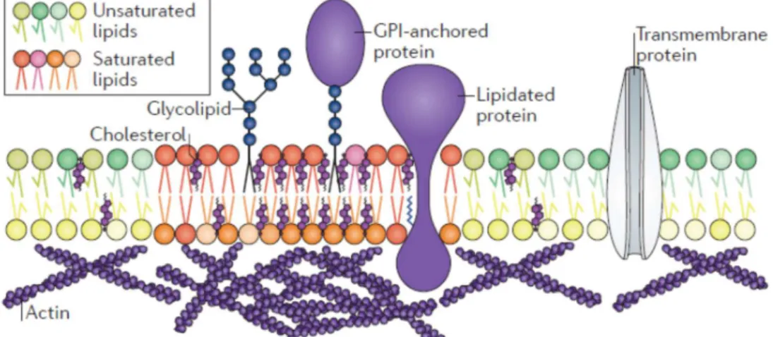
Several authors later suggested that lipid domains are responsible for the sorting of certain membrane proteins [20, 21]. Simons and Ikonen then recognized the potentially important role of cholesterol for the formation of lipid rafts and the functional part that domains might play within the membrane [18]. It is believed that lipid domains, which are enriched with saturated lipids and therefore have a different composition than the surrounding membrane (Figure 1.5), play a key role in the binding of specific proteins to the cell membrane, and therefore play a major role in fundamental processes of the cell membrane, like signal transduction [18, 22, 23]. A widely accepted definition of “rafts” was coined in 2006 [24]:
“Membrane rafts are small (10–200 nm), heterogeneous, highly dynamic, sterol- and sphingolipid-enriched domains that compartmentalize cellular processes. Small rafts can sometimes be stabilized to form larger platforms through protein-protein and protein-lipid interactions.”
Because of their supposed prominent role in cell membrane functionality, the formation and properties of lipid domains have been increasingly studied within the past years. This has typically been done by investigating the phase behaviour of lipids and lipid mixtures. A
1.1 THE CELL MEMBRANE
5
large number of studies exist where phase diagrams of various lipid mixtures have been probed [25-32] or where properties (nucleation and growth, diffusion coefficients, line tension, shape, interactions between domains) of lipid domains were investigated [33-41].
Figure 1.5: Lipid domains are enriched in saturated lipids and cholesterol. They can bind to specific proteins. Image taken from Ref. [42].
Lipids can form different structures, and depending on various external factors, such as temperature, they transition from one structure into another. A very important structure is the so-called liquid-disordered (Ld) phase, which represents the state that most lipids assume in the cell membrane. In accordance with the fluid mosaic model [5], the Ld phase has a low order and packing density, allowing the lipids and other membrane constituents to easily move around. When a bilayer is cooled below its main transition temperature, the so-called solid-ordered (So) or Lβ phase is formed (also called the gel phase). In contrast to the Ld
phase, this is a highly ordered phase.
The main transition or melting temperature Tm of lipids depends mainly on the length and number of double bonds of the fatty acid chains. The shorter they are and the more double bonds they have, the lower is the melting temperature. This is the reason why a saturated lipid like DPPC (1,2-dipalmitoyl-sn-glycero-3-phosphocholine, see Figure 1.6) has a much higher melting temperature (41 °C) than an unsaturated lipid like DOPC (1,2-dioleoyl-sn- glycero-3-phosphocholine, see Figure 1.6) (–18 °C) [43].
When the binary system DOPC/DPPC is cooled below its phase transition temperature, DPPC forms domains consisting of the So phase, while DOPC remains in the Ld phase [28,
1INTRODUCTION
6
44-47]. In order to decrease interactions between the head groups, the DPPC chains are tilted with respect to the bilayer plane [48]. If cholesterol is added to the system, the phase behaviour changes dramatically. Upon cooling, the system phase separates into two liquid phases, the liquid-disordered (Ld) and liquid-ordered (Lo) phase [31]. Due to its high concentration of saturated lipids and cholesterol, the latter phase is believed to be an excellent model for lipid rafts [42, 49], which is why this particular model system is one of the most investigated phase separating mixtures and has been extensively studied by Veatch et. al. [26, 27, 32] and others.
Figure 1.6: Chemical structures of the two phospholipids DPPC (saturated) and DOPC (unsaturated).
Considerable differences exist between these three phases (Figure 1.7). The So phase has a very high translational and conformational order [50], i.e., the lipids cannot diffuse easily within the bilayer, and the fatty acid chains are extended. This leads to a high packing density and an increased bilayer thickness. In the Ld phase, on the other hand, the order is drastically reduced. The lipid molecules have more translational as well as conformational freedom. The Lo phase is a special case, where the addition of cholesterol increases the conformational order of the fatty acid chains (compared to the Ld phase) [25], while at the same time the high lateral mobility of the lipids in the Ld phase is retained [50, 51].
While the biological relevance of the Lo phase is clear, the So phase has received less attention. However, based on the importance of specific lipid species (ceramides) in certain signaling processes such as apoptosis, several studies have recently looked at the formation of So domains in lipid membranes [52, 53]. Their results suggest that cells might be able to
DPPC Saturated Tm = 41 °C
DOPC Unsaturated Tm = –18 °C
1.1 THE CELL MEMBRANE
7
adjust the packing density of domains according to their needs, which could partly explain why membranes contain such a high number of different lipids.
Figure 1.7: Representations of the three phases mentioned in the main text. The liquid- ordered phase contains cholesterol molecules.
Despite extensive research, it has to be noted that there is still no definitive proof that lipid rafts exist [54, 55]. While macroscopic domains can be observed in model membranes, the postulated sizes of domains in live cell membranes are much smaller (10–200 nm), and therefore they cannot be resolved by light microscopy. Recently, cryogenic electron microscopy has been employed to image nanoscopic domains in model membranes and in membranes derived from cells [56, 57] by exploiting the thickness mismatch between the Ld
and Lo phases. This was an important step towards proving that lipid rafts exist. However, although cell derived membranes have compositions which are comparable to the ones found in real cell membranes, they still lack many important characteristics, such as the interaction with the cytoskeleton.
Since cell membranes have such a complex structure, computer simulations, such as molecular dynamics (MD) studies, are a powerful tool to investigate lipid interactions and domain formation [58, 59]. Zhuang et. al. [60] have extensively studied the interactions between various lipid types, showing that results obtained from simulations and experiments agree reasonably well. The formation of lipid domains has been simulated in binary [61] as well as in ternary mixtures [62, 63] (Figure 1.8), confirming the increased order in the domain phases. Rosetti et. al. [63] showed that the mismatch between the saturated and the unsaturated lipid chains is an important factor for domain formation. Shahane et. al. [64]
looked at lateral pressure profiles for different bilayer systems. The pressure within the bilayer is of particular importance, since it could play an important role in protein function
Liquid-ordered (Lo) Liquid-disordered (Ld)
Solid-ordered (So)
1INTRODUCTION
8
[65]. The lateral pressure profile for a pure POPC (1-palmitoyl-sn-2-oleoyl-glycero-3- phosphocholine) bilayer is shown in Figure 1.9. While positive pressures indicate repulsive forces, negative values indicate attractive ones. The most negative values are found between the hydrophilic heads and hydrophobic tails of the lipids. Here, the bilayer tries to reduce the contact points between the hydrophobic and hydrophilic regions. In the middle of the bilayer, repulsive forces act between the fatty acid chains to maximize entropy. Patra [66] has investigated the influence of cholesterol on the lateral pressure in DPPC bilayers. They showed that cholesterol increases the pressure within the bilayers, a result which is related to the increased rigidity when cholesterol is added.
Figure 1.8: MD simulation of the formation of Lo domains in a ternary lipid mixture. The saturated lipids are depicted in green, the unsaturated lipids in red and cholesterol in gray.
Scale bar is 5 nm. Image taken from Ref. [62].
Figure 1.9: Lateral pressure profile for a pure POPC bilayer. Image taken from Ref. [64].
0 μs 3 μs 10 μs 20 μs
1.1 THE CELL MEMBRANE
9
The complexity of the simulated systems is gradually increasing [67]. It will be possible to investigate interactions of cell membranes with the cytoskeleton in future computer simulations [68].
1.1.2. Membrane viscosity
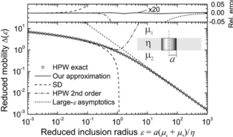
Lipid domains were also used to probe membrane properties, such as membrane viscosity [33, 69]. The viscosity of a membrane has an effect on many processes (most of all, transport) and its quantification is highly sought after [70-72]. Many calculations rely on the Saffman- Delbrück (SD) approximation [73], which relates the diffusion coefficient 𝐷 to the two- dimensional membrane viscosity 𝜂 and the coupling of the bulk fluid. 𝐷 can be measured by tracing membrane inclusions, which can be particles or, as mentioned before, lipid domains. Unfortunately, the SD approximation was originally developed for proteins, and is only valid in a limited range, namely for 𝑟 < 𝑙. Here, 𝑟 is the inclusion size and 𝑙 = 𝜂 /(2𝜇) is a length scale which characterizes the respective diffusion of momentum at the interface and into the bulk, as 𝜇 is the bulk viscosity of the surrounding medium. So, the SD approximation is valid for small inclusions or a large membrane viscosity. This problem was studied by Hughes et. al. [74], who developed a model (HPW model) that is valid for all inclusion sizes. In 2008, Petrov and Schwille [75] presented a simple expression based on Saffman’s and Hughes’ earlier works. All of these models assume that the interface is incompressible. Figure 1.10 illustrates the applicability ranges for all models.
Figure 1.10: Applicability ranges of the models relating diffusion coefficients and membrane viscosity mentioned in the main text (“Our approximation” is Petrov’s and Schwille’s expression). Image taken from Ref. [75].
1INTRODUCTION
10
In Figure 1.10, 𝜀 = ∗ ∗ stands for the reduced inclusion radius, 𝑎 denotes the inclusion radius, 𝜇 and 𝜇 are the bulk viscosities of the surrounding fluids and 𝜂 is the two- dimensional membrane viscosity. The HPW model calculates the exact values of the reduced mobility ∆(𝜀) (y-axis), shown with circles in Figure 1.10. Additionally, four different approximations for ∆(𝜀) are shown:
Saffman-Delbrück (SD; only valid for 𝜀 < 0.1):
∆ (𝜀) = 𝑙𝑛 2 𝜀 − 𝛾 𝛾 = 0.5772 stands for the Euler constant.
HPW 2nd order (only valid for 𝜀 < 0.6):
∆ (𝜀) = 𝑙𝑛 2
𝜀 − 𝛾 +4𝜀 𝜋 −𝜀
2 𝑙𝑛 2 𝜀
Large-𝜀 asymptotics (only valid for 𝜀 > 30):
∆ (𝜀) = 𝜋 2𝜀
Petrov and Schwille (“Our approximation”):
∆ (𝜀) = 𝑙𝑛 2
𝜀 − 𝛾 +4𝜀 𝜋 − 𝜀
2 𝑙𝑛 2
𝜀 ∗ 1 − 𝜀
𝜋 𝑙𝑛 2
𝜀 + 𝑐 𝜀 /(1 + 𝑐 𝜀 ) The values for the parameters are: 𝑐 = 0.73761, 𝑏 = 2.74819, 𝑐 = 0.52119 and 𝑏 = 0.51465.
As can be seen in Figure 1.10, Petrov’s and Schwille’s expression is valid for the whole range of 𝜀, providing a simple and exact solution. Depending on the membrane composition, it can be used to measure the viscosity of the Ld as well as of the Lo phase. The viscosity of the Lo
phase is particularly relevant for protein function.
1.2 TECHNIQUES FOR STUDYING LIPID MEMBRANES
11
1.2. Techniques for studying lipid membranes
Because of the intricate organisation of cell membranes, a widely used technique to study their properties is by investigating simplified model membranes. These can consist of different lipids and have varying complexity. Model membranes can be produced by several methods, two of which will be introduced here.
1.2.1. Black lipid membranes (BLMs)
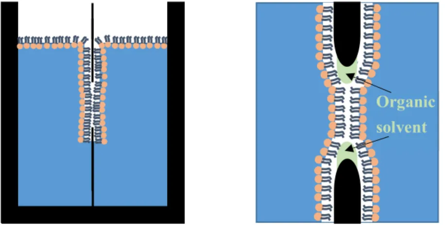
BLMs (Figure 1.11) were first introduced over 50 years ago [76] and since then have been applied in several studies, such as membrane capacitance measurements [77-79]. Two preparation methods for BLMs exist. Both rely on the use of organic solvents, and although the bilayers produced with the second method are usually considered “solvent-free”, it is very likely that small amounts of solvents remain within the bilayer. In the first method, the lipids are dissolved in an organic solvent (e.g. decane), and a small aperture surrounded by an aqueous phase is “painted” with this solution. In the second method, the so-called Montal- Mueller technique [80], a septum with an aperture is placed within and a monolayer of lipids is created on an aqueous phase. The level of the aqueous phase on one side of the aperture is then raised above the aperture, depositing the first monolayer. This is followed by raising the aqueous phase level on the other side of the aperture, resulting in a bilayer.
Figure 1.11: Black lipid membrane, produced by the Montal-Mueller technique. Small amounts of organic solvent remain within the bilayer.
Organic solvent
1INTRODUCTION
12
BLMs are planar and free-standing, which is a huge advantage compared to e.g. supported lipid bilayers (SLBs), where the interactions between the support and the bilayer have to be taken into account. In spite of these advantages, BLMs are often only stable for less than an hour, which limits their application to a great extent.
1.2.2. Giant unilamellar vesicles (GUVs)
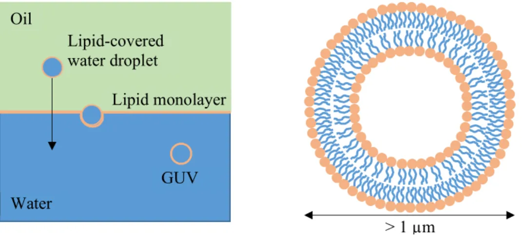
GUVs (Figure 1.12) are nowadays the most widely used technique to produce model membranes. They are circular structures filled with an aqueous phase and bounded by a single (“unilamellar”) bilayer. They usually have a size of 1 – 100 µm [81], which is in the same range as most biological cells [82]. GUVs can be used for multiple applications [81], but have particularly often been used to study mechanical properties of membranes [28, 83] and for phase separation experiments [26-28, 33, 34]. Major findings include membrane shape changes upon the reduction of tension [83] and the observation of different liquid phases in various lipid compositions [26].
GUVs are often produced by electroformation [84], which is a highly reproducible approach.
Another method involves assembling the lipids at an oil-water interface and then letting lipid- covered water droplets pass across this interface [85] (Figure 1.12). This principle is used in the cDICE (continuous droplet interface crossing encapsulation) method [86], which allows for efficient production of GUVs with tuneable size. However, it was recently shown that when using cDICE, cholesterol is not incorporated into the bilayer in sufficient amounts [87], probably because of the presence of residual oil in the vesicles. Recently, Dürre et. al. have overcome this obstacle by adding a second oil layer to the setup [88], thereby increasing the incorporation efficiency of cholesterol to 25 – 35%.
Giant plasma membrane vesicles (GPMVs) are vesicles formed directly from the plasma membrane of cultured cells and are therefore of much higher complexity than GUVs, which usually consist of only a few different lipid species. It has been shown in various studies that GPMVs also undergo phase transitions comparable to the ones observed in GUVs [89-91], not only proving that vesicles with complex compositions can indeed form heterogeneous structures, but also that simple bilayers like GUVs are adequate model systems to study phase separations.
Unfortunately, the composition of GPMVs highly depends on the growth conditions [92], which makes experimental results occasionally difficult to interpret [93]. Furthermore, and
1.2 TECHNIQUES FOR STUDYING LIPID MEMBRANES
13
although giant vesicles have the advantage of being very stable and allowing tension control, their curvature makes imaging more difficult and curvature effects cannot be isolated.
Figure 1.12: Giant unilamellar vesicle, produced by letting lipid-covered water droplets pass across a lipid-covered oil-water interface.
Oil
Water
Lipid monolayer
GUV Lipid-covered water droplet
> 1 µm
1.3 OUTLINE
14
1.3. Outline
The goal of this work was to gain more insight into the organization of model phospholipid membranes. To this end, the phase separation of phospholipid bilayers was investigated by a novel experimental technique. Particular emphasis was put on questions related to materials science, i.e., the phase behaviour of phospholipid bilayers, lipid domain formation and growth and the effects of cholesterol on membrane properties.
Chapter 1 provided a general introduction into the complexity of cell membranes, lipid phase behaviour and model membrane studies, including their shortcomings. Many open questions remain in the field of membrane heterogeneity and lipid phase behaviour. Furthermore, the presence and influence of organic solvents in model lipid membrane studies is an unresolved issue.
In Chapter 2, a novel technique to produce model membranes will be introduced. Its advantages compared to other techniques will be presented.
Chapter 3 focuses on the formation of solid domains in a binary phospholipid system and the effects of various oil solvents on lipid phase behaviour. Additionally, the influence of so- called line-active molecules will be examined.
The complexity of the system is increased in Chapters 4 and 5, where the effects of cholesterol on phase separation are investigated. To achieve this, two different approaches were used. First, cholesterol was dissolved in the oil solvent together with the other lipids. In the second method, a cholesterol-free bilayer was first formed, and cholesterol was then added to the bilayer using a carrier molecule. This strategy revealed fundamental differences into how cholesterol is incorporated into model membranes.
Finally, Chapter 6 recapitulates the results of this thesis and outlines directions for possible future work.
15
Chapter 2
2 . m m
Large area model biomembranes
2.1. Introduction
As mentioned in section 1.2., the current methods to produce model membranes all suffer from individual disadvantages. Naturally, it is desired to have a platform that combines all of the best features of the aforementioned techniques. This has been achieved by large area model biomembranes (LAMBs) (Figure 2.1) [94]. This recently developed technique relies on an adapted thin-film balance setup with a bikewheel film holder (microchip; Figure 2.1b).
The film holder has been adapted from the original design in [95] to allow for a more uniform drainage across the lipid film. The advantages of this platform include fine tension control, the possibility of creating free-standing, planar, and large area (up to 0.78 mm2) membranes, and the ability of investigating transport properties across the membranes. Even the influence of curvature can be studied, despite the natural planarity of the membranes. This is made possible by independent access to both sides of the bilayer. Thereby, the initially planar membranes can be curved, e.g. by introducing an osmotic pressure gradient. In the next sections, the design of the setup and the experimental procedure to produce LAMBs will be described in detail.
2.2. Design of the thin-film balance setup
The bikewheel film holders are fabricated on demand by Micronit Microfluidics (Netherlands) using photolithography. They consist of two glass borosilicate slides. The channels are etched into one of the glass slides by HF. The hole in the centre of the bikewheel is drilled using a diamond drill. Its size can be varied, but in this work it was kept fixed at a
2LARGE AREA MODEL BIOMEMBRANES
16
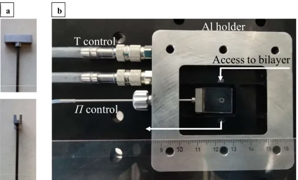
diameter of 1 mm. 24 channels are oriented radially from the hole and connected to larger entrance channels. The film holders are glued to titanium holders (Figure 2.2a).
Figure 2.1: (a): LAMB setup. A differential pressure transducer is coupled with a syringe pump to precisely measure and control the pressure within the thin-film balance cell. The bilayer is formed within a 1 mm hole of the bikewheel film holder shown in (b). Image in (b) is taken from Ref. [94].
Figure 2.2: (a): The bikewheel film holders are glued to titanium holders. (b): The bikewheel film holder is placed in an aluminium pressure chamber with different compartments to ensure access to the bilayer. The chamber is connected to temperature and pressure controls.
Image taken from Ref. [94].
Differential P transducer
Syringe pump with PID control
Lipid bilayer
a b
T control
Π control
Al holder
Access to bilayer
a b
2.3 EXPERIMENTAL PROCEDURE
17
To access both sides of the bilayer independently, the film holder is placed in a pressure chamber with different compartments (Figure 2.2b). The pressure chamber is made out of aluminium and allows for temperature control. Tubing connects the titanium holder to a syringe pump. The pump is connected to a Baratron pressure transducer, which controls the disjoining pressure 𝛱 in the film.
2.3. Experimental procedure
2.3.1. Materials
OTS (octadecyltrichlorosilane), n-hexadecane, n-decane and squalene are bought from Acros Organics (USA). Triolein is bought from Sigma-Aldrich. NaCl (99.99%, metals basis) and NaHCO3 are obtained from Alfa Aesar, and CaCl2 is purchased from Sigma-Aldrich. Deionized water (Milli-Q, Merck-Millipore, resistivity < 18.2 MΩcm) is used to prepare all buffers. Lipids are obtained from Avanti Polar Lipids.
2.3.2. Bikewheel and lipid preparation
First, the surface of the bikewheels has to be hydrophobized, in order to ensure stable lipid film formation. This is achieved by silanization in 1 mM OTS in n-hexadecane for a day, after having cleaned the bikewheels in a base bath (NaOH in ethanol).
The lipids in chloroform are mixed in the desired ratio and dried under nitrogen and vacuum.
They are then dissolved in the desired oil solvent at a concentration of 2.5 – 5 mg/ml.
Concentrations below 2.5 mg/ml result in unstable oil films. The lipid-oil mixture is sonicated for several hours before use to ensure that the lipids are fully dissolved in the organic solvent.
For oil mixtures, better results were observed when the lipids were first dissolved in squalene, and n-hexadecane was added afterwards in the appropriate amount.
After filling the bikewheel film holders with the lipid-oil mixture, they are placed within the pressure chamber and connected to a syringe for pressure control. For temperature dependent experiments, the pressure chamber is pre-heated to the desired temperature. The physiological salt buffer (150 mM NaCl, 2mM CaCl2, 0.2 mM NaHCO3) (pH = 7.4) is filtered with 0.2 µm pore filters and then added into the pressure chamber. Before adding the buffer,
2LARGE AREA MODEL BIOMEMBRANES
18
the pressure is slightly increased, to keep the buffer from entering the channels of the bikewheel.
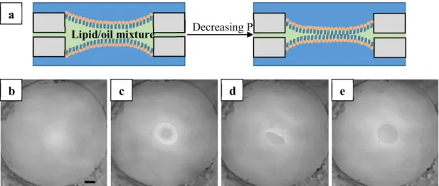
2.3.3. LAMB formation
The steps for LAMB formation are shown in Figure 2.3. First, a thick oil film is formed within the small hole in the centre of the bikewheel (Figure 2.3b). This film is then slowly drained by decreasing the pressure. Interference patterns occur when the oil film is sufficiently thin (Figure 2.3c). Ultimately, a bilayer is formed (Figure 2.3d-e).
By measuring the thickness of the formed bilayers, it was shown that the composition of binary lipid bilayers is in accordance with the composition of the bulk lipid mixtures [96].
2.3.4. Influence of different organic solvents
During this thesis, it was observed that organic solvents (Figure 2.4) have a huge impact on the bilayer properties. A closer examination of the effects of organic solvents on bilayer phase behaviour will be undertaken in Chapter 3. Here, we focus on bilayer stability.
Figure 2.3: (a): Representation of the drainage process of a thick oil film upon decreasing the pressure. (b): Thick oil film. (c): Interference patterns occur when the oil film is sufficiently thin. (d)-(e): Bilayer formation. Scale bar is 100 µm.
a
b c d e
Lipid/oil mixture Decreasing P
2.3 EXPERIMENTAL PROCEDURE
19
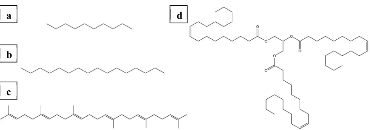
Figure 2.4: Different organic solvents. (a): n-Decane. (b): n-Hexadecane. (c): Squalene. (d):
Triolein.
Different organic solvents require different hydrophobization procedures. It is not a trivial task to optimize the surface functionalization of the bikewheel to the requirements of the solvent. An iteration process is necessary, which can take several days, depending on the duration of the hydrophobization procedure. It was found that for simple alkanes like n- decane or n-hexadecane, hydrophobization in 1 mM OTS is sufficient. Bulkier molecules like squalene require a stronger hydrophobization (10 mM OTS). For solvents of even higher molecular mass (such as triolein), no appropriate surface functionalization could be found.
Bilayers formed with these solvents were not stable.
Additionally, it was observed during the temperature dependent measurements that the long- term bilayer stability upon cooling is increased when using mixtures of n-hexadecane and squalene instead of pure n-hexadecane. This could be due to the higher heat capacity of squalene (𝐶 , = 1321.66 𝐽 ∗ 𝑚𝑜𝑙 𝐾 ) compared to the one of n-hexadecane (𝐶 , = 611.73 𝐽 ∗ 𝑚𝑜𝑙 𝐾 ).
2.3.5. Exchange of the surrounding phase
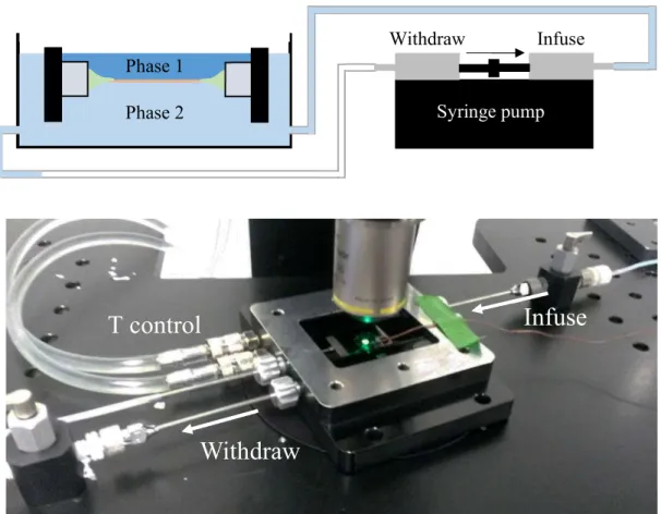
One of the advantages of the LAMB setup is that the environment on both sides of the bilayer can be changed easily and independently after its formation. This is not always possible in other systems. GUVs, for example, allow access only from one side.
The setup to exchange the surrounding phase is shown in Figure 2.5. A pump is used which enables simultaneous infusion of the new and withdrawal of the old phase. This is necessary to ensure a constant volume of the surrounding phase in the pressure chamber, so that a
a
b
c
d
2LARGE AREA MODEL BIOMEMBRANES
20
constant pressure is acting on the bilayer. If only the phase on one side of the bilayer needs to be exchanged, the compartments in the pressure chamber need to be sealed from each other with high-viscosity vacuum grease.
It was shown previously that for the addition of Magainin II, an antimicrobial peptide, a ramp profile (0.01 ml/min to 0.1 ml/min in 10 min) was the most appropriate solution to ensure bilayer stability [97]. During the course of the present thesis, the exchange setup was used to add cholesterol to the bilayer (for further details see Chapter 5). No difference with respect to bilayer stability was observed when the new phase was added at 0.1 ml/min from the beginning. This faster infusion reduced the duration of the measurements, which was an advantage especially for the phase separation experiments, where the duration of light exposure is a critical factor due to the inevitable photobleaching of the fluorophores.
Figure 2.5: Setup to exchange the surrounding phase. A syringe pump is used which enables simultaneous infusion of the new and withdrawal of the old phase. The setup can be used to access only one (as shown in the top image) or both sides of the bilayer.
Phase 1
Syringe pump Phase 2
Infuse Withdraw
Withdraw
Infuse
T control
2.3 EXPERIMENTAL PROCEDURE
21
2.3.6. Asymmetric bilayers
Biological membranes are typically asymmetric, i.e. they have different compositions on the inner and outer leaflet. For example, the outer leaflet of erythrocytes contains more phosphatidylcholines than the inner leaflet [9, 98]. Therefore, and to increase the biological significance of the results, the production of asymmetric model membranes is a very dynamic field of research [99-101].
A promising approach to produce asymmetric LAMBs is to use the phase exchange setup described in the previous section. By sealing the top and bottom layer of the bilayer from each other, the bottom layer can be accessed independently by a new phase. This has been used in preliminary experiments, described below.
First, DPPC was dissolved in salt buffer, following the procedure described in [102]:
1. 0.006 g DPPC were weighed in a clear vial and pre-heated for 15 min at 56 °C.
2. 200 µL Milli-Q water were added (pre-heated at 56 °C).
3. This lipid paste was stirred at 200 rpm, while keeping it at 56 °C, until the lipids were dry to the naked eye.
4. This was repeated three times.
5. 1.6 mL salt buffer was added (pre-heated at 56 °C).
The bikewheel film holder was sealed with vacuum grease prior to bilayer formation to prevent fluid exchange between the top and bottom phase. The experiments were conducted at room temperature. A symmetric bilayer (pure DOPC, containing 1 mol% of a fluorescent lipid) was formed. Then, DPPC (in salt buffer at a concentration of 5 mM) was added by exchanging the bottom phase. After the addition of 1 mL DPPC, black domains form in the bilayer (Figure 2.6). Due to the high packing density in the solid-ordered DPPC phase, the fluorescent lipid is excluded from this phase and the domains appear black.
In another experiment, coalescence of two of these domains was observed (Figure 2.7), which is normally not seen for solid-ordered domains. A possible reason for this behaviour could be that the line tension of the domains, which depends on various factors, is increased if they only appear on one leaflet, thereby increasing the coalescing forces.
2LARGE AREA MODEL BIOMEMBRANES
22
Figure 2.6: DOPC bilayer after the addition of 1 mL DPPC (in salt buffer, 5 mM) into the bottom phase. The image was taken at room temperature. Magnification: 20x.
Figure 2.7: Coalescence of two black domains. Scale bar is 20 µm.
These preliminary experiments show that asymmetric, large area bilayers can be produced by a relatively simple approach.
0 s 5 s 8 s
9 s 10 s 11 s
23
Chapter 3
3 . m m
Domain size regulation in
phospholipid model membranes using linactant and oil molecules
The formation of domains in multicomponent lipid mixtures has been suggested to play a role in moderating certain functions of cells. Understanding how domain size may be regulated by both hybrid lipid molecules and impurities is important, both for understanding real biological processes, and for developing model systems where domain size can be regulated to enable systematic studies of domain formation kinetics and thermodynamics.
Here, we study how line-active hybrid phospholipids and oil molecules which swell the bilayer influence the phase separation in planar, free-standing lipid bilayers consisting of DOPC and DPPC. First, we find that n-hexadecane increases domain size by a factor of 5 compared to the smallest observed domains, while n-decane, a shorter alkane, leads to smaller domains. Secondly, POPC (1-palmitoyl-sn-2-oleoyl-glycero-3-phosphocholine), a line-active hybrid lipid, reduces the domain size when added in small amounts. Lastly, despite the regulation of domain size by both, we find that the phase transition temperature is influenced only by oil molecules, but not by linactants. This suggests that oil molecules have a greater effect on the phase separation in lipid bilayers than linactant molecules, a conclusion that is confirmed by the dependence of the area fraction of the fluid phase on oil composition and linactant content. The incorporation of linactant and oil molecules into this binary membrane model system makes domain size regulation over a wide range of length scales (several tens of microns) possible.
3DOMAIN SIZE REGULATION IN PHOSPHOLIPID MODEL MEMBRANES USING LINACTANT AND OIL MOLECULES
24
3.1. Introduction
Lipid organization within biological membranes has received huge attention in the last two decades, especially since 1997, when lipid “rafts” were first mentioned in the literature [18].
Because of their proposed importance in fundamental cell membrane processes, the formation and properties of lipid domains have been increasingly studied within the past years. Although lipid ‘rafts’ in real cells are nanometer scale and may be transient in nature [55, 103], the raft hypothesis was built upon model membrane studies related to understanding phase separation on the microscale. Similar to the domains in lipid monolayers [104, 105], a phase coexistence between a dense, liquid-ordered (Lo) and a liquid-disordered (Ld) lipid phase is believed to underpin raft assembly in living cells [55, 103]. The understanding of how to control phase behaviour of lipids and lipid mixtures can hence help rationalize the formation of these domains.
Experimentally observed domain sizes range from nano- to micrometers and depend on a complex interplay of various factors, with some analogy to the formation of nano- and microemulsions in bulk. Apart from composition and the distance from the binode and the critical point [34, 106], an important factor is the line tension between the different lipid phases [106-110]. It plays a role akin to interfacial tension in bulk systems, and is governed by the thickness (height) mismatch, hydrophobic interactions and the spontaneous curvature between the domain and the surrounding phase [111]. In particular, line tension increases quadratically with the thickness mismatch [112], and hence strongly depends on bilayer composition. It has been shown that the addition of line-active molecules (linactants), such as hybrid lipids composed of a saturated and an unsaturated tail, reduces both line tension and the average domain size [107, 108, 113-116]. Since lipid domains are enriched with saturated phospholipids, it is hypothesized that linactants concentrate at the interface between the domain and the surrounding membrane, decreasing the line tension and energy penalty for forming such an interface. Most prior experimental studies using linactants focus on liquid-liquid phase separations. The interaction of linactants with solid phases remains unclear. The most studied system for solid- liquid phase transitions is a mixture of a saturated lipid (e.g. DPPC) and an unsaturated lipid (e.g. DOPC). When cooling this system below its phase transition temperature, solid domains enriched with saturated lipid appear. Although solid-liquid phase transitions are commonly observed in model membranes [27, 32, 38, 52, 117], relatively less work has been done on domain size regulation in these systems. Solid domains can play significant roles in various cell processes, such as apoptosis [52], and it is paramount that their formation and growth mechanisms are thoroughly understood.
3.1 INTRODUCTION
25
Apart from linactants, there are other molecules which influence bilayer structure and properties to a great extent. Cell membranes are in contact with a wide variety of molecules, such as oily lipid droplets [118, 119], and since some methods to form a model bilayer necessitate the use of a solvent for the lipids [94, 120, 121], oil molecules have been the subject of numerous studies [122-127]. The need for solubility makes it at the same time difficult to remove the oil from the lipid bilayer once formed. Attention has focused on how this residual oil affects bilayer structure and phase separation temperatures [122, 123, 126, 128]. An important result of these studies was that phase separation temperatures are increased when residual oil is present in the bilayer. However, its influence on domain size has not been examined thoroughly, especially for solid-liquid phase transitions. Likewise, membrane viscosity measurements in different oil solvents are scarce.
Results observed for different experimental techniques, both with respect to the size of the domains observed and whether or not equilibrium conditions are reached have been key to much of the discussions in literature [103, 106, 129-131]. The consensus seems to be that in cells mainly nanodomains are present, although micrometer domains have also been observed, depending on the technique. Stable micrometer sized domains have been observed for compositions closer to the binodes and using fluorescence microscopy [103]. Nanosized domains, stable or transient in nature, are more likely related to compositions close to a critical point [111]. Yet both the nano- and macroscopic domains are an emanation of the same underlying tendencies for phase separation. Phenomena such as coalescence and Ostwald ripening have been observed [103, 106, 110], but most studies in literature correspond to situations where the domains are kinetically trapped into non-equilibrium states as observation time of these domains are typically limited.
In the present work we investigate how linactants and selected oil molecules regulate macroscopic domain size in solid-liquid phase separations. By doing so, we turn the presence of oil, which is usually considered to be a disadvantage, into a possible advantage to create model bilayers which have domains as large as 50 micrometers making the effects readily studied by standard fluorescence microscopy. We use a thin film balance, which is a modification of the Montal-Mueller technique [80], where the pressure and tension in the film are controlled, to produce large-area model biomembranes (LAMBs) [94], which are stable in time. We then study the effects of a hybrid lipid (POPC) and of three different oils (n-hexadecane, squalene and n-decane) on bilayer phase separation of DOPC and DPPC, focusing on domain size and transition temperature, and also measuring membrane viscosity in three different oil conditions. It is well known that the oils used in the formation of bilayers remain present in the bilayer depending upon their water and lipid solubility and the size of
3DOMAIN SIZE REGULATION IN PHOSPHOLIPID MODEL MEMBRANES USING LINACTANT AND OIL MOLECULES
26
the solvent molecule relative to the length of the alkyl chain of the lipids [132]. Here, we use oils and mixtures thereof that are either about the same size as the alkyl chains in DOPC/DPPC (n-hexadecane) or that are significantly larger (squalene [133]) or smaller (n- decane). Squalene also has more saturated bonds and the lowest solubility in most lipids bilayers. However, for mixtures of lipids, less information is available onto how oils affect the bilayer structure and phase equilibria. LAMBs are planar, free-standing and have an area of up to 1 mm2, thereby facilitating fluorescence imaging of microscale domains and decoupling effects from curvature. We adopt a stringent protocol where the phase separation is induced by a relatively slow temperature quench. Moreover, the planar interface of LAMBs facilitates tracking of domains over relatively long time scales to evaluate further changes of the domain size due to potential effects of coalescence or Ostwald ripening. We find that the nature and length of the oil chains has a strong influence on phase separation and domain size, while the inclusion of linactant molecules regulates domain size over a narrow concentration window without modifying the transition temperature.
3.2. Experimental
3.2.1. Materials
The phospholipids 1,2-dioleoyl-sn-glycero-3-phosphocholine (DOPC), 1,2-dipalmitoyl-sn- glycero-3-phosphocholine (DPPC), 1-palmitoyl-sn-2-oleoyl-glycero-3-phosphocholine (POPC), and 1,2-dioleoyl-sn-glycero-3-phosphoethanolamine-N-(lissamine rhodamine B sulfonyl) (ammonium salt) (Rh-DOPE) were obtained from Avanti Polar Lipids. Squalene, n-hexadecane, n-decane, and octadecyltrichlorosilane (OTS) were purchased from Acros Organics (USA). NaCl (99.99%, metals basis) and NaHCO3 were obtained from Alfa Aesar.
CaCl2 was purchased from Sigma-Aldrich. Deionized water (Milli-Q, Merck-Millipore, resistivity < 18.2 MΩcm) was used to prepare all buffers.
3.2.2. Sample preparation
Phospholipids, initially stored in chloroform, are dried under nitrogen before being placed under vacuum, and resuspended in either n-hexadecane, squalene or n-decane following the same procedure as in previous work [94] and as described in Chapter 2. The fraction of DPPC
3.2 EXPERIMENTAL
27
in the solvent mixture was kept constant at 40 mol%. The fraction of the fluorescent lipid added to the lipid mixture was between 0.5 and 1.5 mol%.
3.2.3. Experimental setup
The creation of large-area model biomembranes (LAMBs), including the fabrication and functionalization of the microfluidic chips and the setup of the pressure control system, follows the same protocol as explained previously (Chapter 2). Membrane tensions of the resulting bilayers are kept high, and are in the range of 1-5 mN/m. First, we determined the phase transition temperature of each bilayer composition. We define the phase transition temperature Tm as the temperature when the first domain appeared. At least three measurements were conducted per lipid mixture to determine Tm. Bilayers were formed above the expected melting point of the lipid mixture, and then cooled at a constant cooling rate of 0.8 °C/min. This is a relatively slow cooling rate compared to the time scales of lipid diffusion (1-80 µm2/s in free-standing membranes [106]).
To observe the long-time domain formation kinetics, bilayers were formed 3-4 °C above the previously determined phase transition temperature Tm of the lipid mixture, and then cooled at a cooling rate of 0.8 °C/min to about 1 °C below Tm. Bilayers were kept at this temperature to observe domain growth. We define a domain diameter, D, as the average of the major and minor axis length, measured at about 1 °C below the phase transition temperature. For the system under investigation we observe that 20-25 minutes after nucleation the domains stop growing and a stable diameter is reached. The average domain diameter was then calculated by taking the average of the diameters of at least three domains. The growth of domains formed at the edge of the bilayer or too close to other domains was influenced by the boundary effects and therefore these domains were excluded.
3.2.4. Image analysis
In order to analyse the domain dynamics, a MATLAB (Mathworks Inc., Boston, Massachusetts) code was written. Fluorescence images were cropped, inverted, bandpass filtered and thresholded. The domains were tracked with an adaptation of the famous IDL particle tracking software written by David Grier, John Crocker, and Eric Weeks. The MATLAB adaptation was written by Daniel Blair and Eric Dufresne and is freely available
3DOMAIN SIZE REGULATION IN PHOSPHOLIPID MODEL MEMBRANES USING LINACTANT AND OIL MOLECULES
28
[134]. Examples of processed images for the tracking algorithm are shown in Figures 3.1 and 3.2.
Figure 3.1: (a): Original fluorescence microscopy image of a bilayer formed from a 3:2 DOPC:DPPC lipid mixture in n-hexadecane. (b): Cropped and inverted image with enhanced contrast.
To derive the ratio between the area occupied by the solid DPPC domains (black areas) and the total area of the bilayer (light grey + black areas), the images are analysed according to the following procedure. We first extract the total area of the bilayer and we use it to define the region of interest (ROI) for the detection of the solid domains. For the extraction of the total area, each image is preprocessed by applying cropping, brightness adjustment, smoothing (namely a filter replacing each pixel with the average value of the surrounding 3x3 square) and using a “rolling ball” background subtraction [135]. The image is then binarized via thresholding, to obtain a mask of the total area of the lipid bilayer. We visually inspect the result, to verify correspondence between the sharp edge of the bilayer and the detected mask. When required, we remove spurious detections and we fill occasional undetected pixels inside the bilayer. The total area of the bilayer is then used to define the ROI for the detection of the solid domains, on the original images. The preprocessing steps are repeated, on such a region, with the addition of contrast limited adaptive histogram equalization (CLAHE [136]). Each image is binarized by means of thresholding and visually inspected to correct for spurious detections. Finally, the black pixels are counted and the ratio between darker solid domains and total area of the lipid bilayer is derived.
a b
3.2 EXPERIMENTAL
29
The analysis is performed using an in-house code written in ImageJ macro language, based on the native Fiji plugins Brightness/Contrast Adjustment, Smooth, Subtract Background, Analyze Particles, as well as Stephan Saalfeld’s CLAHE plugin [137].
Figure 3.2: Example of a processed image with domain tracks (coloured points in the domain centres). The corresponding original fluorescence microscopy image is shown in Figure 3.1a.
Domains close to the image edges (light blue square) or domains that are too close to neighbouring domains (light red squares) are not tracked.
3.2.5. Membrane viscosity
The translational diffusion coefficient 𝐷 was determined by first calculating the mean- squared displacement (MSD) from the domain tracks and then fitting it to the following equation:
𝑀𝑆𝐷 = 〈𝑟 (𝜏)〉 = 4 ∗ 𝐷 ∗ 𝜏
Here, 𝜏 denotes the delay. Prior to the fitting, the MSD was corrected for drift. Domains close to the image edges and domains that were very close to neighbouring domains were not tracked, since their movement was hindered by each other.
![Figure 1.2: Representation of the fluid mosaic model. Image taken from Ref. [1].](https://thumb-eu.123doks.com/thumbv2/1library_info/5289691.1676822/19.892.205.694.651.945/figure-representation-fluid-mosaic-model-image-taken-ref.webp)
![Figure 1.9: Lateral pressure profile for a pure POPC bilayer. Image taken from Ref. [64]](https://thumb-eu.123doks.com/thumbv2/1library_info/5289691.1676822/25.892.255.641.717.959/figure-lateral-pressure-profile-popc-bilayer-image-taken.webp)