Optical coherence tomography and fundus
autofluorescence findings in presumed congenital simple retinal pigment epithelium hamartoma
Abstract
Aim:Presumed congenital simple retinal pigment epithelium hamartoma is a rare benign lesion of the macula that mimics congenital hypertrophy
Prabu Baskaran
1Dhananjay Shukla
2of the retinal pigment epithelium (RPE) and combined hamartoma of
Parag Shah
3the retina and the RPE; newer imaging modalities can help in diagnosis.
We report three patients with presumed congenital simple RPE hamar- toma, and describe the enhanced-depth imaging optical coherence
tomography (EDI-OCT) and fundus autofluorescence (FAF) findings. 1 Aravind Eye Hospital and Postgraduate Institute of Methods: Two patients were asymptomatic; one had an intraocular
foreign body in addition to the hamartoma. All had a similar jet black,
Ophthalmology, Pondicherry, India
elevated lesion in the macula, sparing the fovea. EDI-OCT showed a
2 Ratan Jyoti Netralaya, Gwalior, India characteristic hyperreflective layer with complete optical shadowing of
the deeper layers; FAF showed pronounced hypoautofluorescence of
the lesion. 3 Aravind Eye Hospital and
Postgraduate Institute of Conclusion:Multimodal imaging with FAF and EDI-OCT can help to dif-
ferentiate simple RPE hamartoma from similar RPE lesions, and may Ophthalmology, Coimbatore, India
serve as a useful adjunct to clinical diagnosis of this rare tumor. We present the second largest series of presumed congenital simple RPE hamartoma, and – to the best of our knowledge – the first report of FAF findings of this tumor.
Keywords:congenital simple RPE hamartoma, RPE tumors, autofluorescence, optical coherence tomography
Introduction
Tumors of the retinal pigment epithelium (RPE) are rare.
They are classified into four groups, congenital hamar- toma of the RPE, congenital hypertrophy of the RPE (CHRPE), combined hamartoma of retina and RPE (CHRRPE) and adenoma or adenocarcinoma of the RPE [1]. The differentiation between these conditions is primarily based on clinical examination. Spectral domain optical coherence tomography with enhanced depth imaging (EDI-OCT) has been described as an effective and non-invasive tool in diagnosing a variety of these RPE lesions [2], [3]. Fundus autofluorescence imaging (FAF) is a promising but seldom utilized tool for diagnosis of RPE tumors [4]. We report FAF and EDI-OCT findings in three patients with presumed congenital hamartoma of the RPE.
Case descriptions
Case 1
A 25-year-old man presented with pain and redness OD one week following chisel-and-hammer injury. His best- corrected visual acuity (BCVA) was 20/30 OD and 20/20 OS. Intraocular pressures (IOP) were within normal limits OU. Anterior segment examination OD showed a full-thickness self-sealed corneal tear with an underlying iris hole and focal cataract. Fundus examination revealed a single, round, elevated, jet black lesion nasal to the fo- vea (mimicking a foreign body) on the surface of the retina (Figure 1a). In addition, there was an intraocular foreign body (IOFB) in the vitreous cavity (inferonasal quadrant) obscured by surrounding exudates (Figure 2a). B scan ultrasonography and X-ray confirmed the presence of this IOFB and ruled out a second IOFB. Examination of OS was unremarkable. EDI-OCT was performed (Spectralis HRA + OCT; Heidelberg Engineering, Heidelberg, Ger- many): The macula was scanned with 19 parallel horizont- al 9-mm raster lines within a 5x20 degree rectangle centered on the fovea, each line being composed of 100 averaged scans. The parafoveal lesion was evaluated
1/4 GMS Ophthalmology Cases 2017, Vol. 7, ISSN 2193-1496
Case Report
OPEN ACCESS
Figure 1: (a, d, g) Fundus photographs of case 1, case 2 and case 3 respectively, showing presumed congenital simple RPE hamartoma close to fovea without any hemorrhage or exudation in the surrounding retina. (b, e, h) EDI-OCT images showing hyperreflective surface of the lesion with total abrupt optical shadowing of the deeper layers. Choroidal thickness is normal.
(c, f, i) FAF imaging showing marked, uniform hypoautofluorescence of the lesions with isoautofluorescent perimeter.
for thickness, configuration, and effects on retinal and choroidal tissue. Choroidal thickness by EDI-OCT was measured manually at four sites, just adjacent to the tu- mor edge in the superior, nasal, inferior and temporal directions on raster lines passing through the lesion;
these readings were averaged to obtain the mean choroidal thickness around the hamartoma. EDI-OCT re- vealed an irregular, elevated, hyperreflective lesion with complete optical shadowing of the underlying retina as well as choroid; the mean choroidal thickness was within normal limits (304 µm) around the hamartoma [5]
(Figure 1b). Fundus autofluorescence was obtained using a confocal scanning laser ophthalmoscope (Heidelberg Retinal Angiograph 2, Heidelberg Engineering, Heidelberg, Germany), with an excitation wavelength of 488 nm and a barrier filter of 500 nm. A series of 50–90 images was averaged to obtain a high-quality picture. FAF imaging showed pronounced and discrete hypoautofluorescence of the lesion, with normofluorescent surrounding retina (Figure 1c). Pars plana vitrectomy with foreign body re- moval was performed OD (Figure 2b). On his last follow- up visit at three months, his BCVA was maintained at 20/30 with attached retina. The fundi of the parents and other siblings were examined and found to be unremark- able.
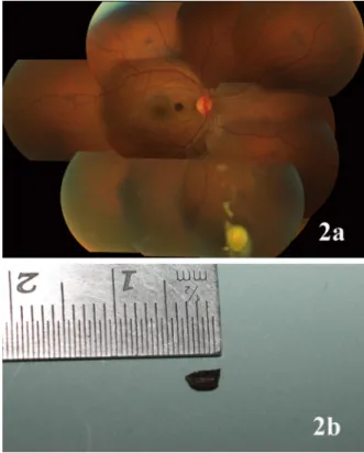
Figure 2: (a) Fundus montage of case 1 showing the IOFB obscured by surrounding exudates in the inferior periphery.
(b) Metallic IOFB following removal by pars plana vitrectomy.
2/4 GMS Ophthalmology Cases 2017, Vol. 7, ISSN 2193-1496
Baskaran et al.: Optical coherence tomography and fundus autofluorescence ...
Cases 2 and 3
Case 2 was a 60-year-old woman who reported with poor vision OU despite undergoing cataract surgery OS. BCVA was 20/200 OD and 20/40 OS. Case 3 was a 63-year-old woman complaining of defective vision OD for six months:
BCVA was 20/30 OD and 20/20 OS. Both patients had remarkably similar findings on biomicroscopy OU: A visu- ally significant cataract OD and pseudophakia OS. Fundus examination OD was normal in both patients. Fundus examination OS revealed a jet black hamartoma similar to case 1 in both patients: close to the fovea nasally in case 2 and temporal to it in case 3 (Figure 1d,g). EDI-OCT and FAF (TRC-50EX mydriatic camera, Topcon medical systems, Tokyo, Japan) findings in these cases were similar to the case 1 (Figure 1e–f,h–i). The choroidal thickness around the hamartoma was unremarkable in both patients (243 µm in case 2 and 238 µm in case 3) [5].
Discussion
The original description of congenital hamartoma of the RPE was given by Laqua in 1981, based on his clinical observations in two cases [6]. In 1989, Gass published a review on focal congenital anomalies of the RPE [1].
He described RPE hamartomas as focal, nodular jet black lesions that involve the full thickness of the retina and spill onto the inner retinal surface in an umbrella-like fashion. In the largest series – 5 cases – by Shields et al., most cases were asymptomatic due to extrafoveal location [7]. The authors noted subtle feeder vessels clinically in all five patients (which were not seen an- giographically), surface wrinkling in four patients, and vitreous cells and exudation in one patient. All patients were stable over 1–16 years of follow-up. Sub-foveal location of the hamartoma results in poor vision [8]. In our first case, the hamartoma clinically mimicked an IOFB;
easily differentiated by B-scan ultrasonography and X-ray.
Further, EDI-OCT findings in all the cases were highly characteristic, similar to the findings previously reported on time domain OCT [2], [3], [8]. However, differentiation from the more closely mimicking diseases, i.e. CHRPE and CHRRPE, may be tricky. The EDI OCT and FAF findings of presumed congenital simple RPE hamartoma are however different from these lesions. CHRPE shows in- creased reflectivity of the RPE layer with thinning of overlying retina and loss of photoreceptors in the region of the flat CHRPE and normal underlying choroid on EDI OCT [2], [3]. CHRRPE shows a prominent epiretinal membrane with thickening and disorganization of the underlying retina, and typical saw-tooth or folded patterns of vitreoretinal traction on OCT [2], [3]. Fundus autofluor- escence of CHRPE shows marked hypoautofluorescence similar to simple RPE hamartoma in about half of the cases; the lacunae are however common, and reveal mild-moderate hyperautofluorescence attributable to bare sclera [4]. CHRRPE shows hypofluorescence corres-
ponding to heavily pigmented lesion containing melanin, and fuzzy hyperautofluorescence corresponding to the epiretinal membrane [9]. We present the second largest series of presumed congenital simple RPE hamartoma, and – to the best of our knowledge – the first report of FAF findings in this tumor. To summarize, multimodal imaging with FAF and EDI-OCT can help to differentiate simple RPE hamartoma from similar RPE lesions, and may serve as a useful adjunct to clinical diagnosis of this rare tumor.
Notes
Competing interests
The authors declare that they have no competing in- terests.
References
1. Gass JD. Focal congenital anomalies of the retinal pigment epithelium. Eye (Lond). 1989;3(Pt 1):1-18. DOI:
10.1038/eye.1989.2
2. Shields CL, Manalac J, Das C, Saktanasate J, Shields JA. Review of spectral domain-enhanced depth imaging optical coherence tomography of tumors of the retina and retinal pigment epithelium in children and adults. Indian J Ophthalmol. 2015 Feb;63(2):128-32. DOI: 10.4103/0301-4738.154384 3. Shields CL, Materin MA, Shields JA. Review of optical coherence
tomography for intraocular tumors. Curr Opin Ophthalmol. 2005 Jun;16(3):141-54. DOI: 10.1097/01.icu.0000158258.01681.40 4. Shields CL, Pirondini C, Bianciotto C, Harmon SA, Shields JA.
Autofluorescence of congenital hypertrophy of the retinal pigment epithelium. Retina (Philadelphia, Pa). 2007 Oct;27(8):1097-100.
DOI: 10.1097/IAE.0b013e318133a174
5. Chhablani J, Rao PS, Venkata A, Rao HL, Rao BS, Kumar U, Narayanan R, Pappuru RR. Choroidal thickness profi le in healthy Indian subjects. Indian J Ophthalmol. 2014 Nov;62(11):1060- 1063. DOI: 10.4103/0301-4738.146711
6. Laqua H. Tumors and tumor-like lesions of the retinal pigment epithelium. Ophthalmologica. 1981;183(1):34-8. DOI:
10.1159/000309131
7. Shields CL, Shields JA, Marr BP, Sperber DE, Gass JD. Congenital simple hamartoma of the retinal pigment epithelium: a study of five cases. Ophthalmology. 2003 May;110(5):1005-11. DOI:
10.1016/S0161-6420(03)00087-3
8. Shukla D, Ambatkar S, Jethani J, Kim R. Optical coherence tomography in presumed congenital simple hamartoma of retinal pigment epithelium. Am J Ophthalmol. 2005 May;139(5):945-7.
DOI: 10.1016/j.ajo.2004.11.037
9. Bruè C, Saitta A, Nicolai M, Mariotti C, Giovannini A. Epiretinal membrane surgery for combined hamartoma of the retina and retinal pigment epithelium: role of multimodal analysis. Clin Ophthalmol. 2013;7:179-84. DOI: 10.2147/OPTH.S39909
3/4 GMS Ophthalmology Cases 2017, Vol. 7, ISSN 2193-1496
Baskaran et al.: Optical coherence tomography and fundus autofluorescence ...
Corresponding author:
Prabu Baskaran, M.S., D.N.B.
Aravind Eye Hospital, Thavalakuppam, Cuddalore Main Road, Pondicherry – 605007, India, Phone:
0413-2619100
prabubaskaran@gmail.com
Please cite as
Baskaran P, Shukla D, Shah P. Optical coherence tomography and fundus autofluorescence findings in presumed congenital simple retinal pigment epithelium hamartoma. GMS Ophthalmol Cases.
2017;7:Doc27.
DOI: 10.3205/oc000078, URN: urn:nbn:de:0183-oc0000785
This article is freely available from
http://www.egms.de/en/journals/oc/2017-7/oc000078.shtml Published:2017-10-25
Copyright
©2017 Baskaran et al. This is an Open Access article distributed under the terms of the Creative Commons Attribution 4.0 License. See license information at http://creativecommons.org/licenses/by/4.0/.
4/4 GMS Ophthalmology Cases 2017, Vol. 7, ISSN 2193-1496
Baskaran et al.: Optical coherence tomography and fundus autofluorescence ...