Article
Properdin Modulates Complement Component Production in Stressed Human Primary Retinal Pigment Epithelium Cells
Nicole Schäfer
1, Hannah N. Wolf
1, Anne Enzbrenner
1, Juliane Schikora
1, Maria Reichenthaler
1, Volker Enzmann
2,3,†and Diana Pauly
1,*
,†1
Experimental Ophthalmology, Eye Clinic, University Hospital Regensburg, 93053 Regensburg, Germany;
Nicole.Schaefer@vkl.uni-regensburg.de (N.S.); Hannah.Wolf@stud.uni-regensburg.de (H.N.W.);
A.enzbrenner@googlemail.com (A.E.); Juliane.Schikora@stud.uni-regensburg.de (J.S.);
Maria.reichenthaler@googlemail.com (M.R.)
2
Department of Ophthalmology, University Hospital of Bern, University of Bern, 3010 Bern, Switzerland;
Volker.Enzmann@insel.ch
3
Department of Biomedical Research, University of Bern, 3010 Bern, Switzerland
* Correspondence: Diana.Pauly@ukr.de
† These authors share senior authorship.
Received: 21 July 2020; Accepted: 22 August 2020; Published: 26 August 2020
Abstract: The retinal pigment epithelium (RPE) maintains visual function and preserves structural integrity of the retina. Chronic dysfunction of the RPE is associated with retinal degeneration, including age-related macular degeneration (AMD). The AMD pathogenesis includes both increased oxidative stress and complement dysregulation. Physiological sources of oxidative stress in the retina are well known, while complement sources and regulation are still under debate. Using human primary RPE (hpRPE) cells, we have established a model to investigate complement component expression on transcript and protein level in AMD-risk and non-risk hpRPE cells. We evaluated the effect of properdin, a complement stabilizer, on the hpRPE cell-dependent complement profile exposed to oxidative stress. hpRPE cells expressed complement components, receptors and regulators.
Complement proteins were also stored and secreted by hpRPE cells. We associated AMD-risk single nucleotide polymorphisms with an increased secretion of complement factors D (CFD) and I (CFI). Furthermore, we detected hpRPE cell-associated complement activation products (C3a, C5a) independent of any extracellularly added complement system. Exogenous properdin increased the mRNA expression of CFI and CFD, but decreased levels of complement components (C1Q, C3), receptors (C3AR, C5AR1, CD11B) and inflammation-associated transcripts (NLRP3, IL1B) in hpRPE cells exposed to oxidative stress. This properdin effect was time-dependently counter regulated. In conclusion, our data unveiled a local, genotype-associated complement component production in hpRPE cells, regulated by exogenous properdin. The local complement production and activation via blood-independent mechanisms can be a new therapeutic target for AMD.
Keywords: retinal pigment epithelium; complement system; properdin; AMD-risk genotype;
intracellular; cell-associated; inflammasome; oxidative stress; human
1. Introduction
As early as 2005, single nucleotide polymorphisms (SNP) in the complement factor H (CFH) gene were identified as genetic risk factors for age-related macular degeneration (AMD), the major cause of visual impairment in the Western world [1,2]. Today, it is known that at least eight of these AMD-risk factors reside in different genes encoding the complement system and enhanced complement deposition
Antioxidants2020,9, 793; doi:10.3390/antiox9090793 www.mdpi.com/journal/antioxidants
Antioxidants2020,9, 793 2 of 19
was observed in AMD-affected eyes [3–6]. However, we still miss a satisfactory answer how these SNPs or the complement system as a whole contributes to AMD.
The complement system is a pathway of the innate immune system, consisting of over 40 proteins, which are consecutively activated. Properdin, is the only known stabilizer of the complement system [7].
It binds to the central, activating protein complex of the cascade and prolongs its half-life by 5–10 times. Next to stabilizing the central C3 convertase, properdin has also a potential role as a pattern recognition molecule activating the complement pathway. The whole complement cascade ensures a first line defense against pathogens and modified cells producing alarm molecules (anaphylatoxins), tagging cells/microorganisms (opsonins) or disrupting cell membranes (membrane attack complex) [8].
Additionally, non-canonical intracellular functions of complement components (“the complosome”) have been described in T-cells, neutrophils, pancreatic β -cells and others [9–11]. Cell-associated or intracellular complement activity modulated cell metabolism, autophagy, survival, and differentiation in these different cell types [10,12–14]. However, so far the complement system has not been further investigated as a cell-dependent/autocrine pathway in relation to AMD so far.
Two major advanced stages of AMD can occur simultaneously in one patient or even in a single eye: Choroidal neovascularization (CNV) and geographic atrophy (GA) [15,16]. These completely different disease patterns cause either disruption or loss of the retinal pigment epithelium (RPE).
Besides genetics, clinical data suggested additional external stimuli, for example oxidative stress or aging processes [17,18], promoting different pathological outcomes in AMD. This needs to be taken into account investigating the role of complement in RPE and AMD.
The RPE forms the blood–retinal barrier, which separates the retina from the systemic circulation and the immune system [19]. The RPE acts as a regulatory, secretory epithelium supporting the retina.
It locally secretes complement components as C1q, complement factor B (CFB), complement component 4 (C4), CFI, and CFH [20–23]. We and others showed that complement secretion is modified by external stress [20–26]. Additionally, generation of complement activation products, such as anaphylatoxins and opsonins, by healthy and stressed RPE cells independent of any external complement source is described [21,24,26,27]. Recently, it was reported, that endogenous CFH and anaphylatoxins contribute to transcriptional and metabolic homeostasis of RPE cells [28–30]. In RPE cells complement anaphylatoxins receptor signaling is involved in eye morphogenesis [31], sub-RPE deposits [32], pro-inflammatory RPE reaction [33–35], PI3/Akt-pathway activation [29], and stress-mediated lipid accumulation in RPE cells [36]. Together this indicates an involvement of autocrine complement reactivity in housekeeping mechanisms maintaining RPE physiology. However, it is not known in detail how this is controlled and how it contributes to retinal degeneration.
In the present study, we tested whether human primary RPE (hpRPE) cells produce and activate complement components in dependence of their genotype and exogenous properdin stress. We demonstrated that hpRPE cells positive for a homozygous AMD-risk SNP within complement genes secreted more complement proteins than non-carriers. Thereby, we supposed that the complement stabilizer properdin modifies the local complement homeostasis in stressed hpRPE cells. We described that hpRPE cell-dependent complement levels were time-dependently changed by oxidative stress and properdin addition.
2. Materials and Methods
2.1. Cultivation and Treatment of hpRPE
The research complies with the human research act (HRA) stating that small quantities of bodily
substances removed in the course of transplantation may be anonymized for research purposes
without consent (HRA chapter 5, paragraph 38, Switzerland). hpRPE were prepared from left and
right eyes of 15 anonymized donors (Table 1) as previously described [37,38]. Briefly, hpRPE cells
were harvested from the eyecup after enzymatic digestion and centrifuged with 259 × g at 4
◦C for
5 min. hpRPE cells were cultivated in Dulbecco’s modified Eagle’s medium/Nutrient Mixture F-12
Antioxidants2020,9, 793 3 of 19
(DMEM/F 12 GlutaMax, Thermo Fisher Scientific, Dreieich, Germany, #31331-028) containing 5%
fetal bovine serum (FBS, Thermo Fisher Scientific, #10500-064), 1% Penicillin-Streptomycin (Thermo Fisher Scientific, #15070-063), 1% N1 Medium Supplement (Merck, Darmstadt, Germany, #N6530), 10 mM MEM non-essential amino acids (Thermo Fisher Scientific, #11140-035), 0.25 mg/mL Taurine (Merck, #T0625), 4.5 mg/mL Glucose solution (Thermo Fisher Scientific, #G524940-01), 0.013 ng/mL Triiodothyronine (Merck, #T2877), 0.02 µg/mL Hydrocortisone (Merck, #H0888), 20 ng/mL human basic growth factor (hbFGF, R&D Systems, Minneapolis, MN, USA, #13256029) and 1 mg/mL human epidermal growth factor (hEGF, Thermo Fisher Scientific, #PHG0311) in laminin-coated Transwell
®inserts under standard conditions (37
◦C, 5% CO
2, 80% humidity).
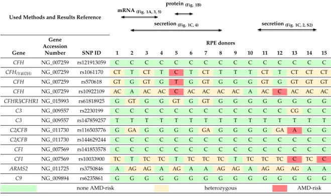
Table 1. Human primary retinal pigment epithelium (hpRPE) cell genotyping and experimental assignment in this study.
Used Methods and Results Reference
protein(Fig. 1B) mRNA(Fig. 1A, 3, 5)
conjugated anti-species antibodies were used for detection (1 h, PBS-T): goat anti-mouse (1:5000, Jackson ImmunoResearch, West Grove, PA, USA, #115-035-164), rabbit anti-goat (1:5000, Jackson ImmunoResearch, #305-035-003). Visualization was performed by WesternSure PREMIUM Chemiluminescent Substrate (LI-COR, Bad Homburg, Germany, #926-95000) in a Fluor Chem FC2 Imaging System (Alpha Innotech, San Leandro, CA, USA).
2.5. Immunohistochemistry
Immunostaining was performed as described previously [22]. PBS-washed, paraformaldehyde- fixated (4%, 20 min; Merck, # 100496) hpRPE cells were blocked 3% BSA (Carl Roth, Karlsruhe, Germany, #8076/ PBS-T, 1 h). Antigens were detected using primary antibodies anti-RPE65 (1:200;
Abcam, Cambridge, UK #ab13826) or anti-bestrophin (1:200; Novus Biologicals, Littleton, CO, USA
#NB300-164), visualized with goat anti-mouse ALEXA Fluor 594 (1:500; Thermo Fisher Scientific,
#A32742) and complemented with nuclei staining (DAPI; Vector Labs, Burlingame, CA, USA, #H- 2000,). Cells were covered with fluorescence mounting medium (Agilent, Boeblingen, Germany,
#S302380-2).
2.6. Statistics
Statistical analysis was performed using GraphPad Prism 7 (GraphPad Software Inc, San Diego, CA, USA). We estimated a normal Gaussian distribution. Significance levels were determined by the parametric, unpaired t-test. Welch‘s test was used to correct for unequal sample distribution variance.
All data are expressed as mean ± standard deviation (SD) unless stated otherwise. Detailed information about specific n-values, implemented statistical tests and coding of significance levels are provided in the respective figures and figure legends.
3. Results
RPE cells are capable of producing cell-derived complement components as we showed recently [22,42]. In the present study we assume that an externally added complement regulator, properdin, can modulate stress-dependent RPE cell-derived complement components.
3.1. Human Primary RPE Cells as a Model System in Cell Culture
We investigated human primary RPE (hpRPE) cells, isolated and cultivated from 15 different donor eyes (Table 1). Studied hpRPE cells were vital and pigmented as well as positive for the RPE markers RPE65 and bestrophin (Figure S1A–G). Pigmentation of the hpRPE cells was stable from 4 to 14 days of cultivation (after removal of non-adherent cells at day 2).
Ex vivo cultivation of hpRPE of both eyes resulted in a maximum of 6 transwell filters with a growth area of 1.12 cm
2from each donor. This limited the study and not all investigations could be performed with the hpRPE cells from the same donor. An overview of the used cell preparations and experiments is given in Table 1.
Table 1. Human primary retinal pigment epithelium (hpRPE) cell genotyping and experimental assignment in this study.
Used Methods and Results Reference
protein (Fig. 1B)
mRNA (Fig. 1A, 3, 5)
secretion (Fig. 1C, 4) secretion (Fig. 1C, 2, S2)
Gene
Accession RPE donors
Gene Number SNP ID 1 2 3 4 5 6 7 8 9 10 11 12 13 14 15 CFH
NG_007259 rs121913059 C C C C C C C C C C C C C C C
CFH(Y402H)
NG_007259 rs1061170 CT T CT T C T CT T T T CT T CT CT CT
CFH
NG_007259 rs570618 GT G GT G T G GT G G G GT G GT GT GT conjugated anti-species antibodies were used for detection (1 h, PBS-T): goat anti-mouse (1:5000, Jackson ImmunoResearch, West Grove, PA, USA, #115-035-164), rabbit anti-goat (1:5000, Jackson ImmunoResearch, #305-035-003). Visualization was performed by WesternSure PREMIUM Chemiluminescent Substrate (LI-COR, Bad Homburg, Germany, #926-95000) in a Fluor Chem FC2 Imaging System (Alpha Innotech, San Leandro, CA, USA).
2.5. Immunohistochemistry
Immunostaining was performed as described previously [22]. PBS-washed, paraformaldehyde- fixated (4%, 20 min; Merck, # 100496) hpRPE cells were blocked 3% BSA (Carl Roth, Karlsruhe, Germany, #8076/ PBS-T, 1 h). Antigens were detected using primary antibodies anti-RPE65 (1:200;
Abcam, Cambridge, UK #ab13826) or anti-bestrophin (1:200; Novus Biologicals, Littleton, CO, USA
#NB300-164), visualized with goat anti-mouse ALEXA Fluor 594 (1:500; Thermo Fisher Scientific,
#A32742) and complemented with nuclei staining (DAPI; Vector Labs, Burlingame, CA, USA, #H- 2000,). Cells were covered with fluorescence mounting medium (Agilent, Boeblingen, Germany,
#S302380-2).
2.6. Statistics
Statistical analysis was performed using GraphPad Prism 7 (GraphPad Software Inc, San Diego, CA, USA). We estimated a normal Gaussian distribution. Significance levels were determined by the parametric, unpaired t-test. Welch‘s test was used to correct for unequal sample distribution variance.
All data are expressed as mean ± standard deviation (SD) unless stated otherwise. Detailed information about specific n-values, implemented statistical tests and coding of significance levels are provided in the respective figures and figure legends.
3. Results
RPE cells are capable of producing cell-derived complement components as we showed recently [22,42]. In the present study we assume that an externally added complement regulator, properdin, can modulate stress-dependent RPE cell-derived complement components.
3.1. Human Primary RPE Cells as a Model System in Cell Culture
We investigated human primary RPE (hpRPE) cells, isolated and cultivated from 15 different donor eyes (Table 1). Studied hpRPE cells were vital and pigmented as well as positive for the RPE markers RPE65 and bestrophin (Figure S1A–G). Pigmentation of the hpRPE cells was stable from 4 to 14 days of cultivation (after removal of non-adherent cells at day 2).
Ex vivo cultivation of hpRPE of both eyes resulted in a maximum of 6 transwell filters with a growth area of 1.12 cm
2from each donor. This limited the study and not all investigations could be performed with the hpRPE cells from the same donor. An overview of the used cell preparations and experiments is given in Table 1.
Table 1. Human primary retinal pigment epithelium (hpRPE) cell genotyping and experimental assignment in this study.
Used Methods and Results Reference
protein (Fig. 1B) mRNA (Fig. 1A, 3, 5)
secretion (Fig. 1C, 4) secretion (Fig. 1C, 2, S2)
Gene
Accession RPE donors
Gene Number SNP ID 1 2 3 4 5 6 7 8 9 10 11 12 13 14 15 CFH
NG_007259 rs121913059 C C C C C C C C C C C C C C C
CFH(Y402H)
NG_007259 rs1061170 CT T CT T C T CT T T T CT T CT CT CT
CFH
NG_007259 rs570618 GT G GT G T G GT G G G GT G GT GT GT
secretion(Fig. 1C, 4)conjugated anti-species antibodies were used for detection (1 h, PBS-T): goat anti-mouse (1:5000, Jackson ImmunoResearch, West Grove, PA, USA, #115-035-164), rabbit anti-goat (1:5000, Jackson ImmunoResearch, #305-035-003). Visualization was performed by WesternSure PREMIUM Chemiluminescent Substrate (LI-COR, Bad Homburg, Germany, #926-95000) in a Fluor Chem FC2 Imaging System (Alpha Innotech, San Leandro, CA, USA).
2.5. Immunohistochemistry
Immunostaining was performed as described previously [22]. PBS-washed, paraformaldehyde- fixated (4%, 20 min; Merck, # 100496) hpRPE cells were blocked 3% BSA (Carl Roth, Karlsruhe, Germany, #8076/ PBS-T, 1 h). Antigens were detected using primary antibodies anti-RPE65 (1:200;
Abcam, Cambridge, UK #ab13826) or anti-bestrophin (1:200; Novus Biologicals, Littleton, CO, USA
#NB300-164), visualized with goat anti-mouse ALEXA Fluor 594 (1:500; Thermo Fisher Scientific,
#A32742) and complemented with nuclei staining (DAPI; Vector Labs, Burlingame, CA, USA, #H- 2000,). Cells were covered with fluorescence mounting medium (Agilent, Boeblingen, Germany,
#S302380-2).
2.6. Statistics
Statistical analysis was performed using GraphPad Prism 7 (GraphPad Software Inc, San Diego, CA, USA). We estimated a normal Gaussian distribution. Significance levels were determined by the parametric, unpaired t-test. Welch‘s test was used to correct for unequal sample distribution variance.
All data are expressed as mean ± standard deviation (SD) unless stated otherwise. Detailed information about specific n-values, implemented statistical tests and coding of significance levels are provided in the respective figures and figure legends.
3. Results
RPE cells are capable of producing cell-derived complement components as we showed recently [22,42]. In the present study we assume that an externally added complement regulator, properdin, can modulate stress-dependent RPE cell-derived complement components.
3.1. Human Primary RPE Cells as a Model System in Cell Culture
We investigated human primary RPE (hpRPE) cells, isolated and cultivated from 15 different donor eyes (Table 1). Studied hpRPE cells were vital and pigmented as well as positive for the RPE markers RPE65 and bestrophin (Figure S1A–G). Pigmentation of the hpRPE cells was stable from 4 to 14 days of cultivation (after removal of non-adherent cells at day 2).
Ex vivo cultivation of hpRPE of both eyes resulted in a maximum of 6 transwell filters with a growth area of 1.12 cm
2from each donor. This limited the study and not all investigations could be performed with the hpRPE cells from the same donor. An overview of the used cell preparations and experiments is given in Table 1.
Table 1. Human primary retinal pigment epithelium (hpRPE) cell genotyping and experimental assignment in this study.
Used Methods and Results Reference
protein (Fig. 1B) mRNA (Fig. 1A, 3, 5)
secretion (Fig. 1C, 4) secretion (Fig. 1C, 2, S2)
Gene
Accession RPE donors
Gene Number SNP ID 1 2 3 4 5 6 7 8 9 10 11 12 13 14 15 CFH
NG_007259 rs121913059 C C C C C C C C C C C C C C C
CFH(Y402H)
NG_007259 rs1061170 CT T CT T C T CT T T T CT T CT CT CT
CFH
NG_007259 rs570618 GT G GT G T G GT G G G GT G GT GT GT
secretion(Fig. 1C, 2, S2)
conjugated anti-species antibodies were used for detection (1 h, PBS-T): goat anti-mouse (1:5000, Jackson ImmunoResearch, West Grove, PA, USA, #115-035-164), rabbit anti-goat (1:5000, Jackson ImmunoResearch, #305-035-003). Visualization was performed by WesternSure PREMIUM Chemiluminescent Substrate (LI-COR, Bad Homburg, Germany, #926-95000) in a Fluor Chem FC2 Imaging System (Alpha Innotech, San Leandro, CA, USA).
2.5. Immunohistochemistry
Immunostaining was performed as described previously [22]. PBS-washed, paraformaldehyde- fixated (4%, 20 min; Merck, # 100496) hpRPE cells were blocked 3% BSA (Carl Roth, Karlsruhe, Germany, #8076/ PBS-T, 1 h). Antigens were detected using primary antibodies anti-RPE65 (1:200;
Abcam, Cambridge, UK #ab13826) or anti-bestrophin (1:200; Novus Biologicals, Littleton, CO, USA
#NB300-164), visualized with goat anti-mouse ALEXA Fluor 594 (1:500; Thermo Fisher Scientific,
#A32742) and complemented with nuclei staining (DAPI; Vector Labs, Burlingame, CA, USA, #H- 2000,). Cells were covered with fluorescence mounting medium (Agilent, Boeblingen, Germany,
#S302380-2).
2.6. Statistics
Statistical analysis was performed using GraphPad Prism 7 (GraphPad Software Inc, San Diego, CA, USA). We estimated a normal Gaussian distribution. Significance levels were determined by the parametric, unpaired t-test. Welch‘s test was used to correct for unequal sample distribution variance.
All data are expressed as mean ± standard deviation (SD) unless stated otherwise. Detailed information about specific n-values, implemented statistical tests and coding of significance levels are provided in the respective figures and figure legends.
3. Results
RPE cells are capable of producing cell-derived complement components as we showed recently [22,42]. In the present study we assume that an externally added complement regulator, properdin, can modulate stress-dependent RPE cell-derived complement components.
3.1. Human Primary RPE Cells as a Model System in Cell Culture
We investigated human primary RPE (hpRPE) cells, isolated and cultivated from 15 different donor eyes (Table 1). Studied hpRPE cells were vital and pigmented as well as positive for the RPE markers RPE65 and bestrophin (Figure S1A–G). Pigmentation of the hpRPE cells was stable from 4 to 14 days of cultivation (after removal of non-adherent cells at day 2).
Ex vivo cultivation of hpRPE of both eyes resulted in a maximum of 6 transwell filters with a growth area of 1.12 cm
2from each donor. This limited the study and not all investigations could be performed with the hpRPE cells from the same donor. An overview of the used cell preparations and experiments is given in Table 1.
Table 1. Human primary retinal pigment epithelium (hpRPE) cell genotyping and experimental assignment in this study.
Used Methods and Results Reference
protein (Fig. 1B) mRNA (Fig. 1A, 3, 5)
secretion (Fig. 1C, 4) secretion (Fig. 1C, 2, S2)
Gene
Accession RPE donors
Gene Number SNP ID 1 2 3 4 5 6 7 8 9 10 11 12 13 14 15 CFH
NG_007259 rs121913059 C C C C C C C C C C C C C C C
CFH(Y402H)
NG_007259 rs1061170 CT T CT T C T CT T T T CT T CT CT CT
CFH
NG_007259 rs570618 GT G GT G T G GT G G G GT G GT GT GT
GeneAccession RPE donors
Gene Number SNP ID 1 2 3 4 5 6 7 8 9 10 11 12 13 14 15
CFH NG_007259 rs121913059 C C C C C C C C C C C C C C C
CFH(Y402H) NG_007259 rs1061170 CT T CT T C T CT T T T CT T CT CT CT
CFH NG_007259 rs570618 GT G GT G T G GT G G G GT G GT GT GT
CFH NG_007259 rs10922109 AC A AC AC C AC AC AC AC A AC C AC AC AC CFHR3/CFHR1 NG_015993 rs61818925 G GT G G GT G GT G G G G G G G G
C3 NG_009557 rs2230199 C C C C C C C C C C C C CG C C
C3 NG_009557 rs147859257 T T T T T T T T T T T T T T T
C2/CFB NG_011730 rs116503776 G GA G G G G GA G G G G GA A G G
C2/CFB NG_011730 rs144629244 C C C C C C C C C C C C C C C
CFI NG_007569 rs141853578 C C C C C C C C C C C C C C C
CFI NG_007569 rs10033900 TC T TC TC T TC TC TC T TC TC TC C TC C ARMS2 NG_011725 rs3750846 A AG AG A AG A A AG AG A AG AG AG A A
C9 NG_009894 rs6235861 G G G G G G G G G G G G G G G
none AMD-risk heterozygous AMD-risk
Before treatment, FBS concentration was reduced to 0% within 3 days (5%–2.5%–1.25%, respectively). hpRPE cells were apically treated either with 0.5 mM H
2O
2, or 0.5 mM H
2O
2and 50 µ g/mL properdin (Complement Technology, Tyler, TC, USA, #A139) for 4, 10, or 24 h. Supernatants (apical and basal) were taken before and after treatment, subsequently frozen in liquid nitrogen and stored at − 80
◦C.
2.2. hpRPE Genotyping
DNA was isolated from sclera slices of the respective donor eyes using ReliaPrep™ FFPE gDNA Miniprep System (Promega, Mannheim, Germany, #A2351). PCR for amplification of DNA sequences containing relevant AMD-associated complement SNPs was performed using in-house generated primers (Table S1) and the following cycle steps, according to the MIQE guidelines [39]:
denaturation (95
◦C, 1 min), annealing (60
◦C, 1 min), elongation (72
◦C, 1 min), 33 cycles. Afterwards, DNA-sequencing was performed by GeneArt (Thermo Fisher Scientific) using either forward or reverse in-house primers, respectively (Table S1).
2.3. RT-qPCR and PCR
mRNA was isolated using the NucleoSpin
®RNA kit (Macherey-Nagel, Düren, Germany, #740955)
and transcribed into cDNA with the QuantiTect
®Reverse Transcription Kit (Qiagen, Hilden, Germany,
Antioxidants2020,9, 793 4 of 19
#205313). Transcripts of complement components, regulators, receptors and inflammation-associated markers were analyzed (i) by PCR (as described in Section 2.2.) and subsequent 2% agarosegel separation for cDNA fragment visualization, or (ii) by qPCR using Rotor-Gene SYBR
®Green PCR Kit (Qiagen) in a Rotor Gene Q 2plex cycler (Qiagen) either with QuantiTect Primer Assays (Qiagen, Table S2), or in-house designed primers for IL1B, VIM and SMA1 (Metabion, Planedd, Germany, Table S1). Data were normalized to the housekeeper GAPDH expression and fold change was calculates using 2(-Delta Delta C(T)) method [40]. Data are visualized on a linear scale using log2-transformed scores [41].
2.4. Multiplex Complement Secretion Assay
Complement components in the cellular supernatants after 11–14 days of cultivation were quantified using the MILLIPLEX MAP Human Complement Panel (Merck, #HCMP1MAG-19K,
#HCMP2MAG-19K) in accordance to the manufacturer’s protocol. The read out of the multiplex assay was performed in a Magpix instrument (Luminex, Austin, TX, USA).
2.5. Western Blot
Lysates of hpRPE cells were generated using RIPA buffer (Sigma-Aldrich, Munich, Germany,
#R0278) with protease and phosphatase inhibitors (1:100, Sigma-Aldrich, #P8340). Samples were dissolved in reducing Laemmli sample buffer, denatured (95
◦C, 10 min) and separated in a 12%
SDS-PAGE and transferred on to an activated PVDF membrane using a wet blotting system. Membranes were blocked (1 h, 5% bovine serum albumin (BSA)/PBS-T) and incubated with the primary antibodies (overnight, 5% BSA/PBS-T): Rabbit anti-GAPDH-HRP (1:1000, Cell Signaling Technology, Beverly, MA, USA, #3683), mouse anti-C3 (1:100, Progen, Heidelberg, Germany, #61019), goat anti-CFI (1:250, Quidel, San Diego, CA, USA, #A313), mouse anti-C3a (1:50, Hycult, Uden, Netherlands, #HM2074) and mouse anti-C5a-biotin (1:250, Biozol, Eching, Germany, #BLD-518306). Peroxdiase-conjugated anti-species antibodies were used for detection (1 h, PBS-T): goat anti-mouse (1:5000, Jackson ImmunoResearch, West Grove, PA, USA, #115-035-164), rabbit anti-goat (1:5000, Jackson ImmunoResearch, #305-035-003).
Visualization was performed by WesternSure PREMIUM Chemiluminescent Substrate (LI-COR, Bad Homburg, Germany, #926-95000) in a Fluor Chem FC2 Imaging System (Alpha Innotech, San Leandro, CA, USA).
2.6. Immunohistochemistry
Immunostaining was performed as described previously [22]. PBS-washed, paraformaldehyde-fixated (4%, 20 min; Merck, # 100496) hpRPE cells were blocked 3% BSA (Carl Roth, Karlsruhe, Germany, #8076/ PBS-T, 1 h). Antigens were detected using primary antibodies anti-RPE65 (1:200; Abcam, Cambridge, UK #ab13826) or anti-bestrophin (1:200; Novus Biologicals, Littleton, CO, USA #NB300-164), visualized with goat anti-mouse ALEXA Fluor 594 (1:500; Thermo Fisher Scientific, #A32742) and complemented with nuclei staining (DAPI; Vector Labs, Burlingame, CA, USA, #H-2000,). Cells were covered with fluorescence mounting medium (Agilent, Boeblingen, Germany, #S302380-2).
2.7. Statistics
Statistical analysis was performed using GraphPad Prism 7 (GraphPad Software Inc, San Diego,
CA, USA). We estimated a normal Gaussian distribution. Significance levels were determined by
the parametric, unpaired t-test. Welch‘s test was used to correct for unequal sample distribution
variance. All data are expressed as mean ± standard deviation (SD) unless stated otherwise. Detailed
information about specific n-values, implemented statistical tests and coding of significance levels are
provided in the respective figures and figure legends.
3. Results
RPE cells are capable of producing cell-derived complement components as we showed recently [22,42]. In the present study we assume that an externally added complement regulator, properdin, can modulate stress-dependent RPE cell-derived complement components.
3.1. Human Primary RPE Cells as a Model System in Cell Culture
We investigated human primary RPE (hpRPE) cells, isolated and cultivated from 15 different donor eyes (Table 1). Studied hpRPE cells were vital and pigmented as well as positive for the RPE markers RPE65 and bestrophin (Figure S1A–G). Pigmentation of the hpRPE cells was stable from 4 to 14 days of cultivation (after removal of non-adherent cells at day 2).
Ex vivo cultivation of hpRPE of both eyes resulted in a maximum of 6 transwell filters with a growth area of 1.12 cm
2from each donor. This limited the study and not all investigations could be performed with the hpRPE cells from the same donor. An overview of the used cell preparations and experiments is given in Table 1.
3.2. hpRPE Cells Produce Complement Components
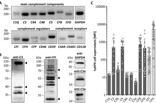
All tested hpRPE cells were positive for transcripts of complement components C1Q, C3, C4A, C4B, C5, CFB, and CFD, as well as complement regulators CFI, CFH, properdin (CFP), CD46, and CD59 (Figure 1A). hpRPE cells expressed also complement receptors for complement activation products like the anaphylatoxin receptors C3AR and C5AR1 as well as the subunit CD11B of complement receptor 3, which interacts with the C3 cleavage products (iC3b, C3d, C3dg) upon activation (Figure 1A).
hpRPE cells produced complement proteins as C3 and CFI shown by Western blot (Figure 1B).
This raised the question, if complement receptors of hpRPE cells could be modulated in an autocrine signaling pathway if complement activation products are generated by these cells. Analyzing hpRPE cell lysates in Western blot, we found hpRPE cell-derived complement activation products C3a, C5a and further C3 cleavage products (C3d, C3dg) in untreated hpRPE cells independent from any external complement sources, indicating a possible autocrine cleavage and function of hpRPE cell-derived activated complement components (Figure 1B and Figure S2 ).
We also determined secreted complement components in the cell culture supernatants of hpRPE
preparations from both eyes 11–14 days after isolation (Figure 1C). Thereby, we detected C3, C3b, C4,
CFB, CFD and the regulators CFI as well as CFH in the supernatants (Figure 1C). C1q, C5 and the
complement activation products C4b and C5a were either not secreted or below the assay detection
limits (Figure 1C). We also identified differences of complement secretion levels between hpRPE
cell preparations of the left and right eye (Figure S3). However, we cannot judge the influence of
retinal phenotype and cell preparation differences. Apical and basal complement secretion levels were
comparable (Figure S3), but due to low cell numbers and reduced cell division of hpRPE a confluent
monolayer was not always observable. Furthermore, we cannot exclude passive diffusion of the
complement components secreted apically and basally through the membrane filter (Figure S3).
Antioxidants2020,9, 793 6 of 19
Antioxidants 2020, 9, x FOR PEER REVIEW 6 of 18
Figure 1. hpRPE cells express and secrete complement components. (A) mRNA expression of main
complement components of the classical (C1Q), central (C3) classical/lectin (C4A, C4B), terminal (C5) and alternative (CFB, CFD) complement pathway was detected in hpRPE cells. hpRPE cells expressed also mRNA of soluble (CFI, CFH, CFP) and membrane bound (CD46, CD59) complement regulators, and transcripts of complement receptors: anaphylatoxin receptors (C3AR, C5AR1), opsonin receptor CR3 subunit (CD11B). Shown expression data of donor 1 are representative for mRNA experiments in hpRPE cells of donors 1–4 (Table 1). (B) Activation products of the central complement component C3 [(blot left, arrows) C3b (115 kDa), C3dg (39 kDa) and C3d (35 kDa)] were detected in hpRPE.
Multiple bands were identified in hpRPE cells using an anti-complement regulator CFI antibody (blot center, arrows for full CFI (80 kDa) and two CFI disulfide linked chains (50 kDa, 30 kDa)).
Anaphylatoxins C3a and C5a (blot right) were found in cell lysates of hpRPE cells. Examples of whole blots are depicted in (B) for donor 5 (C5a) and donor 6 (C3, C3a, CFI). (C) hpRPE cells secrete C3, C4, CFB, CFD and the regulators CFI as well as CFH into the cell culture supernatant (pooled data of two eyes, apical and basal supernatant are shown, details for hpRPE cells of donors 11–16 are presented in Figure S3). Mean with standard deviation is shown. Dotted line depicts blank control. MFI—mean fluorescence intensity.
3.3. AMD-Risk SNPs in Complement Genes Increase hpRPE Cell-Dependent Complement Secretion
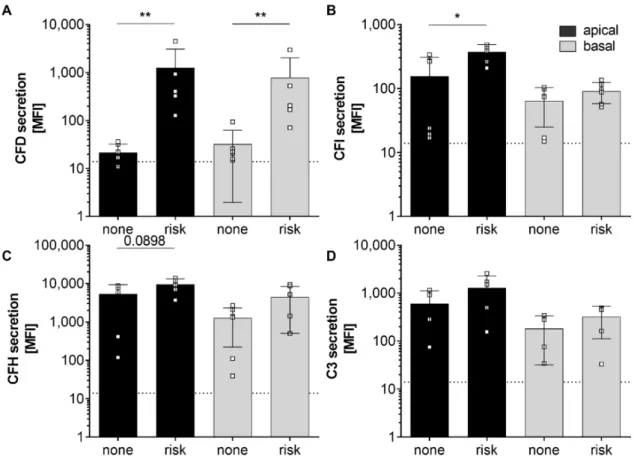
SNPs within genes of the complement cascade were previously associated with the risk of retinal degeneration and changed retinal/RPE physiology [4,43,44]. We analyzed 13 complement-associated risk SNPs in the donor tissues to evaluate the effect of these SNPs on hpRPE cell-derived complement secretion. We found 4 of 15 donors positive for 1 – 3 homozygous AMD-risk SNPs either in the CFH, C2/CFB or CFI gene loci and all donors were positive for 1–5 heterozygous AMD-risk SNPs (Table 1).
Furthermore, we compared complement secretion levels of hpRPE cells positive for at least one homozygous AMD-risk SNPs SNP (donors 12, 13, 15) and non-homozygous AMD-risk SNP hpRPE cells (donors 10, 11, 14) (Table 1, Figure 2). Thereby, significantly higher CFD and CFI levels in supernatants of hpRPE cells with the AMD-associated risk SNPs in complement genes were found (Figure 2A,B). A comparable tendency was also observed for complement regulators CFH and the central complement component C3, which were increased in supernatants of hpRPE with the AMD- risk genotypes after 14 days of cultivation compared to non-risk cells (Figure 2C,D). Only the secretion of components of the alternative pathway was affected by AMD-risk SNPs in complement genes under the described experimental conditions. However, we cannot definitely state that the
Figure 1. hpRPE cells express and secrete complement components. (A) mRNA expression of main complement components of the classical (C1Q), central (C3) classical/lectin (C4A, C4B), terminal (C5) and alternative (CFB, CFD) complement pathway was detected in hpRPE cells. hpRPE cells expressed also mRNA of soluble (CFI, CFH, CFP) and membrane bound (CD46, CD59) complement regulators, and transcripts of complement receptors: anaphylatoxin receptors (C3AR, C5AR1), opsonin receptor CR3 subunit (CD11B). Shown expression data of donor 1 are representative for mRNA experiments in hpRPE cells of donors 1–4 (Table 1). (B) Activation products of the central complement component C3 [(blot left, arrows) C3b (115 kDa), C3dg (39 kDa) and C3d (35 kDa)] were detected in hpRPE. Multiple bands were identified in hpRPE cells using an anti-complement regulator CFI antibody (blot center, arrows for full CFI (80 kDa) and two CFI disulfide linked chains (50 kDa, 30 kDa)). Anaphylatoxins C3a and C5a (blot right) were found in cell lysates of hpRPE cells. Examples of whole blots are depicted in (B) for donor 5 (C5a) and donor 6 (C3, C3a, CFI). (C) hpRPE cells secrete C3, C4, CFB, CFD and the regulators CFI as well as CFH into the cell culture supernatant (pooled data of two eyes, apical and basal supernatant are shown, details for hpRPE cells of donors 11–16 are presented in Figure S3). Mean with standard deviation is shown. Dotted line depicts blank control. MFI—mean fluorescence intensity.
3.3. AMD-Risk SNPs in Complement Genes Increase hpRPE Cell-Dependent Complement Secretion
SNPs within genes of the complement cascade were previously associated with the risk of retinal degeneration and changed retinal/RPE physiology [4,43,44]. We analyzed 13 complement-associated risk SNPs in the donor tissues to evaluate the effect of these SNPs on hpRPE cell-derived complement secretion. We found 4 of 15 donors positive for 1–3 homozygous AMD-risk SNPs either in the CFH, C2/CFB or CFI gene loci and all donors were positive for 1–5 heterozygous AMD-risk SNPs (Table 1).
Furthermore, we compared complement secretion levels of hpRPE cells positive for at least one
homozygous AMD-risk SNPs SNP (donors 12, 13, 15) and non-homozygous AMD-risk SNP hpRPE
cells (donors 10, 11, 14) (Table 1, Figure 2). Thereby, significantly higher CFD and CFI levels in
supernatants of hpRPE cells with the AMD-associated risk SNPs in complement genes were found
(Figure 2A,B). A comparable tendency was also observed for complement regulators CFH and the
central complement component C3, which were increased in supernatants of hpRPE with the AMD-risk
genotypes after 14 days of cultivation compared to non-risk cells (Figure 2C,D). Only the secretion
of components of the alternative pathway was affected by AMD-risk SNPs in complement genes
under the described experimental conditions. However, we cannot definitely state that the hpRPE culture always resulted in the same cell numbers on the transwell filters investigated (even the same preparation protocol was used).
Antioxidants 2020, 9, x FOR PEER REVIEW 7 of 18
hpRPE culture always resulted in the same cell numbers on the transwell filters investigated (even the same preparation protocol was used).
Figure 2.
Secretion of alternative complement pathway components was increased in hpRPE cells positive for homozygous age-related macular degeneration (AMD)-risk single nucleotide polymorphisms (SNPs) (risk; donors 12, 13, 15). Significant higher levels of secreted (A) complement factors D (CFD), (B) complement factors I (CFI) and in tendency higher (C) complement factor H (CFH) and (D) C3 secretion levels were detected in apical (black) and basal (grey) supernatants of hpRPE cells with homozygous AMD-risk SNPs (risk; donors 12, 13, 15) compared to hpRPE cells without a homozygous AMD-risk SNP (none; donors 10, 11, 14) in complement genes (Table 1). Mean with standard deviation is shown. * p < 0.05, ** p < 0.01 unpaired t-test with Welch‘s correction. Dotted line depicts blank control. MFI—mean fluorescence intensity.
3.4. Complement Regulator Properdin Increased Expression of Complement Proteases in Stressed hpRPE Cells
We showed recently that oxidative stress increased the expression of complement regulator properdin in RPE cells [22]. In the present study, we investigated the effect of externally added properdin on stressed hpRPE cells. hpRPE cells were treated either with H
2O
2alone or with combined H
2O
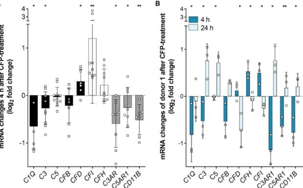
2and properdin. Transcript levels of markers for epithelial–mesenchymal transition (EMT), complement components, regulators and receptors were compared between these treatment groups (Figures 3 and S1). After 4 h of properdin treatment, hpRPE cells showed a tendency of increased EMT markers (Figure S1). Simultaneously, transcripts for complement proteases CFD and CFI were significantly upregulated in hpRPE cells (Figure 3A), while other complement components, which are involved in the general complement cascade (C1Q, C3), and complement receptors (C3AR, C5AR1, CD11B) were downregulated (Figure 3A). This effect slightly differed in the four tested hpRPE preparations of donors 1–4 pooled in herein, but a genotype-dependent reaction was not observable due to limitations of the cohort size. All tested hpRPE preparations were negative for a homozygous AMD-risk SNP within complement genes (Table 1). Properdin treatment of stressed cells did not change the expression levels of C4, CD59, CD46, and CFP (data not shown).
Figure 2. Secretion of alternative complement pathway components was increased in hpRPE cells positive for homozygous age-related macular degeneration (AMD)-risk single nucleotide polymorphisms (SNPs) (risk; donors 12, 13, 15). Significant higher levels of secreted (A) complement factors D (CFD), (B) complement factors I (CFI) and in tendency higher (C) complement factor H (CFH) and (D) C3 secretion levels were detected in apical (black) and basal (grey) supernatants of hpRPE cells with homozygous AMD-risk SNPs (risk; donors 12, 13, 15) compared to hpRPE cells without a homozygous AMD-risk SNP (none; donors 10, 11, 14) in complement genes (Table 1). Mean with standard deviation is shown. * p < 0.05, ** p < 0.01 unpaired t-test with Welch‘s correction. Dotted line depicts blank control. MFI—mean fluorescence intensity.
3.4. Complement Regulator Properdin Increased Expression of Complement Proteases in Stressed hpRPE Cells
We showed recently that oxidative stress increased the expression of complement regulator
properdin in RPE cells [22]. In the present study, we investigated the effect of externally added
properdin on stressed hpRPE cells. hpRPE cells were treated either with H
2O
2alone or with combined
H
2O
2and properdin. Transcript levels of markers for epithelial–mesenchymal transition (EMT),
complement components, regulators and receptors were compared between these treatment groups
(Figure 3 and Figure S1). After 4 h of properdin treatment, hpRPE cells showed a tendency of
increased EMT markers (Figure S1). Simultaneously, transcripts for complement proteases CFD and
CFI were significantly upregulated in hpRPE cells (Figure 3A), while other complement components,
which are involved in the general complement cascade (C1Q, C3), and complement receptors (C3AR,
C5AR1, CD11B) were downregulated (Figure 3A). This effect slightly differed in the four tested hpRPE
preparations of donors 1–4 pooled in herein, but a genotype-dependent reaction was not observable
due to limitations of the cohort size. All tested hpRPE preparations were negative for a homozygous
Antioxidants2020,9, 793 8 of 19
AMD-risk SNP within complement genes (Table 1). Properdin treatment of stressed cells did not change the expression levels of C4, CD59, CD46, and CFP (data not shown).
Antioxidants 2020, 9, x FOR PEER REVIEW 8 of 18
hpRPE cells of donor 1 were used to compare the early (4 h) and late (24 h) effect of external properdin on transcription levels (Figure 3B). We found time-dependent differences of complement expression in properdin-treated cells for all investigated transcripts. Transcription tendencies detected after 4 h were always reversed after 24 h. For example, complement proteases CFD and CFI were decreased and mRNA of complement receptors C3AR, C5AR1, and CD11B were increased after 24 h (Figure 3B).
In summary, our expression data suggested an early anti-inflammatory and a late pro- inflammatory transcriptional response of stressed hpRPE cells upon properdin treatment.
Figure 3. Complement regulator properdin changed complement mRNA expression in stressed
hpRPE cells. (A) H
2O
2stressed hpRPE cells were treated with properdin (CFP) for 4 h. mRNA expression of main complement components C1Q,
C3, C5, and CFB (black) as well as complementreceptors C3AR, C5AR1, and CD11B (grey) was decreased in comparison to stressed cells (dotted line).
CFD (black) and complement regulators CFI and CFH (white) mRNA expression was increased in
stressed, properdin-treated cells compared to stressed cells only. Data from donors 1–4 are presented.
Mean with standard deviation is shown. * p < 0.05, ** p < 0.01 unpaired t-test with Welch‘s correction of stressed versus stressed, properdin-treated cells. (B) H
2O
2stressed hpRPE cells of donor 1 were treated with properdin for 4 h (dark blue) or 24 h (light blue). mRNA Expression of complement components and receptors was reversed after 24 h compared to 4 h. Mean with standard deviation is shown. * p < 0.05, ** p < 0.01 unpaired t-test with Welch‘s correction of 4 h versus 24 h properdin treatment.
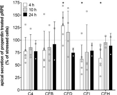
Consistent with the transcription results secretion of complement components CFD, CFI, and CFH was time-dependently changed in stressed, properdin-treated hpRPE cells compared to stressed cells (Figure 4). CFD levels in the apical supernatant were increased 4 h after properdin treatment and declined over time. This was in accordance with the mRNA transcription analyses (Figure 3). In contrast to the transcription results, where we described an early increase of CFI and CFH mRNA, the protein levels of CFI and CFH were rather reduced in the supernatant of properdin-treated hpRPE cells (Figure 4). This suggested an intracellular accumulation of these proteins following properdin interaction as described previously for stressed ARPE-19 cells [22]. C4 and CFB secretion was not significantly changed by properdin addition (Figure 4). C3 secretion could not be reliably determined, as the added properdin resulted in unspecific signals in the used commercial multiplex complement assay.
Figure 3. Complement regulator properdin changed complement mRNA expression in stressed hpRPE cells. (A) H
2O
2stressed hpRPE cells were treated with properdin (CFP) for 4 h. mRNA expression of main complement components C1Q, C3, C5, and CFB (black) as well as complement receptors C3AR, C5AR1, and CD11B (grey) was decreased in comparison to stressed cells (dotted line). CFD (black) and complement regulators CFI and CFH (white) mRNA expression was increased in stressed, properdin-treated cells compared to stressed cells only. Data from donors 1–4 are presented. Mean with standard deviation is shown. * p < 0.05, ** p < 0.01 unpaired t-test with Welch‘s correction of stressed versus stressed, properdin-treated cells. (B) H
2O
2stressed hpRPE cells of donor 1 were treated with properdin for 4 h (dark blue) or 24 h (light blue). mRNA Expression of complement components and receptors was reversed after 24 h compared to 4 h. Mean with standard deviation is shown. * p <
0.05, ** p < 0.01 unpaired t-test with Welch‘s correction of 4 h versus 24 h properdin treatment.
hpRPE cells of donor 1 were used to compare the early (4 h) and late (24 h) effect of external properdin on transcription levels (Figure 3B). We found time-dependent differences of complement expression in properdin-treated cells for all investigated transcripts. Transcription tendencies detected after 4 h were always reversed after 24 h. For example, complement proteases CFD and CFI were decreased and mRNA of complement receptors C3AR, C5AR1, and CD11B were increased after 24 h (Figure 3B).
In summary, our expression data suggested an early anti-inflammatory and a late pro-inflammatory transcriptional response of stressed hpRPE cells upon properdin treatment.
Consistent with the transcription results secretion of complement components CFD, CFI, and
CFH was time-dependently changed in stressed, properdin-treated hpRPE cells compared to stressed
cells (Figure 4). CFD levels in the apical supernatant were increased 4 h after properdin treatment
and declined over time. This was in accordance with the mRNA transcription analyses (Figure 3). In
contrast to the transcription results, where we described an early increase of CFI and CFH mRNA,
the protein levels of CFI and CFH were rather reduced in the supernatant of properdin-treated
hpRPE cells (Figure 4). This suggested an intracellular accumulation of these proteins following
properdin interaction as described previously for stressed ARPE-19 cells [22]. C4 and CFB secretion
was not significantly changed by properdin addition (Figure 4). C3 secretion could not be reliably determined, as the added properdin resulted in unspecific signals in the used commercial multiplex complement assay.
Antioxidants 2020, 9, x FOR PEER REVIEW 9 of 18
Figure 4. Complement protein secretion was modulated in stressed hpRPE cells by properdin.
Complement protein secretion into the apical supernatant 4 h (white), 10 h (grey) and 24 h (black) after properdin treatment of H
2O
2stressed hpRPE is shown as percentage of the secretion level of stressed, non-properdin treated cells (dotted line). We observed a time-dependent change in CFD levels (significantly increased after 4 h) in apical supernatants of stressed properdin-treated hpRPE cells. Decreased concentrations of complement regulators CFI and CFH were detected after 4 h of properdin treatment. Shown are data of donors 2, 4, and 8 for 4 h; donors 3, 5, and 6 for 10 h; donor 1 for 24 h. Mean with standard deviation is shown. * p < 0.05, ** p < 0.01 t-test with Welch‘s correction of stressed hpRPE versus stressed, properdin-treated hpRPE.
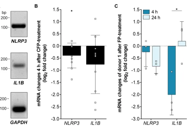
3.5. Inflammasome-Associated Transcription Levels were Reduced by Properdin in Stressed hpRPE Cells Recently, we showed an increase of inflammasome activation following oxidative stress in ARPE-19 cells [22]. To answer the question how the complement regulator properdin acts on inflammasome-associated mRNA expression in stressed RPE cells, we investigated NLRP3 and IL1Β transcripts in properdin-treated and non-treated stressed hpRPE cells (Figure 5). NLRP3 and IL1Β expression was decreased after 4 h in properdin-treated cells compared to only H
2O
2-stressed cells (Figure 5B). This confirmed the suggested early anti-inflammatory effect of properdin on H
2O
2- stressed hpRPE cells (Figure 3A). After 24 h NLRP3 transcripts were still reduced, but IL1Β were counter regulated in comparison to 4 h with increased IL1Β levels (Figure 5C).
Figure 4. Complement protein secretion was modulated in stressed hpRPE cells by properdin.
Complement protein secretion into the apical supernatant 4 h (white), 10 h (grey) and 24 h (black) after properdin treatment of H
2O
2stressed hpRPE is shown as percentage of the secretion level of stressed, non-properdin treated cells (dotted line). We observed a time-dependent change in CFD levels (significantly increased after 4 h) in apical supernatants of stressed properdin-treated hpRPE cells.
Decreased concentrations of complement regulators CFI and CFH were detected after 4 h of properdin treatment. Shown are data of donors 2, 4, and 8 for 4 h; donors 3, 5, and 6 for 10 h; donor 1 for 24 h.
Mean with standard deviation is shown. * p < 0.05, ** p < 0.01 t-test with Welch‘s correction of stressed hpRPE versus stressed, properdin-treated hpRPE.
3.5. Inflammasome-Associated Transcription Levels were Reduced by Properdin in Stressed hpRPE Cells
Recently, we showed an increase of inflammasome activation following oxidative stress in
ARPE-19 cells [22]. To answer the question how the complement regulator properdin acts on
inflammasome-associated mRNA expression in stressed RPE cells, we investigated NLRP3 and IL1B
transcripts in properdin-treated and non-treated stressed hpRPE cells (Figure 5). NLRP3 and IL1B
expression was decreased after 4 h in properdin-treated cells compared to only H
2O
2-stressed cells
(Figure 5B). This confirmed the suggested early anti-inflammatory effect of properdin on H
2O
2-stressed
hpRPE cells (Figure 3A). After 24 h NLRP3 transcripts were still reduced, but IL1B were counter
regulated in comparison to 4 h with increased IL1B levels (Figure 5C).
Antioxidants2020,9, 793 10 of 19
Antioxidants 2020, 9, x FOR PEER REVIEW 10 of 18
Figure 5.
Inflammasome-associated mRNA transcripts were differentially expressed in stressed, properdin-treated hpRPE cells. (A) Inflammasome NLRP3 and pro-inflammatory cytokine IL1Β mRNA was detected in hpRPE cells. Shown expression data of donor 1 are representative for mRNA experiments in hpRPE cells of donors 1–4 (Table 1). (B) Properdin treatment of H
2O
2-stressed hpRPE cells showed a significant reduced expression of NLRP3 and IL1Β after 4 h. Data from donors 1–4 are presented. (C) H
2O
2-stressed hpRPE cells of donor 1 were treated with properdin for 4 h (dark blue) or 24 h (light blue). Reduced NLRP3 mRNA expression was accelerated over time and IL1Β transcripts were significantly increased after 24 h compared to 4 h properdin incubation. Mean with standard deviation is shown. * p < 0.05, ** p < 0.01 unpaired t-test with Welch‘s correction of 4 h versus 24 h properdin treatment.
4. Discussion
AMD is a multifactorial pathological process in the retina resulting from alteration of the RPE.
Chronic inflammatory changes in the RPE can lead to blood–retinal barrier breakdown and disease progression. Thereby, the innate immunity including the complement system plays a pivotal role.
Our study demonstrated that components of the complement system, a part of the innate immune system are produced and secreted by RPE cells. The local complement quantity and activity was modulated by external stress affecting the RPE and contributing to a disturbed blood–retinal barrier.
4.1. Biological Relevance of hpRPE In Vitro Experiments
RPE cells are differentiated and polarized cells, which form a monolayer and are part of the blood–retinal barrier between the photoreceptors and Bruch’s membrane. The main functions of the RPE are: (i) Absorption of light and protection against photo-oxidation, (ii) secretion of immune modulators and growth factors as well as (iii) phagocytosis and recycling of shed photoreceptor [19].
These activities can be altered under non-physiological conditions. In this study, we used hpRPE cells from human donors in order to assure as much in vivo-like RPE cell characteristics as possible in our in vitro experiments. Different genotypes, environmental influences and in vivo differentiation had an impact on the variability and biological relevance of our results. We investigated hpRPE cells from the right and the left eye of 15 different donors, which reflected the RPE phenotype in the human eye [45]. This attached high importance to our results compared to results obtained from immortal, manipulated cell lines [46–48] (RPE in vitro systems were excellently reviewed in [19]). Optimal
Figure 5. Inflammasome-associated mRNA transcripts were differentially expressed in stressed, properdin-treated hpRPE cells. (A) Inflammasome NLRP3 and pro-inflammatory cytokine IL1B mRNA was detected in hpRPE cells. Shown expression data of donor 1 are representative for mRNA experiments in hpRPE cells of donors 1–4 (Table 1). (B) Properdin treatment of H
2O
2-stressed hpRPE cells showed a significant reduced expression of NLRP3 and IL1B after 4 h. Data from donors 1–4 are presented. (C) H
2O
2-stressed hpRPE cells of donor 1 were treated with properdin for 4 h (dark blue) or 24 h (light blue). Reduced NLRP3 mRNA expression was accelerated over time and IL1B transcripts were significantly increased after 24 h compared to 4 h properdin incubation. Mean with standard deviation is shown. * p < 0.05, ** p < 0.01 unpaired t-test with Welch‘s correction of 4 h versus 24 h properdin treatment.
4. Discussion
AMD is a multifactorial pathological process in the retina resulting from alteration of the RPE.
Chronic inflammatory changes in the RPE can lead to blood–retinal barrier breakdown and disease progression. Thereby, the innate immunity including the complement system plays a pivotal role. Our study demonstrated that components of the complement system, a part of the innate immune system are produced and secreted by RPE cells. The local complement quantity and activity was modulated by external stress affecting the RPE and contributing to a disturbed blood–retinal barrier.
4.1. Biological Relevance of hpRPE In Vitro Experiments
RPE cells are differentiated and polarized cells, which form a monolayer and are part of the blood–retinal barrier between the photoreceptors and Bruch’s membrane. The main functions of the RPE are: (i) Absorption of light and protection against photo-oxidation, (ii) secretion of immune modulators and growth factors as well as (iii) phagocytosis and recycling of shed photoreceptor [19].
These activities can be altered under non-physiological conditions. In this study, we used hpRPE cells
from human donors in order to assure as much in vivo-like RPE cell characteristics as possible in our
in vitro experiments. Different genotypes, environmental influences and in vivo differentiation had an
impact on the variability and biological relevance of our results. We investigated hpRPE cells from
the right and the left eye of 15 different donors, which reflected the RPE phenotype in the human
eye [45]. This attached high importance to our results compared to results obtained from immortal,
manipulated cell lines [46–48] (RPE in vitro systems were excellently reviewed in [19]). Optimal culture conditions resulted in physiological cell morphology and pigmentation of the ex vivo supplied cells [45]. Pigmentation of the RPE cells was a marker for differentiation, not often detected in cultured human RPE cell lines [49].
4.2. hpRPE as a Source for Complement Components in the Eye
Previous work identified the RPE as one of the major sources of complement transcripts in the mouse retina [42,50]. Here, we presented for the first time a comprehensive description of transcribed complement components (C1Q, C3, C4, C5, CFB, CFD), regulators (CFI, CFH, CFP, CD46, CD59) and receptors (C3AR, C5AR1, CD11B) in cultivated hpRPE cells. In accordance with our results C3, CFH, and complement receptor C5AR have already been shown to be expressed in the immortalized retinal pigment epithelial cell line ARPE-19 and in primary RPE cells [22,35,51]. RPE cells derived from induced pluripotent stem cells (iPSC) express mRNA for complement activators C3, C5, CFB, and complement inhibitors CFH, CFI, CD46, CD55, CD59, CLUSTERIN, and VITRONECTIN [23]. Single cell transcriptomic analysis in human organoids also showed CFH, C3, and C2 expression in RPE cells [52].
Similar to hpRPE, non-liver dependent complement gene expression was also described for intestinal epithelial cells, podocytes and other cell types [42,53,54].
In addition to complement transcripts, we also detected complement components C3, C3b, C4, CFB, CFD as well as complement regulators CFI and CFH in supernatants of hpRPE cells independent of any external complement sources. This was in line with previously published data for post-confluent ARPE-19 cells which secreted C3, C3a, C4bp, CFI, and CFH [20–22] and iPSC-derived RPE cells that secreted CFB [23,26]. C3 was previously also found in supernatants of primary mouse RPE cells [50]. Therewith, our and other results suggest a local RPE cell-dependent source of the complement components independent from liver-derived complement proteins in the eye.
hpRPE cell-dependent complement secretion was regulated by environmental, external stimuli.
We documented variable complement levels in supernatants comparing secretion of left and right eye hpRPE of the same donor. In contrast to inherited eye diseases, where often both eyes are affected to a similar extent, acquired vision loss can occur in different degrees in the left and right eye. For AMD it is known that the disease progression can be dissimilar between both eyes [55,56]. Our data support this monocular specific disease progression also on the level of hpRPE cell-dependent complement secretion.
4.3. AMD-Risk SNPs in Complement Genes Enhanced hpRPE Cell-Dependent Complement Secretion RPE dysfunction in AMD is associated with SNPs in complement system genes. An international AMD-genome wide association study listed six complement gene loci as top priority AMD-relevant risk candidates based on biological and statistical evidence [4,57,58]. Nevertheless, detailed studies of how precisely certain AMD-risk genotypes modify complement function and protein expression are still lacking.
Here, we found that these SNPs within the complement genes modulate local complement secretion.
hpRPE obtained from donors positive for at least one homozygous complement risk SNP secreted higher levels of complement components than hpRPE cells heterozygous for the complement-associated AMD-risk SNPs. The increase was significant for alternative complement pathway components CFD/
CFI and a tendency was observed for CFH/C3. Related research showed that AMD-risk SNP enhances complement activity in the RPE and the retina. Higher levels of C5a in Bruch’s membrane and increased membrane attack complex deposition in eyes homozygous for the high-risk CFH genotype Y402H were identified [43,44]. Complement dysregulation was also described for a homozygous risk-SNP in the ARMS2 gene [59]. In contrast, blood-derived CFI was reduced in AMD patients with a CFI SNP compared to patients without the SNP [60].
At least two mechanistic models are possible how genetic polymorphism in genes of the
complement system could modify the ability of hpRPE cells to secrete components: (i) Polymorphism
Antioxidants2020,9, 793 12 of 19