Acute stromal keratitis in clinics – are we missing microsporidia?
Abstract
Purpose:To report 3 cases of microsporidial stromal keratitis presenting as a diagnostic dilemma to a tertiary eye care center in north India.
Javed Hussain Farooqui
1Methods:Three eyes of 3 patients underwent therapeutic keratoplasty
for microsporidial stromal keratitis. A decision for early surgery was
Manisha Acharya
1Arpan Gandhi
2taken as the patients were not responding to conventional medical
management and were worsening clinically. The diagnosis of microspo-
Umang Mathur
1ridia was made by corneal scraping and confirmed on histopathological evaluation of the corneal button.
1 Cornea, Cataract and Refractive Surgery, Dr.
Results:Out of the 3 patients, one maintained a clear graft, one had a recurrence and one had graft rejection, 6 months postoperatively. The
Shroff’s Charity Eye Hospital, New Delhi, India
patients were not started on steroids in the postoperative period and were given topical antibiotics and polyhexamethylene biguanide (PHMD).
Oral Albendazole 400 mg was also given twice a day for a month. 2 Laboratory Services, Dr.
Shroff’s Charity Eye Hospital, New Delhi, India
Conclusion:Many questions remained unanswered about the manage- ment protocol of stromal keratitis caused by microsporidia. The role of topical steroids, antifungal agents, oral Albendazole needs to be dis- cussed. Clinicians should be aware of recurrences which may mimic as rejections. There needs to be more awareness regarding microsporidia as a cause of acute stromal keratitis, so that its not overlooked or un- derdiagnosed.
Keywords:microsporidial stromal keratitis, microsporidia
Introduction
Since the time it was first described by Ashton [1] in 1973 as a cause of keratitis, microsporidiosis has come a long way as being recognized as a potential sight-threatening organism. In literature, the pattern of involvement has been described as either keratoconjunctivitis in immuno- compromised individuals or stromal keratitis in immuno- competent individuals [2]. Even though the treatment of keratoconjunctivitis is straightforward, the stromal kera- titis variant poses a diagnostic and therapeutic challenge.
Often misdiagnosed as herpes simplex keratits [3], it also closely mimics bacterial or fungal keratitis, with no spe- cific ulcer pattern having been described till date [4]. We report a series of 3 eyes of 3 patients who presented to us with acute stromal keratitis and were eventually diag- nosed with microsporidial stromal keratitis and treated accordingly. To the best of our knowledge, no major case series of microsporidial stromal keratitis has been de- scribed from north India.
Case descriptions
Case 1
A 73-year-old male patient presented with pain, watering, photophobia, and blurring of vision in the right eye since
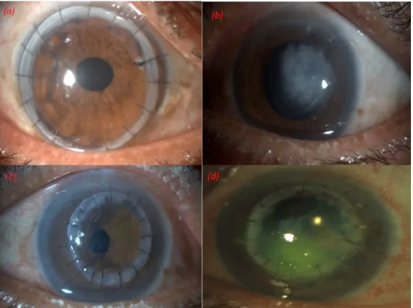
one year and in the left eye since 4 months. He gave history of a foreign body sensation, which commenced by itself with no history of trauma or recurrence in the left eye. He consulted a tertiary eye center for his right eye and records revealed a presence of keratitis with posterior segment involvement for which penetrating keratoplasty (PKP) with pars plana vitrectomy in the right eye was done and vitreous biopsy confirmed the presence of microspor- idial spores. His unaided visual acuity at presentation was 20/200 in the right eye and 20/120 in the left eye.
Slit lamp examination of the right eye showed a 10.5 mm clear, compact graft with intact sutures and a well-formed anterior chamber (Figure 1a). Posterior chamber intraocu- lar lens was noted with glaucomatous optic atrophy. The left eye examination showed an epithelial defect with a whitish grey midstromal infiltrate in the mid periphery measuring 5 mm x 5 mm with ill-defined margins (Fig- ure 1b). Corneal sensations were intact. The anterior chamber had 1+ flare and cells. Intraocular pressure was considered normal on digital tonometry. Corneal scraping and microbiological work-up was done which revealed microsporidial spores. The patient was started on poly- hexamethylene biguanide (PHMB) 0.02% 2-hourly along with lubricating and cycloplegic eye drops. When the pa- tient didn’t improve over a 1-week period, a decision of early therapeutic PKP was taken and 7.5 mm donor graft was used (Figure 1c). The surgery was uneventful and the patient was continued on PHMB and 0.5% moxifloxa-
Case Report
OPEN ACCESS
Figure 1: (a) Right eye showing a compact graft, with intact sutures and well-formed anterior chamber.
(b) Left eye showing epithelial defect with whitish grey midstromal infiltrate in the mid periphery measuring 5 mm x 5 mm with ill-defined margins. (c) One week post therapeutic penetrating keratoplasty.
(d) Six months postoperatively, midstromal infiltrate measuring 4 mm x 4 mm with an overlying epithelial defect.
cin eye drops 2-hourly after the surgery. In the subsequent months, unaided visual acuity improved to 20/200 in the left eye. Unfortunately, 6 months postoperatively, the patient presented with hand movement vision and corneal scraping revealed a recurrence of microbiologically proven microsporidial keratitis with retinal detachment (Fig- ure 1d).
Case 2
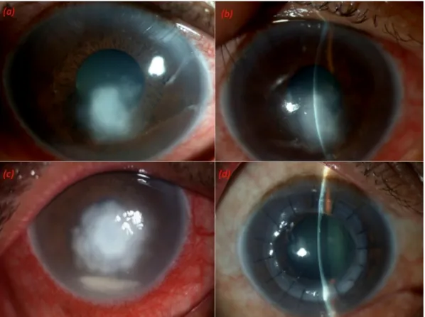
A 55-year-old male patient presented in 2018 with pain, watering, photophobia, and blurring of vision in the right eye since 1 week. He had had a similar episode 3 weeks back and had been treated with anti-virals elsewhere.
His symptoms had improved and subsequently he had discontinued treatment. His unaided visual acuity was 20/200 in the right eye and 20/40 in the left eye. Slit lamp examination of the right eye showed a midstromal greyish infiltrate measuring 4 mm x 3 mm with intact overlying epithelium (Figure 2a). Corneal scraping was inconclusive and the patient was started on antivirals based on clinical suspicion. The patient did not improve on his day 3 follow-up (Figure 2b) and further worsened on day 7 with increase in the infiltrate size and appear- ance of a hypopyon (Figure 2c). Repeat scrapings were done on day 3 and day 7. The second scraping also did not aid in diagnosis and there was no growth on culture as well till then. A ‘drug holiday’ was given with the clinical suspicion of an atypical organism. This was confirmed on
subsequent scraping, wherein microsporidial spores were identified. The patient underwent therapeutic PKP with a 8 mm graft. The postoperative period was uneventful with the patient maintaining a clear graft and a visual acuity of 20/120 over a period of 6 months postoperative (Figure 2d).
Case 3
A 22-year-old male patient presented with pain, watering, photophobia, and blurring of vision in the left eye for the last 3 weeks. His visual acuity in the right eye was 20/20 and in the left eye was counting fingers at 2 meters. Slit lamp examination of the left eye showed an epithelial defect of 7 mm x 7 mm, multifocal subepithelial infiltrates and central 4 mm x 4 mm thinning of 70%. The anterior chamber was formed with a hypopyon of 0.5 mm (Fig- ure 3a). A working diagnosis of viral keratitis was made and antivirals were started. When the patient didn’t re- spond to treatment after 1 week, corneal scraping was done which revealed microsporidial spores. The patient was taken up for early therapeutic PKP, and an 8 mm eccentric graft was taken (Figure 3b). The patient did well for the first 3 months, but returned in the fourth month with fall in vision, graft edema and signs of rejection (Figure 3c). There was no infiltrate and high dose intra- venous steroids were given for 3 days and topical steroids stepped up to hourly interval. Graft edema did not subside at 6 months (Figure 3d), and the patient was then given
Figure 2: (a) Right eye showing mid stromal greyish infiltrate measuring 4 mm x 3 mm with intact overlying epithelium.
(b) Third day picture with increase in the size of the infiltrate. (c) One week picture showing appearance of hypopyon along with the preexisting infiltrate. (d) Six months postoperative picture showing clear graft.
Figure 3: (a) Left eye showing epithelial defect 7 mm x 7 mm, multifocal sub epithelial infiltrates and central 4 mm x 4 mm with a hypopyon of 0.5 mm. (b) Post therapeutic PKP, 8 mm eccentric graft at 1-week follow-up.
(c) Fourth month postoperative picture showing stromal edema and signs of graft rejection. (d) Six months postoperative picture showing failed graft.
Farooqui et al.: Acute stromal keratitis in clinics – are we missing ...
Figure 4: (a) Microsporidial spores (black arrow) seen as well-defined oval reddish bodies on Ziehl Neelsen stain of the corneal button derived from the first patient. (b) Gram’s stain of the second patient showing microsporidial spores (black arrow). (c) Potassium hydroxide (KOH) 10% depicting well defined microsporidial spores (black arrow),
obtained from corneal scraping from the third patient.
the option of repeat keratoplasty. The patient has not yet consented to the procedure.
Postoperative regimen
All 3 patients were given 1% prednisolone acetate 6 times a day along with a cycloplegic and 0.5% moxifloxacin 4 times a day in the initial period. The first patient was prescribed 0.2% PHMB for the first month at a 2-hourly interval. The other two patients did not receive PHMB because of availability issues. Oral Albendazole 400 mg twice a day was given for the first 3 weeks after surgery.
Corneal scraping and laboratory work-up
As protocol, all the patients underwent corneal scrapings according to standard techniques mentioned in the text [5]. Slides were sent for Gram’s, Giemsa and potassium hydroxide 10%. The material was also inoculated on fresh blood agar, chocolate agar, Saburaud’s dextrose agar (SDA), non-nutrient agar with an overlay ofEscherichia coliand brain heart infusion broth. The corneal buttons derived from surgery were also sent for microbiological and histopathological evaluation. Figure 4 shows pictures of various laboratory slides derived from the three pa- tients.
Discussion
India is home to a quarter of the world’s blind population [6]. Various studies have estimated corneal blindness to account for between 3 to 14% of total blindness in India, and almost 59% of this is because of keratits and its se- quel [7], [8], [9]. It is to our surprise that out of such a high number of keratitis cases being reported from India, no major series of microsporidia has been reported from north India. All the major series are from south India from limited centers only [10], [11]. This prompts us to think that this condition is being misdiagnosed or missed al- together. Due to the paucity of reports, we still don’t have treatment protocols set up for treating patients with mi- crosporidial stromal keratitis. In our patients, we were unsure about the use of topical steroids in the postopera- tive period. Since microsporidia has recently been classi- fied as a fungus [3], the use of topical steroids can be debated. We did not start any of our patients on topical antifungals and wonder if there is a role of these drugs in long-term safety and graft survival. Also, PHMB has been described in previous reports [11], but its limited availability has hampered its use in clinical practice. Even though it’s available in big cities and towns, efforts need to be made to make them easily available in smaller cities as well. Along with this, clear guidelines should be set for the use of oral Albendazole. Its role in systemic microspo- ridiosis has been established, but most of the studies mention no benefit to ocular pathology with the use of
oral Albendazole [10]. We feel unnecessary overmedica- tion of patients is not warranted and work should be done to assess the efficacy of Albendazone for ocular microspo- ridiosis.
Our last patient went into rejection at 3 months. We cannot, with certainty, say if that patient had rejection or recurrence of the disease. Microsporidiosis is known to have recurrence, and may mimic as rejection [3]. In such cases, as a repeat procedure, a full thickness graft would be better than endothelial keratoplasty to avoid recur- rence. In our first and third case, a 1 mm clear margin was taken all around the infiltrate during host trephina- tion, but this seems inadequate for microsporidia and both patients had recurrence. For the second patient, a clear margin of more than 1 mm was kept and perhaps this led to his graft survival. Although there is no way to confirm this hypothesis, these are points that should be kept in mind while operating on these patients.
Many questions still remain unanswered about microspo- ridial stromal keratitis. We hope that this report provokes thought and discussion and helps us understand the disease better. We also look forward to ophthalmologists formulating universal treatment protocols for the manage- ment of this disease.
Notes
Competing interests
The authors declare that they have no competing in- terests.
Acknowledgments
We appreciate the help of Dr. Vaibhav Pednekar and Dr.
Mitali Kekan in patient work-up and data gathering.
References
1. Ashton N, Wirasinha PA. Encephalitozoonosis (nosematosis) of the cornea. Br J Ophthalmol. 1973 Sep;57(9):669-74. DOI:
10.1136/bjo.57.9.669
2. Sharma S, Das S, Joseph J, Vemuganti GK, Murthy S.
Microsporidial keratitis: need for increased awareness. Surv Ophthalmol. 2011 Jan-Feb;56(1):1-22. DOI:
10.1016/j.survophthal.2010.03.006
3. Pradhan S, Mascarenhas J, Srinivasan M. Microsporidial stromal keratitis masquerading as acute graft rejection. Cornea. 2015 Mar;34(3):353-4. DOI: 10.1097/ICO.0000000000000337
4. Rauz S, Tuft S, Dart JK, Bonshek R, Luthert P, Curry A.
Ultrastructural examination of two cases of stromal microsporidial keratitis. J Med Microbiol. 2004 Aug;53(Pt 8):775-81. DOI:
10.1099/jmm.0.45524-0
5. Garg P, Rao GN. Corneal ulcer: diagnosis and management.
Community Eye Health. 1999;12(30):21-3.
6. Farooqui JH. Postgraduate training program in ophthalmology in India: what’s lacking? J Clin Ophthalmol Res. 2015;3:111-2. DOI:
10.4103/2320-3897.156613
7. Murthy GV, Vashist P, John N, Pokharel G, Ellwein LB. Prevelence and causes of visual impairment and blindness in older adults in an area of India with a high cataract surgical rate. Ophthalmic Epidemiol. 2010 Aug;17(4):185-95. DOI:
10.3109/09286586.2010.483751
8. Murthy GV, Gupta S, Ellwein LB, Munoz SR, Bachani D, Dada VK.
A population-based eye survey of older adults in a rural district of Rajasthan: I. Central vision impairment, blindness, and cataract surgery. Ophthalmology. 2001 Apr;108(4):679-85. DOI:
10.1016/s0161-6420(00)00579-0
9. Dandona L, Dandona R, Srinivas M, Giridhar P, Vilas K, Prasad MN, John RK, McCarty CA, Rao GN. Blindness in the Indian state of Andhra Pradesh. Invest Ophthalmol Vis Sci. 2001
Apr;42(5):908-16.
10. Sabhapandit S, Murthy SI, Garg P, Korwar V, Vemuganti GK, Sharma S. Microsporidial Stromal Keratitis: Clinical Features, Unique Diagnostic Criteria, and Treatment Outcomes in a Large Case Series. Cornea. 2016 Dec;35(12):1569-74. DOI:
10.1097/ICO.0000000000000939
11. Vemuganti GK, Garg P, Sharma S, Joseph J, Gopinathan U, Singh S. Is microsporidial keratitis an emerging cause of stromal keratitis? A case series study. BMC Ophthalmol. 2005 Aug;5:19.
DOI: 10.1186/1471-2415-5-19
Corresponding author:
Dr. Javed Hussain Farooqui
Cornea, Cataract and Refractive Surgery, Dr. Shroff’s Charity Eye Hospital, 5027, Kedarnath Marg, Daryaganj, New Delhi – 110002, India
jhfarooqui@gmail.com
Please cite as
Farooqui JH, Acharya M, Gandhi A, Mathur U. Acute stromal keratitis in clinics – are we missing microsporidia? GMS Ophthalmol Cases.
2020;10:Doc01.
DOI: 10.3205/oc000128, URN: urn:nbn:de:0183-oc0001288
This article is freely available from
https://www.egms.de/en/journals/oc/2020-10/oc000128.shtml Published:2020-02-14
Copyright
©2020 Farooqui et al. This is an Open Access article distributed under the terms of the Creative Commons Attribution 4.0 License. See license information at http://creativecommons.org/licenses/by/4.0/.
Farooqui et al.: Acute stromal keratitis in clinics – are we missing ...