Imaging tamoxifen retinopathy using spectral-domain optical coherence tomography
Abstract
A case of tamoxifen retinopathy examined with spectral-domain optical coherence tomography (SD-OCT) is presented. The typical refractile
Albert Caramoy
1Paula Scholz
1deposits are located between ganglion cell layer and inner plexiform
Sascha Fauser
1layer in SD-OCT. A defect on the outer retinal layer with disruption of
Bernd Kirchhof
1the photoreceptor layer with sharp edges is seen. The still attached posterior hyaloids gives evidence of other pathomechanism involved in the outer retinal defect than that of macular hole, as suggested in
the literature. 1 University of Cologne, Center
of Ophthalmology, Keywords:tamoxifen retinopathy, drug induced retinopathy, toxicity,
imaging, spectral domain optical coherence tomography, breast cancer
Department of Vitreo-Retinal Surgery, Cologne, Germany
Introduction
Tamoxifen is a non-steroidal antiestrogen drug, used as adjuvant treatment of estrogen receptor positive breast cancer. Although generally tolerable, tamoxifen retino- pathy has been described in the literature as a rare complication. The first cases of tamoxifen retinopathy were described by Kaiser-Kupfer in 1978 [1]. In that case a high dose of tamoxifen was used. Since then the dose of tamoxifen has been reduced, however tamoxifen ret- inopathy has been also described using a relatively low dose of 20–40 mg daily [2], [3].
Since the introduction of optical coherence tomography (OCT), it is possible to examine tamoxifen retinopathy more closely. The characteristics of tamoxifen retinopathy in OCT have been described by several authors [4], [5], [6], [7], [8]. In most of these cases a foveal cyst with disruption of photoreceptor layer was seen in OCT. Al- though the exact pathogenesis of such foveal cysts is unclear, mechanisms suggesting the formation of a macular hole in such patients have been assumed. In this case report, we show a case of tamoxifen retinopathy as seen in spectral-domain OCT (SD-OCT). In particular we examine closely the vitreous, since this has been suggested as the cause of macular hole formation [9].
Case presentation
A 49-year-old woman was referred to our clinic due to maculopathy and visual deterioration. In the medical history she had taken tamoxifen 20 mg daily for 4½ years (cumulative dose 33 g) following mastectomy 6 years ago. Visual acuity deterioration has been marked since 11 months, tamoxifen maculopathy was diagnosed by the referring ophthalmologist, and tamoxifen was stopped
9 months ago. In the medical history she was using topiramate, trazodone, sertraline, and omeprazole due to depression and gastroesophageal reflux disease. She was allergic to sulfonamide.
The distant visual acuity was 20/40 for the right eye and 20/100 for the left eye. Reading visual acuity was 20/32 for the right eye and 20/50 for the left eye. On the Amsler grid a central scotoma on the left eye was stated. She indicated that deterioration of central scotoma developed over a few days, even after tamoxifen was discontinued.
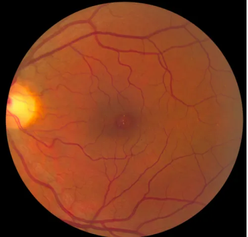
On ophthalmic examinations the media were clear. The optic disc and vessels were normal. On the macula on both eyes there were refractile deposits (Figure 1 and Figure 2). The fluorescein angiography showed a hyper- fluorescent area in the macular region in the early phase, which slowly dissipated in the late phase, correlated to a window defect. In SD-OCT the refractile deposits could be localized in the region between ganglion cell layer and inner plexiform layer (Figure 3). There was a hyporeflective area on the fovea in the outer retinal layer with sharp edges (Figure 4). The vitreous was still attached to the retina on both eyes (Figure 3 and Figure 5).
Discussion
The characteristics of tamoxifen retinopathy have been described as macular edema, which can be associated with refractile deposits. In histopathological studies these refractile deposits have been suggested as axonal degen- eration [10], primarily where the nerve fiber layer is abundant and close to a rich capillary bed, such as in paramacular region. Tamoxifen has been suggested to bind on polar lipids due to its amphiphilic nature. It can accumulate in lysosomes and induce cell damage through oxidative damage [11]. Since tamoxifen might act as a
Figure 1: Fundus photography of the right eye. Refractile deposits can be seen on the macula. Tiny drusen-like deposits are also noted elsewhere in the macular region and in the periphery.
Figure 2: Fundus photography of the left eye. Refractile deposits are seen in the macular region.
Caramoy et al.: Imaging tamoxifen retinopathy using spectral-domain ...
Figure 3: Spectral-domain optical coherence tomography of the right eye. The black arrow showed the hyperreflective substances between the ganglion cell layer and inner plexiform layer correspond with the refractile deposits. The arrowhead showed the
still attached posterior hyaloid.
Figure 4: Spectral-domain optical coherence tomography of the left eye. A defect in the outer retinal layer with sharp edges can be seen. The posterior hyaloid is still attached.
Figure 5: Spectral-domain optical coherence tomography of the right eye. A disruption of the photoreceptor layer can be seen.
The arrowhead showed the still attached posterior hyaloid.
glutamate receptor antagonist in retinal pigment epithe- lium [12], other mechanism involving glutamate increase followed by axonal degeneration and Müller cells impair- ment has been suggested, too [13].
In OCT, foveal cysts or defects in the outer retinal layer can be seen. Some authors discussed that these changes might represent an idiopathic macular hole [14] or lamellar hole formed by the atrophic retina. But no atten- tion was given to the vitreous. The posterior hyaloid membrane is important in the pathogenesis of an idiopathic macular hole [9]. Or in case of lamellar hole, an epiretinal membrane formed after posterior hyaloid detachment is prerequisite [15]. In our case, the posterior hyaloids was still attached to the retina, and no epiretinal membrane was seen. When all 37 scans of SD-OCT were examined, there was no connection between the defects in the outer retinal layer and the vitreous. Therefore we hypothesize that the pathogenesis of these changes is different from an idiopathic macular hole or a lamellar macular hole. One possible explanation might be impaired or atrophic Müller cells, as suggested by Gualino et al.
[13].
Therapy of such outer retinal defects is unknown. Due to similarities with idiopathic macular holes, two cases were treated with pars plana vitrectomy, internal limiting membrane (ILM) peeling and SF6tamponade [16]. How- ever when those cases are carefully examined, no refract- ile deposits can be seen in the fundus. The authors dis- cussed that these cases might also be idiopathic macular holes in patients taking tamoxifen rather than cases with tamoxifen retinopathy. Another case presented by Bern- stein et al. 2007 was a persistent macular hole with crystalline maculopathy, which closed after pars plana vitrectomy, membrane peel, and C3F8tamponade [8]. In our case visual deterioration was noted over the last few months, even after tamoxifen was discontinued. The reason for this is unclear. One possible explanation might be, if there are mechanisms involved similar to idiopathic macular hole formation. In that case tamoxifen retino- pathy might be a predisposing factor.
We saw in the SD-OCT that there is no connection between the outer retinal defect and the vitreous in our patient. In a review written by Smiddy et al. 2004 [17], they proposed that hyperplastic glial cells are also able to proliferate along the posterior hyaloid, sealing occult macular hole prior to posterior hyaloid detachment (cf.
Figure 6 in Smiddy et al. 2004 [17]). In that case the tis- sue bridge overlying the outer retinal defect might repre- sent these secondary proliferated hyperplastic glial cells.
Macular edema caused by tamoxifen resolved over time after discontinuation of the drug, but refractile deposits persisted [18], [19], [20]. Longitudinal SD-OCT studies on such patients do not exist. In our case, the patient had already discontinued taking tamoxifen for about 9 months. It is uncertain, if the macular changes are revers- ible.
In conclusion, we presented a case of tamoxifen retino- pathy with an attached posterior vitreous. The current understanding is that a posterior vitreous detachment is
a prerequisite for the development of a macular hole, so that the development of an outer retinal layer defect in the case of a tamoxifen retinopathy might have a different pathomechanism than that of an idiopathic macular hole.
Up to date there are only 3 cases described in the litera- ture justifying macular hole surgery. Therefore when dis- cussing the benefits and risks of such procedure with the patient, one must bear in mind that the prognosis is un- clear.
Notes
Competing interests
The authors declare that they have no competing in- terests.
References
1. Kaiser-Kupfer MI, Lippman ME. Tamoxifen retinopathy. Cancer Treat Rep. 1978;62(3):315-20.
2. Vinding T, Nielsen NV. Retinopathy caused by treatment with tamoxifen in low dosage. Acta Ophthalmol (Copenh).
1983;61(1):45-50. DOI: 10.1111/j.1755-3768.1983.tb01393.x 3. Chern S, Danis RP. Retinopathy associated with low-dose
tamoxifen. Am J Ophthalmol. 1993;116(3):372-3.
4. Mauget-Faÿsse M, Gambrelle J, Quaranta-El Maftouhi M. Optical coherence tomography in tamoxifen retinopathy. Breast Cancer ResTreat. 2006;99(1):117-8. DOI: 10.1007/s10549-006-9187- y
5. Srikantia N, Mukesh S, Krishnaswamy M. Crystalline
maculopathy: a rare complication of tamoxifen therapy. J Cancer Res Ther. 2010;6(3):313-5. DOI: 10.4103/0973-1482.73332 6. Ritter C, Renner AB, Wachtlin J, Bechrakis NE, Krause L.
Tamoxifen-Retinopathie: Eine Fallserie von klinischen und funktionelle Daten [Tamoxifen retinopathy: a case series of clinical and functional data]. Ophthalmologe. 2008;105(6):544- 9. DOI: 10.1007/s00347-007-1677-8
7. Hager T, Hoffmann S, Seitz B. Ungewöhnliches Erscheinungsbild einer tamoxifenassoziierten Makulopathie [Unusual symptoms for tamoxifen-associated maculopathy]. Ophthalmologe.
2010;107(8):750-2. DOI: 10.1007/s00347-010-2150-7 8. Bernstein PS, DellaCroce JT. Diagnostic & therapeutic challenges.
Tamoxifen toxicity. Retina. 2007;27(7):982-8. DOI:
10.1097/IAE.0b013e31813c69f7
9. Johnson MW. Posterior vitreous detachment: evolution and complications of its early stages. Am J Ophthalmol.
2010;149(3):371-82.e1. DOI: 10.1016/j.ajo.2009.11.022 10. Kaiser-Kupfer MI, Kupfer C, Rodrigues MM. Tamoxifen
retinopathy. A clinicopathologic report. Ophthalmology.
1981;88(1):89-93.
11. Toler SM. Oxidative stress plays an important role in the pathogenesis of drug-induced retinopathy. Exp Biol Med (Maywood). 2004;229(7):607-15.
12. Mäenpää H, Mannerström M, Toimela T, Salminen L, Saransaari P, Tähti H. Glutamate uptake is inhibited by tamoxifen and toremifene in cultured retinal pigment epithelial cells. Pharmacol Toxicol. 2002;91(3):116-22. DOI: 10.1034/j.1600-
0773.2002.910305.x Caramoy et al.: Imaging tamoxifen retinopathy using spectral-domain ...
13. Gualino V, Cohen SY, Delyfer MN, Sahel JA, Gaudric A. Optical coherence tomography findings in tamoxifen retinopathy. Am J Ophthalmol. 2005;140(4):757-8. DOI:
10.1016/j.ajo.2005.04.042
14. Cronin BG, Lekich CK, Bourke RD. Tamoxifen therapy conveys increased risk of developing a macular hole. Int Ophthalmol.
2005;26(3):101-5. DOI: 10.1007/s10792-005-5424-3 15. Caramoy A, Fauser S, Kirchhof B. Spontaneous progression of a
full-thickness macular microhole to a lamellar macular hole in spectral domain optical coherence tomography. GMS Ophthalmol Cases. 2011;1:Doc02. DOI: 10.3205/oc000002. Available from:
http://www.egms.de/static/de/journals/oc/2011-1/
oc000002.shtml
16. Chung SE, Kim SW, Chung HW, Kang SW. Estrogen antagonist and development of macular hole. Korean J Ophthalmol.
2010;24(5):306-9. DOI: 10.3341/kjo.2010.24.5.306 17. Smiddy WE, Flynn HW Jr. Pathogenesis of macular holes and
therapeutic implications. Am J Ophthalmol. 2004;137(3):525- 37. DOI: 10.1016/j.ajo.2003.12.011
18. Bentley CR, Davies G, Aclimandos WA. Tamoxifen retinopathy: a rare but serious complication. BMJ. 1992;304(6825):495-6.
DOI: 10.1136/bmj.304.6825.495
19. Chang T, Gonder JR, Ventresca MR. Low-dose tamoxifen retinopathy. Can J Ophthalmol. 1992;27(3):148-9.
20. Heier JS, Dragoo RA, Enzenauer RW, Waterhouse WJ. Screening for ocular toxicity in asymptomatic patients treated with tamoxifen. Am J Ophthalmol. 1994;117(6):772-5.
Corresponding author:
Dr. Albert Caramoy
University of Cologne, Center of Ophthalmology, Department of Vitreo-Retinal Surgery, Kerpener Str. 62, 50924 Cologne, Germany, Phone: 0049-0221-478 43 08, Fax: 0049-0221-478 35 26
acaramoy@yahoo.co.uk
Please cite as
Caramoy A, Scholz P, Fauser S, Kirchhof B. Imaging tamoxifen retinopathy using spectral-domain optical coherence tomography. GMS Ophthalmol Cases. 2011;1:Doc07.
DOI: 10.3205/oc000007, URN: urn:nbn:de:0183-oc0000078
This article is freely available from
http://www.egms.de/en/journals/oc/2011-1/oc000007.shtml Published:2011-12-08
Copyright
©2011 Caramoy et al. This is an Open Access article distributed under the terms of the Creative Commons Attribution License
(http://creativecommons.org/licenses/by-nc-nd/3.0/deed.en). You are free: to Share — to copy, distribute and transmit the work, provided the original author and source are credited.