J. Perinat. Med. 37 (2009) 370–373•Copyrightby Walter de Gruyter•Berlin•New York. DOI 10.1515/JPM.2009.051
Article in press - uncorrected proof
Changes in symphysis pubis width during labor
Shahnoza Rustamova, Mladen Predanic, Melanie Sumersille and Wayne R. Cohen*
Departments of Obstetrics and Gynecology, Jamaica Hospital Medical Center and the Weill Cornell Medical College, New York, USA
Abstract
We studied changes in the width of the symphysis pubis in 32 women examined serially by ultrasound during labor. Measurements were made at the superior border of the symphysis and at its narrowest breadth in the latent phase, the active phase, and the second stage of labor. There was a significant increase in the width of the symphysis between the first and second stages of labor at both measured levels. Widening was observed in 94%
at the superior symphyseal breadth and in 59% at the narrowest. Of those cases in which the width of the sym- physis increased, there was a large spectrum of change, ranging from 9 to 98% of the original width at the nar- rowest measurement site and from 2 to 139% at the superior breadth. There was a strong inverse correlation between maternal age and the degree of symphyseal widening in nulliparas, but not in multiparas. We con- clude that labor is associated with a substantial widening of the symphysis pubis in most, but not all women.
Keywords:Labor; pelvic joints; symphysis pubis.
Introduction
Evaluation of the structure of the pelvis has, in various forms, always been used to guide obstetric practice. The bony architecture of the pelvis is in fact a crucial element in determining the mechanism of labor and the likelihood of safe vaginal delivery. A clear understanding of the structure and dynamic anatomy of the pelvis is therefore important if the most informed decisions about labor and delivery are to be made.
Considerable rigidity and strength of the pelvic artic- ulations are necessary for normal locomotion and sup-
*Corresponding author:
Wayne R. Cohen, MD Jacobi Medical Center 1400 Pelham Parkway South Room 1N30
Bronx
New York 10461 USA
Tel.:q1-718-918-4567 Fax:q1-718-918-8765 E-mail: wayne.cohen@nbhn.net
port of the axial skeleton, and under most circumstances there is minimal movement in the sacroiliac joints or the symphysis pubis. There is considerable evidence, nev- ertheless, that some relaxation of the pelvic joints occurs during pregnancyw1, 2, 10, 15x. With specific regard to the symphysis pubis, several studies have shown that its width increases in most women during pregnancyw1, 2, 5, 16, 17x, although this finding has not been universal w11x.
While the symphysis pubis probably widens during pregnancy, it is less clear whether and to what extent the symphyseal gap expands during labor, as a possible adaptation to foster passage of the fetus. Borell and Fernstro¨m w6, 7x concluded, based on an extensive series of intrapartum X-ray studies, that widening of the symphysis was not a common or important part of the expansion of pelvic diameters observed during labor, and other investigators found minimal intrapartum change in the symphyseal breadthw4, 17x. Some studies are, how- ever, consistent with substantial widening of the sym- physis during laborw8, 10x.
To help clarify this issue, we serially studied a sample of women during labor to assess the dynamic parturitio- nal features of the symphysis pubis. We used ultrasound to measure the span of their symphysis at two anatomic levels three times during the course of labor.
Materials and methods
Authorization for this observational study was obtained from the Jamaica Hospital Medical Center’s Institutional Review Board. A convenience sample of 32 subjects with singleton pregnancies at gestational weeks 37–42 was recruited in early labor. All had cephalic presentations. We excluded women with a prior cesa- rean delivery, a history of symphysis pubis separation, or current symphyseal pain. After obtaining informed consent, a series of three measurements of the symphysis pubis width was obtained between uterine contractions in each subject: 1) in the latent phase (cervical dilatation 1–4 cm); 2) in the active phase (cervical dilatation 5–9 cm); and 3) in the second stage, within one hour of complete cervical dilatation. All patients were actively bear- ing-down during second stage contractions. The phase of labor in which the measurements were made was determined accord- ing to Friedman’s criteriaw9x.
To insonate the symphysis pubis, we used a GE LOGIQ 200 PRO ultrasound unit, with a 3.5 MHz 3Cb transabdominal trans- ducer (General Electric Healthcare, Waukesha, WI, USA). With the subject supine and her hips flexed about 158, the probe was placed at the upper border of the symphysis and angled caudad.
In this manner a clear view of the upper portion of the symphy- seal cleft and its bordering pubic bones was obtained in all subjects.
Rustamova et al., Changes in symphysis pubis width during labor 371
Article in press - uncorrected proof
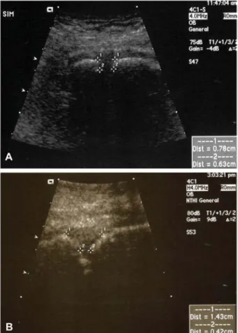
Figure 1 Ultrasonic view of the superior portion of the sym- physis pubis demonstrating the two measurement levels used in the study in a ‘‘T-shaped’’ (A) and a ‘‘Y-shaped’’ (B) superior symphyseal conformation.
Table 1 Characteristics of the sample (ns32).
Age (years) 25.7"8
Parity (%, multiparas) 62%
Body mass index (kg/m2) 29.7"4.4 Race/ethnicity n (%)
Black 11 (34%)
Hispanic 16 (50%)
South Asian 5 (15%)
Delivery mode
Spontaneous vaginal n (%) 29 (91%)
Cesarean n (%) 3 (9%)
Oxytocin use n (%) 3 (16%)
Birth weight (g) 3401"367
Gestational age (weeks) 39.3"1.7
Two measurements were acquired at each examination using the electronic calipers of the machine. The first was the distance between the medial margins of the superior pubic rami at the upper boundary of the symphysis, i.e., immediately inferior to the inferred location of the superior pubic ligament (superior symphyseal breadth); the second was made at the narrowest visualized portion of the symphyseal gap (narrowest symphyseal breadth) (Figure 1). All measurements were made in real-time by a single author; later, a second author blinded to the subject’s identity or stage of labor verified that the images were of good quality and that the calipers had been placed uniformly in each case.
Pertinent information about the subjects was abstracted from their electronic medical records (E&C Medical Intelligence, Inc., New York, NY, USA). All data were entered into a statistical data base (JMP Statistical Software 7.0; SAS Institute, Cary, NC, USA) for analysis. Differences among the measurements obtained at the three intervals during labor were sought using one-way repeated measures analysis of variance (ANOVA). The Student-Newman-Keuls test was employed to identify differenc- es between individual groups in the ANOVA. Pearson correlation coefficients were calculated to assess the relationship between the change in the symphyseal width and other continuous
variables, including birth weight, maternal body mass index (BMI), age, and parity. Data are presented as mean"standard deviation, unless otherwise specified.
Results
Characteristics of the study sample are presented in Table 1. Age of subjects ranged from 14 to 44 years, and they were predominantly multiparous (62%). Three had oxytocin for labor augmentation, and three were deliv- ered by cesarean during the second stage because of concerns about abnormal fetal heart rate patterns. The remaining 29 patients delivered spontaneously. Birth weights ranged from 2870 to 4200 g.
The difference in the symphyseal gap among the three measurement groups was highly significant for both the superior and the narrowest symphyseal breadth (SB) measurements (Table 2). Multiple comparison testing showed that this difference was accounted for by the change between measurements done in the latent phase and active phase when compared to the second stage measurements. The difference in symphyseal breadth between the latent and active phases of the first stage of labor was not significant. There was no correlation between the degree of expansion of the symphysis in the active phase and the degree of attained cervical dilata- tion (Ps0.344).
Although the overall changes were significantly differ- ent, the symphyseal cleft did not expand in all patients.
Widening was observed in 94% at the superior SB meas- urement and in 59% at the narrowest SB. Among cases in which symphyseal gap distension was noted, the median change in the SB dimension was 28% of the original superior diameter and 31% of the narrowest diameter. In some subjects the observed SB change was minimal, while in others the original gap more than dou- bled in width (Table 3). The changes in the SB measure- ments during labor did not differ by parity, and the latent phase SB measurements were not significantly different between nulliparas and multiparas.
372 Rustamova et al., Changes in symphysis pubis width during labor
Article in press - uncorrected proof
Table 2 Symphyseal breadth (cm).
Symphyseal breadth Latent phase Active phase Second stage P-value
Narrowest 0.99"0.47 1.05"0.47 1.21"0.59 0.007
Superior 1.60"0.50 1.66"0.49 2.07"0.56 0.0001
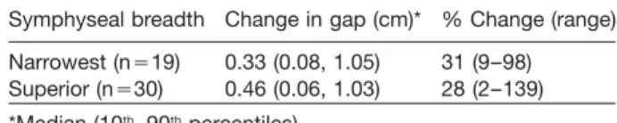
Table 3 Change in symphyseal breadth in cases in which it expanded during labor.
Symphyseal breadth Change in gap (cm)* % Change (range) Narrowest (ns19) 0.33 (0.08, 1.05) 31 (9–98) Superior (ns30) 0.46 (0.06, 1.03) 28 (2–139)
*Median (10th, 90thpercentiles).
The SB diminished between the latent phase and se- cond stage at the narrowest measurement strait in 16%
and at the superior SB in 6% of subjects. It remained unchanged in 25% of cases at the narrow dimension and in none at the superior measurement. In those women in whom the symphyseal gap decreased, the median reduction in diameter was 0.3 cm. The largest observed change was an increase of 1.13 cm at the superior SB and 1.5 cm at the narrowest SB.
There was not a significant correlation between the change in the symphyseal breadth according to birth weight, parity or maternal BMI. When we examined the relationship between the change in the symphyseal width between the latent phase and the second stage accord- ing to maternal age and parity, we found a strong neg- ative correlation in nulliparas (rs–0.85, Ps0.0004) but not in multiparas (rs0.22, Ps0.345).
Discussion
The width of the symphysis increased significantly overall in our sample during the second stage of labor. While the mean SB was greater in the active phase than in the latent phase of the first stage, this trend was not signif- icant. Our data are consistent with those of other invest- igators who suggested that the symphysis may widen during labor w8, 10, 16x. We also have confirmed that changes in the symphysis occur to a varying degree in most women, but are not universal.
Most previous efforts to assess changes in the bony pelvis during labor were done with radiography. Thorp and Frayw16x, for example, showed widening of the sym- physeal gap in 44% of cases in the first stage of labor compared to an evaluation done in the third trimester pri- or to labor. Brehm and Weiraukw8xfound an increase in the width of the symphysis in 54% of patients X-rayed before and after delivery. However, Youngw17xfound no appreciable effect of labor on the symphyseal breadth, and Ohlse´nw12xidentified a change of only about 1 mm
in a few patients. Differences among results of various studies might relate to the difficulty of correcting for magnification in radiographic images, which is influenced by the distance of the measured object from the film. It is also difficult to compare results of studies because measurements were not always taken with subjects in a standardized position. Different postures may alter object-film distances, and change pelvic dimensions. We used ultrasound because it allowed uniformity in posi- tioning of the subjects, in addition to being riskless and causing no discomfort.
Ultrasound was shown by Bjo˜rkland et al. in an ana- tomic model to have precision equivalent to that of radio- graphy in measuring the symphysis pubisw3x. They used ultrasound to study the pubic symphysis during labor at the time of engagement and then in the second stage w4x. They found a very modest increase (averaging -1 mm) in the symphyseal breadth in most subjects, and no change or a narrowing in 9%. Their findings may have been compromised by the fact that subjects were not always studied in the same posture at both time points in labor. Moreover, some had experienced pelvic pain during the pregnancy studied, suggesting that they may have had symphyseal pathology.
Our subjects were evaluated in the supine position with mild hip flexion for all three measurements. None com- plained of pubic pain during pregnancy, labor or the postpartum period. We did not define the subjects’ pelvic types in our study, and were therefore unable to deter- mine whether pelvic architecture influenced changes in the symphyseal breadth during parturition.
Each delivery causes some alteration of the symphy- seal bone and fibrocartilage. In addition to inducing degenerative arthritic changes, each pregnancy might render the pelvic joints more lax w13x. In addition, age causes changes in the symphysis, regardless of parity w13x. This age-associated loss of elasticity and progres- sive fibrosis in the joint might result in reduced ability of the pelvis in reproductively older nulliparas to mold opti- mally as the fetus attempts its odyssey down the birth canal. Our data showing an inverse relation between age and the parturitional widening of the symphyseal gap in nulliparas, but not multiparas, would be consistent with this notion. The multipara may have changes in the sup- porting ligaments that render the symphysis more dis- tensible regardless of age.
Our measurements are generally consistent with sym- physeal gaps noted by several other investigators using
Rustamova et al., Changes in symphysis pubis width during labor 373
Article in press - uncorrected proof
radiographs or ultrasound w1, 8, 10, 11, 14x. Measure- ment can be difficult to standardize because of the fre- quently observed pubic bone surface irregularities and the fact that the bony margins of the symphysis are not always parallel. Also, the profile of the superior portion of the symphysis can take two general shapes (Figure 1b). In some women the medial edges of the pubic bones descend caudad in an arc, giving the upper portion of the symphysis a Y-shaped appearance; in others the bor- ders of the symphysis are parallel throughout, conferring a T-shape to the superior SB. To address the potential difficulty this might introduce in comparing dimensions between subjects, we used two measurements: the dis- tance between the superior pubic rami, and the narrow- est portion of the symphyseal gap visualized in our scans. The minimal SB was not influenced by the con- figuration of the upper portion of the symphysis. Both measurements changed in the same direction in most subjects, regardless of the symphyseal joint shape. The fact that the superior SB enlarged in more subjects than the narrowest SB suggests that symphyseal widening may not occur symmetrically.
In summary, we have demonstrated that the pubic symphysis widens during labor in most women, and that these changes can be readily assessed by ultrasound.
The dynamic properties of the joint are presumably a physiologic adaptation to ease the passage of the fetus through the birth canal. It seems most likely that the observed changes are a passive response to the forces created by the descending fetus, acting on the joint and its surrounding ligaments, the compliance of which has been increased by hormonal changes of pregnancy. We speculate that the extensibility of the pelvic joints during the second stage of labor may be an important factor in determining the probability of a safe vaginal delivery.
References
w1x Abramson D, Roberts SM, Wilson PD. Relaxation of the pelvic joints in pregnancy. Surg Gynecol Obstet. 1934;
58:595–613.
w2x Bahlmann F, Merz E, Macchiella D, Weber G. Sonogra- phische darstellung des symphysenspaltes zur beurteilung
eines symphysenschadens in der schwangerschaft und post partum. Z Geburtshilfe Perinatol. 1993;197:27–30.
w3x Bjo˜rklund K, Bergstro¨m S, Lindgren PG, Ulmsten U. Ultra- sonographic measurement of the symphysis pubis: A potential method of studying symphyseolysis in pregnan- cy. Gynecol Obstet Invest. 1996;42:151–3.
w4x Bjorklund K, Lindgren PG, Bergstrom S, Ulmsten U. Sono- graphic assessment of symphyseal joint distention intra partum. Acta Obstet Gynecol Scand. 1997;76:227–32.
w5x Bjo˜rklund K, Nordstrom ML, Bergstrom S. Sonographic assessment of symphyseal joint distention during preg- nancy and post partum with special reference to pelvic pain. Acta Obstet Gynecol Scand. 1999;78:125–30.
w6x Borell U, Fernstro¨m I. The movements at the sacro-iliac joints and their importance to changes in the pelvic dimen- sions during parturition. Acta Obstet Gynecol Scand.
1957;36:42–57.
w7x Borell U, Fernstro¨m I. Pelvimetric method for the assess- ment of pelvic ‘mouldability’. Acta Radiol. 1957;47:365–9.
w8x Brehm W, Weirauk HV. Separation of the symphysis pubis during labor. Am J Obstet Gynecol. 1928;15:187–91.
w9x Friedman EA. Labor: Clinical Evaluation and Management, second edition. Appleton-Century-Crofts, New York, 1978.
w10x Garagiola DM, Tarver RD, Gibson L, Rogers RE, Wass JL.
Anatomic changes in the pelvis after uncomplicated vagi- nal delivery: a CT study on 14 women. Am J Rhinol. 1989;
153:1239–41.
w11x Heyman J, Lundqvist A. The symphysis pubis in pregnan- cy and parturition. Acta Obstet Gynecol Scand. 1932;12:
191–226.
w12x Ohlse´n H. Moulding of the pelvis during labour. Acta Radiol Diagn. 1973;14:417–34.
w13x Putschar WG. The structure of the human symphysis pubis with special consideration of parturition and its sequelae. Am J Phys Anthropol. 1976;45:589–94.
w14x Scriven MW, Jones DA, McKnight L. The importance of pubic pain following childbirth: a clinical and ultrasono- graphic study of diastasis of the pubic symphysis. J R Soc Med. 1995;88:28–30.
w15x Thoms H. Relaxation of the symphysis pubis in pregnancy.
J Am Med Assoc. 1936;106:1364–6.
w16x Thorp D, Fray W. The pelvic joints during pregnancy and labor. J Am Med Assoc. 1938;11:1162–6.
w17x Young J. Relaxation of the pelvic joints in pregnancy. J Obstet Gynaec Br Emp. 1940;47:493–524.
The authors stated that there are no conflicts of interest regard- ing the publication of this article.
Received June 19, 2008. Revised October 27, 2008. Accepted November 7, 2008. Previously published online March 17, 2009.