Infection prevention in catheter-interventional treatment of children and adults
Infektionsprophylaxe bei katheterinterventionellen Behandlungen von Kindern und Erwachsenen
Abstract
Catheter-interventional treatment is a growing field in pediatric cardi- ology and cardiology, replacing an increasing number of operations.
Heike Schneider
1Thomas Paul
1This article provides an overview of the general practice of hygienic measures and antimicrobial prophylaxis in the cardiac catheterization laboratory to prevent post-procedural infection, particularly if foreign material is utilized.
1 Department of Pediatric Cardiology, Intensive Care Medicine and Neonatology, Keywords:interventional pediatric cardiology, cardiology, infection
prevention
University Medical Center Göttingen, Göttingen, Germany
Zusammenfassung
Die katheterinterventionelle Behandlung ist ein stetig wachsendes Feld der Kinderkardiologie und Kardiologie, das zunehmend Operationen ersetzt. Dieser Artikel gibt einen Überblick über die allgemeine Handha- bung von hygienischen Maßnahmen und antimikrobieller Prophylaxe im Herzkatheterlabor, um postprozedurale Infektionen zu verhindern, insbesondere wenn Fremdmaterial eingesetzt wird.
Schlüsselwörter:Interventionelle Kinderkardiologie, Kardiologie, Infektionsprophylaxe
Introduction
Within the past 20 years, significant progress has been achieved in catheter-interventional treatment of children and adults with congenital cardiac heart disease as well as in patients with acquired cardiac lesions.
These achievements were made possible by the develop- ment of special cardiac implants or devices primarily de- signed for treatment of congenital heart defects and valve lesions, which have subsequently been introduced into clinical practice for patients of all ages.
Nowadays, modifications of these devices are used in adults to close persistent foramen ovale to prevent em- bolic stroke.
In contrast, vascular stents were primarily developed to treat peripheral vascular stenosis and stenotic coronary arteries in the adult population, but have also been used in children with vessel stenosis, such as coarctation of the aorta or peripheral pulmonary artery stenosis, and even to keep a ductus arteriosus patent in infants with either pulmonary or systemic duct-dependent circulations.
Today, these developments allow minimally invasive treatment of a large number of congenital and acquired heart defects by catheter interventions. A significant proportion of cardiac surgery is already being replaced by (and in the future will even more commonly replace) catheter interventions; this enables patients to be mobi- lized earlier and start rehabilitation sooner, with less surgical trauma and pain. Limitations, such as the small size of children, and associated problems; such as vascu- lar access, will be overcome via combined surgical and interventional hybrid procedures.
Examples of catheter-interventional treatment options for congenital and acquired heart defects
Balloon valvuloplasty of stenotic semilunar valves
Balloon dilation is the main treatment option for valvar pulmonary stenosis, with good long-term outcome for patients of all age groups. The rate of re-stenosis is low.
Complications are rare and are usually limited to vascular access sites [1]. Aortic valve stenosis in children can also be treated well by valvuloplasty and a significant reduction of the transvalvular gradient can be achieved [2]. These procedures can be performed in the cardiac catheteriza- tion laboratory without additional measures regarding hygiene standards. Pre-procedural antibiotic prophylaxis is not required [3].
Electrophysiological studies and catheter ablation
Nowadays a large number of cases of supraventricular and ventricular tachycardia can be effectively treated and cured in adults as well as in children by means of radiofre- quency or cryothermal catheter ablation [4]. These pro- cedures can be performed in the cardiac catheter or electrophysiological laboratory under aseptic conditions and without additional hygiene measures. Pre-procedural antibiotic prophylaxis is not required [3].
Catheter-interventional procedures with implantation of non-biological foreign material and implantation of cardiac pacemakers and internal
cardioverter/defibrillators (ICD)
Higher requirements for hygienic conditions exist for these procedures to prevent peri-interventional infection or endocarditis. Ideally, these interventions should be per- formed in specially designed hybrid suites or in an oper- ating room, where surgical procedures can be performed under aseptic conditions with the option of utilizing high- quality fluoroscopy and angiography.
In the conventional cardiac catheterization laboratory, a mobile laminar airflow system positioned at the aseptic catheterization table or operation field (2020 communi- cation with A Kramer; unreferenced, see “Notes”) can be employed as an alternative to prevent contamination of the device by airborne microorganisms.
During implantation, peri-interventional prophylactic anti- biotic prophylaxis with a single dose of cefazolin or cefur- oxime is recommended; data concering infection-preven- tion effectiveness, however, are currently unavailable [3], [5], [6]. After the complete closure of a heart defect, en- docarditis prophylaxis (as secondary prophylaxis, e.g., before a dental procedure) is recommended for 6 months, until complete endothelialization of the device is achieved [3], [7]. Further recommendations are given in the follow- ing according to the particular cardiac defect.
Patent ductus arteriosus
If the ductus arteriosus remains patent, a left-to-right shunt exists that can result in left ventricular volume overload, depending on diameter and length of the duc- tus, with the risk of developing pulmonary arterial hyper- tension. Today, the method of choice after the neonatal period is to close the ductus by catheter intervention.
Several different occluder systems are available for closing the ductus, such as occluding coils and specially- designed double-disc occluder systems made of nitinol.
Almost any ductus independent of size and length can be treated in the catheterization laboratory with these occluders [8].
Access to the ductus can be gained either in an antegrade fashion via the femoral vein through the right atrium, right ventricle and the pulmonary trunk, or in a retrograde ap- proach via the aorta. Size of the child and morphology of the ductus are important parameters to consider when selecting the ideal device. A residual shunt after device occlusion is infrequent, and a secondary intervention is rarely necessary. Peri-interventional antibiotic prophylaxis with cefuroxime is recommended [3]. Potential complica- tions include embolization of the device, obstruction of the aorta or the left pulmonary artery, vascular injury, and hemolysis, which is usually transient.
Interventional duct occlusion in preterm infants and neonates has been decribed. In this age group, however, the procedure is associated with a significantly higher rate of complications. After complete closure of the ductus without residual shunting, endocarditis prophylaxis is re- commended for 6 months [7], [8].
Atrial septal defects
A tissue defect within the atrial septum is present. The atrial septal defect of the secundum type is located centrally within the oval fossa. A left-to-right shunt exists between the atria, resulting in volume overload of the right atrium, right ventricle, and the pulmonary circulation.
As typical sequelae, pulmonary arterial hypertension can develop in early adulthood with secondary decrease of right ventricular function. In addition, types of supra- ventricular tachycardia, such as atrial flutter or atrial fi- brillation, may occur.
The method of choice today to close an atrial septal defect of the secundum type is interventional closure with a double-disc device (Figure 1). There is a wide array of occluders on the market with a diversity of designs and different sizes from several manufacturers [9].
In children, the procedure is performed often under gen- eral anesthesia, as continuous monitoring of the pro-ce- dure by transesophageal echocardiography is used (Fig- ure 2). Monitoring by transthoracic echocardiographic guidance under sedation is the alternative. Complications are rare and include device embolization, cardiac perfor- ation, thromboembolic events, air embolization as well as supraventricular tachyarrhythmias. Peri-interventional antibiotic prophylaxis with cefuroxime is recommended [3].
After the procedure. endocarditis prophylaxis is recom- mended for 6 months, until complete endothelialisation of the device is achieved. In addition, anti-platelet therapy with aspirin at a low dose of 2–5 mg/kg is advised to prevent thrombus formation on the occluder. For adoles- cents and adults, dual antiplatelet therapy with aspirin 100 mg and clopidogrel 75 mg is administered for 6 to 12 months [9].
Ventricular septal defects
The ventricular septal defect is the most common congen- ital heart defect. Ventricular septal defetcs can occur
alone or in combination with other congenital heart de- fects. A defect in the perimembranous region of the inter- ventricular septum is the most prevalent manifestation.
A left-to-right shunt exists at the ventricular level; its quantity depends on the size of the defect and the level of the pulmonary vascular resistance. In general, in- creased pulmonary blood flow with volume overload of the left atrium and ventricle is present. If left untreated, large defects can lead to pulmonary arterial hypertension within the first year of life. Irreversible remodeling of the pulmonary arteries with a progressive increase in vascular resistance can result in reversal shunting via the ventricular defect (Eisenmenger reaction) [10]. In general, ventricular septal defects can be treated by catheter in- tervention. Limitations mainly include the size of the child and the risk of AV block in perimembranous ventricular septal defects in children <20 kg. As in atrial septal de- fects, peri-interventional antibiotic prophylaxis with cefur- oxime is administered, followed by 6 months of endocardi- tis prophylaxis as well as antiplatelet therapy with aspirin at a dose between 2 to 5 mg/kg to prevent apposition of thrombi on the occluder. For adolescents and adults, a dual anitplatelet regimen consisting of aspirin 100 mg and clopidogrel 75 mg for 6–12 months is recommended [10].
Percutaneous pulmonary valve implantation
In patients with complex congenital heart defects who underwent surgery including a conduit between the right ventricle and the pulmonary artery trunk, degenerative processes often occur that lead to significant pulmonary regurgitation and/or obstruction of the right ventricular outflow tract.
As an alternative to a surgical conduit replacement, a catheter-interventional procedure has been developed.
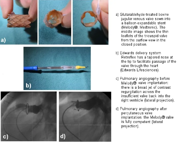
A bovine jugular venous valve, treated with glutaralde- hyde, is sewn into a balloon-expandable valve (Melody®, Medtronic, Minneapolis, Minnesota) (Figure 3). The valved stent can be implanted into the failing conduit with a 22 F delivery system. The Melody®valve has already been im- planted successfully in >12,000 patients worldwide. Initial limitations included the size of the conduit ≤22 mm and a body weight >30 kg. Today, implantation in smaller children is technically feasable with application of a combined catheter-interventional and hybrid pediatric- cardiac-surgical procedure, even in children with a body weight of 10 kg [11]. For larger conduits, native or patched right ventricular outflow tracts, Edwards Sapien valves (Edwards, Lifescience, Irvine, California), originally developed for the calcified aortic valve of the elderly, are increasingly used in the pulmonary position in patients with operated congenital heart disease requiring a smaller delivery system (Figure 3). Large self-expanding valves are nearing clinical approval. The implantation is per- formed under strict aseptic conditions. For implantation, peri-interventional antibiotic prophylaxis with cefazolin or cefuroxime is recommended; at least 3 doses are typ-
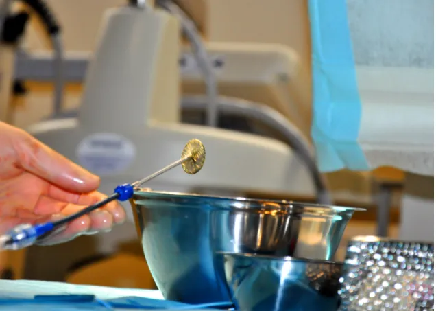
Figure 1: A double-disc device (Occlutech ASD Occluder, Occlutech, Jena, Germany) for closure of an atrial septal defect is already screwed onto the delivery system.
Figure 2: Catheter-interventional closure of an atrial septal defect of the secundum type in a 6-year-old boy
Figure 3: Percutaneous pulmonary valve implantation in a 17-year-old adolescent after surgical implantation of a 22-mm bioprosthetic valved Hancock® conduit (Medtronic) in a patient previously operated on for tetralogy of Fallot and pulmonary atresia: degenerative changes of the conduit ten years after his last open-heart surgery led to significant pulmonary regurgitation
and stenosis, resulting in the indication for pulmonary valve implantation.
ically administered. The incidence of infection or endo- cardits after percutaneous pulmonary valve implantation has been reported to be approximately 3% per patient- year [12]. Uniform protocols, however, are lacking [3], [5], [6]. After valve implantation, life-long endocarditis prophylaxis is recommended [7], and in general antiplate- let therapy with aspirin 2–5 mg/kg (maximum 100 mg) for at least one year.
Transcatheter aortic valve implantation (TAVI)
The most common cardiac lesion in adults is degenerative aortic valve stenosis, a disease of the elderly population.
To minimize the significant risk of open-heart surgery for aortic valve replacement, due to the patients’ advanced age and often associated multimorbidity, transcatheter aortic valve implantation has been developed. Similar to the percutaneous pulmomary valve implantation, a bal- loon- expandable or a self-expanding valve mounted on a stent can be implanted either transfemorally or trans-
apically (in the hybrid suite). This procedure has been shown to be superior to pharmaceutical therapy in high- risk patients. Recently, in patients with an intermediate operative risk, transfemoral aortic valve implantation has not been found to be inferior to surgery [13]. Peri-inter- ventional antibiotic prophylaxis is also performed and supported by the ESC, with cephalosporin monotherapy being used most frequently [14], [15].
Edge-to-Edge-Repair of AV valves
Severe regurgitation of the atrioventricular valves in el- derly patients is associated with a poor prognosis.
Therefore, minimally invasive procedures to reconstruct the mitral valve have been developed. Using the Mitra- Clip® system (Abbott Vascular, Abbott Park, Illinois), catheter interventional ‘edge-to-edge- repair’ can be performed for severe mitral regurgitation. During the procedure, dehiscent valve leaflets are clippped together.
Again, peri-operative antibiotic prophylaxis is recommend- ed for this procedure [15]. Recently, several other system, e.g., the Cardioband™ Mitral System and Cardioband™
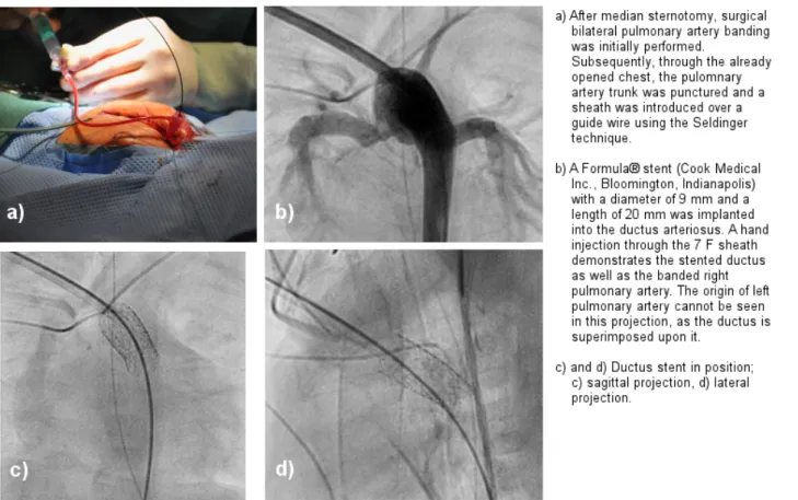
Figure 4: Combined pediatric-cardiac-surgical and catheter-interventional procedure in a hypotrophic newborn baby (body weight 2 kg) with hypopolastic left heart syndrome
Tricuspid Reconstruction System or the Pascal™ system (Edwards Lifesciences, Irvine California), have been de- veloped for treatment of atrioventricular valve regurgita- tion. In the near future, transcatheter valves are expected to enter the market, targeting atrio-ventricular valve im- plantation.
Hybrid procedures
Hybrid procedures are performed in close collaboration between cardiologists and cardiac surgeons, to overcome limitations of vascular access to the heart, to expands the potential of interventional therapy, and reduce the risk of the procedure (Figure 4). These procedures should be performed in a specially designed hybrid suite to meet the hygienic standards of the operating room. For these procedures, peri-procedural antibiotic prophylaxis is also recommended [5], [6].
Notes
Competing interests
The authors declare that they have no competing in- terests.
Acknowledgement
Alternatives were discussted verbally in the chapter
“cardioverter/defibrillators (ICD)”:
Heike Schneider, Thomas Paul (Department of Pediatric Cardiology, Intensive Care Medicine and Neonatology, University Medical Center Göttingen, Germany). Conver- sation with: Prof. Dr. med. Axel Kramer (Institute of Hy- giene and Environmental Medicine, University Medicine Greifswald, Germany). 2020 Dec 09.
References
1. Jux C, Weil J, Oberhoffer, R Bennink G. Leitlinie Pädiatrische Kardiologie: Valvuläre Pulmonalstenose. Düsseldorf: dgpk; 2017.
Available from: http://www.kinderkardiologie.org/fileadmin/user_
upload/Leitlinien/LL_valvPstnachVSS.pdf
2. Michel-Behnke I, Murin P, Sarikouch S. S2k-Leitlinie Pädiatrische Kardiologie: Kongenitale Aortenklappenstenose. Düsseldorf:
dgpk; 2020. Available from: http://www.kinderkardiologie.org/
fileadmin/user_upload/Leitlinien/LL_vAS_2020_11_25_final.pdf 3. Knirsch W. Empfehlungen der Arbeitsgemeinschaft
Interventionelle Kardiologie der Deutschen Gesellschaft für Pädiatrische Kardiologie (DGPK) zur präinterventionellen Antibiotika-Prophylaxe bei diagnostischen und interventionellen Herzkatheteruntersuchungen. Düsseldorf: dgpk; 2016. Available from: http://www.kinderkardiologie.org/fileadmin/user_upload/
Stellungnahmen/Antibiotika_Periinterventionelle_
Endokarditisprophylaxe_final_20160510__002_.pdf
4. Paul T, Gebauer R, Kriebel T, Schneider H, Janousek J. Leitlinie Pädiatrische Kardiologie: Tachykarde Herzrhythmusstörungen.
Düsseldorf: dgpk; 2018. Available from: http://
www.kinderkardiologie.org/fileadmin/user_upload/Leitlinien/
Leitlinie_Tachykardien_DGPK_2018_11_28_final.pdf 5. Bratzler DW, Dellinger EP, Olsen KM, Perl TM, Auwaerter PG,
Bolon MK, Fish DN, Napolitano LM, Sawyer RG, Slain D, Steinberg JP, Weinstein RA; American Society of Health-System
Pharmacists; Infectious Disease Society of America; Surgical Infection Society; Society for Healthcare Epidemiology of America.
Clinical practice guidelines for antimicrobial prophylaxis in surgery. Am J Health Syst Pharm. 2013 Feb;70(3):195-283. DOI:
10.2146/ajhp120568
6. Baddour LM, Epstein AE, Erickson CC, Knight BP, Levison ME, Lockhart PB, Masoudi FA, Okum EJ, Wilson WR, Beerman LB, Bolger AF, Estes NA 3rd, Gewitz M, Newburger JW, Schron EB, Taubert KA; American Heart Association Rheumatic Fever, Endocarditis, and Kawasaki Disease Committee; Council on Cardiovascular Disease in Young; Council on Cardiovascular Surgery and Anesthesia; Council on Cardiovascular Nursing;
Council on Clinical Cardiology; Interdisciplinary Council on Quality of Care; American Heart Association. Update on cardiovascular implantable electronic device infections and their management:
a scientific statement from the American Heart Association.
Circulation. 2010 Jan;121(3):458-77. DOI:
10.1161/CIRCULATIONAHA.109.192665
7. Wilson W, Taubert KA, Gewitz M, Lockhart PB, Baddour LM, Levison M, Bolger A, Cabell CH, Takahashi M, Baltimore RS, Newburger JW, Strom BL, Tani LY, Gerber M, Bonow RO, Pallasch T, Shulman ST, Rowley AH, Burns JC, Ferrieri P, Gardner T, Goff D, Durack DT; American Heart Association Rheumatic Fever, Endocarditis, and Kawasaki Disease Committee; American Heart Association Council on Cardiovascular Disease in the Young;
American Heart Association Council on Clinical Cardiology;
American Heart Association Council on Cardiovascular Surgery and Anesthesia; Quality of Care and Outcomes Research Interdisciplinary Working Group. Prevention of infective endocarditis: guidelines from the American Heart Association: a guideline from the American Heart Association Rheumatic Fever, Endocarditis, and Kawasaki Disease Committee, Council on Cardiovascular Disease in the Young, and the Council on Clinical Cardiology, Council on Cardiovascular Surgery and Anesthesia, and the Quality of Care and Outcomes Research Interdisciplinary Working Group. Circulation. 2007 Oct;116(15):1736-54. DOI:
10.1161/CIRCULATIONAHA.106.183095
8. Lindinger A, Kozlik-Feldmann R, Cesnjevar R. Leitlinie Pädiatrische Kardiologie: Persistierender Ductus arteriosus.
Düsseldorf: dgpk; 2015. Available from: http://
www.kinderkardiologie.org/fileadmin/user_upload/Leitlinien/
LL.PDA.aktualisiert.final.AS.VSS.29.4.15.pdf
9. Jux C, Vogel MJ, Scheewe J, Weil J. S2k-Leitlinie Pädiatrische Kardiologie: Vorhofseptumdefekt. Düsseldorf: dgpk; 2019.
Available from: http://www.kinderkardiologie.org/fileadmin/user_
upload/Leitlinien/2019_11_27_LL_ASD.total_
final.27.10.2019.li.pdf
10. Gorenflo M, Schirmer KR, Dewald O. Leitlinie Pädiatrische Kardiologie: Der isolierte Ventrikelseptumdefekt. Düsseldorf:
dgpk; 2019. Available from: http://www.kinderkardiologie.org/
fileadmin/user_upload/Leitlinien/2019_11_27_
VSDnachFulda20.9.2019.final.li.pdf
11. Nordmeyer J, Ewert P, Gewillig M, AlJufan M, Carminati M, Kretschmar O, Uebing A, Dähnert I, Röhle R, Schneider H, Witsenburg M, Benson L, Gitter R, Bökenkamp R, Mahadevan V, Berger F. Acute and midterm outcomes of the post-approval MELODY Registry: a multicentre registry of transcatheter pulmonary valve implantation. Eur Heart J. 2019 07;40(27):2255-64. DOI: 10.1093/eurheartj/ehz201 12. McElhinney DB, Sondergaard L, Armstrong AK, Bergersen L,
Padera RF, Balzer DT, Lung TH, Berger F, Zahn EM, Gray RG, Hellenbrand WE, Kreutzer J, Eicken A, Jones TK, Ewert P.
Endocarditis After Transcatheter Pulmonary Valve Replacement.
J Am Coll Cardiol. 2018 12;72(22):2717-28. DOI:
10.1016/j.jacc.2018.09.039
13. Kuck KH, Eggebrecht H, Elsässer A, Hamm C, Haude M, Ince H, Katus H, Möllmann H, Naber CK, Schunkert H, Thiele H, Werner N. Qualitätskriterien zur Durchführung der kathetergestützten Aortenklappenimplantation (TAVI). Aktualisierung des Positionspapiers der Deutschen Gesellschaft für Kardiologie.
Kardiologe. 2016;10:282-300. DOI: 10.1007/s12181-016- 0082-4
14. Harding D, Cahill TJ, Redwood SR, Prendergast BD. Infective endocarditis complicating transcatheter aortic valve implantation.
Heart. 2020 04;106(7):493-8. DOI: 10.1136/heartjnl-2019- 315338
15. Habib G, Lancellotti P, Antunes MJ, Bongiorni MG, Casalta JP, Del Zotti F, Dulgheru R, El Khoury G, Erba PA, Iung B, Miro JM, Mulder BJ, Plonska-Gosciniak E, Price S, Roos-Hesselink J, Snygg- Martin U, Thuny F, Tornos Mas P, Vilacosta I, Zamorano JL; ESC Scientific Document Group. 2015 ESC Guidelines for the management of infective endocarditis: The Task Force for the Management of Infective Endocarditis of the European Society of Cardiology (ESC). Endorsed by: European Association for Cardio-Thoracic Surgery (EACTS), the European Association of Nuclear Medicine (EANM). Eur Heart J. 2015 Nov;36(44):3075- 128. DOI: 10.1093/eurheartj/ehv319
Corresponding author:
PD Dr. Heike Schneider
Department of Pediatric Cardiology, Intensive Care Medicine and Neonatology, University Medical Center Göttingen, Göttingen, Robert-Koch-Str. 40, 37075 Göttingen, Germany, Phone: +49 551-3962580, Fax:
+49 551-3922561
heike.schneider@med.uni-goettingen.de
Please cite as
Schneider H, Paul T. Infection prevention in catheter-interventional treatment of children and adults. GMS Hyg Infect Control.
2021;16:Doc14.
DOI: 10.3205/dgkh000385, URN: urn:nbn:de:0183-dgkh0003858
This article is freely available from
https://www.egms.de/en/journals/dgkh/2021-16/dgkh000385.shtml Published:2021-03-24
Copyright
©2021 Schneider et al. This is an Open Access article distributed under the terms of the Creative Commons Attribution 4.0 License. See license information at http://creativecommons.org/licenses/by/4.0/.