Kalkulierte parenterale Initialtherapie bakterieller Infektionen: Bakterielle Endokarditis
Zusammenfassung
Dies ist das zwölfte Kapitel der von der Paul-Ehrlich-Gesellschaft für Chemotherapie e.V. (PEG) herausgegebenen S2k Leitlinie „Kalkulierte
Pascal M. Dohmen
1Klaus Friedrich Bodmann
2parenterale Initialtherapie bakterieller Erkrankungen bei Erwachsenen – Update 2018“ in der 2. aktualisierten Fassung.
Wolfgang Graninger
3Die bakterielle Endokarditis ist gekennzeichnet durch eine gleichblei-
bende Inzidenz, aber eine Veränderung in der betroffenen Patientenpo-
Pramod Shah
4pulation durch den Einsatz von Klappenprothesen, Fremdmaterialien
Florian Thallhammer
5wie Schrittmachern und den zunehmenden Einsatz invasiver medizini- scher Maßnahmen. Damit verbunden ist eine Veränderung des Erreger-
spektrums hin zu Staphylokokken auch bei der Nativklappen-Endokar- 1 Klinik und Poliklinik für Herzchirurgie,
ditis. Dieses Kapitel gibt Empfehlungen für das interdsiziplinäre Mana-
gement der bakteriellen Endokarditis von der Diagnostik über die Prä- Universitätsmedizin Rostock, Deutschland
vention zur Therapie mit dem Fokus auf der Antibiotika-Therapie und
evaluiert die aktuellen Empfehlungen der ESC aus deutscher Sicht. 2 Klinik für Internistische Intensiv- und Notfallmedizin und Klinische Infektiologie, Klinikum Barnim GmbH, Werner Forßmann Krankenhaus, Eberswalde, Deutschland
3 Wien, Österreich 4 Frankfurt am Main,
Deutschland
5 Klinische Abteilung für Infektiologie und
Tropenmedizin, Medizinische Universität Wien, Österreich
Einleitung
Die bakterielle Endokarditis wurde zum ersten Mal im 17. Jahrhundert durch Morgagni [1] erwähnt. Die Ätiologie dieser Infektion wurde erst 100 Jahre später durch Roki- tansky beschrieben. Hier wurden Bakterien identifiziert, die sich innerhalb embolisierter Vegetationen befanden [2]. Heute ist die bakterielle Endokarditis weiterhin eine Erkrankung mit erheblicher Morbidität und Letalität, auch wenn sich die antimikrobiellen und chirurgischen Inter- ventionsmaßnahmen substantiell verbessert haben [3].
Die Häufigkeit der bakteriellen Endokarditis beträgt 1 Fall pro 1.000 internistische Krankenhausaufnahmen [4].
Die jährliche Inzidenz der bakteriellen Endokarditis nativer Herzklappen liegt in Europa bei etwa 3 Fällen auf 100.000 Einwohner [5]. Wie verschiedene Studien gezeigt haben, ist die Inzidenz der bakteriellen Endokarditis in den letzten Jahrzehnten nahezu konstant geblieben [6];
dies ist auf eine Umverteilung prädisponierender Faktoren
zurückzuführen. Eine systematische Übersichtsarbeit aus mehreren westeuropäischen Ländern zeigte eine deutli- che Zunahme von Patienten mit Herzklappenprothese- nendokarditis sowie einer Endokarditis auf Basis einer degenerativen Herzklappenerkrankung oder intravenösem Drogenabusus [7]. Durch den Anstieg von invasiven me- dizinischen Prozeduren hat die Zahl der Bakteriämien und damit der Anteil der bakteriellen Healthcare-asso- ciated Endokarditiden zugenommen [8]. Diese Zunahme von bis zu 34% an allen Endokarditiden [9] hat ebenfalls zur Folge, dass während der letzten Jahre eine Verschie- bung des Durchschnittsalters um 10–15 Jahre nach oben stattfand [10], [11].
Klinik
Ein großes Problem stellt die lange Verzögerung von dem Auftreten erster unerkannter Symptome und dem Anfang
einer mikrobiellen Invasion des endokardialen Herzgewe- bes bei intravaskulär implantierten Fremdkörpermateria- lien wie Herzklappenprothesen, Schrittmacherelektroden, Micra Kardiokapseln, Okkludern und Kunstherzen dar.
Insbesondere in Deutschland kommt der hohe Anteil an Kultur-negativen Endokarditiden hinzu, welche die defini- tive Diagnose erschweren. Die daraus resultierende Ver- zögerung der Einleitung einer adäquaten Antibiotika- Therapie kann erheblich dazu beitragen, dass sich Mor- bidität und Letalität erhöhen [12]. Klassische Leitsympto- me sind oft schwer zu beurteilen, wie z.B. ein Herzge- räusch bei einem bisher kardiologisch nicht untersuchten Patienten oder unspezifische Symptome wie subfebrile Temperaturen, Fieber, Nachtschweiß, Gewichtsverlust, Appetitlosigkeit, allgemeine Abgeschlagenheit, Myalgien und Arthralgien. Nicht selten sind erste Symptome bereits Zeichen eingetretener Komplikationen, wie die progre- diente Belastungsdyspnoe und anschließende Orthopnoe, die auf eine Klappendestruktion mit Ventilfunktionsverlust durch eine Herzklappeninsuffizienz mit einer bedeutsa- men Volumenbelastung hinweist. Zeichen der zentralen septischen Embolisierung können als Erstsymptome neurologische Ausfälle sein. Bei Rechtsherzendokarditis kann es zu Symptomen einer pulmonalen Embolisierung kommen. Weiterhin können periphere Mikro- oder Makro- embolien in Kombination mit immunologischen Phäno- menen vorhanden sein, z.B. Osler-Knötchen, Jayneway- Läsionen und Splinter-Hämorrhagien.
Beim Vorliegen von Risikofaktoren wie intravenösem Drogenabusus oder bei Patienten mit intravaskulär im- plantierten Fremdmaterialien muss auch bei unspezifi- schen Symptomen differentialdiagnostisch immer an eine bakterielle Endokarditis gedacht werden. Zur klinischen Beurteilung sollten bei diesen Patienten immer die modi- fizierten Duke-Kriterien angewendet werden [13] sowie gegebenenfalls ein Endokarditis-Expertenteam aus einem nahegelegenen Klinikum der Maximalversorgung einbe- zogen werden [14].
Prävention
Da die bakterielle Endokarditis eine gefährliche und schwierig zu behandelnde bakterielle Infektion ist, wird bei Patienten mit erhöhtem Risiko seit über 50 Jahren eine Endokarditis-Prophylaxe durchgeführt. Im Jahre 2002 wurden die Indikationen für diese Prophylaxe im Rahmen einer Risiko-Nutzen-Analyse [15] neu definiert. Diese Er- gebnisse wurden in den vergangenen Jahren sukzessiv in alle internationalen Leitlinien übernommen [16], [17], [18], [19], [20].
In den im Jahr 2007 erschienenen Leitlinien der American Heart Association [16] wurden die neuen Indikationen für eine Durchführung der Endokarditis-Prophylaxe imple- mentiert [21]. Entscheidend war, dass man sich heute ausschließlich auf evidenzbasierte Daten beruft. Die zuvor formulierten Leitlinien basierten auf Tierversuchen und Expertenmeinungen und nicht auf prospektiven, rando- misierten und doppelblind durchgeführten Studien, die
heute zu fordern sind. Durch die Veränderung der Indika- tion der Endokarditis-Prophylaxe wird diese heute nur noch bei Patienten mit höchstem Risiko für eine bakteri- elle Endokarditis durchgeführt.
Im Jahr 2008 hat das britische National Institute for He- alth and Care Excellence (NICE) die Empfehlung gegeben, sowohl bei Patienten mit zahnärztlichen als auch bei nicht zahnärztlichen Eingriffen unabhängig vom Risiko auf eine Endokarditis-Prophylaxe zu verzichten [22]. Eine nach dieser Empfehlung durchgeführte epidemiologische Stu- die in Großbritannien zeigte einen Anstieg der Zahl der bakteriellen Endokarditiden nach Inkrafttreten der NICE- Richtlinien aus dem Jahr 2008 [23]. Eine im Jahr 2012 in Großbritannien durchgeführte Erhebung ergab, dass die Mehrheit der Kardiologen und Herzchirurgen die Notwendigkeit einer Antibiotika-Prophylaxe bei Patienten mit Herzklappenprothesen befürwortet [24]. Eine weitere im Vereinigten Königreich durchgeführte Studie zeigte einen signifikanten Anstieg der Inzidenz von bakteriellen Endokarditiden, sowohl bei Hochrisiko- wie auch bei Niedrigrisikopatienten, so dass die Einführung der NICE- Richtlinien 2008 nicht als alleinige Ursache dieser Tatsa- che herangezogen werden kann [8].
Die interdisziplinäre Betreuung
Die interdisziplinäre Behandlung von Patienten mit bak- terieller Endokarditis beginnt mit einer guten Kommuni- kation zwischen dem zuweisenden niedergelassenen Arzt und dem aufnehmenden Krankenhausarzt. Innerhalb des Krankenhauses muss die Interdisziplinarität fortge- führt werden.
Die Voraussetzung für die Aufnahme eines Patienten in die multizentrische randomisierte SYNTAX-Studie, die sich Patienten mit koronarer Herzerkrankung gewidmet hat, war die interdisziplinäre Zusammenarbeit zwischen Kardiologen und Herzchirurgen [25]. Dieses Konzept ei- nes fachübergreifenden Herzteams zur Festlegung des Prozederes der Therapie wurde für Höchstrisiko-Patienten, die an einer Aortenklappenerkrankung leiden, übernom- men. Hierbei wird interdisziplinär festgelegt, welcher Pa- tient einen herkömmlichen chirurgischen Aortenklappen- Ersatz bekommt und welcher Patient einer Transkatheter- Aortenklappen-Implantation (TAVI) zugeführt werden soll.
Dieses Team, bestehend aus Kardiologen und Herzchirur- gen, wurde durch einen Anästhesiologen erweitert [26].
Die Konzeption ist heute fester Bestandteil der Europäi- schen Richtlinien.
Bisher sahen die kardiologischen Leitlinien für die Behand- lung von Patienten mit bakterieller Endokarditis ein inter- disziplinäres Ärzteteam bestehend aus Kardiologen, Mi- krobiologen, Infektiologen und Herzchirurgen vor. Auf- grund der vorhandenen Komplexität dieser Erkrankung sollten weitere Experten wie Neurologen, Neurochirurgen, Radiologen, Nuklearmediziner und Anästhesiologen sowie klinische Pharmakologen und im Falle von kongenitalen Vitien, Kinderkardiologen und Kinderherzchirurgen hinzu- gezogen werden. Durch die Einführung dieser Maßnah-
men konnte in einer französischen Studie die Letalität einer Endokarditis signifikant von 18,5% auf 8,2% gesenkt werden [27]. Durch Übernahme dieses Konzeptes konnte auch andernorts das Überleben von Patienten mit bakte- rieller Endokarditis maßgeblich verbessert werden [28].
Im Jahr 2014 wurden diese Vorgaben auch in die Leit- linien für die Behandlung von Patienten mit Herzklappen- erkrankung [19] der American Heart Association/ des American College of Cardiology mit einer Klasse Ib-Emp- fehlung übernommen. Die European Society of Cardiology passte sich bei der Erstellung der im Jahre 2015 veröf- fentlichten ESC Leitlinien zur Behandlung von Patienten mit bakterieller Endokarditis diesen Vorgaben an [29].
Bildgebung
Die Grundlage in der bildgebenden Diagnostik stellt die Echokardiografie in der transthorakalen und vor allen Dingen in der transösophagealen Untersuchungsform dar. Diese Untersuchungsmethode ist von großer Bedeu- tung für die präoperative Diagnostik, für das präoperative Monitoring, für die Evaluation des operativen Ergebnisses sowie für die postoperative Verlaufskontrolle. Aufgrund der Komplexität der heutigen Endokarditis mit einem Anstieg der Healthcare-assoziierten bakteriellen Endokar- ditis werden jedoch weitere bildgebende Verfahren benö- tigt, wie die Magnetresonanztomographie, Multislice Computertomographie oder nuklearmedizinische Ver- fahren wie die Einzelphotonen-Emissions-Tomographie (SPECT) und die18F-Flurdesoxyglucose (FDG)-Positronen- Emissions-Tomographie. Auf die Notwendigkeit der Echokardiographie als Basisuntersuchung wurde bereits hingewiesen. Sie nimmt eine Schlüsselstellung in Bezug auf Diagnostik und Verlaufsbeurteilung ein. Die trans- ösophageale Echokardiografie ist der transthorakalen bezüglich der Sensitivität signifikant überlegen [30], [31], insbesondere bei Fragestellungen, die die Trikuspidal- und die Pulmonalklappe bei implantierten Fremdkörper- materialien betreffen. In Bezug auf eine floride Endokar- ditis ist die Spezifität der Echokardiographie jedoch ein- geschränkt [32]. Bei klinischen Verdachtskriterien wie z.B. unklarem Fieber mit positiven Blutkulturen, bei denen typische Erreger nachgewiesen worden sind, ist eine Ab- klärung einschließlich einer echokardiographischen Un- tersuchung jedoch unbedingt notwendig. Wird eine aktiv bestehende Endokarditis nachgewiesen, ist eine wöchent- liche echokardiographische Verlaufskontrolle sinnvoll.
Im Hinblick auf den prozentualen Anstieg positiver Blut- kulturen durch die zunehmende Implementation von Fremdmaterialien durch invasive Prozeduren in unsere Patienten und durch die immer häufiger durchgeführten weniger invasiven TAVI sind weiterführende bildgebende Verfahren wie oben beschrieben von zunehmender Wichtigkeit. So haben z.B. Studien gezeigt, dass zerebrale septische Embolien in 33% der Patienten mit Endokarditis ohne klinische Symptomatik vorkommen [33]. Auch diese Erkenntnis konnte nur durch die Anwendung weiterer bildgebender Verfahren gewonnen werden.
Erregernachweis
Der Nachweis des ursächlichen Erregers ist für eine schnelle und zielgerichtete Therapie entscheidend. Von größter Bedeutung ist eine korrekte Abnahme von min- destens drei Blutkulturpaaren (aerob und anaerob) zu unterschiedlichen Zeitpunkten über eine periphere Ve- nenpunktion unter strikter Einhaltung der aseptischen Bedingungen für die Venenpunktion und die Entnahme der Probe. Venöse Blutkulturen sind arteriellen aufgrund strömungsdynamischer Faktoren überlegen.
Da bei einer bakteriellen Endokarditis eine kontinuierliche Bakteriämie vorliegt, kann die Abnahme der Blutkulturen jederzeit stattfinden, ohne die Körpertemperatur zu be- rücksichtigen. Wichtig ist die Tatsache, dass Fieberspit- zen, die durch einen Zerfall von Erregern entstehen, sogar einen ungünstigen Zeitpunkt zur Abnahme der Blutkultu- ren darstellen, da zu diesem Zeitpunkt die Erregerdichte im Blut besonders gering ist. Die abgenommenen Blutkul- turen müssen umgehend in ein mikrobiologisches Labor weitergeleitet werden.
In bis zu 30% der Fälle bleiben Blutkulturen ohne Erreger- nachweis, was häufig durch eine vor der Kulturabnahme initiierte Antibiotika-Therapie verursacht wird. Bei nicht kritisch kranken Patienten mit Endokarditis und negativen Blutkulturen sollte die Antibiotika-Therapie für einen Zeitraum von 48 Stunden unterbrochen werden, um dann bessere Voraussetzungen für einen Erregernachweis bei abzunehmenden Blutkulturen zu erzielen. Eine weitere Ursache negativer Blutkulturen kann das Vorliegen von schwer kultivierbaren Erregern wie zum BeispielBartonel- laspp.,Coxiellaspp.,Mycoplasmaspp.,Chlamydiaspp., Tropheryma whipplei, Pilzen und Erregern der HACEK- Gruppe sein. Die HACEK-Gruppe umfasstHaemophilus spp.,Actinobacillus actinomycetemcomitans,Cardiobac- terium hominis,Eikenella corrodens und Kingellaspp.
Es ist wichtig, dem mikrobiologischen Labor die Verdachts- diagnose einer Endokarditis mitzuteilen, um eine adäqua- te Untersuchung der Materialien zu gewährleisten. Aktuell gibt es kulturunabhängige Techniken zur Optimierung der mikrobiologischen Diagnostik durch Anwendung molekularbiologischer und serologischer Methoden. Die Fluoreszenz-in-situ-Hybridisierung (FISH) kombiniert mo- lekularbiologische und histologische Techniken, die ebenfalls beim Nachweis von Erregern hilfreich sein können. Neben der Polymerase-Kettenreaktion (PCR) aus Vollblut oder Serum zum Erregernachweis können auch Biopsien von Herzklappen, Herzklappenprothesen oder peripheren Embolien weiterhelfen. Aufgrund einer fehlen- den Standardisierung ist die Relevanz dieser Methoden zurzeit noch unklar.
Desweitern kann die Sonikation von Prothesen, sprich Herzschrittmacher oder Herzklappen, den Nachweis von Erregern um 30% steigern.
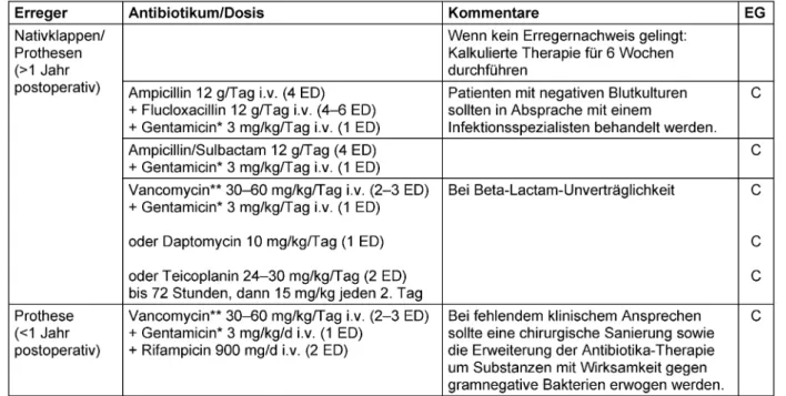
Tabelle 1: Empirische Therapie der kulturnegativen bakteriellen Endokarditis bei vorheriger Antibiotika-Therapie bzw. bis zum Erhalt der Blutkulturergebnisse. Bei kulturnegativer Endokarditis ohne vorherige Antibiotika-Therapie sollte ein Infektionsexperte
zur Rate gezogen werden.
Antimikrobielle Therapie
Bei einem kritischen Allgemeinzustand des Patienten wird umgehend, aber stets nach Entnahme der Blutkultu- ren, mit einer empirischen Antibiotika-Therapie begonnen.
Bei Nativklappenendokarditis sowie der späten Endokar- ditis nach Herzklappenersatz (>1 Jahr nach der Operation oder Intervention) sind vor allem Methicillin-sensible Staphylococcus-aureus-Stämme (MSSA), verschiedene Streptokokken-Spezies undEnterococcus faecaliszu er- warten (Tabelle 1) Eine Hilfestellung für die kalkulierte antimikrobielle Therapie kann das mikrobiologische Er- gebnis aus der ersten Infektionsepisode geben. Bei der frühen Endokarditis nach Herzklappenersatz (<1 Jahr nach der Intervention oder Operation) ist dagegen gehäuft an Methicillin-resistenteStaphylococcus-aureus-Stämme (MRSA), Koagulase-negative Staphylokokken aber auch gramnegative Bakterien zu denken. Die empirisch begon- nene Therapie sollte nach Vorliegen der mikrobiologi- schen Ergebnisse, wenn möglich, modifiziert werden.
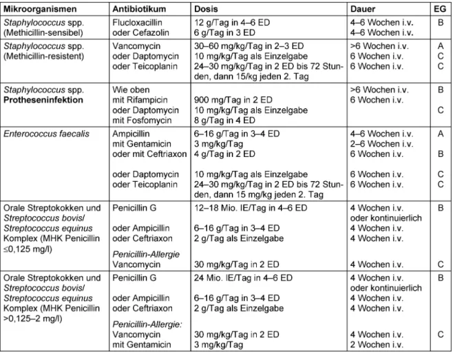
Therapieschemata für die häufigsten Endokarditis-Erreger finden sich in Tabelle 2. Weitere detaillierte Therapieemp- fehlungen sowie Empfehlungen zum Management von Komplikationen zur chirurgischen Indikationsstellung und zur Fortführung der Antibiotika-Therapie nach chirurgi- scher Sanierung finden sich in den Leitlinien der Europäi- schen Gesellschaft für Kardiologie [29].
Bewertung der Empfehlungen zur Antibiotika-Therapie der
Endokarditis der Europäischen Gesellschaft für Kardiologie (ESC) von 2015
Wesentliche Änderungen in der Neuauflage der europäi- schen Leitlinien betreffen die Endokarditis-Prophylaxe, das interdisziplinäre „Endokarditis-Team“, die Erweiterung der Bildgebungsmodalitäten und die Ergebnisse einer ersten randomisierten Studie zur operativen Behandlung der bakteriellen Endokarditis.
Die europäischen Leitlinien halten an einer Antibiotika- Prophylaxe bei Risikoeingriffen von Patienten mit prädis- ponierenden kardialen Faktoren fest. Erste Studien unter- stützen dieses Vorgehen, da sich nach der Einführung der NICE-Richtlinien in 2008 [21] negative Effekte durch die extreme Einschränkung der Indikation der Antibiotika- Prophylaxe in Großbritannien gezeigt hatten [23], [24].
Die Empfehlung, bei der Nativklappenendokarditis empi- risch mit Ampicillin und Flucloxacillin zu therapieren, re- sultiert nicht aus Studiendaten, sondern aus der Notwen- digkeit, in der Initialtherapie Staphylokokken, Streptokok- ken undEnterococcus faecaliszu erfassen. Da nicht in allen europäischen Ländern Ampicillin/Sulbactam zur parenteralen Therapie verfügbar ist und die Kombination Amoxicillin/Clavulansäure aus Toxizitätsgründen nicht ausreichend hoch dosiert werden kann, wurde die Kom- bination Aminopenicillin plus Flucloxacillin empfohlen.
Da in Deutschland Ampicillin/Sulbactam zur parenteralen
Tabelle 2: Übersicht über die gebräuchlichen Antibiotika bei bakterieller Endokarditis
Therapie zur Verfügung steht, wären 4x 3 g Ampicillin/Sul- bactam eine gleichwertige Alternative, die den Vorteil hat, mit nur einem Arzneimittel bzw. einer fixen Kombination, umgehen zu müssen.
Chirurgische Therapie und Nachsorge
Die Einführung eines interdisziplinären Endokarditis- Teams hat gezeigt, dass die chirurgische Therapie inte- graler Bestandteil des gesamten Therapiekonzeptes der bakteriellen Endokarditis ist und nicht erst dann durchge- führt werden darf, wenn es zu einem Versagen der Anti- biotika-Therapie gekommen ist. Die klassischen Indika- tionen zur operativen Sanierung, die bei etwa 50% aller Patienten angewendet werden müssen, sind eine entstan- dene schwere Herzinsuffizienz, die irreversible und infek-
tionsbedingte Zerstörung kardialer Strukturen und das Auftreten von Embolien, neu aufgetretene AV-Blockierun- gen und Endokarditiden bei Herzklappenprothesen, Transkatheterherzklappen, Schrittmachersystemen und Verschlusssystemen. Die für eine operative Risikostratifi- zierung notwendigen Scores (EuroSCORE II und STS- Score) sind hierzu nur eingeschränkt geeignet, da sie primär für eine koronare Revaskularisation oder einen degenerativen Herzklappenersatz entwickelt wurden. Für eine chirurgische Intervention bei bakterieller Endokarditis wurden spezielle Score-Systeme entwickelt, wie zum Beispiel der De-Feo-Score und der International Collabo- ration on Endocarditis-Score (ICE-Score).
Der Vorteil der frühzeitigen chirurgischen Sanierung ist, dass speziell bei der nativen Mitral- oder Trikuspidalklap- pen-Endokarditis, Herzklappen erhaltend operiert werden kann, ggf. sogar minimalinvasiv. Die Indikation für eine frühe operative Sanierung ist häufig schwierig und eine
wichtige Aufgabe des „interdisziplinären Endokarditis- Teams“. Eine randomisierte Studie an einem Niedrig- Risiko-Kollektiv mit kleinen Fallzahlen zeigte, dass durch einen chirurgischen Eingriff innerhalb von 48 Stunden die Embolisationsrate bis 6 Monate nach der Operation signifikant gesenkt werden konnte. Das Überleben der Patienten wurde jedoch nicht beeinflusst.
Eine spezielle Patientenpopulation stellen die Patienten dar, die mit einer TAVI versorgt werden. Um diese Patien- ten in Zukunft optimal therapieren zu können, müssen neue Strategien entwickelt werden.
Grundprinzip der chirurgischen Sanierung ist die Entfer- nung des kompletten infektiösen Materials und Gewebes, um dann unter adäquater Antibiotika-Therapie eine Aus- heilung der Endokarditis zu erzielen. In Österreich wird die adäquate Antibiotika-Therapie teilweise in Form einer ambulanten parenteralen Antibiotika-Therapie (APAT) durchgeführt. In Deutschland hat sich diese Therapieform bisher nicht durchgesetzt.
Bildgebungen (wie zum Beispiel echokardiographische Verfahren), Blutkulturen und serologische Untersuchun- gen sollten den Therapieerfolg kontrollieren und wenn nötig dazu beitragen, dass die Therapiemaßnahmen entsprechend adjustiert werden.
Anmerkung
Dies ist das zwölfte Kapitel der von der Paul-Ehrlich-Ge- sellschaft für Chemotherapie e.V. (PEG) herausgegebenen S2k Leitlinie „Kalkulierte parenterale Initialtherapie bak- terieller Erkrankungen bei Erwachsenen – Update 2018“
in der 2. aktualisierten Fassung.
Nach Veröffentlichung der 1. Version der Leitlinie wurden von der Arbeitsgruppe folgende Dosierungsvorschläge aktualisiert (Tabelle 1: Empirische Therapie der kultur- negativen bakteriellen Endokarditis bei vorheriger Anti- biotika-Therapie bzw. bis zum Erhalt der Blutkultur- ergebnisse):
• Gentamicin-Dosierung in Kombination mit Ampicillin, Ampicillin/Sulbactam bzw. Vancomycin: 1x 3 mg/kg ANSTATT 3x 3 mg/kg
• Rifampicin-Dosierung in Kombination mit Vancomycin:
Rifampicin 900 mg i.v. (2 ED) ANSTATT Rifampicin 900 mg i.v. (3 ED)
Interessenkonflikte
Die Autoren erklären, dass sie keine Interessenkonflikte in Zusammenhang mit diesem Artikel haben.
Literatur
1. Glynn TR. The Mumleian Lectures on Infective endocarditis mainly in its clinical aspects. Lancet. 1903;161(4155):1073-7. DOI:
10.1016/S0140-6736(01) 71890-9
2. Kirkes WS. On some of the principal effects resulting from the Detachment of Fibrinous Deposits from the interior of the heart, and their mixture with the circulating blood. Med Chir Trans.
1852;35:281-324.
3. Krul MM, Vonk AB, Cornel JH. Trends in incidence of infective endocarditis at the Medical Center of Alkmaar. Neth Heart J.
2015 Nov;23(11):548-54. DOI: 10.1007/s12471-015-0743-0 4. Tauchnitz C. Mikrobielle (bakterielle) Endokarditis. In: Hahn H,
Falke D, Kaufmann SHE, Ullmann U, editors. Medizinische Mikrobiologie und Infektiologie. Berlin-Heidelberg: Springer;
2005. p. 911-5. DOI: 10.1007/3-540-26529-5_113
5. Hoen B, Alla F, Selton-Suty C, Béguinot I, Bouvet A, Briançon S, Casalta JP, Danchin N, Delahaye F, Etienne J, Le Moing V, Leport C, Mainardi JL, Ruimy R, Vandenesch F; Association pour l’Etude et la Prévention de l’Endocardite Infectieuse (AEPEI) Study Group.
Changing profile of infective endocarditis: results of a 1-year survey in France. JAMA. 2002 Jul;288(1):75-81. DOI:
10.1001/jama.288.1.75
6. Mathew J, Addai T, Anand A, Morrobel A, Maheshwari P, Freels S. Clinical features, site of involvement, bacteriologic findings, and outcome of infective endocarditis in intravenous drug users.
Arch Intern Med. 1995 Aug 7-21;155(15):1641-8. DOI:
10.1001/archinte.1995.00430150125013
7. Tleyjeh IM, Abdel-Latif A, Rahbi H, Scott CG, Bailey KR, Steckelberg JM, Wilson WR, Baddour LM. A systematic review of population-based studies of infective endocarditis. Chest. 2007 Sep;132(3):1025-35. DOI: 10.1378/chest.06-2048
8. Benito N, Pericas JM, Gurguí M, Mestres CA, Marco F, Moreno A, Horcajada JP, Miró JM. Health Care-Associated Infective Endocarditis: a Growing Entity that Can Be Prevented. Curr Infect Dis Rep. 2014 Nov;16(11):439. DOI: 10.1007/s11908-014- 0439-4
9. Benito N, Miró JM, de Lazzari E, Cabell CH, del Río A, Altclas J, Commerford P, Delahaye F, Dragulescu S, Giamarellou H, Habib G, Kamarulzaman A, Kumar AS, Nacinovich FM, Suter F, Tribouilloy C, Venugopal K, Moreno A, Fowler VG Jr; ICE-PCS (International Collaboration on Endocarditis Prospective Cohort Study) Investigators. Health care-associated native valve endocarditis: importance of non-nosocomial acquisition. Ann Intern Med. 2009 May;150(9):586-94. DOI: 10.7326/0003- 4819-150-9-200905050-00004
10. Lomas JM, Martínez-Marcos FJ, Plata A, Ivanova R, Gálvez J, Ruiz J, Reguera JM, Noureddine M, de la Torre J, de Alarcón A; Grupo Andaluz para el Estudio de las Infecciones Cardiovasculares (Andalusian Group for the Study of Cardiovascular Infections) at the Sociedad Andaluza de Enfermedades Infecciosas (SAEI).
Healthcare-associated infective endocarditis: an undesirable effect of healthcare universalization. Clin Microbiol Infect. 2010 Nov;16(11):1683-90. DOI: 10.1111/j.1469-0691.2009.03043.x 11. DeSimone DC, Tleyjeh IM, Correa de Sa DD, Anavekar NS, Lahr
BD, Sohail MR, Steckelberg JM, Wilson WR, Baddour LM.
Temporal trends in infective endocarditis epidemiology from 2007 to 2013 in Olmsted County, MN. Am Heart J. 2015 Oct;170(4):830-6. DOI: 10.1016/j.ahj.2015.07.007 12. Westphal N, Plicht B, Naber C. Infective endocarditis –
prophylaxis, diagnostic criteria, and treatment. Dtsch Arztebl Int.
2009 Jul;106(28-29):481-9; quiz 490. DOI:
10.3238/arztebl.2009.0481
13. Li JS, Sexton DJ, Mick N, Nettles R, Fowler VG Jr, Ryan T, Bashore T, Corey GR. Proposed modifications to the Duke criteria for the diagnosis of infective endocarditis. Clin Infect Dis. 2000 Apr;30(4):633-8. DOI: 10.1086/313753
14. Botelho-Nevers E, Thuny F, Casalta JP, Richet H, Gouriet F, Collart F, Riberi A, Habib G, Raoult D. Dramatic reduction in infective endocarditis-related mortality with a management-based approach. Arch Intern Med. 2009 Jul;169(14):1290-8. DOI:
10.1001/archinternmed.2009.192
15. Danchin N, Duval X, Leport C. Prophylaxis of infective endocarditis: French recommendations 2002. Heart. 2005;
91(6):715-8. DOI: 10.1136/hrt.2003.033183
16. Wilson W, Taubert KA, Gewitz M, Lockhart PB, Baddour LM, Levison M, Bolger A, Cabell CH, Takahashi M, Baltimore RS, Newburger JW, Strom BL, Tani LY, Gerber M, Bonow RO, Pallasch T, Shulman ST, Rowley AH, Burns JC, Ferrieri P, Gardner T, Goff D, Durack DT; American Heart Association Rheumatic Fever, Endocarditis, and Kawasaki Disease Committee; American Heart Association Council on Cardiovascular Disease in the Young;
American Heart Association Council on Clinical Cardiology;
American Heart Association Council on Cardiovascular Surgery and Anesthesia; Quality of Care and Outcomes Research Interdisciplinary Working Group. Prevention of infective endocarditis: guidelines from the American Heart Association: a guideline from the American Heart Association Rheumatic Fever, Endocarditis, and Kawasaki Disease Committee, Council on Cardiovascular Disease in the Young, and the Council on Clinical Cardiology, Council on Cardiovascular Surgery and Anesthesia, and the Quality of Care and Outcomes Research Interdisciplinary Working Group. Circulation. 2007 Oct;116(15):1736-54. DOI:
10.1161/CIRCULATIONAHA.106.183095
17. Habib G, Hoen B, Tornos P, Thuny F, Prendergast B, Vilacosta I, Moreillon P, de Jesus Antunes M, Thilen U, Lekakis J, Lengyel M, Müller L, Naber CK, Nihoyannopoulos P, Moritz A, Zamorano JL;
ESC Committee for Practice Guidelines. Guidelines on the prevention, diagnosis, and treatment of infective endocarditis (new version 2009): the Task Force on the Prevention, Diagnosis, and Treatment of Infective Endocarditis of the European Society of Cardiology (ESC). Endorsed by the European Society of Clinical Microbiology and Infectious Diseases (ESCMID) and the International Society of Chemotherapy (ISC) for Infection and Cancer. Eur Heart J. 2009 Oct;30(19):2369-413. DOI:
10.1093/eurheartj/ehp285
18. Gould FK, Elliott TS, Foweraker J, Fulford M, Perry JD, Roberts GJ, Sandoe JA, Watkin RW; Working Party of the British Society for Antimicrobial Chemotherapy. Guidelines for the prevention of endocarditis: report of the Working Party of the British Society for Antimicrobial Chemotherapy. J Antimicrob Chemother. 2006 06;57(6):1035-42. DOI: 10.1093/jac/dkl121
19. Nishimura RA, Otto CM, Bonow RO, Carabello BA, Erwin JP 3rd, Guyton RA, O’Gara PT, Ruiz CE, Skubas NJ, Sorajja P, Sundt TM 3rd, Thomas JD, Anderson JL, Halperin JL, Albert NM, Bozkurt B, Brindis RG, Creager MA, Curtis LH, DeMets D, Guyton RA, Hochman JS, Kovacs RJ, Ohman EM, Pressler SJ, Sellke FW, Shen WK, Stevenson WG, Yancy CW; American College of Cardiology; American College of Cardiology/American Heart Association; American Heart Association. 2014 AHA/ACC guideline for the management of patients with valvular heart disease: a report of the American College of Cardiology/American Heart Association Task Force on Practice Guidelines. J Thorac Cardiovasc Surg. 2014 Jul;148(1):e1-e132. DOI:
10.1016/j.jtcvs.2014.05.014
20. Naber CK; Paul-Ehrlich-Gesellschaft für Chemotherapie; Deutsche Gesellschaft für Kardiologie, Herz- und Kreislaufforschung;
Deutsche Gesellschaft für Thorax-, Herz- und Gefässchirurgie;
Deutsche Gesellschaft für Infektiologie; Deutsche Gesellschaft für Internistische Intensivmedizin und Notfallmedizin; Deutsche Gesellschaft für Hygiene und Mikrobiologie. S2-Leitlinie zur Diagnostik und Therapie der infektiösen Endokarditis [S2 Guideline for diagnosis and therapy of infectious endocarditis].
Z Kardiol. 2004 Dec;93(12):1005-21. DOI: 10.1007/s00392- 004-0183-0
21. Duval X, Leport C. Prophylaxis of infective endocarditis: current tendencies, continuing controversies. Lancet Infect Dis. 2008 Apr;8(4):225-32. DOI: 10.1016/S1473-3099(08) 70064-1 22. Centre for Clinical Practice at NICE. Prophylaxis against infective
endocarditis: antimicrobial prophylaxis against infective endocarditis in adults and children undergoing interventional procedures. London: National Institute for Health and Clinical Excellence; 2008. (NICE clinical guideline; 64). Available from:
https://www.nice.org.uk/guidance/cg64/evidence/full-guideline- pdf-196759981
23. Thornhill MH, Dayer MJ, Forde JM, Corey GR, Chu VH, Couper DJ, Lockhart PB. Impact of the NICE guideline recommending cessation of antibiotic prophylaxis for prevention of infective endocarditis: before and after study. BMJ. 2011 May;342:d2392.
DOI: 10.1136/bmj.d2392
24. Dayer MJ, Jones S, Prendergast B, Baddour LM, Lockhart PB, Thornhill MH. Incidence of infective endocarditis in England, 2000-13: a secular trend, interrupted time-series analysis.
Lancet. 2015 Mar 28;385(9974):1219-28. DOI:
10.1016/S0140-6736(14) 62007-9
25. Serruys PW, Morice MC, Kappetein AP, Colombo A, Holmes DR, Mack MJ, Ståhle E, Feldman TE, van den Brand M, Bass EJ, Van Dyck N, Leadley K, Dawkins KD, Mohr FW; SYNTAX Investigators.
Percutaneous coronary intervention versus coronary-artery bypass grafting for severe coronary artery disease. N Engl J Med. 2009 Mar;360(10):961-72. DOI: 10.1056/NEJMoa0804626 26. Joint Task Force on the Management of Valvular Heart Disease
of the European Society of Cardiology (ESC); European Association for Cardio-Thoracic Surgery (EACTS); Vahanian A, Alfieri O, Andreotti F, Antunes MJ, Barón-Esquivias G,
Baumgartner H, Borger MA, Carrel TP, De Bonis M, Evangelista A, Falk V, Iung B, Lancellotti P, Pierard L, Price S, Schäfers HJ, Schuler G, Stepinska J, Swedberg K, Takkenberg J, Von Oppell UO, Windecker S, Zamorano JL, Zembala M. Guidelines on the management of valvular heart disease (version 2012). Eur Heart J. 2012 Oct;33(19):2451-96. DOI: 10.1093/eurheartj/ehs109 27. Botelho-Nevers E, Thuny F, Casalta JP, Richet H, Gouriet F, Collart
F, Riberi A, Habib G, Raoult D. Dramatic reduction in infective endocarditis-related mortality with a management-based approach. Arch Intern Med. 2009 Jul;169(14):1290-8. DOI:
10.1001/archinternmed.2009.192
28. Chirillo F, Scotton P, Rocco F, Rigoli R, Borsatto F, Pedrocco A, De Leo A, Minniti G, Polesel E, Olivari Z. Impact of a multidisciplinary management strategy on the outcome of patients with native valve infective endocarditis. Am J Cardiol.
2013 Oct;112(8):1171-6. DOI: 10.1016/j.amjcard.2013.05.060
29. Habib G, Lancellotti P, Antunes MJ, Bongiorni MG, Casalta JP, Del Zotti F, Dulgheru R, El Khoury G, Erba PA, Iung B, Miro JM, Mulder BJ, Plonska-Gosciniak E, Price S, Roos-Hesselink J, Snygg- Martin U, Thuny F, Tornos Mas P, Vilacosta I, Zamorano JL;
Document ReviewersErol Ç, Nihoyannopoulos P, Aboyans V, Agewall S, Athanassopoulos G, Aytekin S, Benzer W, Bueno H, Broekhuizen L, Carerj S, Cosyns B, De Backer J, De Bonis M, Dimopoulos K, Donal E, Drexel H, Flachskampf FA, Hall R, Halvorsen S, Hoen B, Kirchhof P, Lainscak M, Leite-Moreira AF, Lip GY, Mestres CA, Piepoli MF, Punjabi PP, Rapezzi C, Rosenhek R, Siebens K, Tamargo J, Walker DM. 2015 ESC Guidelines for the management of infective endocarditis: The Task Force for the Management of Infective Endocarditis of the European Society of Cardiology (ESC). Endorsed by: European Association for Cardio-Thoracic Surgery (EACTS), the European Association of Nuclear Medicine (EANM). Eur Heart J. 2015 Nov;36(44):3075- 128. DOI: 10.1093/eurheartj/ehv319
30. Evangelista A, Gonzalez-Alujas MT. Echocardiography in infective endocarditis. Heart. 2004; 90(6):614-7. DOI:
10.1136/hrt.2003.029868
31. Vilacosta I, Olmos C, de Agustín A, López J, Islas F, Sarriá C, Ferrera C, Ortiz-Bautista C, Sánchez-Enrique C, Vivas D, San Román A. The diagnostic ability of echocardiography for infective endocarditis and its associated complications. Expert Rev Cardiovasc Ther. 2015 Nov;13(11):1225-36. DOI:
10.1586/14779072.2015.1096780
32. Mügge A. Echocardiographic detection of cardiac valve vegetations and prognostic implications. Infect Dis Clin North Am. 1993 Dec;7(4):877-98.
33. Misfeld M, Girrbach F, Etz CD, Binner C, Aspern KV, Dohmen PM, Davierwala P, Pfannmueller B, Borger MA, Mohr FW. Surgery for infective endocarditis complicated by cerebral embolism: a consecutive series of 375 patients. J Thorac Cardiovasc Surg.
2014;147(6):1837-44. DOI: 10.1016/j.jtcvs.2013.10.076
Korrespondenzadresse:
Prof. Dr. Pascal M. Dohmen
Klinik und Poliklinik für Herzchirurgie, Universitätsmedizin Rostock, Schillingallee 35, 18057 Rostock, Deutschland pascal.dohmen@med.uni-rostock.de
Bitte zitieren als
Dohmen PM, Bodmann KF, Graninger W, Shah P, Thallhammer F.
Kalkulierte parenterale Initialtherapie bakterieller Infektionen:
Bakterielle Endokarditis. GMS Infect Dis. 2020;8:Doc08.
DOI: 10.3205/id000052, URN: urn:nbn:de:0183-id0000522
Artikel online frei zugänglich unter
https://www.egms.de/en/journals/id/2020-8/id000052.shtml Veröffentlicht:26.03.2020
Copyright
©2020 Dohmen et al. Dieser Artikel ist ein Open-Access-Artikel und steht unter den Lizenzbedingungen der Creative Commons Attribution 4.0 License (Namensnennung). Lizenz-Angaben siehe
http://creativecommons.org/licenses/by/4.0/.
Calculated initial parenteral treatment of bacterial infections: Bacterial endocarditis
Abstract
This is the twelfth chapter of the guideline “Calculated initial parenteral treatment of bacterial infections in adults – update 2018” in the 2nd
Pascal M. Dohmen
1Klaus Friedrich Bodmann
2updated version. The German guideline by the Paul-Ehrlich-Gesellschaft für Chemotherapie e.V. (PEG) has been translated to address an inter-
national audience.
Wolfgang Graninger
3The bacterial endocarditis is characterised by a constant incidence but
a shift in the patient population due to the use of prosthetic heart valves
Pramod Shah
4Florian Thallhammer
5and foreign materials like pacemakers and the increasing application of invasive medical procedures. This is linked to a change in the pre-
dominant infecting organisms towards staphylococci. This chapter gives 1 Klinik und Poliklinik für Herzchirurgie,
recommendations for the interdisciplinary management of infective endocarditis from the diagnostic workup over prevention to therapy with a focus on antibiotic therapy.
Universitätsmedizin Rostock, Germany
2 Klinik für Internistische Intensiv- und Notfallmedizin und Klinische Infektiologie, Klinikum Barnim GmbH, Werner Forßmann Krankenhaus, Eberswalde, Germany
3 Vienna, Austria
4 Frankfurt am Main, Germany 5 Klinische Abteilung für
Infektiologie und
Tropenmedizin, Medizinische Universität Wien, Vienna, Austria
Introduction
Bacterial endocarditis was first mentioned in the 17th century by Morgagni [1]. The aetiology of this infection was only described 100 years later by Rokitansky. It identified bacteria which existed within embolized vege- tation [2]. Today, bacterial endocarditis continues to be a disease with significant morbidity and mortality, al- though antimicrobial and surgical intervention has im- proved substantially [3]. The incidence of bacterial endo- carditis is 1 case per 1,000 hospitalizations in internal medicine [4].
The annual incidence of bacterial endocarditis of native heart valves in Europe is around 3 cases per 100,000 inhabitants [5]. As several studies have shown, the inci- dence of bacterial endocarditis has remained almost constant in recent decades [6]; this is due to a redistribu- tion of predisposing factors. A systematic review from
several Western European countries showed a significant increase in patients with prosthetic heart endocarditis and endocarditis due to degenerative heart valve disease or intravenous drug abuse [7]. The increase in invasive medical procedures has increased the number of bac- teremias and thus the proportion of bacterial healthcare- associated endocarditis [8]. This increase of up to 34%
in all endocarditis cases [9] also results in the average age increasing 10–15 years in recent years [10], [11].
The hospital setting
A major problem is the long delay between the first occur- rence of unrecognized symptoms and the onset of micro- bial invasion of the endocardium in intravascularly im- planted foreign body materials such as heart valve pros- theses, pacemaker electrodes, micra cardiocapsules,
occluders, and artificial hearts. In Germany in particular, culture-negative endocarditis is common, further compli- cating definitive diagnosis. The resulting delay to initiating of adequate antibiotic treatment can significantly increase morbidity and mortality [12]. Classic guiding symptoms are often difficult to assess, such as a heart murmur in a patient who has not previously been examined by a cardiologist or non-specific symptoms such as sub-febrile temperature, fever, night sweats, weight loss, loss of ap- petite, general fatigue, myalgias and arthralgias. Not in- frequently, initial symptoms are already signs of incipient complications, such as progressive stress dyspnoea and subsequent orthopnea, which indicates valve destruction with loss of valve function due to a heart valve insuffi- ciency with significant volume overload. Signs of central septic embolization may be the first symptoms of neuro- logical deficits. Right-sided endocarditis may cause symptoms of pulmonary embolization. Furthermore, peripheral micro- or macroemboli may be present in combination with immunological phenomena, for example Osler nodules, Jayneway lesions and splinter hemor- rhages.
If risk factors are present, such as intravenous drug abuse or in patients with intravascular implants made from for- eign materials, when carrying out a differential diagnosis bacterial endocarditis must always be a consideration, even with non-specific symptoms. For clinical evaluation, the modified Duke criteria should always be used in these patients [13] and, if appropriate, a team of endocarditis experts from a nearby hospital providing maximal medical care should be included [14].
Prevention
Since bacterial endocarditis is a dangerous and difficult- to-treat bacterial infection, endocarditis prophylaxis has been used in risk patients for over 50 years. In 2002, the indications for such prophylactic care were redefined as part of a risk-benefit analysis [15]. These results have been successively incorporated into all international guidelines in recent years [16], [17], [18], [19], [20].
The 2007 American Heart Association guidelines [16]
implemented the new indications for endocarditis prophy- laxis [21]. The decisive factor was that nowadays such guidance relies exclusively on evidence-based data. Pre- viously the guidelines were based on animal studies and expert opinions, rather than prospective, randomized and double-blind studies as one would expect today. Due to the changes to the indication of endocarditis prophylaxis, it is now only performed in patients at the highest risk of bacterial endocarditis.
In 2008, the National Institute for Health and Care Excel- lence (NICE) recommended refraining from endocarditis prophylaxis for both dental and non-dental patients [22].
An epidemiological study in the UK conducted following this recommendation showed an increase in the number of bacterial endocarditis cases after the NICE guidelines came into force in 2008 [23]. A survey conducted in the
UK in 2012 found that the majority of cardiologists and cardiac surgeons advocated the need for antibiotic pro- phylaxis in patients with heart valve prostheses [24]. An- other UK study showed a significant increase in the incid- ence of bacterial endocarditis in both high-risk and low- risk patients, so the introduction of NICE guidelines in 2008 cannot be taken as the sole cause of this fact [8].
Interdisciplinary care
The interdisciplinary treatment of patients with bacterial endocarditis begins with good communication between the referring GP and the hospital doctor in charge of ad- mission. The interdisciplinary approach must be continued within the hospital.
The prerequisite for including a patient in the SYNTAX multicentre randomized study, which looked at patients with coronary heart disease, was interdisciplinary collab- oration between cardiologists and cardiac surgeons [25].
This concept of a multidisciplinary cardiac team to deter- mine the course of treatment has been adopted for highest-risk patients suffering from aortic valve disease.
In such cases, an interdisciplinary team determines which patient will receive conventional surgical aortic valve re- placement and which patient will be sent for transcatheter aortic valve implantation (TAVI). This team, consisting of cardiologists and cardiac surgeons, has been expanded to include an anesthesiologist [26]. This concept today is an integral part of European guidelines.
Up until now, cardiology guidelines for treating patients with bacterial endocarditis required an interdisciplinary team of physicians including cardiologists, microbiologists, infectiologists and cardiac surgeons. Due to the complex- ity of this condition, there was a call for additional experts to be consulted, such as neurologists, neurosurgeons, radiologists, nuclear medicine specialists and anesthesi- ologists, as well as clinical pharmacologists and in the case of congenital disorders, pediatric cardiologists and pediatric cardiac surgeons. The introduction of these measures has significantly reduced the mortality of endo- carditis from 18.5% to 8.2% in a French study [27]. The survival of patients with bacterial endocarditis has been significantly improved elsewhere too by adopting this concept [28]. In 2014, these guidelines were also incor- porated into the Guidelines for the Treatment of Patients with Heart Valve Disease [19] by the American Heart As- sociation/American College of Cardiology with a class 1b recommendation. The European Society of Cardiology complied with these guidelines when preparing the ESC guidelines for the treatment of patients with bacterial endocarditis published in 2015 [29].
Imaging
The core diagnostic imaging procedure in transthoracic and, above all, in transesophageal examinations is echocardiography. This examination method is of great
importance for pre-operative diagnosis, for pre-operative monitoring, for evaluating the outcome of surgery as well as for post-operative follow-up. However, due to the complexity of today’s endocarditis with an increase in healthcare-associated bacterial endocarditis, additional imaging techniques are needed, such as magnetic reson- ance imaging, multislice computed tomography or nuclear medical techniques such as single-photon emission tomography (SPECT) and 18F-fluorodeoxyglucose (FDG) positron emission tomography. The necessity of echocar- diography as a core examination has already been pointed out. It occupies a key position in terms of diagnosis and assessment. Transesophageal echocardiography is signi- ficantly superior in sensitivity to transthoracic echocardio- graphy [30], [31], especially when tricuspid and pulmo- nary valves in implanted foreign body materials are con- cerned. However, with respect to florid endocarditis, the specificity of echocardiography is limited [32]. In the presence of clinically suspicious factors however, for ex- ample an unclear fever with positive blood cultures in which typical pathogens have been detected, clarification including an echocardiographic examination is absolutely necessary. If active endocarditis is detected, weekly echocardiographic follow-up is useful.
Advanced imaging procedures as described above are of increasing importance in view of the percentage increase in positive blood cultures due to the increasing implemen- tation of foreign materials in patients through invasive procedures and the more and more frequently performed less invasive TAVIs. Studies have shown, for example, that cerebral septic embolisms occur in 33% of patients with endocarditis without clinical symptoms [33]. This fact only became clear through the use of advanced imaging methods.
Pathogen detection
Proof of the causative pathogen is crucial for rapid and targeted treatment. Correctly obtaining at least three blood culture pairs (aerobic and anaerobic) at different times via peripheral venipuncture, with strict adherence to the aseptic conditions for venipuncture and removal of the sample, is of paramount importance. Venous blood cultures are superior to arterial cultures due to fluid dy- namic factors.
Because bacterial endocarditis constitutes a continuous bacteremia, blood cultures can be obtained at any time without the need for taking body temperature into ac- count. It is important to note that fever peaks, caused by decomposition of pathogens, represent an unfavorable time for obtaining blood cultures because pathogen density in the blood is particularly low at this point in time.
The obtained blood cultures must be immediately forward- ed to a microbiological lab.
In up to 30% of cases, pathogens cannot be identified in blood cultures, often due to antibiotic treatment before cultures are obtained. In patients with endocarditis and negative blood cultures who are not critically ill, antibiotic
treatment should be interrupted for a period of 48 hours in order to improve conditions for detecting pathogens in a subsequent blood culture. Another cause of negative blood cultures may be the presence of difficult-to-cul- tivate pathogens such asBartonellaspp.,Coxiellaspp., Mycoplasmaspp.,Chlamydiaspp.,Tropheryma whipplei, fungi and HACEK group pathogens. The HACEK group in- cludesHaemophilusspp.,Actinobacillus actinomycetem- comitans,Cardiobacterium hominis,Eikenella corrodens andKingellaspp. It is important to inform the microbiolo- gical lab if endocarditis is suspected in order to ensure an adequate examination of the materials. Currently there are culture-independent techniques for the optimization of microbiological diagnostics through application of molecular biological and serological methods. Fluores- cence in-situ hybridization (FISH) combines molecular biology and histological techniques, which may also be helpful in detecting pathogens. Biopsies of heart valves, heart valve prostheses or peripheral embolisms can also help to detect pathogens in addition to the polymerase chain reaction (PCR) based on whole blood or serum. Due to a lack of standardization, the relevance of these methods is still unclear.
In addition, the sonication of prostheses, i.e. cardiac pacemakers or heart valves, can increase the detection of pathogens by 30%.
Antimicrobial therapy
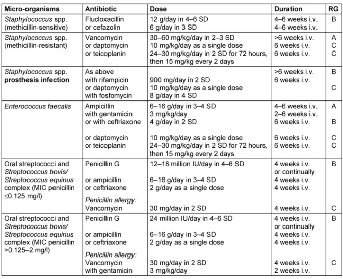
If the general condition of a patient is critical, empirical antibiotic treatment is started immediately but always after obtaining blood cultures. For native valve endocardit- is and late endocarditis following heart valve replacement (>1 year after surgery or intervention), above all methicil- lin-sensitive Staphylococcus aureus strains (MSSA), various streptococcal species andEnterococcus faecalis should be expected (Table 1). The microbiological results from the first infection episode may help inform further calculated antimicrobial treatment. However, in early endocarditis after heart valve replacement (<1 year after intervention or surgery) this is more likely to be associated with methicillin-resistantStaphylococcus aureusstrains (MRSA), coagulase-negative staphylococci and also Gram- negative bacteria. Initial empirical treatment should, if possible, be modified once microbiological results are available. Treatment schemes for the most common en- docarditis agents can be found in Table 2. Further de- tailed treatment recommendations as well as recommen- dations regarding complications indicating a need for surgical intervention and for the continuation of antibiotic treatment after surgical restoration can be found in the guidelines of the European Society of Cardiology [29].
Table 1: Empiric treatment of culture-negative bacterial endocarditis with prior antibiotic treatment or until blood culture results are ready. For culture-negative endocarditis without prior antibiotic treatment, an infection expert should be consulted.
Evaluation of Recommendations for Antibiotic Treatment of
Endocarditis by the European Society of Cardiology (ESC) 2015
Significant changes to the European guidelines include endocarditis prophylaxis, the interdisciplinary “endocardit- is team”, the expansion of imaging modes and the results of a first randomized study on the surgical treatment of bacterial endocarditis.
The European guidelines continue to recommend antibi- otic prophylaxis for risky interventions in patients with predisposing cardiac factors. Preliminary studies support this approach, as after the introduction of the NICE guidelines in 2008 [21] negative effects had been shown by the extreme restriction of the indication of antibiotic prophylaxis in the United Kingdom [23], [24].
The recommendation to empirically treat with ampicillin and flucloxacillin in native valve endocarditis does not result from study data but from the need to cover staphyl- ococci, streptococci andEnterococcus faecalisin initial treatment. Because ampicillin/sulbactam is not available in all European countries for parenteral treatment and the combination of amoxicillin/clavulanic acid cannot be dosed adequately for toxicity reasons, the combination of aminopenicillin plus flucloxacillin was recommended.
Since ampicillin/sulbactam is available for parenteral treatment in Germany, 4x 3 g of ampicillin/sulbactam would be an equivalent alternative, which has the advant- age of having to deal with only one drug or a fixed com- bination.
Surgical treatment and aftercare
The introduction of interdisciplinary endocarditis teams has shown that surgical treatment is an integral part of the overall treatment concept in bacterial endocarditis and should not only take place once antibiotic treatment has failed. The classic indications for surgical restoration which must be used in approximately 50% of all patients are the development of cardiac insufficiency, irreversible and infection-related destruction of cardiac structures and the appearance of emboli, newly-emergent AV blockages and endocarditis in heart valve prostheses, transcatheter heart valves, pacemaker systems and closure systems. The scores necessary for surgical risk stratification (EuroSCORE II and STS score) are of limited suitability in this, as they were primarily developed for coronary revascularization or degenerative heart valve replacement. For surgical intervention in bacterial endo- carditis, special score systems have been developed, such as the De-Feo Score and the International Collabor- ation on Endocarditis Score (ICE Score).
The advantage of early surgical restoration is that espe- cially in native mitral or tricuspid valve endocarditis, heart valves can be operated on, possibly even in minimally invasive ways. Determining indication for early surgical restoration is often difficult and an important task for the
“interdisciplinary endocarditis team”. A randomized study in a low-risk population with small case numbers showed that surgical intervention reduced the rate of embolism significantly within 48 hours up to 6 months after surgery.
However, patient survival was unaffected.
Patients who are provided with TAVI represent a special patient population. In order to be able to optimally treat
Table 2: Overview of the common antibiotics for bacterial endocarditis
these patients in the future, new strategies must be de- veloped.
The basic principle of surgical restoration is the complete removal of the infectious material and tissue in order to cure the patient’s endocarditis with adequate antibiotic treatment. In Austria, adequate antibiotic treatment is partly carried out as out-patient parenteral antibiotic therapy (OPAT). In Germany, this form of treatment has not yet made inroads.
Imaging techniques (such as echocardiography), blood cultures and serological examinations should serve to monitor treatment success and, if necessary, contribute to appropriate adjustment of therapeutic measures.
Note
This is the twelfth chapter of the guideline “Calculated initial parenteral treatment of bacterial infections in adults – update 2018” in the 2ndupdated version. The German guideline by the Paul-Ehrlich-Gesellschaft für Chemo- therapie e.V. (PEG) has been translated to address an international audience.
Following the publication of the 1stversion of the guideline in German, these dosage suggestions were updated by the working group (Table 1: Empiric therapy of culture- negative bacterial endocarditis with previous antibiotic therapy or until the blood culture results are obtained):
• gentamicin dosage in combination with ampicillin, ampicillin/sulbactam or vancomycin: 1x 3 mg/kg INSTEAD OF 3x 3mg/kg
• rifampicin dosage in combination with vancomycin: ri- fampicin 900 mg iv (2 SD) INSTEAD OF rifampicin 900 mg iv (3 SD)
Competing interests
The authors declare that they have no competing in- terests.
References
1. Glynn TR. The Mumleian Lectures on Infective endocarditis mainly in its clinical aspects. Lancet. 1903;161(4155):1073-7. DOI:
10.1016/S0140-6736(01) 71890-9
2. Kirkes WS. On some of the principal effects resulting from the Detachment of Fibrinous Deposits from the interior of the heart, and their mixture with the circulating blood. Med Chir Trans.
1852;35:281-324.
3. Krul MM, Vonk AB, Cornel JH. Trends in incidence of infective endocarditis at the Medical Center of Alkmaar. Neth Heart J.
2015 Nov;23(11):548-54. DOI: 10.1007/s12471-015-0743-0 4. Tauchnitz C. Mikrobielle (bakterielle) Endokarditis. In: Hahn H,
Falke D, Kaufmann SHE, Ullmann U, editors. Medizinische Mikrobiologie und Infektiologie. Berlin-Heidelberg: Springer;
2005. p. 911-5. DOI: 10.1007/3-540-26529-5_113
5. Hoen B, Alla F, Selton-Suty C, Béguinot I, Bouvet A, Briançon S, Casalta JP, Danchin N, Delahaye F, Etienne J, Le Moing V, Leport C, Mainardi JL, Ruimy R, Vandenesch F; Association pour l’Etude et la Prévention de l’Endocardite Infectieuse (AEPEI) Study Group.
Changing profile of infective endocarditis: results of a 1-year survey in France. JAMA. 2002 Jul;288(1):75-81. DOI:
10.1001/jama.288.1.75
6. Mathew J, Addai T, Anand A, Morrobel A, Maheshwari P, Freels S. Clinical features, site of involvement, bacteriologic findings, and outcome of infective endocarditis in intravenous drug users.
Arch Intern Med. 1995 Aug 7-21;155(15):1641-8. DOI:
10.1001/archinte.1995.00430150125013
7. Tleyjeh IM, Abdel-Latif A, Rahbi H, Scott CG, Bailey KR, Steckelberg JM, Wilson WR, Baddour LM. A systematic review of population-based studies of infective endocarditis. Chest. 2007 Sep;132(3):1025-35. DOI: 10.1378/chest.06-2048
8. Benito N, Pericas JM, Gurguí M, Mestres CA, Marco F, Moreno A, Horcajada JP, Miró JM. Health Care-Associated Infective Endocarditis: a Growing Entity that Can Be Prevented. Curr Infect Dis Rep. 2014 Nov;16(11):439. DOI: 10.1007/s11908-014- 0439-4
9. Benito N, Miró JM, de Lazzari E, Cabell CH, del Río A, Altclas J, Commerford P, Delahaye F, Dragulescu S, Giamarellou H, Habib G, Kamarulzaman A, Kumar AS, Nacinovich FM, Suter F, Tribouilloy C, Venugopal K, Moreno A, Fowler VG Jr; ICE-PCS (International Collaboration on Endocarditis Prospective Cohort Study) Investigators. Health care-associated native valve endocarditis: importance of non-nosocomial acquisition. Ann Intern Med. 2009 May;150(9):586-94. DOI: 10.7326/0003- 4819-150-9-200905050-00004
10. Lomas JM, Martínez-Marcos FJ, Plata A, Ivanova R, Gálvez J, Ruiz J, Reguera JM, Noureddine M, de la Torre J, de Alarcón A; Grupo Andaluz para el Estudio de las Infecciones Cardiovasculares (Andalusian Group for the Study of Cardiovascular Infections) at the Sociedad Andaluza de Enfermedades Infecciosas (SAEI).
Healthcare-associated infective endocarditis: an undesirable effect of healthcare universalization. Clin Microbiol Infect. 2010 Nov;16(11):1683-90. DOI: 10.1111/j.1469-0691.2009.03043.x 11. DeSimone DC, Tleyjeh IM, Correa de Sa DD, Anavekar NS, Lahr
BD, Sohail MR, Steckelberg JM, Wilson WR, Baddour LM.
Temporal trends in infective endocarditis epidemiology from 2007 to 2013 in Olmsted County, MN. Am Heart J. 2015 Oct;170(4):830-6. DOI: 10.1016/j.ahj.2015.07.007 12. Westphal N, Plicht B, Naber C. Infective endocarditis –
prophylaxis, diagnostic criteria, and treatment. Dtsch Arztebl Int.
2009 Jul;106(28-29):481-9; quiz 490. DOI:
10.3238/arztebl.2009.0481
13. Li JS, Sexton DJ, Mick N, Nettles R, Fowler VG Jr, Ryan T, Bashore T, Corey GR. Proposed modifications to the Duke criteria for the diagnosis of infective endocarditis. Clin Infect Dis. 2000 Apr;30(4):633-8. DOI: 10.1086/313753
14. Botelho-Nevers E, Thuny F, Casalta JP, Richet H, Gouriet F, Collart F, Riberi A, Habib G, Raoult D. Dramatic reduction in infective endocarditis-related mortality with a management-based approach. Arch Intern Med. 2009 Jul;169(14):1290-8. DOI:
10.1001/archinternmed.2009.192
15. Danchin N, Duval X, Leport C. Prophylaxis of infective endocarditis: French recommendations 2002. Heart. 2005;
91(6):715-8. DOI: 10.1136/hrt.2003.033183
16. Wilson W, Taubert KA, Gewitz M, Lockhart PB, Baddour LM, Levison M, Bolger A, Cabell CH, Takahashi M, Baltimore RS, Newburger JW, Strom BL, Tani LY, Gerber M, Bonow RO, Pallasch T, Shulman ST, Rowley AH, Burns JC, Ferrieri P, Gardner T, Goff D, Durack DT; American Heart Association Rheumatic Fever, Endocarditis, and Kawasaki Disease Committee; American Heart Association Council on Cardiovascular Disease in the Young;
American Heart Association Council on Clinical Cardiology;
American Heart Association Council on Cardiovascular Surgery and Anesthesia; Quality of Care and Outcomes Research Interdisciplinary Working Group. Prevention of infective endocarditis: guidelines from the American Heart Association: a guideline from the American Heart Association Rheumatic Fever, Endocarditis, and Kawasaki Disease Committee, Council on Cardiovascular Disease in the Young, and the Council on Clinical Cardiology, Council on Cardiovascular Surgery and Anesthesia, and the Quality of Care and Outcomes Research Interdisciplinary Working Group. Circulation. 2007 Oct;116(15):1736-54. DOI:
10.1161/CIRCULATIONAHA.106.183095
17. Habib G, Hoen B, Tornos P, Thuny F, Prendergast B, Vilacosta I, Moreillon P, de Jesus Antunes M, Thilen U, Lekakis J, Lengyel M, Müller L, Naber CK, Nihoyannopoulos P, Moritz A, Zamorano JL;
ESC Committee for Practice Guidelines. Guidelines on the prevention, diagnosis, and treatment of infective endocarditis (new version 2009): the Task Force on the Prevention, Diagnosis, and Treatment of Infective Endocarditis of the European Society of Cardiology (ESC). Endorsed by the European Society of Clinical Microbiology and Infectious Diseases (ESCMID) and the International Society of Chemotherapy (ISC) for Infection and Cancer. Eur Heart J. 2009 Oct;30(19):2369-413. DOI:
10.1093/eurheartj/ehp285
18. Gould FK, Elliott TS, Foweraker J, Fulford M, Perry JD, Roberts GJ, Sandoe JA, Watkin RW; Working Party of the British Society for Antimicrobial Chemotherapy. Guidelines for the prevention of endocarditis: report of the Working Party of the British Society for Antimicrobial Chemotherapy. J Antimicrob Chemother. 2006 06;57(6):1035-42. DOI: 10.1093/jac/dkl121