Functional and structural characterization of LeuO, a
pleiotropic LysR-type transcription regulator in Escherichia coli
Inaugural-Dissertation zur
Erlangung des Doktorgrades
der Mathematisch-Naturwissenschaftlichen Fakultät der Universität zu Köln
vorgelegt von Susann Marlies Fragel
aus Berlin
Köln, September 2018
Berichterstatter: Prof. Dr. Karin Schnetz (Gutachter) Prof. Dr. Niels Gehring
Tag der mündlichen Prüfung: 05.09.2018
Nothing in life is to be feared, it is only to be understood. Now is the time to understand more, so that we may fear less.
Marie Curie
Contents ... 4
Zusammenfassung ... 1
Abstract ... 2
1 Introduction ... 3
1.1 LysR-type transcription regulators ... 3
1.2 Structure of LysR-type transcription regulators (LTTRs) ... 4
1.3 Regulation of target genes by LysR-type regulators ... 7
1.4 LeuO... 9
1.5 Objectives of this thesis ... 11
2 Results ... 12
2.1 Screen for hyperactive LeuO mutants ... 12
2.2 Crystal structure of the LeuO effector-binding domain (EBD) ... 15
2.3 Characterization of additional LeuO mutants ... 19
2.4 The LeuO DNA-binding domain (DBD) is functional ... 23
2.5 LeuO binds to the promoter regions of cas, yjjQ and leuO ... 24
2.6 DNase I footprinting by LeuO, LeuO-S120D and the LeuO-DBD ... 25
2.7 DNA-binding consensus sequence of LeuO ... 32
2.8 Regulation of presumptive target loci by LeuO ... 33
2.9 Autoregulation of PleuO by hyperactive LeuO mutants ... 35
2.10 Is LeuO activity modulated by an effector? ... 37
3 Discussion ... 42
3.1 DNA-binding of LeuO and LeuO-S120D ... 42
3.2 Is LeuO activity regulated by an effector? ... 43
3.3 The spectrum of LeuO target loci ... 45
3.4 How does LeuO regulate transcription? ... 46
3.5 Approaches for analyzing LysR-type transcriptional regulators ... 47
4 Materials and Methods ... 48
4.1 Media, antibiotics and bacterial cultivation ... 48
4.2 Standard molecular techniques ... 48
4.3 Bacterial strains, plasmids and oligonucleotides ... 48
4.4 CaCl
2-competent cells and transformation ... 56
4.5 Electrocompetent cells and electroporation ... 56
4.6 Random mutagenesis screen ... 57
4.7 Site-directed mutagenesis by overlap PCR ... 57
4.8 Chromosomal integration into attB sites ... 57
4.9 Gene deletion and insertion using λ-Red mediated recombination ... 58
4.10 Chromosomal two-step mutagenesis using λ-Red mediated recombination ... 58
4.11 Phage transduction ... 59
4.12 Expression analysis ... 59
4.13 Protein purification of LeuO and LeuO mutants ... 60
4.14 SDS-PAGE ... 60
4.15 Crystallization of LeuO-EBD and LeuO-S120D-EBD ... 61
4.16 Electrophoretic shift assay (EMSA) ... 61
4.17 DNase I footprinting ... 62
4.18 Identification of a consensus LeuO DNA-binding site ... 63
5 References ... 66
Danksagung ... 71
Erklärung ... 72
Zusammenfassung
LeuO ist ein konservierter LysR-Typ Transkriptionsregulator (LTTR) in Escherichia coli und anderen Gammaproteobakterien. LeuO reguliert Gene, die unter anderem für Pathogenität und Stressantworten verantwortlich sind. Viele der LeuO-regulierten Gene werden durch das globale Repressorprotein H-NS reprimiert und co-reguliert. Daher wird LeuO als globaler Antagonist von H- NS angesehen. LTTRs regulieren ihre Zielgene typischerweise als Tetramere, die sich aus zwei Dimeren der N-terminalen DNA-Bindungsdomänen (DBDs) und zwei Dimeren der C-terminalen Effektorbindungsdomänen (EBDs) zusammensetzen. Viele der bisher beschriebenen LTTRs benötigen einen Effektor, der strukturelle Veränderungen induziert. Diese strukturellen Veränderungen führen zur Änderung der DNA-Bindung des LTTR und ermöglichen die Aktivierung oder Repression spezifischer Zielgene. Die Regulation der LeuO Aktivität und deren strukturelle Merkmale sind jedoch weitgehend unbekannt.
In dieser Arbeit wurde der CRISPR-assoziierte cas Promotor als ideales Reporter identifiziert, da er
sensitiv Veränderungen der LeuO-Aktivität abbildet. Mit diesem Reporter wurden in einem
genetischen Screen neun hyperaktive Mutanten von LeuO identifiziert, welche den Reporter bei
basaler Expression voll aktivieren. Die gelösten Kristallstrukturen der EBDs des wild-typischen LeuO
und der hyperaktiven Ser120Asp Mutante zeigen, dass die Mutationen, die Hyperaktivität bewirken,
in der Dimerisierungsoberfläche der EBD lokalisiert sind. Eine Überlagerung der Strukturen der EBDs
deutet darauf hin, dass die S120D-Mutation eine strukturelle Veränderung induziert, welche einen
Effektor-gebundenen Zustand nachahmt. Diese strukturelle Veränderung kann sich dann auf die
sterische Anordnung der DBDs übertragen und somit die DNA-Bindeeigenschaften beeinflussen. Eine
DNase I Footprint Analyse der LeuO-Bindestellen in der Region des cas Promotors zeigt eine
hochspezifische DNA-Bindung der LeuO Mutante (S120D), welche außerdem im Vergleich zum wild-
typischen LeuO ein deutlicheres Bindemuster an die DNA zeigt. Eine detaillierte Analyse dieses
Bindemusters deckte eine "Core-Site" auf, welche im Weiteren die Definition eines palindromischen
DNA-Bindemotifs ermöglichte. Die Daten deuten darauf hin, dass LeuO möglicherweise eine andere
Gruppe von Zielgenen reguliert, wenn ein Effektor gebunden ist, der strukturelle Veränderungen des
Proteins auslöst. Die Analyse von hyperaktiven Mutanten, welche vermutlich eine Effektor-
gebundene Form abbilden, kann zum Verständnis des molekularen Mechanismus der
Transkriptionsregulation durch LeuO beitragen.
Abstract
LeuO is a conserved LysR-type transcriptional regulator (LTTR) of global function in Escherichia coli and other Gammaproteobacteria. LeuO regulates genes related to pathogenicity and stress adaptation, many of which are co-regulated by the nucleoid-associated global repressor protein H- NS. Therefore, LeuO is considered a general H-NS antagonist. LTTRs typically are tetramers that consist of two dimeric N-terminal DNA-binding domains (DBDs) and two dimeric C-terminal effector- binding domains (EBDs). Many LTTRs described so far require an effector which induces structural changes that lead to alteration of the LTTR activity in repression or activation of specific target genes.
However, the regulation of LeuO activity and its structural features are poorly understood.
In this work, I characterized the CRISPR-associated cas promoter as the most sensitive reporter for
monitoring LeuO activity. Using this reporter nine LeuO mutants were identified in a genetic screen
that render LeuO hyperactive. The solved crystal structures of the EBDs of wild-type LeuO and one of
the hyperactive mutants (Serine 120 to aspartic acid exchange) revealed that all mutations causing
hyperactivity localize to the dimerization interface of the EBD. Comparison and superposing of the
structures of the wild-type and S120D EBDs suggests that the mutation of S120D induces a structural
change that mimics an effector-bound state. This structural change is presumed to transmit to the
arrangement of the DBDs and changes the binding to the DNA. DNase I footprinting of the cas
promoter region presented here revealed a highly specific DNA-binding and a more distinct DNA-
binding pattern of the S120D mutant compared to the wild-type LeuO. A detailed analysis of these
footprints unraveled a “core-site” of palindromic sequence, which allowed the definition of a specific
DNA-binding sequence motif for LeuO. The data indicate that LeuO might regulate a different set of
target genes when an effector is bound. This is relevant in understanding the molecular mechanism
of transcriptional regulation by LeuO and the role of LeuO as a global regulator.
1 Introduction
Bacteria face harsh and quickly changing environmental conditions encountering various stresses and changing nutrition sources. In order to adapt to, and survive in different environments, bacteria like E. coli have evolved a complex gene regulation network (Lee et al., 2012). The regulation of transcription by transcription factors allows an individual control of a single gene or of specific subsets of genes in response to an environmental stimulus. Activating transcription factors bind close to the transcription start site and orchestrate the formation of the transcription initiation complex.
These activators can directly recruit the RNA polymerase to the promoter or they induce structural changes of the DNA promoter region, enabling the binding and initiation of transcription by RNA polymerase (Browning & Busby, 2016). Most repressive transcription factors inhibit transcription initiation by steric hindrance, in which the repressor DNA-binding denies access of the RNA polymerase to the promoter. A repressor can also block transcription initiation by targeting the activating transcription factor. Contrary, a transcriptional factor can also obstruct a potential repressor and hence indirectly activate transcription (Browning & Busby, 2016).
The family of LysR-type transcriptional regulators (LTTRs) is most prevalent in bacteria. The LysR-type transcriptional regulators activate or repress diverse sets of genes related to virulence, stress responses and motility (Maddocks & Oyston, 2008). The activity of LTTRs is controlled by an effector that binds to an “effector-binding cleft” or elsewhere, or by modification of amino acids. LeuO is a conserved LysR-type transcription regulator in Enterobacteriaceae regulating many targets related to stress adaptation, pathogenicity and biofilm formation (Henikoff et al., 1988, Dillon et al., 2012, Guadarrama et al., 2014b). However, the control of LeuO activity and the mechanism of its regulation have not been studied in detail. For LeuO, no effector or condition is known that modulates its activity. Further, the intrinsic structural properties of LeuO and its DNA-binding mode are open questions. In this work, a functional and structural analysis of the LysR-type transcription regulator LeuO was conducted.
1.1 LysR-type transcription regulators
LysR-type transcription regulators (LTTRs) represent the largest family of transcription factors,
resembling 16% of the overall repertoire of transcription factors in bacteria (Tropel & van der Meer,
2004). They are highly conserved and orthologues have been identified in Archaea and eukaryotic
organisms (Henikoff et al., 1988, Schell, 1993, Maddocks & Oyston, 2008). To date, 46 LTTRs are
present in Escherichia coli K12 (Karp et al., 2014) and these LTTRs are involved in the regulation of
diverse cellular functions as oxidative stress responses, metabolic pathways, virulence and biofilm
type-regulators are homo-tetramers and their activity is controlled by structural changes within the tetrameric structure. These structural changes can be induced by effectors or the modification of amino acid residues. Effectors can bind to a specific “binding pocket”, in the interface of two dimers or elsewhere on the tetramer. Different effectors that bind to the LTTR can regulate different target loci or even change the LTTR from an activator to a repressor at a given target loci (Laishram &
Gowrishankar, 2007). In general, LTTRs regulate expression of a target gene by binding two distinct sites in the promoter region matching the degenerated consensus binding motif T-11N-A (Maddocks
& Oyston, 2008).
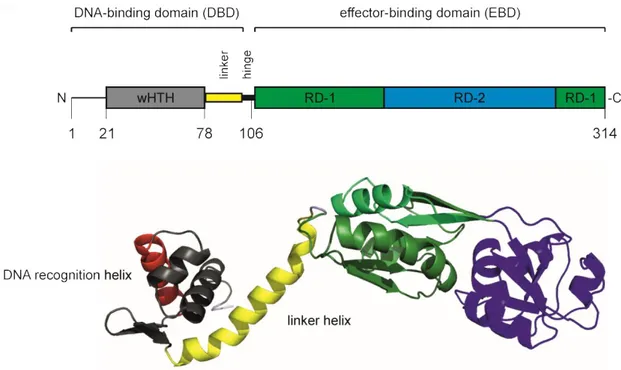
1.2 Structure of LysR-type transcription regulators (LTTRs)
LTTR monomers comprises of approximately 300 amino acids. They consist of a characteristic N- terminal DNA-binding domain (DBD) with a winged helix-turn-helix (wHTH) domain that connects via a helical linker and a flexible hinge to a C-terminal effector-binding domain (EBD, also called regulatory domain, RD; Figure 1)(Maddocks & Oyston, 2008, Momany & Neidle, 2012) The DNA- binding domain is highly conserved and the wHTH domain comprises of three core helices, forming a helical bundle with an almost triangular outline. The specific DNA target is determined by the α3 DNA-recognition helix and forms a DNA-protein interface by insertion into the major groove of the DNA (Aravind et al., 2005). The “wing” is a β-strand hairpin unit between helix α3 and α4 and usually interacts with the minor groove of the DNA (Aravind et al., 2005). Two DBDs can dimerize via their α4 helical linker and bind to a palindromic DNA sequence in an inverted arrangement of the DBDs.
The homology of the C-terminal effector-binding domain (EBD) in LTTRs is less well conserved on the amino acid sequence level, but the EBDs have a conserved domain structure. The EBDs exhibit a typical fold with two
/
subdomains (also called RD-1 and RD-2 domains) that connect by two extended, antiparallel cross-over β-strands. A cleft locates between the two subdomains of the EBD (RD-1 and RD-2) and for some LTTRs effectors are described to bind in this cleft. Two EBDs dimerize in antiparallel head-to-tail orientation and form a conserved dimerization interface (Figure 2). In this dimerization interface, likewise effector-binding was described (Jiang et al., 2018).
Several structures of LTTRs are solved so far, and a typical LTTR tetramer can be described as a dimer
of dimers, where a dimer comprises of two monomers, dimerizing via their EBDs and displaying an
extended and a compact conformation of their DBDs, as shown by the structure of a structure model
of LeuO in Figure 2. The assembly of the tetramer arises when the compact DBD subunits interact
with the extended DBD subunits of the other EBD dimer and contact is made by their linker helices
(Henikoff et al., 1988, Maddocks & Oyston, 2008, Momany & Neidle, 2012, Taylor et al., 2012).
Figure 1: The domain structure of a monomeric LysR-type regulator.
LTTR monomers consist of an N-terminal winged HTH DNA-binding domain (DBD) connected via a helical linker and a hinge to a C-terminal effector-binding domain (EBD). The DNA recognition helix is shown in red. The α4 helical linker, depicted in yellow is important for dimerization of the DBDs. A “cleft” exists between the two regulatory subdomains (RD-1 and RD-2) where a putative effector binds or the modification of an amino acid residue may alter the protein structure. However, alternative effector-binding sites exist for some LTTRs in the interface of two EBD. Depicted is the monomeric structure of LeuO as example of a LysR-type transcriptional regulator (uniprot entry: P10151; https://swissmodel.expasy.org/repository/uniprot/P10151). The figure was adapted from (Lerche et al., 2016) and generated using PyMOL (The PyMOL Molecular Graphics System).
The tetrameric LTTR harbors two dimers of the DBD that bind to two adjacent imperfect palindromic sites of the DNA, a high affinity DNA binding site (generally called RBS, regulatory binding site) and an alternative low-affinity DNA-binding site (ABS, activator binding site) (Toledano et al., 1994, Wek &
Hatfield, 1988, Rhee et al., 1998). For most LTTRs, this target recognition and binding to the DNA is independent of a bound effector. However, the binding of an effector or the mutation of an amino acid residue can induce structural changes of the EBDs and subsequently change the distance or the angle of the two DBDs, as shown for OxyR (Figure 2D) (Jo et al., 2015). This steric re-arrangement alters the DNA-binding pattern, the DBD dimer bound to the high affinity DNA-binding site is maintained, while the second DBD dimer “slides” along the DNA to an alternative DNA-binding site.
This “sliding” along the sequence, leads to a different DNA-binding pattern, resulting in a differential
regulation of gene expression. LTTRs regulate transcription of target genes by this “sliding-dimer”-
mechanism, as described for example for DntR (Lerche et al., 2016, Toledano et al., 1994). The
mechanism of the sliding dimer was also shown by DNase I footprinting for BenM, ArgP and OxyR
(Bundy et al., 2002, Laishram & Gowrishankar, 2007, Kullik et al., 1995).
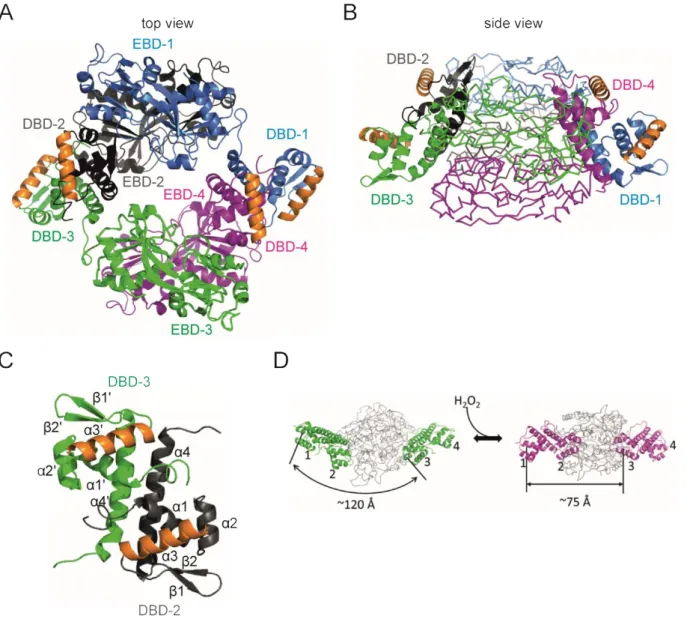
Figure 2: Predicted tetrameric structure of LeuO.
A top view (A) and a side view (B) of the predicted LeuO tetramer structure with monomeric LeuO proteins highlighted in blue, grey, green and magenta (uniprot entry: P10151;
https://swissmodel.expasy.org/repository/uniprot/P10151 based on (Lunin et al., 2005)). Dimerization is shown of the effector-binding domains EBD-1 (blue) with EBD-2 (grey) and dimerization of EBD-3 (green) with EBD-4 (magenta). The DNA-binding domain DBD-1 dimerizes with DBD-4 and the DBD-2 dimerizes with DBD-3.
The α3 DNA-recognition helices are marked in orange. (C) A structural model of the DBD-dimer. The individual monomers of the DBDs are depicted in grey and green with the α3 DNA-recognition helices marked in orange.
The dimerization of the DBDs is mediated by the α4 linker helices. (D) The reduced (left side) and oxidized form of OxyR (right side) shows the structural change transmitted to the DBDs upon oxidization of the protein (Jo et al., 2015). The α3 helices of the DBDs are indicated by number and the distance between DBD-1 and DBD-3 is 120 Å in the reduced full-length protein. The DBD-1/DBD-2 dimer and the DBD-3/DBD-4 dimer should get closer by 45 Å during the transition to the oxidized state (Jo et al., 2015). The figure was generated using PyMOL (The PyMOL Molecular Graphics System).
1.3 Regulation of target genes by LysR-type regulators
Structural changes in the LTTR tetramer can be induced by binding of an effector or a modification of an amino acid residue. The LTTR BenM in Acinetobacter sp. controls the aromatic compound metabolism by regulating genes in the β-ketoadipate pathway (Bundy et al., 2002). In absence of an effector, BenM binds to the promoter of benA (encoding a benzoate-dioxygenase) and represses transcription. In the presence of the metabolites benzoate and muconate structural changes of BenM shift the binding to a different DNA-binding site of the benA promoter region and BenM synergistically activates the transcription of benA expression. In this way, the benzoate degradation is directly coupled to the presence of benzoate in the cell (Bundy et al., 2002).
Modifications of an amino acid are possibly caused by a change of pH or by oxidation. An example for the control of an LTTR by amino acid modification is the hydroxylation of the cysteine residue (C199) in OxyR, leading to structural changes of the tetrameric OxyR. Consequently, the OxyR-regulated small RNA oxyS is activated which regulates the oxidative stress response (Toledano et al., 1994, Jo et al., 2015, Kullik et al., 1995). The OxyR-C199D mutant described in (Jo et al., 2015) mimics an oxidized form of the protein that renders the protein active. For AphB in Vibrio cholera the N100E mutation renders the pH-sensitive transcription regulator active at non-permissive pH 8.5. The structures of wild-type AphB and of mutant N100E revealed structural changes of the tetrameric AphB that render AphB-N100E constitutively active at its target loci (Taylor et al., 2012). Mutations in the EBD of LysR-type regulators can render the LTTR constitutively active, suggesting that the mutation mimics an effector-bound state and induce the same structural change as the effector.
Constitutively active mutants were also described for the LTTRs AmpR, CysB, DarR, NahR, TsaR, and XapR (Balcewich et al., 2010, Colyer & Kredich, 1994, Jones et al., 2018, Schell et al., 1990, Monferrer et al., 2010, Jørgensen & Dandanell, 1999).
Effectors are usually metabolites or ions and can bind either to the cleft between the two RD subdomains of the EBD or to a secondary binding site in the dimeric interface of two EBDs (Jiang et al., 2018). In some cases, different effectors are bound by the LTTR.
These effectors can alter the transcription regulation of the LTTR as a direct read out of a stimulus in
the cell. For example, to prevent toxic levels of arginine in the cell, the LTTR ArgP binds the effector
arginine and activates the transcription of the arginine exporter encoding argO. When lysine is bound
to ArgP transcription of argO is inhibited and also additional ArgP target genes are inhibited
(Laishram & Gowrishankar, 2007). The LTTR NdhR coordinates the balance of carbon and nitrogen
metabolism in cyanobacteria. For NdhR two effector binding sites were identified, where different
effectors can bind (Jiang et al., 2018). One binding site is located in the cleft of the effector-binding
domain between two RD subdomains and binds 2-PG (2-phosphoglycolate, depicted in green; Figure
3). A secondary binding site is located in the dimeric interface of two EBDs of NdhR and binds 2-OG (2-oxoglutarate, shown in blue; Figure 3). Binding of either one of the effectors to different sites of the protein allows a fine-tuning in the regulation, depending on the incoming signal. NdhR adopts a repressor conformation upon 2-OG binding. The binding of 2-PG to the cleft induces structural changes and leads to the activation of gene expression. When the repressing effector is bound, the activating effector is no longer able to bind due to the structural changes (Jiang et al., 2018).
It is noteworthy that some of the described LTTRs map next to genes they regulate, linking their location to their function. For example, BenM regulates the transcription of the divergent benA gene (Maddocks & Oyston, 2008, Bundy et al., 2002). In the E. coli genome about 50% of the LTTRs map divergently to their target genes. The LTTR IlvY is located divergent to its regulated target gene ilvC (Wek & Hatfield, 1988). The ilvC gene encodes a ketol-acid reductoisomerase for branched-chain amino acid synthesis (Rhee et al., 1998).
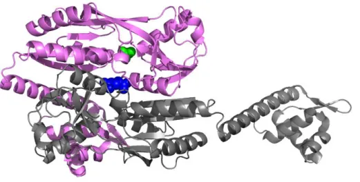
Figure 3: The binding of effectors to different sites of the LTTR NdhR.
Depicted is dimer of the NdhR tetrameric structure with two different effectors (indicated in green and blue) bound to different sites in the structure (Jiang et al., 2018). The effector 2-PG (in green) binds to the cleft between the two RD subdomains of NdhR and induces gene expression. The effector 2-OG (in blue) binds to a secondary binding site in the dimer interface between two EBDs and acts as co-repressor of NdhR (Jiang et al., 2018).
1.4 LeuO
LeuO is a pleiotropic LysR-type transcriptional regulator and conserved in Gammaproteobacteria (Henikoff et al., 1988, Hernández-Lucas & Calva, 2012, The UniProt Consortium, 2017). LeuO has been best studied in the species Escherichia coli and Salmonella enterica, as well as in Vibrio cholera.
The LeuO proteins are 90% identical in the Enterobacteriaceae and 50% identical to LeuO in V.
cholera and LeuO is presumed to act as a tetramer, like other LTTRs (Guadarrama et al., 2014b).
LeuO regulates more than 100 loci in E. coli and S. enterica, as identified by microarray transcriptome analyses and genomic SELEX screening (Dillon et al., 2012, Shimada et al., 2011, Stratmann et al., 2012, Ishihama et al., 2016). LeuO activates and represses loci related to acid-stress resistance, pathogenicity, multi-drug efflux, and biofilm formation (Henikoff et al., 1988, Dillon et al., 2012, Guadarrama et al., 2014b).
Among many targets, LeuO activates the transcription of CRISPR-cas bacterial defense system, important for defense against foreign DNA and phage infection (Medina-Aparicio et al., 2011, Pul et al., 2010, Westra et al., 2012, Hernández-Lucas & Calva, 2012). Further, LeuO plays a positive role in the regulation of the yjjQ-bglJ operon, encoding FixJ/NarL-type transcriptional regulators and the bgl operon, encoding proteins for uptake and utilization of aryl-β,D-glucosides (Ueguchi et al., 1998, Stratmann et al., 2008). In Vibrio, LeuO inhibits the expression of the cholera toxin (CT) and the toxin co-regulated pilus (TCP). Furthermore, leuO expression is activated by the membrane bound transcription regulator ToxR, which links leuO expression to various stimuli that activate the ToxR- dependent virulence regulon of V. cholerae (Ayala et al., 2018, Bina et al., 2013, Ante et al., 2015a, Ante et al., 2015b).
Many LeuO-activated target loci, like the CRISPR-cas operon, are repressed by the global repressor H- NS (heat-stable nucleoid structuring protein) and its paralogue StpA. LeuO presumably competes with H-NS for DNA-binding and delimits spreading and thus the formation of the repressing oligomeric H-NS complex (De la Cruz et al., 2007, Chen & Wu, 2005, Westra et al., 2010). LeuO is therefore considered an H-NS antagonist (Ueguchi et al., 1998, De la Cruz et al., 2007). However, in several pathways LeuO has been identified as additional repressor of H-NS-repressed gene loci and thus seems to functionally cooperate with H-NS or functions as a “back-up” of H-NS. For example, LeuO causes repression via HilE of pathogenicity island 1 (SPI-1) in Salmonella when repression by H- NS is impaired (Espinosa & Casadesús, 2014).
The leuO gene (GenBank Accession number U00096.3 K12: 84,368 to 85,312) is located between the
divergent leuLABCD operon and the ilvIH operon, both encoding enzymes for the synthesis of
branched-chain amino acids (Gemmill et al., 1983, Wang & Calvo, 1993) (Figure 4). In standard
laboratory conditions leuO is silent, but is moderately induced in response to starvation in stationary
growth phase (Majumder et al., 2001, Fang et al., 2000). Transcription of leuO is repressed by H-NS and StpA under standard laboratory conditions (Klauck et al., 1997, Stratmann et al., 2012).
Transcription of leuO is activated by the transcription regulator BglJ-RcsB and by the LysR-type regulator LrhA (Stratmann et al., 2012, Breddermann & Schnetz, 2017). LeuO itself shows a weak positive autoregulation, when expressed in high copy numbers (Chen & Wu, 2005). However, LeuO acts as a negative autoregulator, counteracting the activation by BglJ-RcsB as well as by LrhA, and derepression in hns stpA mutants (Stratmann et al., 2012, Breddermann & Schnetz, 2017). Two distinct LeuO DNA-binding sites in the promoter sequence of leuO have been described (Stratmann et al., 2012).
Although LeuO regulates many target loci, only a degenerated consensus DNA-binding motif was identified displaying the typical weak LysR-type binding motif T-11N-A (Dillon et al., 2012, Maddocks
& Oyston, 2008). The weak DNA-binding specificity might be increased when LeuO activity is altered by an effector or an amino acid modification, as described for other LysR-type regulators. Up to date, no effector or condition has been described that modulates the LeuO activity.
Figure 4: Schematic model of the organization of the leuO promoter region including the binding sites of transcriptional regulators.
The leuO gene is located between the operons ilvIH and leuLABCD, both encoding for enzymes involved in branched-chain amino acid synthesis. LeuO DNA-binding sites are indicated as black boxes, the H-NS nucleation site is shown as red box and the binding site for the heterodimeric activator BglJ-RcsB as grey box (Stratmann et al., 2012). Transcription of leuO is repressed by H-NS and StpA under standard laboratory conditions. The promoter P2 is activated by BglJ-RcsB, while LrhA mainly activates P3 (and weakly P and P1). In hns stpA mutant strains, LeuO acts as an autorepressor, and it counteracts the activation by LrhA and BglJ- RcsB in the wild-type (Stratmann et al., 2012, Breddermann & Schnetz, 2017).
1.5 Objectives of this thesis
LeuO is a pleiotropic LysR-type regulator modulating the transcription of many target loci in Escherichia coli. For other LysR-type regulators effectors or modifications of amino acids have been described which alter the activity of the protein. No effector is known so far for LeuO and the control of LeuO activity is poorly understood. Additionally, the mode of LeuO binding to the DNA and the mechanism of regulation of its target loci remain open questions.
In this study, I identified the cas promoter as the most sensitive reporter for analyzing the activity of LeuO. Using this reporter, I isolated nine LeuO mutants that are highly active causing full activation even when present at low levels. I further characterize the LeuO structure, by crystallization of the effector-binding domains (EBDs) of the wild-type LeuO and one of the hyperactive mutants (S120D).
The structures revealed that all hyperactive LeuO mutants map in the dimerization interface of the EBDs and the S120D mutant causes a structural change.
In addition, I determined the DNA-binding mode of wild-type LeuO, of the hyperactive LeuO-S120D
mutant and of the LeuO-DBD dimer. The DNA-binding specificity of the S120D mutant and of the DBD
dimer is higher than of wild-type LeuO, which enabled the identification of a palindromic DNA-
binding sequence and a specific LeuO DNA-binding motif. Furthermore, using the motif I identified
and initiated the characterization of putative LeuO target loci.
2 Results
2.1 Screen for hyperactive LeuO mutants
For the LysR-type regulator LeuO no effector is known, while for other LysR-type regulators, such as AphB, XapR and OxyR, effectors have been described and amino acid mutations have been characterized that render these regulators constitutively active (Taylor et al., 2012, Jørgensen &
Dandanell, 1999, Jo et al., 2015). Therefore, to characterize LeuO, I set up a screen for LeuO mutants
that are constitutively active. As a reporter, the promoter of the cas (CRISPR-associated) operon,
which is activated by LeuO (Pul et al., 2010, Westra et al., 2010) was fused to lacZ, and this Pcas-lacZ
fusion was integrated into the genome (at the phage λ attachment site attB) in a ΔlacZ ΔleuO Δ(yjjP-
yjjQ-bglJ) background (T1610; Figure 5A). The expression of the Pcas-lacZ reporter was strongly
activated by LeuO (from 6 units to 568 units), when LeuO was expressed at high levels using a
plasmid carrying leuO under the control of the IPTG (isopropyl-β,D-thiogalactopyranoside) inducible
tac promoter (Figure: 5B and 5C), as shown before (Westra et al., 2010). At basal expression levels of
LeuO, without induction of Ptac, LeuO activated Pcas-lacZ moderately (compare 51 units to 6 units of
control, Figure 5B). Thus, the basal expression level of LeuO is a suitable condition to screen for
constitutive LeuO mutants. In this screen, promoter lacZ fusions of the LeuO-activated bgl and yjjQ
promoters were also included as reporters (Schubert, 2013). However the Pcas-lacZ fusion was found
to be the most sensitive reporter. For mutagenesis the leuO gene fragment was amplified by PCR in
parallel reactions, using the non-proofreading Taq polymerase, and the PCR fragments were ligated
into the Ptac expression plasmid (Figure 5A). Transformants of the Pcas-lacZ reporter strain with
these ligations were screened for a Lac-positive phenotype on tryptone X-Gal (5-bromo-4-chloro-3-
indolyl-β-D-galactopyranoside) indicator plates without IPTG (Figure 5A and 5C). Nine different
mutations of single amino acid residues were isolated from 25 independent mutagenesis PCR
reactions. These mutants include 3 independent isolates of LeuO-S128P as well as 2 independent
isolates each of LeuO-H142R, LeuO-Q210R, LeuO-A237V and LeuO-H254R, respectively. The mutants
LeuO-T127I and LeuO-R218C, respectively, were isolated once. The mutant LeuO-M244T was isolated
as a single mutant and as an independent double mutant with the second mutation V230I (Figure
8A). In addition, mutant LeuO-S120D was isolated as a triple mutant with additional amino acid
exchanges at residues E111D and D205N. This triple mutant was isolated using the Pbgl-lacZ reporter
strain (Schubert, 2013) (Figure 8A). To analyze the impact of LeuO Ser120 to Asp exchange, a single
mutant was constructed and used for further analysis. Multiple independent isolations of the same
mutants suggest that the screen for constitutively active LeuO mutants was saturated.
Figure 5: Screen for constitutively active LeuO mutants.
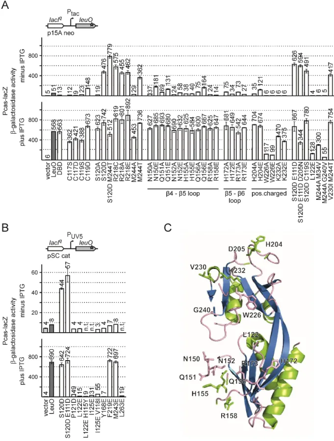
(A) To screen for active LeuO mutants, a Pcas-lacZ fusion was integrated into the chromosomal attB-site of strain T1281 [ΔlacZ ΔleuO Δ(yjjP-yjjQ-bglJ)], and this Pcas-lacZ fusion was used as reporter for LeuO activity (top left side). The leuO gene was amplified by non-proofreading PCR and the leuO fragments were ligated into plasmid pKESK22 (carrying a p15A replication origin, kanamycin resistance) to place leuO under control of the IPTG inducible tac promoter (Ptac) (top right side). Transformants were selected on tryptone X-Gal, kanamycin indicator plates and screened for a Lac-positive phenotype (A bottom). Of a total of 17 Lac+ clones the leuO fragment was sequenced, which revealed 9 different leuO mutants (see Table 3). (B) Activation of Pcas by LeuO mutants. Expression levels of Pcas-lacZ were determined of cultures grown without induction (top panel) and with 1 mM IPTG for induction of leuO expression (bottom panel). Expression was determined in absence (vector control, pKESK22) and in presence of plasmid-provided LeuO (pKETS5) and its mutants S120D (pKESL104), T127I (pKESL74), S128P (pKESL75), H142R (pKESL76), Q210R (pKESL77), R218C (pKESL78), A237V (pKESL80), M244T (pKESL73), and H254R (pKESL101). Cultures for β-galactosidase assays were inoculated from overnight cultures and grown in LB medium with kanamycin to an OD600 of 0.5. Where indicated 1 mM IPTG was added both to the overnight and exponential culture. Mean values of at least 3 independent biological replicates are shown as bars, and error bars indicate standard deviations. (C and D) Spotting of Pcas-lacZ strain (T1281) carrying leuO on a plasmid. Over day cultures were grown in LB with antibiotics to an OD600 ~ 3.0 and diluted to OD600 = 1.0 for spotting. OD600 = 1.0 equals approximately 109 cells per ml, and 10 µl (= 10-2 ml) and 10 µl of the ten-fold serial dilutions (10-3, 10-4, 10-5, 10-6, 10-7, 10-8)
The LeuO mutants conferring a Lac
+phenotype at basal expression levels (without IPTG) were used in a quantitative assay of Pcas-lacZ activation (Figure 5B). To this end, transformants of the Pcas-lacZ reporter strain with plasmids carrying leuO mutants were grown without (no IPTG) and with induction of leuO expression (1 mM IPTG) and the β-galactosidase activities were determined. When LeuO expression was induced (1 mM IPTG), all mutants caused full activation of Pcas-lacZ (Figure 5B, bottom panel). At basal expression levels of leuO, without induction, all LeuO mutants isolated in the screen fully activated the Pcas-lacZ reporter (Figure 5B top panel). The expression levels ranged from 362 to 708 units referring to a 7 to 14-fold stronger activation of Pcas-lacZ than by wild-type LeuO (51 units, Figure 5B top panel). Thus, each of the nine mutations of single amino acid residues render LeuO hyperactive. Interestingly, all the residues locate in the effector-binding domain (EBD, amino acid residue 108 to 314), and not a single mutant mapped to the N-terminal DNA-binding domain (DBD, amino acid residue 1 to 101).
To visualize the increased activity of LeuO-S120D and to further analyze the growth of the mutant, transformants of the Pcas-lacZ reporter strain (T1281) with plasmids expressing wild-type (pKETS5, p15A-ori or pKESMS38, pBR-ori) and mutant LeuO (pKESL104, p15A-ori or pKESMS79, pBR-ori) were spotted in serial dilutions on tryptone X-Gal indicator plates with and without 200 µM IPTG. Empty plasmids served as control (pKESK22, p15A-ori or pKES334, pBR-ori). The plasmids carrying ap15A-ori express moderate levels of LeuO (Figure 5C), while pBR-derived plasmids express high levels of LeuO (Figure 5D), under the control of the IPTG inducible tac promoter. Transformants were grown in LB with antibiotics to an OD
600~3.0 and diluted to OD
600=1.0 (equals approximately 10
9cells per ml).
Ten-fold serial dilutions were made and 10 µl was pipetted per spot of each dilution (= 10
-2, 10
-3, 10
-4, 10
-5, 10
-6, 10
-7, 10
-8per ml).
The Pcas-lacZ strain expressing LeuO showed a blue phenotype when induced with IPTG and a light blue color when the plasmids were not induced. In agreement with the data from β-galactosidase assays, the mutant LeuO-S120D conferred a Lac
+phenotype even when no IPTG was present. Further the cells expressing wild-type LeuO showed a normal growth (up to dilution 10
-8), while the hyperactive LeuO-S120D mutant conferred a growth defect when the expression was induced from the high-copy number pBR-ori plasmid (no growth as of dilution 10
-4). This suggests that high levels of
Figure 5 continued from the previous pagewere spotted on tryptone X-Gal indicator plates, with ampicillin or kanamycin. Plates were supplemented with 200 µM IPTG where indicated. The reporter strain T1281 was transformed with plasmids expressing LeuO or LeuO-S120D under the control of the IPTG inducible lacIq-tac promoter system and either (C) expressed moderate levels of LeuO from a low to medium copy plasmid (p15A origin of replication; pKESK22, control;
pKETS5, LeuO; pKESL104 LeuO-S120D) or (D) high levels of LeuO from a pBR derived plasmid (pKES334, control; pKESMS38, LeuO; pKESMS79 LeuO-S120D).
hyperactive LeuO-S120D are toxic for E. coli cells. Further, all colonies of the dilution 10
-3and 10
-4were Lac
-. The sequencing of leuO in several of these clones revealed an additional mutation W303*.
The additional mutation leads to a truncated protein with lost hyperactivity.
2.2 Crystal structure of the LeuO effector-binding domain (EBD)
The nine mutations causing hyperactivity of LeuO, map in the effector-binding domain (EBD) that extends from residue Ala108 to Arg314, according to a structural model of E. coli LeuO (https://swissmodel.expasy.org/repository/uniprot/P10151) and an alignment of S. enterica LeuO with other LysR-type regulators (Figure 6) (Guadarrama et al., 2014a). To characterize the functional relevance of these residues, the crystal structure of the LeuO-EBD was determined in cooperation with the Lab of Prof. Dr. Ulrich Baumann, Institute for Biochemistry, Cologne (Figure 7). In addition, the structure of the EBD of hyperactive mutant LeuO-S120D was solved in an orthorhombic (space group I222) and a monoclinic (space group C2) crystal form (Figure 7A). Most residues are well resolved in the electron density maps with the exception of the loop connecting strands β4 and β5 (residues 151-158 including helix α6; secondary structures are numbered according to the prediction for the full-length protein), which has weak electron density in the wild-type and in the monoclinic crystal form of the S120D structure, but it could be traced in the orthorhombic S120D crystal form.
Furthermore, the loop between β5 and β6 residues (R173 to E175) is not resolved in the wild-type and monoclinic mutant structures and possesses only weak density in the orthorhombic S120D structure.
The EBD exhibits the typical fold with two / subdomains (also denoted as RD domains) connected by two extended, antiparallel cross-over β-strands β6 and β11. DALI and PDB FOLD searches revealed the effector domain of DntR of Burckholderia sp. as the structurally most similar protein with known structure (PDB ID: 5AE5) (Lerche et al., 2016).
In all three crystal structures, the EBDs form a dimer (Figure 7A) that is predicted to be stable in
solution under standard conditions as judged by the PISA server (Krissinel & Henrick, 2007). This
dimer is similar to those observed in structures of other isolated EBDs as well as in full-length
structures of LysR-type regulators, e.g. in the structures of AphB (Taylor et al., 2012). As anticipated,
there is no sign of a tetramer in all three crystal structures presented here, because tetramerization
requires the α4 linker helix of the DNA-binding domain that is not present in the constructs.
Detailed analysis of the crystal structures by Prof. Dr. Baumann revealed the following features: The three-dimensional structures of wild-type and S120D mutant are similar, however with some important differences (Figure 7D). The root-mean-square (RMS) deviation of all 192 equivalent C
pairs is about 1.7 Å for the superposition of wild-type and each of the two S120D structures, while superposing the latter results in a significantly lower RMS deviation of 1.2 Å. Flexible alignment by the RAPIDO web server (Mosca & Schneider, 2008) reduced the RMS deviation to about 0.8 Å for 175 C pairs, revealing two rigid bodies. The first rigid body essentially consists of -strands 3 and 4 and helices
5 and 10. This becomes more apparent when superposing only domains RD-2 (residues185 to 285), which results in an RMS deviation of the 101 C pairs of 0.4 Å between wild-type and each mutant structure. Superposing in this way the entire structures lead to a good alignment of both S120D mutant structures, while the wild-type differs especially in the position of
-strands 3and 4 as well as helices
5 and 10 of domain RD-1 (Figure 7D). Remarkably, the C atoms of thefirst N-terminally resolved residue (Arg112) have a distance of about 4 Å between wild-type and mutant (Figure 7D). Similar but less pronounced differences between inactive and active EBDs have been observed previously, e.g. in the structures of DntR (PDB ID: 2y7w, 2y7r) (Devesse et al., 2011).
Interestingly, the largest deviations of the three structures in the RD-2 domain occur at the “arginine elbow” at residue 218, of which a mutation to glutamic acid, cysteine or alanine results in hyperactivation.
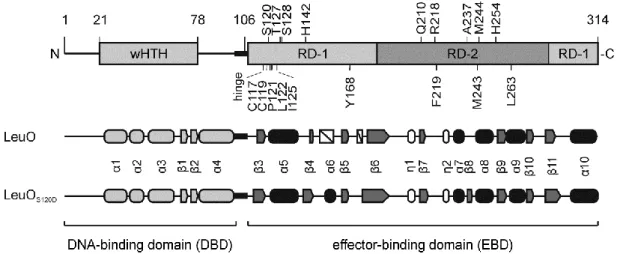
Figure 6: Domain architecture and secondary structure elements of LeuO and LeuO-S120D.
(A) Schematic presentation of the LeuO domain and secondary structure, with the N-terminal DNA-binding domain (DBD) carrying a wHTH motif, and the C-terminal effector-binding domain (EBD), connected by a hinge region. Mutations of residues, which render the protein hyperactive, are shown atop and additional mutations are depicted below the scheme. The secondary structure of the solved crystal structures is depicted in a schematic representation showing α helices as round filled cylinders, η helices as round unfilled cylinders, β sheets as arrows and non-solved areas as white boxes. The secondary structure of the DBD (shown in light grey) is based on a structural model (https://swissmodel.expasy.org/repository/uniprot/P10151).
Intriguingly, the amino acid whose mutation causes hyperactivity, are surface exposed and all map in the putative dimerization interface (Figure 7A). The residues Ser120, Arg218, and Met244 are located in the vicinity of the putative effector-binding cleft, which is located in between the two
/
subdomains. Met244 is close to its counterpart of the second monomer (Figure 7C). Arg218 is reasonably well defined in the wild-type structure and points towards the interior of the putative effector-binding pocket. The mutation of residue Ser120 to Asp induces a reorientation of the side- chain of Arg218 (Figure 7D) with much weaker electron density. Further, in the wild-type protein structure there appears to be a chloride ion bound close to the position in which the carboxylate group of Asp120 is located in the S120D mutant (Figure 7C). The assignment of the electron density peak as chloride is based on the coordination of the ion, the peak height and a weak anomalous signal. The putative chloride ion is coordinated by the backbone amide of Met243, the side-chain hydroxyl group of Ser120, and two water molecules. The vicinity is otherwise rather hydrophobic, with the side-chains of Pro121, Ala242, Met243 and Val214 lining the pocket (Figure 7E). The guanidinium group of Arg218 is about 4.5 Å apart from the putative chloride ion (Figure 7C).
Taken together, crystallization in cooperation with the Lab of Ulrich Baumann and the analysis of the crystal structure by Prof. Dr. Ulrich Baumann suggests the typical fold of the EBD with the two
/subdomains. The structure is quite similar to LysR-type regulator DntR (Lerche et al., 2016) and the structural change between the EBDs of LeuO wild-type and the hyperactive S120D mutant is similar to structural changes described for other LysR-type regulators. For LysR-type regulators DntR, AphB and BenM, the structural changes are induced by an effector binding to the EBD. In the crystal structures of the LeuO-EBD and the hyperactive S120D the side-chain of Arg218 is reoriented at the edge of the presumptive effector-binding cleft, hinting to an effector regulating the LeuO activity.
This putative LeuO effector is also supported by the putative chloride ion binding to the rather
hydrophobic effector binding pocket. The change in the structure, caused by an effector or a
hyperactive mutant, may transmit to the DNA-binding domains and alter for example the angle of
the DNA-binding.
Figure legend next page
Figure 7: Crystal structures of LeuO-EBD and LeuO-S120D-EBD monomer and homodimers by Prof. Dr. Ulrich Baumann.
(A) Cartoon representation of LeuO-S120D-EBD dimer with residues causing hyperactivity when mutated. (B) LeuO-EBD monomer shown in gradient coloring (blue, N-terminus; red C-terminus), with secondary structure elements labeled. A bound chloride ion is shown in green and activating residues are indicated as sticks. (C) Close-up view of dimer LeuO-EBD with bound chloride ion and residues Ser120, Met244 and Arg218 shown as sticks. (D) Superposition of LeuO-EBD in green and LeuO-S120D-EBD crystallized in different space groups in cyan (I222) and purple (C2) in ribbon representation. The C atoms of residue Arg112 have a distance of about 4 Å between wild-type and mutant. (E) Dimer of the LeuO-EBD. The monomeric EBD is shown in gray ribbon (left), with amino acids Pro121, Leu122, Tyr168, Phe219, Met243 (depicted as sticks) lining the pocket that holds the putative chloride ion. The second monomeric EBD is shown in tubes in green, blue, yellow, and orange. Residues that render LeuO inactive are indicated as magenta spheres (Pro121, Leu122, Ile125, Tyr168, and Leu263). Residues that are neutral when mutated compared to wild-type LeuO are depicted in gray spheres (Met243, Phe219, and Cys117). Numbering of residues is according to the full-length protein sequence of LeuO. The Figure was prepared using Chimera (Pettersen et al., 2004).
2.3 Characterization of additional LeuO mutants
Residues Ser120, Arg218 and Met244 map close to the putative effector binding pocket, and are involved in intramolecular and intermolecular contacts. To further characterize these residues, they were mutated to alanine, and Arg218 was in addition mutated to glutamic acid. Further, a LeuO- S120D/M244T double mutant was constructed. The LeuO mutants were constructed in plasmid pKESK22 as before, carrying leuO under control of the IPTG inducible tac promoter (Ptac), a p15A replication origin and a kanamycin resistance. The activity of these LeuO mutants was analyzed using the Pcas-lacZ reporter (strain T1281). LeuO mutants S120A and M244A showed a reduced activation of Pcas compared to wild-type LeuO at basal expression levels (no IPTG, Figure 8A, top panel). Upon induction of their expression (1 mM IPTG) the mutants fully activated Pcas (Figure 8A, bottom panel).
These results show that the hyperactivity is specific for the Ser120 to Asp and Met244 to Thr amino acid exchanges, but not caused by the exchange to alanine (S120A and M244A, Figure 8A). The double mutant LeuO-S120D/M244T remained hyperactive, causing full activation of the Pcas (Figure 8A). Interestingly, mutation of Arg218 to alanine or glutamic acid rendered the protein similarly hyperactive as the Arg218 to cysteine mutation, which was isolated in the screen and caused full activation of Pcas at basal expression levels (Figure 8A).
LeuO harbors a cysteine residue at position 119, located in the cleft of the EBD. A second cysteine at
position 117 is located in the dimer interface of the LeuO-EBDs (Figure 7B and 7E). To test the
relevance of Cys117 and Cys119 for LeuO activity, these residues were mutated to a serine and
aspartic acid, respectively. The mutant C119D showed increased activation of Pcas at basal
expression levels compared to the wild-type (increase from 51 units to 148 units; Figure 8A), and
caused full activation of Pcas at high expression levels. In contrast the other three mutants C117S,
C117D and C119S had a slightly reduced activity even when expressed at high levels (Figure 8A).
Thus, introduction of the negatively charged aspartic acid at residue 119 enhances LeuO activity, which may have a similar effect as the mutation of residue 120 from serine to the negatively charged aspartic acid in the hyperactive mutants S120D.
In the solved LeuO-EBD structure several loops showed a weak electron density, as e.g. between β4 and β5 (residues Q151 to R158) and between β5 and β6 (residues R173 to E175; Figure 7B and 8C).
To analyze these unstructured regions, amino acids in these loops (Asn150, Gln151, Asn152, His155, Gln156, Arg158, His172, Arg173) and additional positively charged residues His204, Trp226 and Lys232, were mutated to alanine and to glutamic acid. The mutants Asn150, Gln151, Asn152, His155, Gln156, Arg158, His172, Arg173, His204 showed full activation of Pcas when their expression was induced with IPTG, irrespective of being mutated to alanine or glutamic acid (Figure 8A). At basal expression levels the LeuO mutants N150E, Q151E, Q156E and H204E showed a slightly increased activation (2 to 3-fold) of the reporter compared to wild-type LeuO. The LeuO mutants N150A, Q151A, N152A, N152E, H155A, H155E, Q156A, R158A and R158E showed a similar activation at basal expression levels as the wild-type (Figure 8A). The activation of Pcas was reduced for LeuO mutants W226A and W226E with induction and abolished without induction possibly due to protein instability or structural changes. The LeuO mutants K232A and K232E had a slightly reduced activity even when expressed at high levels and showed no activation at basal expression levels (Figure 8A).
In the screen for constitutively active LeuO mutants a triple mutant LeuO-S120D/E111D/D205N and a double mutant LeuO-V230I/M244T were isolated. In addition, several double mutants of LeuO (S120D/E111D, S120D/C119S, M244A/M34V, M244A/G240V) were isolated in the process of cloning.
The activation of the Pcas-lacZ by these LeuO mutants was analyzed as well. All LeuO mutant variants of S120D showed full activation of Pcas-lacZ with and without induction and irrespectively of the second or third additional mutation (LeuO-S120D/E111D/D205N, LeuO-S120D/E111D, LeuO- S120D/C119S; Figure 8A). LeuO mutant M244A/M34V showed a similar reduced activation of Pcas as the single mutant of LeuO-M244A, while the activation by LeuO-M244A/G240V was abolished. The additional mutant LeuO-M244T/V230I showed full activation of Pcas-lacZ just as the LeuO-M244T single mutant. Taken together, the additional secondary LeuO mutations were rather negligible, as the activation does not differ significantly from their corresponding single mutant (Figure 8A).
In order to further characterize residues in the cleft of the EBD, several additional residues were
mutated (Figure 6 and 7E). Pro121 was mutated to the negatively charged aspartic acid and Leu122,
Ile125, Tyr168, Phe219, Met243, and Leu263 to negatively charged glutamic acid. The activity of
these mutants was analyzed using a low copy number (pSC origin) expression plasmid carrying leuO
under control of the moderate lacUV5 promoter (Figure 8B). The LeuO mutant L122E was
additionally analyzed expressing the mutant from the Ptac, p15A-ori kanR plasmid. Upon induction of leuO expression mutants F219E and M243E showed a similar activation of the Pcas-lacZ reporter as the wild-type LeuO (Figure 8B, plus IPTG). In contrast, activation of Pcas was strongly decreased for LeuO mutants P121D, L122E, I125E, Y168E and L263E (Figure 8B, plus IPTG). Expressing the LeuO mutant L122E from a medium copy number plasmid (p15A-ori) does not activate the Pcas-lacZ reporter without IPTG and showed reduced activation upon induction, compared to the wild-type (Figure 8A). Thus, these mutants are inactive possibly due to a structural change or protein instability.
Taken together, the analysis of additional LeuO mutants revealed that hyperactivity is specific for the
particular amino acid exchanges S120D and M244T, while the activities of mutants C119D, N150E,
Q151E, Q156E and H204E are moderately increased. Arg218 may be inhibitory since mutation of this
residue to alanine, cysteine, or glutamic acid, causes hyperactivity.
Figure 8: Characterization of additional LeuO mutants.
Activation of the Pcas-lacZ by LeuO mutants was determined in reporter strain T1281. Expression levels were determined of cultures grown without induction (top panel) or with 1 mM IPTG for induction of leuO expression (bottom panel). (A) Pcas-lacZ expression was analyzed of transformants carrying Ptac plasmid for directing expression of wild-type LeuO (pKETS5), LeuO-DBD (pKESMS63), as well as mutants S120D (pKESL104), S120A (pKESL108), R218C (pKESL78), R218A (pKESL238), R218E (pKESL243), M244T (pKESL73), M244A (pKESMS1), S120D/M244T (pKESL107). C117S (pKESMS64), C117D (pKESMS67), C119S (pKESMS65),
Figure 8 continued from the previous page
C119D (pKESMS66), L122E (pKESL106), N150A (pKESL221), N150E (pKESL231), Q151A (pKESL229), Q151E (pKESL232), N152A (pKESL222), N152E (pKESL233), H155A (pKESL223), H155E (pKESL234), Q156A (pKESL224), Q156E (pKESL241), R158A (pKESL228), R158E (pKESL235), H172A (pKESL225), H172E (pKESL236), R173A (pKESL226), R173E (pKESL242), H204A (pKESL230), H204E (pKESL237), W226A (pKESL239), W226E (pKESL240), K232A (pKESL227), K232E (pKESL244), S120D/E111D (pKESL71), S120D/E111D/D205N (pKESL72), S120D/C119D (pKESL105), M244A/M34V(pKESL110), M244A/G240V(pKESL109), V230I/M244T (pKESL79), empty vector pKESK22 served as a control (vector). (B) Pcas-lacZ expression was analyzed in transformants carrying low copy plasmids (pSC-ori) with the lacUV5 promoter (PUV5) directing expression of wild-type LeuO (pKESL39), and mutants S120D (pKESL102), S120D/E111D (pKESL40) P121D (pKESL69), L122E (pKESL103), L122E/H115Y (pKESL68), I125E (pKESL64), I125E/V118I (pKESL65), Y168E (pKESL70), F219E (pKESL67), M243E (pKESL61), and L263E (pKESL62). The empty parent vector of these plasmids served as control (vector). Cultures for β- galactosidase assays were grown to in LB medium with appropriate antibiotics to an OD600 of 0.5. Mean values of at least 3 independent biological replicates are shown as bars; error bars indicate standard deviations. (C) Mutated amino acid residues of unstructured loops of the LeuO protein. The Figure was created using PyMOL (The PyMOL Molecular Graphics System).
2.4 The LeuO DNA-binding domain (DBD) is functional
The LeuO protein in E. coli is presumably a tetramer, like S. enterica LeuO and other LysR-type regulators (Guadarrama et al., 2014a) (predicted structure model from https://swissmodel.expasy.org/repository/uniprot/P10151 shown in Figure 2A and 2B). Tetramers of LysR-type regulators contain two pairs of dimeric effector binding domains (EBD) and two pairs of dimeric DNA-binding domains (DBD; Figure 2A and 2B). Dimerization of the N-terminal DBD is mediated by the α4 helical linker (Figure 2), while the EBDs provide an additional dimerization interface (Figure 7A and Figure 2). In Figure 2A the four monomeric LeuO proteins are highlighted in blue, grey, green and magenta. The EBD-1 (blue) dimerizes with EBD-2 (grey) and the EBD-3 (green) dimerizes with EBD-4 (magenta). The tetrameric complex is completed by the dimerization of DBD-1 with DBD-4 and the dimerization of DBD-2 with DBD-3 (shown in Figure 2B and 2C). The α3 helices (marked in orange) are the DNA recognition helices, forming the DNA-protein interface by insertion into the major groove of the DNA (Aravind et al., 2005) (Figure 2C).
The two DBD dimers within one tetramer presumably bind to two adjacent sites on the DNA.
Therefore a single dimeric DBD may be capable of specifically binding to the DNA as well. To address
this, I tested whether a LeuO DBD-dimer is sufficient to activate the Pcas-lacZ reporter. The DBD (aa1
to aa101) was expressed using the Ptac expression plasmid pKESMS63, and the capability of the DBD
to activate the Pcas-lacZ reporter was analyzed. Upon induction of the DBD expression, full activation
of Pcas-lacZ was observed (Figure 8A). At basal expression levels of the DBD (no IPTG), Pcas
activation was 4-fold lower by the DBD than by wild-type LeuO, but still detectable (Figure 8A,
compare 13 and 51 units). These data show that the DBD alone is sufficient to activate Pcas,
suggesting specific DNA binding by the presumptive LeuO DBD dimer.
2.5 LeuO binds to the promoter regions of cas, yjjQ and leuO
LeuO is an activator of Pcas and PyjjQ and represses its own promoter (PleuO). DNase I footprinting showed two distinct LeuO DNA-binding sites for the promoter regions of cas and leuO and three putative LeuO DNA-binding sites for PyjjQ (Westra et al., 2010, Stratmann et al., 2012) (and unpublished lab data). The LeuO DNA-binding sites are indicated in the schematic view of the promoter regions in Figure 9. To further characterize the kinetics of the LeuO binding to the individual DNA-binding sites, I performed electrophoretic mobility shift assays (EMSA). 80 bp DNA fragments were amplified by PCR, covering the different possible LeuO DNA-binding sites of the promoter regions of Pcas (I and II), PleuO (I and II) and PyjjQ (I, II and III; see schemes in Figure 9).
C-terminally His-tagged LeuO was purified using the ÄKTA fast protein liquid chromatography (FPLC) system. The DNA fragments (20 nM) were incubated with increasing concentrations of LeuO and separated by gel electrophoresis on 8% native polyacrylamide gels.
A distinct band is visible for the DNA fragments when no LeuO protein was added. For the fragment of LeuO DNA binding site II of the cas locus the intensity of the band decreases at a LeuO concentration of 100 nM and a shift is visible. The band intensity of the shift increases at 250 nM and is completely shifted at 500 nM and 1000 nM LeuO (Figure 9A). The DNA fragment of DNA-binding site I of the cas locus shows a decrease of the band intensity at 250 nM, and the shift is visible at protein concentrations 500 nM and at 1000 nM (Figure 9A). The band intensity of both DNA fragments of the DNA-binding sites I and II at the leuO locus decreases at LeuO concentration of 250 nM and is completely shifted at 500 nM and 1000 nM LeuO (Figure 9B). The promoter region of yjjQ contains 3 LeuO DNA-binding sites characterized by DNase I footprinting (unpublished lab data;
Figure 9C). The independent analysis of the single DNA-binding sites shows an unspecific shift for DNA-binding site I, where the band intensity starts to decrease at high protein concentrations of 500 nM and 1000 nM LeuO. Similarly, for DNA-binding site II the band intensity starts to decrease at 250 nM and 500 nM and is shifted at 1000 nM LeuO. The intensity of the band of DNA-binding site III is highly decreased at 250 nM and shifted at 500 nM and 1000 nM LeuO (Figure 9C). For DNA-binding site I and II at the yjjQ locus the band is shifted at rather high protein concentrations indicating a low unspecific binding to these sites (Figure 9C).
Taken together, LeuO binds to two distinct DNA-binding sites at the cas and leuO locus with high
specificity, as shown before (Westra et al., 2010, Stratmann et al., 2012). LeuO binding at the yjjQ
locus was shown only for binding site III at high protein concentrations. However, a more specific
characterization of the DNA-binding sites seems not possible, since the EMSA is not sensitive enough
to characterize differences in the DNA-binding sites.
Figure 9: LeuO binding to DNA-binding sites at the cas, leuO and yjjQ loci.
Binding of LeuO to DNA-binding sites at cas, leuO and yjjQ loci was tested by electrophoretic mobility shift assays (EMSA). A schematic view of the promoter regions of Pcas (A), PleuO (B) and PyjjQ (C) is depicted on top of each panel, with the LeuO DNA-binding sites indicated (I, II and III) (Westra et al., 2010, Stratmann et al., 2012) (and unpublished lab data). The gel pictures of the EMSAs are depicted below each scheme with the DNA-binding sites indicated. The 80 bp DNA fragments were amplified by PCR and 20 nM were incubated without protein or with increasing concentrations of LeuO as indicated (0, 100, 250, 500 and 1000 nM). The samples were separated on 8% native polyacrylamide gels and stained with ethidium bromide.
2.6 DNase I footprinting by LeuO, LeuO-S120D and the LeuO-DBD
Two extended AT-rich DNA-binding sites for LeuO at the cas promoter region (LeuO sites I and II)
have been identified by DNase I footprinting (Westra et al., 2010). In these footprints a rather high
LeuO concentration was used, which corresponds to high levels of wild-type LeuO that is required for
activation of Pcas in vivo. In contrast, only low levels of the hyperactive LeuO-S120D are required for
Pcas activation. Therefore, DNA-binding by S120D may be more specific than by wild-type LeuO. To
test this, DNA-binding of LeuO and its S120D mutant was analyzed by DNase I footprinting using a
broad range of protein concentrations. In addition, to narrow down on the DNA-binding sites, LeuO- DBD was included in the footprinting analysis. The rational of this was that the full-length protein carrying two DBD dimers presumably contacts DNA at two sites and therefore yields an extended footprint. The DBD presumably occupies only half of the full-length LeuO DNA-binding site, which may allow defining a consensus sequence.
For footprinting His-tagged LeuO, LeuO-S120D and LeuO-DBD were purified. DNA fragments covering the full-length LeuO DNA-binding sites I and II of the cas promoter region, were [
32P]-labeled at the top and bottom strand, respectively, and used for footprinting analysis (Figure 10). Footprints of DNA fragments “a” and “b” covering LeuO site II are shown in Figure 11A and 11B, while the footprints of LeuO site I (fragments “c” and “d”) are shown in Figure 11C and 11D. Corresponding to the previously described LeuO site II, DNase I protection by all 3 proteins was detected (Figure 11A to D). A hypersensitive site maps in the middle of this footprint (black triangle) and therefore two half-sites IIa and IIb are labeled in the footprint (IIa & IIb, Figure 11 A and 11B). Strikingly, DNase I protection by LeuO mutant S120D occurred already at very low protein concentrations (31 nM), suggesting that amino acid exchange S120D causes a higher DNA-binding affinity. This higher DNA-binding affinity is reflected by additional protection sites only detected for S120D (Figure 11A, 11C, 11D and 12A).
Intriguingly, the DBD caused a DNase I protection pattern at low concentrations (31 nM) as well, but only at DNA-binding site IIa, while at higher protein concentrations (125 nM) both DNA-binding sites were protected (Figure 11B). For wild-type LeuO a footprint was apparent as of a concentration of 125 nM at both DNA-binding sites, but this footprint is more diffuse, at least at DNA-binding site IIb (Figure 11A and 11B). The DNase I footprints by LeuO-S120D and in particular by LeuO-DBD indicate that DNA-binding site IIa (in LeuO DNA-binding site II) is a high-affinity DNA-binding site, which is called “core site” in the following. This “core site” is palindromic (indicated by inverted arrows and bold letters, Figure 11A and 11B). Palindromic sequences are typical for DNA-binding of transcription regulators with dimeric DBDs (Browning & Busby, 2016). Thus the palindromic sequence of DNA- binding site IIa may represent one of the two DNA contact sites of the tetrameric LeuO. For LeuO site I (footprints of fragments “c” and “d”) all three proteins protected the DNA from DNase I cleavage, confirming previous data (Westra et al., 2010) (Fig footprint C and D). However, no palindromic core site is apparent in LeuO DNA-binding site I (Figure 12A). Furthermore, the DNase I footprint of LeuO- S120D showed an additional hypersensitive site, indicating the binding of at least two tetramers that might form a higher order complex (open arrowhead, Figure 11A).
Taken together, these results confirm the LeuO DNA-binding sites at the cas promoter (Westra et al.,
2010). In addition, hyperactive mutant S120D and the DBD bind with higher affinity to LeuO site II
than wild-type LeuO, Further, the footprint by S120D and DBD is more distinct, and allowed the
identification of the core DNA-binding site in DNA-binding site IIa. The palindromic sequence of the core site suggests that this represents a close to ideal DNA-binding site of LeuO.
Figure 10: Schematic overview of the cas promoter region and DNA fragments for DNase I footprinting with LeuOHis6, LeuO-DBDHis6, and LeuO-S120DHis6.
Indicated are LeuO DNA-binding sites I and II (black boxes) (Westra et al., 2012), the H-NS nucleation site (open box), and the transcription start sites (bent arrows) (Pul et al., 2010). Fragments (“a” to “d”) used for DNase I footprinting cover either one or both LeuO DNA-binding sites, as indicated. The fragments were generated by PCR using primer pairs of which one primer was 5' [32P]-labeled, indicated by an asterisk (fragment “a”: oligonucleotides [32P]-OA477/OA474, fragment “b”: OA477/[32P]-OA475, fragment “c”: [32P]- OA476/OA473, fragment “d”: OA477/[32P]-OA473). Fragments “a” and “c” were labeled at the top strand, and fragments “b” and “d” were labeled at the bottom strand. The [32P]-labeled DNA fragments were incubated with increasing protein concentrations of LeuOHis6, LeuO-DBDHis6 and LeuO-S120DHis6, as indicated.