Function of the CD74 receptor in B cell pro-survival signaling
in the context of chronic lymphocytic leukemia
I n a u g u r a l - D i s s e r t a t i o n zur
Erlangung des Doktorgrades
der Mathematisch-Naturwissenschaftlichen Fakultät der Universität zu Köln
vorgelegt von Romy Barthel
Köln, 2015
Berichterstatter/in: PD Dr. rer. nat. Frank Thomas Wunderlich Prof. Dr. rer. nat. Thorsten Hoppe
Prof. Dr. med. Michael Hallek
Tag der mündlichen Prüfung: 19.01.2015
ZUSAMMENFASSUNG ... 1
ABSTRACT ... 2
1 INTRODUCTION ... 3
1.1 CHRONIC LYMPHOCYTIC LEUKEMIA ... 3
1.1.1 Epidemology and Etiology ... 3
1.1.2 Diagnosis ... 3
1.1.3 Pathophysiology ... 4
1.1.4 Therapy ... 6
1.2 CLL ANIMAL MODELS ... 7
1.2.1 Eµ-TCL1 mice ... 7
1.3 CD74 ... 9
1.3.1 The invariant chain (Ii) part of the MHC class II complex ... 10
1.3.2 The surface receptor CD74 ... 11
1.3.2.1 CD74 receptor signaling ... 11
1.3.3 CD74 physiology ... 13
1.3.4 CD74 pathophysiology ... 14
1.3.4.1 CD74 during Helicobacter pylori infection ... 14
1.3.4.2 CD74 in B cell neoplasia ... 14
1.4 CD74 IN CHRONIC LYMPHOCYTIC LEUKEMIA ... 15
1.5 OBJECTIVE ... 17
2 RESULTS ... 18
2.1 CD74 EXPRESSION IN Eµ-TCL1-TRANSGENIC MICE ... 18
2.2 CD74 IN THE DEVELOPMENT OF TCL1-INDUCED CLL ... 20
2.2.1 Crossbreeding of Eµ-TCL1-transgenic mice with CD74ko mice ... 20
2.2.2 Leukemia development in TCL1+ CD74ko mice ... 21
2.2.3 BCR genetics in TCL1+ CD74ko mice ... 24
2.3 DISTRIBUTION OF MYELOID LINEAGE CELLS IN THE SPLEEN OF TCL1+CD74KO MICE ... 25
2.4 IMPACT OF CD74 DELETION ON APOPTOSIS AND PROLIFERATION OF MALIGNANT B CELLS ... 27
2.4.1 Apoptosis of malignant B cells in TCL1+ CD74ko mice ... 27
2.4.2 Proliferation of malignant B cells in TCL1+ CD74ko mice ... 29
2.5 OVERALL SURVIVAL OF TCL1+CD74KO MICE ... 31
2.6 SYNGENEIC TRANSPLANTATION OF MURINE CLL CELLS INTO CD74KO MICE... 32
2.7 CD74-DEPENDENT REGULATION OF PRO-SURVIVAL PATHWAYS ... 33
2.7.1 Activation of pro-survival pathways in unstimulated murine CLL cells ... 34
2.7.2 Activation of pro-survival pathways in MIF-stimulated murine CLL cells ... 35
2.7.3 Signal transduction upon MIF stimulation in human CLL cells ... 36
2.7.4 CD74 receptor stimulation in murine CLL cells ... 37
2.7.5 MIF signaling involving CD74 co-receptors CXCR2, CXCR4 and CD44 ... 37
2.7.5.2 MIF stimulation of murine CLL cells upon CXCR2- and CXCR4- or CD44 inhibition ... 39
2.8 B CELL DEVELOPMENT IN TCL1+CD74KO MICE ... 41
3 DISCUSSION ... 45
3.1 CD74 EXPRESSION IS UPREGULATED IN Eµ-TCL1-TRANSGENIC MICE ... 45
3.2 CD74 DELETION DOES NOT INFLUENCE DEVELOPMENT IN TCL1-INDUCED CLL ... 45
3.3 AKT KINASE ACTIVATION UPON MIF STIMULATION IS CD74-DEPENDENT ... 47
3.4 TCL1 OVEREXPRESSION ALTERS B CELL DEVELOPMENT IN CD74KO MICE ... 49
3.5 CONCLUSION AND OUTLOOK ... 51
4 MATERIALS ... 52
4.1 INSTRUMENTS ... 52
4.2 CHEMICALS AND REAGENTS ... 53
4.3 SUBSTANCES ... 54
4.4 ANTIBODIES ... 55
4.4.1 Antibodies for Immunoblotting ... 55
4.4.2 Antibodies for cell culture ... 56
4.4.3 Antibodies for flow cytometry ... 56
4.5 MOUSE STRAINS ... 57
4.6 OLIGONUKLEOTIDES ... 57
4.7 SPECIAL REAGENTS AND KITS ... 57
4.8 PRIMARY PATIENT MATERIAL ... 58
4.9 SOFTWARE ... 58
5 METHODS ... 59
5.1 BREEDING ... 59
5.2 GENOTYPING ... 59
5.2.1 DNA-Preparation ... 59
5.2.2 Polymerase-Chain-Reaction (PCR) ... 59
5.2.3 Agarose gel electrophoresis ... 61
5.3 BLOOD ANALYSIS... 61
5.3.1 Blood sampling ... 61
5.3.2 Differential blood count ... 61
5.4 EXTRACTION OF ORGANS ... 62
5.5 CELL CULTURE ... 62
5.5.1 Culture conditions ... 62
5.5.2 Counting ... 62
5.5.3 Freezing and thawing of cells ... 62
5.6 ISOLATION OF PRIMARY MURINE CELLS ... 63
5.6.1 Isolation of primary murine splenocytes ... 63
5.6.2 Isolation of primary murine B cells ... 63
5.7 ISOLATION OF PRIMARY HUMAN CLL CELLS ... 64
5.8 FLOW CYTOMETRY ... 65
5.8.1 Staining of surface proteins ... 65
5.8.2 Staining of intracellular proteins ... 65
5.9 STIMULATION EXPERIMENTS ... 65
5.10 PROTEIN BIOCHEMISTRY ... 66
5.10.1 Preparation of cell lysates ... 66
5.10.2 Protein quantification ... 66
5.10.3 SDS Polyacrylamide gel electrophoresis (PAGE) ... 67
5.10.4 Protein transfer ... 68
5.10.5 Immunoblotting ... 68
5.10.5.1 Detection by chemiluminescence ... 69
5.10.5.2 Detection by fluorescence ... 70
5.11 SYNGENEIC TRANSPLANTATION OF TCL1-CLL CELLS... 70
5.12 QUANTIFICATION OF PROLIFERATING CELLS ... 70
5.13 QUANTIFICATION OF APOPTOSIS ... 71
5.14 IGVH STATUS ... 71
5.15 IMMUNOHISTOCHEMISTRY ... 71
REFERENCES ... 73 ABBREVIATIONS ... I LIST OF FIGURES ... III DANKSAGUNG ... IV ERKLÄRUNG ... V
Zusammenfassung
Das Oberflächenprotein CD74 wird auf der Membran von B-Zellen, Makrophagen und Epithelien exprimiert und kontrolliert viele Bereiche des Immunsystems. Von besonderem Interesse ist die Rezeptorfunktion von CD74 für das Chemokin MIF (macrophage migration inhibitory factor). Bei der Bindung von MIF an CD74 werden - unter anderem über den Korezeptor CD44 - die AKT, MAPK und NF-B Signalwege aktiviert, welche die Zellproliferation anregen und die Apoptose hemmen.
Bei vielen Tumorarten, wie z. B. Magenkarzinomen und B-Zell-Lymphomen, wird eine Überexpression von CD74 beobachtet. Die Funktion von CD74 in B-Zell-Lymphomen wurde im Fall der chronischen lymphatischen Leukämie (CLL) veranschaulicht, bei der nicht nur CD74 sondern auch MIF hoch reguliert sind. CLL tritt im hohen Lebensalter auf und ist die häufigste Leukämieform in Europa und Nordamerika. Das Tumormikromilieu spielt eine zentrale Rolle in der CLL und trägt maßgeblich zum Überleben der CLL-Zellen bei. Daher wird bei der Entwicklung von möglichen Therapien ein besonderes Augenmerk auf das Zusammenspiel der CLL-Zellen mit dem Tumormikromilieu gesetzt.
Die Rolle von CD74 in der CLL wurde meist in primären, humanen CLL-Zellen und Zelllinien in vitro untersucht ohne das Tumormikromilieu zu berücksichtigten. Um den Einfluss von CD74 auf die B-Zell-Onkogenese im Zusammenspiel mit dem Mikromilieu
in vivo genauer zu untersuchen, wurden in dieser Arbeit das CLLMausmodell (Eµ-TCL1-transgen) mit dem CD74-defizienten Mausmodell gekreuzt. In den dadurch generierten TCL1
+CD74
koMäusen wurden dann die CLL-Entwicklung sowie die Veränderung der zellulären Signalwege untersucht und mit der Kontrollgruppe TCL1
+CD74
wtverglichen.
In TCL1
+CD74
koMäusen waren die gemessene Tumorlast im Blut, die Infiltration leukämischer Zellen in lymphatischen Organen und das Überleben der Tiere vergleichbar mit dem der Kontrollgruppe. Des Weiteren wurden Proliferation und Apoptose der CLL Zellen nicht von der CD74-Expression beeinflusst. Stimulations- experimente mit leukämischen Zellen beider Modelle zeigten jedoch, dass die Aktivierung der AKT Kinase durch MIF nur in Gegenwart von CD74 stattfand, während die ERK und NF-B Signalwege CD74-unabhängig waren.
Anhand dieser Ergebnisse konnte in dieser Arbeit erstmals gezeigt werden, dass die
Deletion von CD74, anders als MIF und CD44, die Entwicklung der CLL im
Mausmodell nicht wesentlich beeinflusst. Zusammenfassend lässt sich somit auf eine
untergeordnete Rolle der MIF vermittelten CD74 –Signalwege für das Wachstum und
die Entwicklung von CLL Zellen schließen.
CD74 is a surface protein expressed on B cells, macrophages and many epithelial cells and has been found to control several aspects of the immune system. One of them is its function as surface receptor for the chemokine macrophage migration inhibitory factor (MIF). Signaling through the CD74 receptor and its co-receptor CD44 upon MIF binding leads to activation of the AKT, MAPK and NF-B pathways, and thereby promotes cell proliferation and survival.
Elevated expression of CD74 has been observed in several human cancers e.g. gastric carcinoma and B cell neoplasia. The role of CD74 in B cell neoplasms has been suggested in the case of chronic lymphocytic leukemia (CLL), where both the receptor CD74 and its ligand MIF are upregulated. CLL is one of the most common leukemias found in adults in Europe and North America. The microenvironment plays a central role to CLL development and progression.
So far, studies on the role of CD74 in CLL were based on experiments with primary CLL cells or cell lines in vitro. However, the exact contribution of CD74 to the pathogenesis of CLL remained far from being understood.
To understand the role of CD74 for the pathogenesis of leukemia, this project aimed to determine the influence of the CD74 receptor during B cell lymphomagenesis and the mechanisms underlying CD74-dependent signaling in B cells by using the CLL mouse model (Eµ-TCL1-transgenic). Eµ-TCL1 transgenic mice pro- and deficient for CD74 (TCL1
+CD74
wtand TCL1
+CD74
ko) were generated and monitored for CLL development and activation of pro-survival signaling upon MIF stimulation.
CLL development in TCL1
+CD74
komice was similar to control TCL1
+CD74
wtmice depicted by comparable growth of the leukemic load, development of hepato- splenomegaly and overall survival. Moreover, the apoptosis and proliferation rate of malignant cells from TCL1
+CD74
komice were similar to control mice. Experiments with MIF stimulation in CLL cells showed that MIF induced AKT activation in a CD74 dependent manner, whereas ERK and NF-B activation did not differ between TCL1
+CD74
wtand TCL1
+CD74
kocells.
Taken together this study showed that targeted gene deletion of Cd74 does not influence
the development of CLL in Eµ-TCL1-transgenic mice and suggested that the pathways
mediated by MIF through CD74 are not sufficiently potent to promote growth of CLL
cells.
1 Introduction
1.1 Chronic Lymphocytic Leukemia
Chronic lymphocytic leukemia (CLL) is one of the most common leukemias in Europe and North America. It mostly affects older individuals with an median age of 65 to 70 years and rarely under 50 years of age, with men being twice as often affected as women [1, 2]. The World Health Organization describes CLL as leukemic, lymphocytic lymphoma distinguishable from small lymphocytic leukemia (SLL) by its leukemic appearance [3]. Like other cancers, CLL is caused by genomic damage that alters distinct signaling pathways in B cells which leads to the induction of anti-apoptotic proteins and the downregulation of pro-apoptotic proteins [4]. Dysregulated expression and signaling of these cell death regulators then leads to a progressive accumulation of long-lived and apoptosis-resistant B cells in the peripheral blood, bone marrow and secondary lymphoid tissues [1].
1.1.1 Epidemology and Etiology
The incidence of CLL lies at 3/100.000/year and varies with age and sex structure of the population [4, 5]. Rates of CLL in the population show also significant international variation, with the highest rates in the U.S. and Europe and the lowest rates in Asia [6].
The cause for CLL is still unsure. Large, population-based case-control and cohort studies have shown significant familial aggregation of CLL with first degree relatives being three times more likely to have CLL or other lymphoid neoplasms than the general population [6-8]. While there is evidence for a genetic disposition for CLL, attempts to link genetic aberrations to CLL have been unsuccessful [6]. Additionally, linking CLL incidences with environmental exposure to radiation or other chemicals showed no consistent evidence so far [4]. On the other hand, induction through viral infection, e.g. Epstein- Barr-Virus (EBV) and Merkel cell polyomavirus (MCPyV), is often discussed [9], but could not be proven so far [10].
1.1.2 Diagnosis
The World Health Organization and the guidelines from the international workshop on
CLL defined a count of more than 5x10
9monoclonal CD5-positive B cells per litre
blood, which is consistent for more than 3 months, as a safe diagnosis for CLL [11]. To
differentiate CLL cells from other B cell lymphomas, cell surface marker are used
which characterize the CLL cell phenotype. In flow cytometric analyses CLL cells are simultaneously positive for the surface marker CD19, low levels of CD20, CD23 and aberrant CD5 [12]. Often CLL is found during routine checkup, since many patients do not develop any symptoms [13]. Typical symptoms of CLL are lymphocytosis leading to enlargement of lymphoid organs (e.g. spleen and liver) and the swelling of lymph nodes, weight loss, abdominal pain, night sweat, susceptibility to infection and 30% of patients develop skin irritation. At a later stage defective haematopoiesis leads to anemia resulting in fatigue and weakness, while thrombocytopenia leads to bleeding [14]. Rai and colleagues developed a system of clinical staging CLL that could prospectively distinguish patients according to their overall outlook for survival [15-17].
Later Binet and colleagues added another prognostic classification [18]. Both methods of staging are recognized as simple, yet accurate predictors of survival and are still used in a modified version to group patients with CLL based on physical examination and complete blood counts [3, 11, 19].
In the last years molecular and cellular markers have been identified that also could predict disease progression. Especially the mutational profile of the immunoglobulin genes, cytogenetic abnormalities, serum-based markers like β2-microglobulin and cellular marker like CD38 and ZAP-70 show strong prognostic value [4, 20].
1.1.3 Pathophysiology
One hallmark of CLL cells is the expression of the B cell receptor (BCR) [21]. The
BCR is expressed on the plasma membrane of B cells as a disulfide-bonded complex of
heavy and light immunoglobulin (Ig) chains associated with the Ig and Ig (or
CD79a/CD79b) heterodimer. The BCR is the key molecule for the signaling pathway
involved in B cell proliferation, survival, differentiation, anergy and apoptosis [22]. The
Ig component of the BCR has a unique molecular feature, which marks CLL cells and
determines the indolent or aggressive nature of the disease. In this context CLL can be
divided into two main subsets, based on whether the tumor arose from a B cell prior to
initiation of somatic hypermutation in the Ig variable (V) region genes (unmutated
CLL), or after this process had taken place and then stopped (mutated CLL) [23]. The
unmutated cases show an aggressive disease progression which is often accompanied by
high ZAP-70 expression, while mutated cases show a more indolent form with low
ZAP-70 expression [24]. Furthermore, conventional cytogenetic analyses and
fluorescent
in situ hybridization (FISH) showed genetic aberrations in ~82% of CLLcases [25]. Among those aberrations, four are quite commonly found, del(17q13),
del(11q23), del(13q14) and trisomy 12 [20]. The 17p deletion (del17p) affects the tumor suppressor p53 protein and is associated with a poor prognosis [26], as well as the 11q deletion (del11q), which is mostly accompanied by a mutation in the ATM kinase leading to impaired DNA damage response in the cells [27, 28]. Deletion of 13q (del13q) is associated with a better prognosis and leads to the loss of the microRNA’s miR15 and miR16 [29], which target the anti-apoptotic protein BCL-2 [30]. Despite the heterogeneity in the disease, CLL patients show a common gene expression signature differentiating them from other lymphoid cancers, which led to the conclusion that CLL patients share a common mechanism of transformation or cell of origin [31, 32].
Recently, studies were linking unmutated CLL to unmutated mature CD5-positive B cells and mutated CLL to a distinct CD5- and CD27-positive post–germinal center B cell subset [33].
Another hallmark of CLL is the accumulation of mature B cells that escape programmed cell death and undergo cell cycle arrest in the G0/G1 phase [34]. In accordance, CLL cells show a low proliferation rate and overexpression of the anti- apoptotic proteins of the BCL-2 family (BCL-XL, BAG1, MCL-1) while the pro- apoptotic proteins (BAX, BCL-XS) are under expressed [35]. Despite their apparent longevity
in vivo, culturing of CLL cells in vitro results in spontaneous apoptosis,indicating a supporting role for the microenvironment in CLL cell survival [36].
Interestingly, first studies in the microenvironment of CLL revealed pseudo follicles and cell clusters in lymph nodes and the bone marrow. Those clusters consist of increased numbers of CD4-positive T cells and CLL cells. CD4-positive T cells express the CD40 ligand, which stimulates B cells and induces expression of anti-apoptotic proteins [37, 38]. T cells also secret anti-apoptotic cytokines like Interleukine 4 (IL-4) providing further stimulus for B cells [39]. In fact, stimulation with CD40 ligand and IL-4 prevented CLL cells from apoptosis in vitro [37]. In addition stromal cells, nurse like cells and follicular dendritic cells are found in the microenvironment of CLL cells.
Nurse like cells differentiate from CD14-positive monocytes through the interaction
with CLL cells, which in turn protect CLL cells from apoptosis mediated through the
production of B cell-activating factor of the TNF family (BAFF), the proliferation-
inducing ligand (APRIL) [40] and the secretion of stromal-derived factor 1 (SDF-1) [41,
42]. Bone marrow derived stromal cells and follicular dendritic cells also provide a
complex system of survival signals for CLL cells. For one they express integrins
interacting with CLL cells and secondly secret cytokines like SDF-1 and VEGF (vascular endothelial growth factor) to further enhance CLL survival [43].
Another cytokine playing an important role in the maintenance of the CLL clone is the macrophage migration inhibitory factor (MIF). MIF is a proinflammatory and immunoregulatory cytokine ubiquitously expressed in cells of the mammalian system and has been shown to promote CLL development [44]. On B cells MIF binds to the surface receptors CD74, CXCR2 and CXCR4 and thereby stimulates pro-survival signaling. In CLL it has been shown that binding of MIF to the receptor CD74 leads to the production of Interleukine 8 (IL-8) and the upregulation of the anti-apoptotic protein BCL-2, which in turn promote CLL cell survival [45, 46] (this aspect will be further discussed under 1.4).
Taken together, CLL cells actively shape their microenvironment by producing cytokines and chemokines, and by subverting normal accessory cells to promote leukemia-cell survival, proliferation, and escape from immune surveillance [43].
1.1.4 Therapy
Chronic lymphocytic leukemia is a disease which is not curable. The only curative approach is allogeneic stem cell transplantation, which has a high lethality rate of 25%
and therefore is only used on high-risk patient with good fitness [11, 47]. The management of CLL has changed a lot in the last decades due to a better understanding of the biology of the disease and the approval of new drugs. Treatment is chosen depending on the clinical stage of the disease, cytogenetics, patients fitness and treatment situation [48]. In 2008 the international workshop on CLL updated guidelines for the management of CLL [11]. Patients in clinical Binet stage A and B without active symptomatic disease are not treated but controlled after the “watch and wait”-principle.
First-line treatment for patients in higher stages involves chemotherapy with differing
regimes depending on the patient fitness. Patients with a 17p deletion have a very poor
prognosis and often show resistance to chemotherapy [49]. First-line treatment for these
high-risk patients so far were the purine analog Alemtuzumab in combination with
steroids [50]. Since most patients eventually relapse alternative treatments within
clinical trials are suggested [48]. Recently several novel drugs targeting kinases
involved in the pro-survival signaling of B cells have been approved by regulatory
agencies or are under evaluation. Among them being the BTK inhibitor Ibrutunib [51],
the PI3K
inhibitor Idealisib (GS1101) [52], which showed promising results in
refractory patients. This recent advances show the importance to dissect and understand the biology of the disease to provide the right approach to treat the disease.
1.2 CLL animal models
Mouse models recapitulating human malignancies are valuable tools for pre-clinical studies and to study pathological mechanisms. Several mouse models for CLL have been generated either through mimicking genetic aberrations, deregulated gene expression in CLL or driven by ectopic oncogene expression (reviewed in [53]).
The first mouse resembling human CLL was the Eµ-TCL1-transgenic mouse generated in 2002 by Bichi and colleagues [54]. Since then several transgenic mice have been created that also lead to CLL-like disease. One of them is the APRIL-transgenic mouse, which resembles the elevated levels of the tumor necrosis factor (TNF) family member APRIL found in sera of CLL patients. At 9- to 12-month-old APRIL transgenic mice develop lymphoid tumors that originate from expansion of the peritoneal B-1 B cell population [55]. Another model is the BCL-2xtraf2dn double transgenic mouse, studying the influence of both these molecules in CLL pathogenesis [56]. Both BCL-2 and the TNF-associated factor 2 (TRAF2) have been implicated in mediating CLL cell survival [35, 57]. The single BCL-2 or TRAF2 mutant mice develop lymphadenopathy and splenomegaly with age due to lymphoid cell expansion but only the BCL-2xtraf2dn double transgenic mice develop an age-dependent B cell leukemia resembling human CLL [56]. Next to those, mice mimicking the deletion of 13q14 (e.g. mir-15/16-1
-/-and 14qC3 minimal deleted region (MDR)
-/-mice), the most frequent genetic lesion found in CLL [29], also develop CLL-like disease and provide evidence for the tumor suppressor function of a CLL-associated genetic lesion [58].
The most notable difference between those mouse models is the penetrance of the phenotype which is highest in the Eµ-TCL1 mice (~100%), intermediate in 14qC3 MDR knock-out and APRIL-transgenic mice (40-50%) and lowest in the mir-15/16-1 knock out mice [53]. Due to the complete disease penetrance and the similarities in the developed disease to human CLL, Eµ-TCL1 mice have become the most commonly used model in CLL research.
1.2.1 Eµ-TCL1 mice
TCL1 is a proto-oncogene highly expressed in most B cell and T cell tumors, e.g. CLL
and T-PLL [59, 60]. Under physiological conditions TCL1 is expressed during B cell
development in pre-B cells, antigen-naïve IgM-positive, mantle zone and germinal
centre B cells [61, 62], and during T cell development in early T cells before the T cell receptor is expressed [63].
In CLL, TCL1 expression correlates with aggressive clinical progression and the phenotypic features like unmutated IgV
Hand ZAP-70 expression [59]. In 2002, Bichi and colleagues used the human TCL1 gene to establish a CLL mouse model. For that, a 350 bp sequence of the human TCL1 gene was put under the control of the murine IgV
Hpromotor and the Eµ enhancer to ensure expression in immature and mature B cells.
These mice spontaneously develop B cell hyperplasia first evident in the peritoneal cavity (~ 2 months) and later in the lymph nodes, spleen, bone marrow and blood (~ 3-8 months). Later at around 8-12 months these mice develop a CLL-like disease with an accumulation of CD5-positive B cells in the spleen, liver and lymph nodes [54].
Furthermore, the BCRs of Eµ-TCL1-transgenic mice resemble those from human CLL patients with the more aggressive from of the disease (unmutated IgV
Hgene rearrangements) and exhibited stereotype in IGHV, IGKV and IGLV gene rearrangements [64]. Since its development, the Eµ-TCL1-transgenic mouse has been used by many laboratories to elucidate the functional role of specific molecules in the onset and progression of CLL in vivo (overview in Figure 1), providing new insights into the pathogenic role of those genes in the dysregulation of signaling, proliferation, and apoptosis, and in the aberrant cross-talk with the microenvironment [53].
Figure 1: Study of novel pathogenic mechanisms in the Eµ-TCL1-transgenic mouse model Deletion or overexpression (tg: transgenic) of molecules in the Eµ-TCL1-transgenic mouse model affecting disease phenotype. (bm: bone marrow; TAM: tumor associated macrophages) (from [53])
Functionally, TCL1 interacts with the protein kinase B (AKT) mediated by the PH domain of AKT. The interaction enhances the kinase activity and promotes the nuclear translocation of the AKT kinase, leading to the transduction of anti-apoptotic and proliferative signals [65]. However, the effects of TCL1 on AKT are not sufficient to explain TCL1 oncogenesis in Eµ-TCL1-transgenic mice, since AKT activation itself does not cause B cell neoplasia [66, 67]. Studies on different transgenic mouse models (e.g. APRIL) showed the importance of the NF-B pathway in the development of a CLL-like disease suggesting a role of the NF-B pathway in the pathogenesis of CLL [68]. Interestingly, there are studies showing that TCL1 enhances NF-B activation independent of AKT through direct interaction with IB [67]. Studies on the oncogenic effect of TCL1 expression in B cells are still not completed and also suggest TCL1 as transcriptional regulator of the CREB binding protein p300 and the activating protein 1 (AP-1) [69].
1.3 CD74
The invariant chain (Ii, known as CD74 when expressed on the plasma membrane) is a type II membrane protein first identified as the MHC class II-associated chaperon [70].
It is expressed in HLA class II-positive cells like B cells, monocytes, macrophages, Langerhans cells, dendritic cells, thymic epithelial cells and gastric epithelial cells.
CD74 controls several aspects of the immune system; e.g. B cell development, dendritic cell motility, thymic selection and has been associated with B cell neoplasia and solid tumor development, progression and metastasis [70].
The human CD74 gene (chromosome 5, 9 exons) and the murine counterpart
(chromosome 18, 8 exons) share much homology. Both have two main transcript
variants (p31 and p41), with humans having two further transcripts (p35 and p43),
resulting from an alternative translation start site [71, 72]. The human and the murine
CD74 consist of a 29-46 amino acid NH
2-terminal intracytoplasmic domain, depending
on which of two alternative initiation codons are translated, a 26-amino acid
hydrophobic transmembrane region, and a 160-amino acid extracytoplasmic domain
containing two N-linked carbohydrate chains [73] (Figure 2). In both species the shorter
isoform of CD74 predominates with an estimated ratio of 9:1 [74]. The p33 and the p35
isoform regulate the MHC class II antigen presentation, while the p41 and p43 isoform
encode a thyroglobulin type 1 domain that can bind cathepsins [75].
Figure 2: CD74 structure
The schema shows the intracytoplasmic (IC), transmembrane (TM) and extracellular (EC) domains of CD74. Amino acid numbers refer to the human p35 variant. CLIP: class II-associated invariant chain peptide. ICD: Intracellular domain.(from [73])
1.3.1 The invariant chain (Ii) part of the MHC class II complex
The first and best described function of the invariant chain (Ii) is its part in the major
histocompatibility class II complex (MHC class II), which controls a major component
of the immune system. The invariant chain functions as a chaperone helping with the
proper folding of MHC class II proteins and protection from peptide binding during
transit through the ER [76-78] (reviewed in [79]). In more detail, after synthesis in the
endoplasmic reticulum (ER) the Ii combines with MHC class II heterodimers, where it
assures proper folding and assembly of the MHC class II dimers [80-82]. This complex
then exits the ER and travels through the golgi apparatus to the endosomal
compartment. The cytoplasmic tail of Ii contains two di-leucin-based motifs, which are
essential for efficient sorting [83]. During the transit to the endosomal compartment
binding of the Ii to the MHC class II prevents the unspecific binding of peptides to the
complex [84]. After the Ii-MHC class II complex reaches the endocytic compartment,
the Ii is progressively degraded until only an Ii derived peptide called CLIP (class II
associated invariant chain derived peptide) remains associated to MHC class II. CLIP
then is exchanged for an antigenic peptide. The mature MHC class II-peptide complexes
are then translocated to the cell surface for CD4
+T cell recognition [79].
In 2012 the group from Basha et.al. showed that the Ii also associates with MHC class I molecules in dendritic cells. There Ii directs a subset of MHC class I molecules to the endolysosomal pathway, where dissociation of the Ii and reassembly of MHC class I with antigenic peptides are carried out [85], showing that the Ii is also involved in the cross-presentation pathway of dendritic cells that has a major role in the generation of MHC class I-restricted, cytolytic T cell response to viral protein.
1.3.2 The surface receptor CD74
Several studies showed that 2-5% of the invariant chain (Ii) is found on the cell surface [86, 87]. A small subset of the Ii is modified by the addition of chondroitin sulfate and rapidly transported from the golgi apparatus to the cell surface [88], where it remains for a short time with an estimated surface half-life of ten minutes [89]. Expression of this cell surface protein, designated CD74, is independent from the expression of class II molecules [90, 91]. CD74 is expressed on several MHC class II positive cells (B cells, monocytes, macrophages, Langerhans cells, dendritic cells, thymic epithelium and gastric epithelial cells) but is also found on a number of cells without MHC class II (e.g.
pulmonary alveolar epithelium, colon epithelium) [92, 93]. On the surface CD74 is a receptor for extracellular MIF, D-DT/MIF-2 and bacterial proteins [94-99].
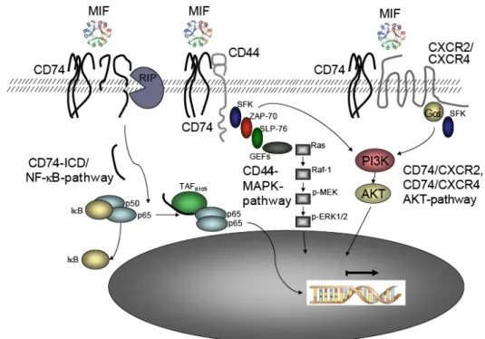
1.3.2.1 CD74 receptor signaling
Studies on the signaling function of surface CD74 revealed several signaling pathways (overview Figure 3). The major part is the identification of CD74 as a high-affinity receptor for the macrophage migration inhibitory factor (MIF) [94]. MIF is a proinflammatory and immunregulatory cytokine, which is ubiquitously expressed in mammalians (reviewed in [100]). Extracellular MIF binds to CD74 and survival signals via the SYK, MAPK, AKT or the NF-κB pathways are transmitted [94, 101, 102].
Those signaling events result in cell proliferation and inhibition of apoptosis [94, 103].
Although it was shown that phosphorylation of the serine residues takes place on the
p35 variant of Ii [104], the short cytoplasmic sequence of CD74 does not appear to
signal directly. It was demonstrates that MIF-induced extracellular signal-regulated
kinase 1 and 2 (ERK1/2) MAP kinase activation is dependent on CD44 in fibroblast,
monocytes, B cells (Raji cell line) and macrophages [105]. CD44 is a structurally
diverse and multivalent co-receptor due to prominent alternative splicing and
posttranslational modifications (e.g., glycosylation). It recruits several kinases (e.g.
receptor tyrosine kinases and non-receptor src family kinases), which eventually all evoke strong MAPK/ERK and PI3K/AKT responses [106].
Studies using a CD74 activating antibody show that activation of CD74 also results in the cleavage of the intracellular cytoplasmic domain of CD74 (CD74-ICD) in B cells [107, 108] by a process called regulated intermembrane proteolysis (RIP). In RIP cleavage of the membrane protein releases a new active peptide which migrates into the nucleus to regulate gene transcription (reviewed in [109, 110]). The RIP of the CD74- ICD is dependent on PI3K/AKT phosphorylation [111] and induces activation of the NF-B p65 and the B cell-enriched co-activator TAF
II105leading to cell proliferation and survival [112, 113].
MIF is also a non-cognate, high-affinity ligand for the chemokine receptors CXCR2 and CXCR4 [101, 114]. Both chemokine receptors belong to the family of seven helix- membrane-spanning G-protein coupled receptors. CXCR2 is the cognate receptor for CXC chemokines such as CXCL8, and CXCR4 is the cognate receptor for SDF1
[115]. It has been shown that CD74 forms complexes with CXCR2 or CXCR4 on the surface of monocytes and T cells [101, 116], which has been suggested to amplify MIF triggered responses in monocytes [101].
Figure 3: CD74- dependent MIF signaling
Schema of MIF induced CD74 signaling. 1: Binding of MIF leads to regulated intermembrane proteolysis (RIP) of the intracellular domain (ICD) of CD74, which then induces activation of the NF-B pathway. 2:
MIF binding to CD74 leads to the recruitment of the co-receptor CD44, which induces activation of the PI3K/AKT and MAPK pathway. 3: CD74 and CXCR2 or CXCR4 form a complex leading to G-protein coupled (Gi) AKT activation upon MIF binding.
1.3.3 CD74 physiology
Development of the CD74-knockout mouse (CD74
ko) revealed the profound effect of CD74 on the MHC class II biology and the immune system [76]. Kept under controlled conditions, CD74 deletion generally did not change the health status of the mice, which was comparable to that of wild type mice [117], only occasional severe wasting was reported [76]. Studies on the CD74
komice revealed decreased levels of MHC class II surface expression on splenic B cells due to misfolding and dimerization of the MHC class II complex, which causes its retention in the endoplasmatic reticulum and vesicles [76, 77]. Since MHC class II plays a critical role in both thymic selection and peripheral expansion of CD4
+T lymphocytes [118], CD74
komice show decreased numbers of mature CD4
+T cells in the thymus and periphery.
Additionally, the loss of CD74 leads to a higher motility of dendritic cells due to an interaction of the motor protein myosin II with the cytoplasmic tail of CD74 in the endosomal compartment of dendritic cells. Upon degradation of CD74 through Cathepsin S the myosin II dissociates from the endosome and binds to actin leading to an enhanced motility of dendritic cells [119, 120].
Finally, studies with CD74
komice showed that CD74 is an essential cofactor for B cell maturation. Splenic B cells from mice lacking CD74 showed a developmental block in an immature state [121]. B cell development in mammalians takes place in the primary lymphoid tissue (e.g. bone marrow, fetal liver) with the formation of immature B cells.
Subsequently final differentiation into mature B cells, that are responsive to antigens,
takes place in the secondary lymphoid tissue (e.g. lymph nodes and spleen) [122]. In
CD74
komice the differentiation block is characterized by an accumulation of B cells in
the transitional stage 1 (T1), marginal zone B cells and a decreased life span of
follicular B cells [123] in the spleen. Mechanistically, activation of surface CD74 leads
to regulated intermembrane proteolytic release (RIP) of the intracellular domain of
CD74 (CD74-ICD) [107, 108] by Sppl2a (Signal peptide peptidase-like 2a) [124]. The
cleaved CD74-ICD then translocates to the nucleus and activates the NF-κB p65
homodimer and the TAF
II105 B cell enriched co-activator [111, 113]. Insufficient NF-
κB activation in CD74 knockout mice then leads to a developmental block of B cellmaturation [125].
1.3.4 CD74 pathophysiology
CD74 expression is increased in diverse tissue injury disorders, such as gastric epithelium during Helicobacter pylori infection [97], ulcerative colitis [126], heart- ischemia-reperfusion injury [127], toxin-induced liver fibrosis [128] and human atherosclerotic plaques [129]. Additionally, CD74 is expressed by a broad range of malignant cells, e.g. in more than 90% of B cell neoplasms [130] and solid tumors including clear renal carcinoma [131], intestinal adenomas [132], lung tumors [133] and breast cancers [73, 134]. Since CD74 is essential for initiating signaling cascades induced by MIF, leading to cell proliferation and cell survival, it is often involved in carcinogenesis and tumor progression, e.g. in gastric carcinoma [135] and B cell neoplasia [130].
1.3.4.1 CD74 during Helicobacter pylori infection
Expression of CD74 is increased in the gastric epithelium during Helicobacter pylori infection [97]. Helicobacter pylori binds directly to CD74 via urease, a common bacterial protein involved in the catalysis of urea [98], leading to increase of CD74 gene and protein expression in the gastric epithelium [97]. Additionally, binding of
Helicobacter pylori to CD74 triggers signaling of the NF-B pathway causing theexpression and secretion of Interleukin-8 [97]. Interleukine-8 (IL-8) is a pro- inflammatory cytokine and potent chemotactic factor for neutrophils, which increases the inflammatory response [70]. MIF is also highly expressed during Helicobacter
pylori infection and binds to the abundant CD74 receptor leading to the activation ofNF-B and ERK1/2 pathways [45, 105], which further promote the production of inflammatory cytokines and the increase of cell proliferation and survival [135, 136].
Together CD74 and MIF might contribute to carcinogenesis in chronic conditions through the upregulation of IL-8, which has its own mechanism leading to increased proliferation and tumor growth and angiogenesis [135].
1.3.4.2 CD74 in B cell neoplasia
CD74 expression is found on many B cell malignancies [130] and is also highly expressed on many cell lines used as models for hematological neoplasms [137].
Functionally, CD74 together with CD44 is essential for initiating signaling cascades
induced by MIF in mature B cells [45]. MIF induces cell entry into S-phase by elevating
cyclin E levels in a CD74-CD44-dependent manner, resulting in cell proliferation. The
same cascade leads to elevated expression of BCL-2, an anti-apoptotic protein supporting cell survival [45, 111]. Moreover, it was demonstrated that CD74 stimulation by MIF recruits the tyrosine kinase receptor, c-Met, to the CD74/CD44 complex and thereby enables the induction of its signaling cascade within the B cell.
This signaling results in secretion of hepatocyte growth factor (HGF), which stimulates the survival of the mature B cell population in an autocrine manner [138].
Another axis inducing B cell survival involves the NF-B pathway. Binding of MIF to CD74 leads to activation of the p65 domain of the NF-B pathway which in turn increases the transcription and expression of TAp63 [139]. The p63 protein shows high sequence and structure homology to p53 [140] and plays a role in development regulation of limbs, skin, most epithelial tissue and epidermal differentiation [141]. In B cells TAp63 binds to the Bcl-2 promotor and increases the expression of the anti- apoptotic protein BCL-2, which in turn leads to cell survival. Taken together, MIF binding to CD74 initiates pro-survival signaling, resulting in proliferation of the mature B-cell population, and their rescue from death [142].
The functional significance of this has especially been studied in Chronic Lymphocytic Leukemia (CLL) where it could be shown that CLL cells overexpress both CD74 and its ligand MIF in comparison to healthy B cells.
1.4 CD74 in Chronic Lymphocytic Leukemia
CD74 and its binding partner MIF are suggested to play a pivotal role in the regulation
of malignant B cell survival in chronic lymphocytic leukemia (CLL) (reviewed in
[142]). CLL cells show an upregulated expression of the surface receptor CD74 as well
as MIF production [44, 46]. Studies using cell lines and CLL cells from patients show
that MIF binding to CD74 on CLL cells leads to an increase in Interleukin-8 (IL-8)
transcription and secretion [46]. IL-8 in turn induces BCL-2 expression, which then
activates the anti-apoptotic pathway in CLL cells, though no effect on proliferation was
observed [142]. IL-8 is a member of the CXC chemokine family, which is important in
autoimmune, inflammatory and infectious diseases [143-145]. In addition, the
chemokine IL-8 itself possesses tumorigenic and proangiogenic properties [146]. In
CLL increased serum levels of IL-8 were shown to have negative prognostic
significance [147]. Thus the signaling cascade induced by the MIF/CD74 axis results in
an important CLL cell survival mechanism, which appears from the very early stages of
the disease [46].
Also, CD74 plays an important role in the homing of CLL cells into the bone marrow.
The bone marrow stroma plays an essential role in B lymphopoiesis by providing survival niches for both normal and leukemic mature B cells [148]. Adhesion of CLL cells to bone marrow stromal cells has been shown to rescue these lymphocytes from apoptosis [149]. With disease progression accumulation of CLL cells into the bone marrow increases, with advanced stage CLL cells showing a higher expression of the VLA-4 integrin compared to early stage cells [150, 151]. The VLA-4 integrin enables retention and survival of CLL cells in the bone marrow, an environment which is enriched with the VLA-4 ligands, VCAM-1, and fibronectin [151]. MIF and CD74 were demonstrated to play a significant role in the regulation of VLA-4 expression in CLL.
Thus, MIF/CD74 and its target gene VLA-4 facilitate migration of CLL cells back to the bone marrow, where they interact with the supportive environment that rescues them from apoptosis [150].
Taken together these results suggest that blocking of CD74 or its ligand MIF, e.g. with an antagonistic anti-CD74 antibody, might inhibit survival of CLL cells and their homing to the bone marrow. In fact, Reinart et al. showed recently that deletion of MIF delays the development of CLL in the mouse model (Eµ-TCL1-transgenic mice) by reducing the survival of CLL cells [44]. Additionally, Fedorchenko et al. showed that deletion of the CD74 co-receptor CD44 reduced the tumor-burden in the CLL mouse model and led to prolonged survival [152]. At the moment the expression of CD74 on B cells is being exploited to develop novel strategies for the therapy of B cell lymphoma.
Labelling of anti-CD74 monoclonal antibodies with radioactivity or cytostatic drugs, to
enhance targeting of the malignant cells, was demonstrated to effectively kill malignant
B cells in vitro and in vivo [153-157].
1.5 Objective
The surface receptor CD74 has been shown to be an important regulator of B cell survival. Binding of the macrophage migration inhibitory factor (MIF) to CD74 regulates the activity of several pro-survival pathways such as PI3K/AKT, MAPK or NF-B in normal and malignant B cells.
Studies on MIF, the high-affinity ligand of CD74, and the CD74 co-receptor CD44 have shown that both molecules promote disease development in the CLL mouse model.
Since CD74 is known to be the mediator of MIF-induced and CD44-mediated intracellular signaling transduction, we postulated a central role for CD74 in CLL development and CLL survival signaling.
So far, studies on the role of CD74 in CLL are based on experiments with primary CLL cells or human cell lines in vitro. Given the strong dependence of CLL cells on the tumor microenvironment, the exact contribution of CD74 to the pathogenesis of CLL is far from being understood. Thus, the CD74 knock out mouse was crossed with the murine CLL-model (Eμ-TCL1-transgenic). Using this model, this project aimed to clarify the influence of the CD74 receptor during B cell oncogenesis and the mechanisms underlying CD74-dependent signaling in B cells.
In detail, the resulting TCL1
+CD74
wtand TCL1
+CD74
komice were analyzed comparing the leukemic load, overall survival and biology of the malignant B cells.
Furthermore, the mechanism of CD74-dependent regulation of pro-survival signaling
was studied using murine malignant B cells from the established mice.
2 Results
2.1 CD74 expression in Eµ-TCL1-transgenic mice
Studies on CLL showed a significantly higher expression of the surface protein CD74 in human CLL cells compared to healthy B cells [46]. The aim of this project was to study the role of the CD74 in CLL development by using the Eµ-TCL1-transgenic CLL mouse model. Therefore, the expression of CD74 in malignant B cells was examined during the development of CLL in the Eµ-TCL1-transgenic mouse model (Figure 4). Splenic B cells from Eµ-TCL1-transgenic mice (TCL1
+) at different leukemic stages (ranging from 22% - 94% CD5
+/CD19
+cells) were isolated. CD74 protein expression was analyzed by immunoblotting. In this experiment the CD74 protein expression did not differ in Eµ-TCL1-transgenic mice compared to wild type control mice (TCL1
wt).
Figure 4: CD74 expression in splenic B cells from wild type and TCL1+ mice
Splenic B cells of wild type (TCL1wt) and TCL1-transgenic (TCL1+) mice, with different leukemic load, were isolated and lysed. Protein lysates were separated using SDS-PAGE and transferred on to a nitrocellulose membrane. Immunodetection was performed using ECL detection. Leukemic load was measured using flow cytometry (percentage of CD5+ B cells in the spleen).
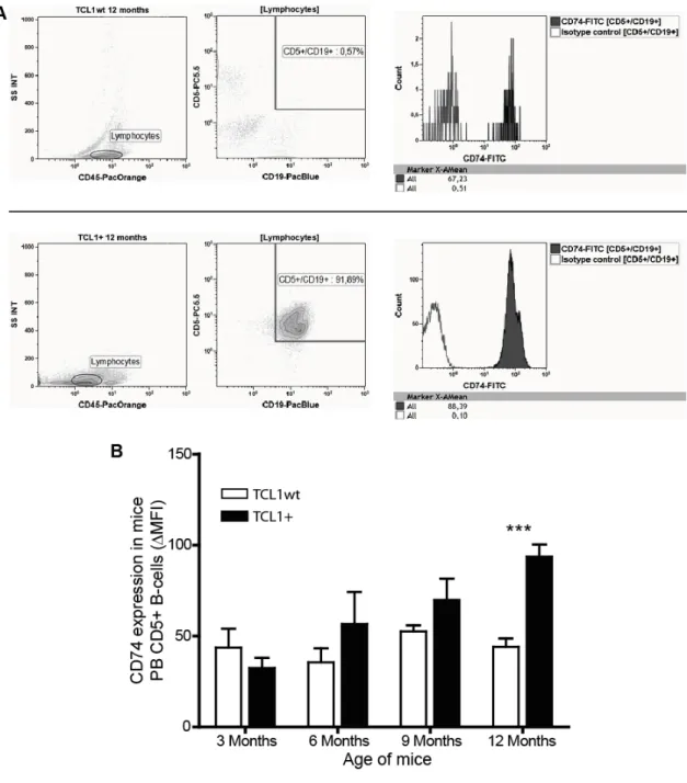
In parallel, blood from TCL1
+mice was taken every 3 months and CD74 expression measured in CD5-expressing B cells by flow cytometry (Figure 5). Here, a significant increase in the mean fluorescence of the CD74 signal was observed in highly leukemic TCL1
+mice compared to TCL1
wtmice (3 months: TCL1
wt43.58±10.4 vs. TCL1
+32.44±5.5; 6 months: TCL1
wt35.56±7.6 vs. TCL1
+56.58±17.6; 9 months: TCL1
wt52.54±3.3 vs. TCL1
+69.83±11.8; 12 months: TCL1
wt43.99±8.8 vs. TCL1
+93.6±6.6
MFI).
Figure 5: Flow cytometric analyses of CD74 expression in malignant B cells from murine blood samples
A: Flow cytometric analysis of CD74 expression in CD5-expressing B cells from murine blood samples in TCL1wt and TCL1+ mice. Blood samples were stained with CD5, CD19, CD45 and CD74 antibodies.
After gating on lymphocytes using the CD45 expression and the side scatter (SS INT), malignant B cells were gated on using both CD5 and CD19 expression. Using the CD5+/CD19+-gate, mean fluorescent intensity (MFI; here depicted as X-AMean) of CD74-FITC signal was measured. The appropriate isotype control was used as control. B:Blood samples of TCL1wt and TCL1+ mice were taken from different age groups and CD74 expression in CD5+ B cells was measured via flow cytometry. ( PB: peripheral blood) [t-test, *** p<0.0005, bars show SEM; TCL1wt n=5; TCL1+ 3 months n=7, 6 months n=4, 9 months n=5, 12 months n=7]
2.2 CD74 in the development of TCL1-induced CLL
Studies on CD74 and its role in the malignant transformation of B cells were mostly carried out in primary human samples and human cell lines. Given the strong dependence of CLL cells on the tumor microenvironment [39, 43, 158, 159], the functional contribution of CD74 to the pathogenesis of CLL within this niche is far from being understood. Thus, the CD74-knockout mouse (CD74
ko) was crossed with the murine CLL model (Eµ-TCL1-transgenic) to study the functional influence of CD74 in the pathogenesis of TCL-1-induced CLL.
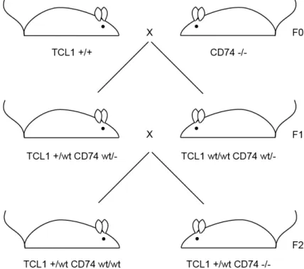
2.2.1 Crossbreeding of Eµ-TCL1-transgenic mice with CD74
komice
Breeding of the CD74
komouse with the Eµ-TCL1-transgenic mouse was done in the animal facility of the Experimental Medicine at the University Hospital of Cologne.
Since a homozygous state of the transgene TCL1 might cause artificial phenotypes, special care in the breeding strategy was taken to avoid a homozygous state of TCL1 in the F2 generation. Animals of the F2 generation with the genotypes TCL1
+/wt Cd74-/-and
TCL1+/wt Cd74wt/wt(from now on called TCL1
+CD74
ko, TCL1
+CD74
wtrespectively) where used for the analyses of leukemic development and survival.
Figure 6: Breeding strategy for TCL1+ with CD74ko mice
B6C3H Eµ-TCL1 mice homozygous for TCL1 were crossed with C57Bl/6J CD74-/- mice. To avoid homozygosity for TCL1 in experimental animals, F1 generations were crossed using TCL1+ mice with TCL1wt mice leading to F2 generations with the preferred genotype.
Littermates with TCL1
wt/wt Cd74wt/wtand TCL1
wt/wt Cd74-/-genotypes (from now on called TCL1
wtCD74
wt, TCL1
wtCD74
korespectively) were used as controls.
The genotypes of the mice were controlled by polymerase chain reaction (PCR) using tail tissue from the mice. Genomic DNA was extracted from the tissue samples and used as templates in the PCR with suitable primer pairs. For Cd74, one primer pair is binding to the exon 1 and exon 4 in the gene and another primer pair binds to the neomycin cassette inserted into the genome to delete Cd74. Figure 7 shows a schema of the PCR strategy to analyze Cd74 expression. Similarly, the TCL1 status was analyzed by using a
TCL1-specific primer pair (data not shown).Figure 7: Genotyping PCR for CD74 status
Genomic DNA was extracted from mice tails and then used for PCR. A: Primer pairs were binding to either exon 1 and 4 (165 bp) or to the neomycin cassette (260 bp). B: Example of PCR products separated by agarose gel electrophoresis.
2.2.2 Leukemia development in TCL1
+CD74
komice
The Eµ-TCL1-transgenic mouse model develops a CLL-like disease with an accumulation of CD5-positive B cells in the peripheral blood, spleen, liver and lymph nodes [54]. Therefore, leukemic load and hepatosplenomegaly in TCL1
+CD74
wtand TCL1
+CD74
komice was measured in order to monitor the development of leukemia.
The leukemic load in the blood of TCL1
+CD74
wtand TCL1
+CD74
komice was
compared at months 3, 6, 9 and 12 by measuring the leukocyte number (WBC) and
amount of malignant B cells (CD5
+/CD19
+). The leukocyte count of both strains
showed similar levels in the course of disease development from 6 to 12 months (6 months: TCL1
+CD74
wt14080±583.7 vs. TCL1
+CD74
ko12370±744.6; 9 months:
TCL1
+CD74
wt14740±891.7 vs. TCL1
+CD74
ko23190±6005; 12 months: TCL1
+CD74
wt28830±3443 vs. TCL1
+CD74
ko37560±7812 cells/µl) (Figure 8). The TCL1
+CD74
kogroup showed a significantly lower leukocyte count at 3 months (TCL1
+CD74
wt14730±367.5 vs. TCL1
+CD74
ko9488±250.1 cells/µl), which was in agreement with published data, that CD74
komice show a lower leukocyte count due to a lower number of mature B cells in the periphery [160].
Figure 8: White blood cell count in TCL1+ CD74wt and TCL1+ CD74ko mice
Blood samples of TCL1+ CD74wt and TCL1+ CD74ko mice were taken every 3 months and the white blood count (WBC) measured using the XE-5000 hematology-analyzer. [t-test, *** p<0.0001, bars show median]
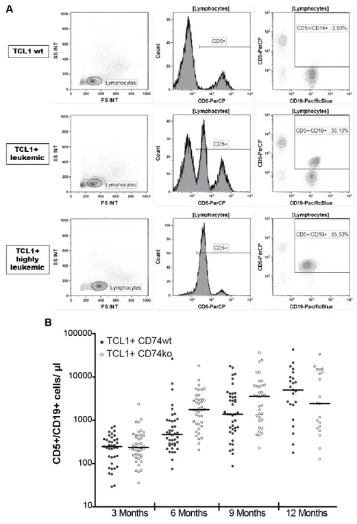
Additionally to the leukocyte count, the amount of malignant CD5-expressing B cells was measured using flow cytometry (Figure 9A). The analysis of CD5-expressing B cells also revealed no significant difference in the number of malignant B cells in TCL1
+CD74
komice compared to TCL1
+CD74
wtmice (3 months: TCL1
+CD74
wt270.2±29.4 vs. TCL1
+CD74
ko354.1±59; 6 months: TCL1
+CD74
wt1512±674.6 vs.
TCL1
+CD74
ko2696±496.7; 9 months: TCL1
+CD74
wt3187±660.6 vs. TCL1
+CD74
ko5617±1292; 12 months: TCL1
+CD74
wt7691±1936 vs. TCL1
+CD74
ko7201±1983
cells/µl) (Figure 9B).
Figure 9: Absolute numbers of CD5-expressing B cells in TCL1+ CD74wt and TCL1+ CD74ko mice
A: Flow cytometric analysis of malignant B cells in murine blood samples from TCL1+ mice in the course of leukemia development. Blood samples were stained with CD5, CD19 and CD45 antibodies.
After gating on lymphocytes using the CD45 expression and the side scatter (SS INT) malignant B cells were gated on using both CD5 and CD19 expression. Malignant B cells show a medium CD5 expression, which is clearly distinguished from T cells with a high CD5 expression (middle panel). B:Blood samples of TCL1+ CD74wt and TCL1+ CD74ko mice were taken every 3 months and used for flow cytometric analyses of CD5-expressing, malignant B cells. Absolute numbers were calculated using the white blood count from Figure 8. [bars show median].
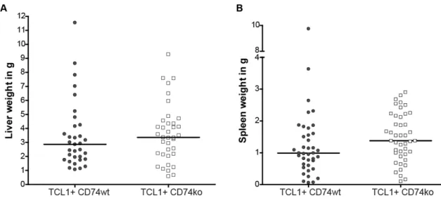
Furthermore, splenomegaly and hepatomegaly were analyzed in both groups, since Eµ-
TCL1-transgenic mice, like CLL-patients, show increased infiltration of malignant cellsinto the lymphoid tissues [54]. As shown in Figure 10, both mouse models developed similar hepatosplenomegaly (liver weight: TCL1
+CD74
wt3.46±0.4g vs. TCL1
+CD74
ko3.63±0.3g; spleen weight: TCL1
+CD74
wt1.34±0.3g vs. TCL1
+CD74
ko1.48±0.1g).
Figure 10: Hepatosplenomegaly in TCL1+ CD74wt and TCL1+ CD74ko mice
A: Analysis of liver weight at time of death in TCL1+ CD74wt (n=33) and TCL1+ CD74ko (n=37) mice.
B: Analysis of spleen weight at time of death in TCL1+ CD74wt (n=33) and TCL1+ CD74ko (n=37). [bars show median]
2.2.3 BCR genetics in TCL1
+CD74
komice
The status of heavy chain gene somatic hyper mutations in the B cell receptor is one of
the prognostic markers for CLL. The Eµ-TCL1-transgenic mouse was described to
develop leukemia resembling the aggressive, unmutated IgV
HCLL cases [54]. The
status of heavy-chain gene somatic hyper-mutations and the immunoglobulin heavy and
light chain usage was analyzed in leukemic, murine samples. Table 1 shows that both
TCL1
+CD74
wtand TCL1
+CD74
komice developed an IgV
Hunmutated B cell clone.
Table 1: BCR-genetics in TCL1+ CD74wt and TCL1+ CD74ko mice Genotype IGHV IGHD IGHJ Mutation status
TCL1+ CD74wt 1-26 1-1 1 unmutated; in-frame; no stop-codon, TCL1+ CD74wt 11-2 2-1 1 unmutated; in-frame; no stop-codon, TCL1+ CD74wt 12-3 3-1 1 unmutated; in-frame; no stop-codon TCL1+ CD74wt 7-3 2-3 1 unmutated; in-frame; no stop-codon
TCL1+ CD74ko 3-8 1-1 1 unmutated; in-frame; no stop-codon TCL1+ CD74ko 12-3 3-3 1 unmutated; in-frame; no stop-codon TCL1+ CD74ko 12-3 3-2 1 unmutated; in-frame; no stop-codon TCL1+ CD74ko 12-3 2-3 1 unmutated; in-frame; no stop-codon TCL1+ CD74ko 6-6 4-1 3 unmutated; in-frame; no stop-codon
2.3 Distribution of myeloid lineage cells in the spleen of TCL1
+CD74
komice
As mentioned before, the microenvironment with its different stimuli is important for
the survival of CLL cells. The study from Reinart et al. showed that deletion of the
CD74 receptor ligand MIF led to a decreased migration of macrophages into the spleen
[44]. To further dissect the role of CD74 in the migration of microenvironmental cells to
the spleen, different cells of the myeloid lineage were analyzed in the spleen of TCL1
+CD74
wtmice and compared to the TCL1
+CD74
kospleens (Figure 11). Using flow
cytometry with antigens specifically expressed on monocytes/macrophages
(CD11b
+/CD18
+) [161], dendritic cells (CD11c
+) [162], granulocytes (Gr-1
+) [163] and
macrophages (F4/80
+) allowed to quantify the amount of these myeloid cells found in
the spleen. Aged mice of control groups, not transgenic for TCL1, were included in the
analysis to compare wild type and CD74
komice. As shown in Figure 11 both TCL1
wtgroups showed similar levels of the tested myeloid lineage cell populations (TCL1
wtCD74
wtCD11b
+/CD18
+: 15.47±8%, CD11c
+: 32.29±19.1%, Gr-1
+: 21.57±7.9%,
F4/80
+:8.46±4.7%; TCL1
wtCD74
koCD11b
+/CD18
+: 17.3±6.5%, CD11c
+: 19.47±1.1%,
Gr-1
+: 38.23±6.4%; F4/80
+:3.58±0.7%). However, a significant increase of monocytes
and granulocytes was observed in the spleen of TCL1
+CD74
komice (CD11b
+/CD18
+:
2.1±0.4% TCL1
+CD74
wtvs. 8.57±2.3% TCL1
+CD74
ko; Gr-1
+: 7.1±1.4% TCL1
+CD74
wtvs. 18.68±4.9% TCL1
+CD74
ko). Dendritic cells were found equally in the
spleens of TCL1
+CD74
wtand TCL1
+CD74
komice (9.69±1.2% TCL1
+CD74
wtvs.
15.33±3.7% TCL1
+CD74
ko). Additionally, F4/80
+macrophages were also found in similar amounts (1.34±0.7% TCL1
+CD74
wtvs. 3.4±2.1% TCL1
+CD74
ko).
Figure 11: Flow cytometric analyses of myeloid cells in the spleen of TCL1+ CD74ko mice A: Flow cytometric analysis of myeloid cells in isolated splenocytes from a TCL1wt CD74ko mouse.
Splenocytes were stained with CD11b, CD11c, CD18, CD45, F4/80 and Gr-1 antibodies. After gating on CD45+-cells, expression of other antigens was analyzed. B: Splenocytes from aged mice (~12 months) were isolated and used for flow cytometric analyses. Different myeloid cell types were distinguished within the CD45+ cell population. [t-test, *p<0.05, **p<0.005, bars show SEM; TCL1wt CD74wt n=3 (n=5 for CD11b+/CD18+ and n=7 for F4/80+), TCL1wt CD74ko n=3 (n=7 for CD11b+/CD18+ and F4/80+), TCL1+ CD74wt n=8, TCL1+ CD74ko n=4 (n=6 for CD11b+/CD18+ and F4/80+)]
To further analyze the number of macrophages in the spleen of leukemic mice, spleen sections were stained with a CD68 antibody specific for macrophages. After staining, slides were scanned and red stained cells were counted at 40-fold magnification (Figure 12). The staining showed similar amounts of macrophages between leukemic TCL1
+CD74
wtand TCL1
+CD74
komice (14.3±3.8 vs. 20±6.1 of CD68
+cells).
Figure 12: Macrophages in the spleen of TCL1+ CD74ko mice
Spleen sections of leukemic mice were stained for CD68 by immunohistochemistry. A: Two examples of the staining (40-fold magnification) per genotype are shown (CD68 positive cells are dark red). B: 10 high power fields per mice were counted and the mean depicted in the box plot. [t-test, n.s. p>0.05, bars show SEM; n=5 per group]