the vascular endothelial growth factor (VEGF) in chronic lymphocytic leukemia (CLL):
Implications to overcome the apoptotic block
Inaugural-Dissertation
zur Erlangung des Doktorgrades
der Mathematisch-Naturwissenschaftlichen Fakultät der Universität zu Köln
vorgelegt von Iris Gehrke aus Leverkusen
Köln
April 2010
Tag der mündlichen Prüfung: 31. Mai 2010
Chronic lymphocytic leukemia (CLL) is an incurable disease, which is char- acterized by an accumulation of monoclonal CD5/CD19/CD23-positive B- lymphocytes, which exhibit a functional apoptotic block. The vascular en- dothelial growth factor (VEGF) is a potent mitogen with the capacity to in- duce angiogenesis by stimulation of vascular endothelial cells. It has been suggested that VEGF has an angiogenesis-independent role in hematological diseases. Also, CLL cells could be shown to express and secrete VEGF and to feature VEGF-receptors (VEGF-R).
Despite their apoptotic-resistance in vivo, CLL cells die within a few days when taken out of their natural microenvironment and are placed under cell culture conditions, strongly suggesting the bone marrow and peripheral blood to be of critical importance in the prevention of apoptotic CLL cell death in vivo. As for example bone marrow stromal cells produce and secrete VEGF, a paracrine feedback loop might be involved in the apoptotic resistance CLL cells feature in vivo.
The aim of this investigation was to elucidate the role of VEGF in the apoptotic resistance of CLL cells, especially in the background of a microenvironmental influence and therefore, to discover potential targets for a CLL cell specific therapeutic approach.
In this study it could be demonstrated that CLL cells, but not healthy B-
cells express the most common VEGF isoforms and exhibit a phosphorylated
VEGF-receptor 2 (VEGF-R2). Phosphorylation was lost with time under cell
culture conditions and went along with a loss of the apoptotic resistance. Since
addition of rhVEGF increased levels of anti-apoptotic proteins, but did not
significantly influence CLL cell survival in vitro, it can be concluded that
VEGF has pro-survival functions, but requires further components derived
from the microenvironment to achieve full apoptotic protection as present in
vivo. Therefore, CLL cells were cocultured together with the bone marrow-
derived stromal cell line HS5, which produced and secreted high levels of
VEGF. This resulted in maintenance of the phosphorylated receptor status
and a prolonged survival of the CLL cells in vitro. Interestingly, healthy
mediated survival-support could be demonstrated, as neutralization of VEGF in CLL/HS5 coculture using a monoclonal VEGF antibody significantly re- duced the survival advantage. In this study it could further be demonstrated that paracrine VEGF, derived from bone marrow stromal cells, rather than CLL cell-derived VEGF is essential, as downregulation of VEGF in HS5 cells by siRNA almost completly abolished the coculture-mediated survival support for CLL cells.
As a possible mechanism of VEGF-mediated survival support the activation of signal transducer and activator of transcription (STAT) 3 via tyr705 phospho- rylation could be demonstrated. This phosphorylation was induced by addition of recombinant human VEGF to CLL cell monoculture as well as by coculti- vation with HS5 cells and was reversible by addition of a VEGF-R inhibitor.
The activation of STAT3 could be demonstrated by an upregulation of the known STAT3 targets Bcl
XLand cyclinD1. The known downstream effector of the VEGF-R2 Akt was neither phosphorylated upon rhVEGF stimulation nor by HS5 coculture.
The VEGF-STAT3 signal transduction pathway can therefore be considered a suitable target for a therapeutic intervention. The tested monoclonal anti- body MAb293 and the VEGF-R inhibitor GW 786034 significantly reduced the survival advantage CLL cells gained from HS5 coculture. The selected STAT3- inhibitor was effective in high concentrations after long incubation times with limited selectivity.
In conclusion, we propose that VEGF action is indispensable in a multi-
part pro-survival complex involving STAT3 and subsequent expression of pro-
survival factors in CLL cells. The VEGF/VEGF-R/STAT3 pathway might
therefore be a promising target for selective therapeutic approaches in CLL.
Die chronische lymphatische Leukämie (CLL) ist eine unheilbare Erkrankung, die sich durch eine Akkumulation monoklonaler CD5/CD19/CD23-positiver B- Lymphozyten auszeichnet, deren Fähigkeit zur Apoptose in vivo hochgradig reduziert ist. Der vaskuläre endotheliale Wachstumsfaktor (VEGF) ist ein po- tentes Mitogen, welches durch Stimulation von Gefäßendothelien Angiogenese auslöst. Es wird angenommen, dass VEGF bei hämatologischen Erkrankungen eine Angiogenese-unabhängige Funktion besitzt. Auch CLL-Zellen exprimieren und sezernieren VEGF und weisen VEGF-Rezeptoren auf.
Trotz ihrer Apoptoseresistenz in vivo sterben CLL-Zellen, die aus ihrer Umge- bung im Blut oder Knochemark isoliert werden, innerhalb weniger Tage in vitro ab, weswegen dem natürlichen Mikromilieu der CLL-Zellen eine wichtige Funk- tion bei der apoptotischen Resistenz zugeschrieben wird. Da Knochemarkstro- mazellen etwa VEGF produzieren, könnte eine parakrine VEGF-Rückkopplung an der Verhinderung des Zelltodes der CLL-Zellen in vivo beteiligt sein.
Das Ziel der vorliegenden Arbeit war, die Rolle von VEGF beim apoptoti- schen Block der CLL-Zellen, insbesondere im Hinblick auf den Einfluss des Mikromilieus zu untersuchen und dadurch potentielle Angriffspunkte für eine zielgerichtete Therapie aufzudecken.
Es konnte gezeigt werden, dass CLL-Zellen im Gegensatz zu gesunden B- Zellen verschiedene VEGF-Isoformen produzieren und phosphorylierte VEGF- Rezeptoren 2 (VEGF-R2) aufweisen. Diese Phosphorylierung ging mit der Zeit in Kultur verloren, was mit einem Verlust der Apoptoseresistenz assoziiert war.
Da eine Zugabe von rekombinantem VEGF (rhVEGF) zu einer verstärkten Ex-
pression anti-apoptotischer Proteine führte, das Überleben der CLL-Zellen in
vitro jedoch nicht verbessern konnte, kann davon ausgegangen werden, dass
weitere Komponenten des Mikromilieus benötigt werden um eine vollständi-
ge Verhinderung von apoptotischem Zelltod, wie sie in vivo vorhanden ist,
zu erzielen. CLL-Zellen wurden deswegen zusammen mit der Knochenmark-
stromazelllinie HS5, welche VEGF exprimiert und sezerniert, kultiviert. Dies
führte zu einer Aufrechterhaltung der VEGF-R2-Phosphorylierung und einem
deutlich verbesserten Überleben der CLL-Zellen. Gleichzeitig profitierten ge-
sunde B-Zellen, welche den VEGF-R2 nur in sehr geringen Mengen aufweisen,
nicht von einer Kokultur mit HS5. Die entscheidende Rolle von VEGF bei der
VEGF-neutralisierenden Antikörpers zu einem reduzierten Überlebensvorteil führte. Zudem konnte gezeigt werden, dass ein parakriner Effekt essentiell ist, da eine siRNA-vermittelte Herunterregulierung von VEGF in HS5-Zellen den Überlebensvorteil der CLL-Zellen fast komplett aufheben konnte.
Als ein möglicher Mechanismus des VEGF-vermittelten Überlebens der CLL- Zellen konnte eine Aktivierung des signal transducers and activators of trans- cription (STAT) 3 durch Tyr705-Phosphorylierung gezeigt werden. Diese wur- de durch Zugabe von rhVEGF in einer CLL-Monokultur und durch Kokul- tivierung mit HS5-Zellen hervorgerufen und konnte durch gleichzeitige Zuga- be eines VEGF-R-Inhibitors wieder rückgängig gemacht werden. Die Akti- vierung von STAT3 wurde durch eine Hochregulierung der STAT3 Zielgene Bcl
XLund cyclinD1 nachgewiesen. Der bekannte intrazelluläre Signalmedia- tor des VEGF-Rezeptors Akt zeigte weder nach exogener VEGF Stimulation noch nach HS5-Kokultur eine verstärkte Phosphorylierung. Die VEGF/VEGF- R/STAT3-Achse eignet sich demnach als potentieller Angriffspunkt für einen zielgerichteten Therapienasatz. Sowohl der monoklonale anti-VEGF Antikör- per MAb293, als auch der VEGF-R-Inhibitor GW 786034 reduzierten das Überleben der CLL-Zellen in einer überlebensfördernden Kokultur mit HS5- Zellen signifikant. Der ausgewählte STAT3-Inhibitor war in hohen Konzentra- tionen und nach langer Inkubationszeit wirksam und begrenzt selektiv.
Zusammenfassend kann VEGF als ein essentieller Bestandteil eines überle-
bensfördernden Zusammenspiels der CLL-Zelle mit ihrem Mikromilieu be-
schrieben werden, bei dem die Aktivierung von STAT3 eine Rolle spielt. Der
VEGF/VEGF-R/STAT3 Signalweg ist demnach ein viel versprechendes Ziel
für eine therapeutische Intervention in der CLL.
Contents i
List of Figures v
List of Tables viii
Abbreviations ix
Acknowledgements xiii
1 Introduction 1
1.1 Chronic lymphocytic leukemia (CLL) . . . . 1
1.1.1 Definition . . . . 1
1.1.2 Epidemiology . . . . 1
1.1.3 Etiology and molecular pathogenesis . . . . 2
1.1.4 Clinical aspects . . . 13
1.2 The vascular endothelial growth factor . . . 20
1.2.1 The VEGF-family . . . 20
1.2.2 The VEGF-receptors . . . 22
1.2.3 VEGF/VEGF-R-signaling . . . 25
1.2.4 Angiogenesis . . . 29
1.2.5 Angiogenesis-independent physiological roles . . . 30
1.2.6 VEGF in cancer . . . 31
1.2.7 Objective . . . 34
2 Results 36 2.1 VEGF status in CLL and healthy B-cells . . . 36
2.1.1 CLL cells express the major VEGF isoforms to a signif- icantly higher extent than healthy B-cells . . . 36
i
2.1.3 CLL cells secrete VEGF to a higher extent than healthy B-cells . . . 38 2.1.4 CLL cells exhibit the VEGF-receptor 2 (VEGF-R2) . . . 39 2.1.5 The VEGF-R2 is constitutively phosphorylated in CLL
cells, but not in healthy B-cells . . . 39 2.1.6 Stimulation with rhVEGF induces increased VEGF ex-
pression in CLL cells . . . 40 2.1.7 Secretion of VEGF by CLL cells increases with time in
culture . . . 41 2.1.8 CLL cell-derived VEGF is not sufficient to stimulate the
VEGF-R2 . . . 42 2.2 Role of VEGF in the apoptotic block of CLL cells . . . 43
2.2.1 rhVEGF induces upregulation of anti-apototic proteins in CLL cells, but not healthy B-cells . . . 44 2.2.2 The anti-apoptotic proteins Mcl1, XIAP and Bcl2 are
reduced with time in culture . . . 44 2.2.3 rhVEGF stimulation does not affect survival of CLL cells 45 2.3 Influence of bone marrow (BM) stromal cells on the VEGF sta-
tus in CLL cells and their survival . . . 46 2.3.1 The BM-derived stromal cell line HS5 produces high
amounts of VEGF . . . 46 2.3.2 VEGF expression is significantly increased by coculture
with HS5 in CLL cells, but not healthy B-cells . . . 47 2.3.3 CLL cells maintain constitutive phosphorylated
VEGF-R2 when cocultured with HS5 . . . 48 2.3.4 Coculture with HS5 supports survival of CLL cells, but
not healthy B-cells . . . 49 2.3.5 Neutralization of VEGF by a monoclonal antibody re-
duces the coculture-mediated survival advantage . . . 51 2.3.6 VEGF-depletion in HS5 by siRNA abolishes the coculture-
mediated survival advantage for CLL cells . . . 52
ii
2.4 Mechanistical background of VEGF-mediated apoptosis preven- tion . . . 55 2.4.1 PCR-array suggests an upregulation of STAT3 and down-
regulation of RB1 and E2F1 upon rhVEGF-stimulation . 56 2.4.2 PCR-array suggested downregulation of E2F1 and RB1
is not reproducible by PCR . . . 57 2.4.3 rhVEGF stimulation does not increase total STAT3 but
induces its phosphorylation on tyr705 . . . 57 2.4.4 Phosphorylation on tyr705 activates STAT3 . . . 59 2.4.5 Coculture with HS5 provokes tyr705 phosphorylation of
STAT3 and its activation in CLL cells . . . 60 2.4.6 Neither rhVEGF nor coculture with HS5 effectuates phos-
phorylation of Akt in CLL cells . . . 62 2.5 Potential of VEGF as therapeutic target in CLL . . . 63
2.5.1 Anti-VEGF antibody treatment does not alter survival in CLL cell monoculture . . . 63 2.5.2 The VEGF-R inhibitor GW 786034 effectively induces
apoptosis in CLL cells in mono- and in coculture . . . . 64 2.5.3 The STAT3 inhibitor S3I-201 requires high concentra-
tions to reduce CLL cell survival . . . 67 2.5.4 Pan-JAK inhibition by Pyridone 6 does not reduce CLL
survival in tested low concentrations . . . 68
3 Discussion 69
3.1 VEGF status in CLL cells and its involvement in apoptosis pre- vention . . . 69 3.2 Influence of the bone marrow-derived stromal cell line HS5 on
VEGF-mediated CLL cell survival . . . 73 3.3 Mechanistical background of VEGF-mediated apoptosis preven-
tion . . . 78 3.4 Potential of VEGF as therapeutic target in CLL . . . 82 3.5 Future directions . . . 87
iii
4.1.1 Instruments . . . 89
4.1.2 Consumables . . . 91
4.1.3 Chemicals and reagents . . . 91
4.1.4 Buffer and solutions . . . 92
4.1.5 Cell culture reagents and media . . . 95
4.1.6 Special reagents and kits . . . 97
4.1.7 Ready-to-use buffers and solutions . . . 97
4.1.8 Antibodies . . . 98
4.1.9 Oligonucleotides . . . 99
4.1.10 Cell lines . . . 99
4.1.11 Primary patient material . . . 100
4.1.12 Software . . . 100
4.2 Methods . . . 101
4.2.1 Cells . . . 101
4.2.2 Molecular biology . . . 104
4.2.3 Protein biochemistry . . . 111
4.2.4 Flow Cytometry . . . 114
4.2.5 Statistics . . . 116
References 118
Ehrenwörtliche Erklärung 160
Curriculum Vitae 161
iv
1 Model for the cellular derivation of CLL. . . . 3
2 Bone marrow microenvironment of the CLL cell. . . . 8
3 VEGF gene and its splice variants . . . 21
4 Structure of VEGF-receptors and Neuropilins . . . 23
5 Intracellular signaling cascades downstream of VEGF-R2 . . . . 28
6 Relative expression levels of VEGF isoforms. . . 37
7 VEGF levels secreted by CLL and healthy B-cells. . . 38
8 VEGF-R2 status in CLL and healthy B-cells. . . 39
9 pVEGF-R2 status in CLL and healthy B-cells. . . 40
10 pVEGF-R2 status in rhVEGF-stimulated CLL and healthy B- cells. . . 40
11 VEGF mRNA levels of rhVEGF-stimulated CLL cells. . . . 41
12 VEGF levels secreted by CLL and healthy B-cells over 5 days. . 42
13 pVEGF-R2 levels in CLL cells during a time course of five days. 43 14 Anti-apoptotic protein levels in CLL and healthy B-cells after rhVEGF stimulation. . . 44
15 Anti-apoptotic protein levels in CLL cell during a time course of 5 days. . . . 45
16 Survival advantage of rhVEGF-stimulated CLL cells. . . 46
17 VEGF status of HS5 cells. . . 47
18 VEGF mRNA fold change in CLL or healthy B-cells by cocul- ture with HS5. . . . 48
19 pVEGF-R2 levels of CLL cells cocultured with HS5 over a time course of three days. . . 49
20 Survival advantage of CLL and healthy B-cells cocultured with HS5. . . 50
v
22 pVEGF-R2 levels upon addition of the VEGF-neutralizing an- tibody MAb293 (mAb). . . . 51 23 Annexin V-FITC/PI staining of CLL cells in mono- and cocul-
ture with and without the addition of anti-VEGF MAb293. . . . 52 24 siRNA-mediated downregulation of VEGF. . . 53 25 CLL cell survival after cocultivation with control-treated and
VEGF siRNA-treated HS5 cells after 24 and 48 hours of coculture. 53 26 Survival of siRNA control-treated or VEGF siRNA-treated HS5
cells after three days. . . 54 27 Survival advantage of CLL cells in coculture with HS5 with and
without physical separation. . . 55 28 PCR array results of unstimulated versus rhVEGF-stimulated
CLL cells. . . 56 29 RB1 and E2F1 mRNA levels of rhVEGF-stimulated CLL cells
relative to unstimulated control. . . 57 30 STAT3 and pSTAT3 levels in CLL cells after rhVEGF stimula-
tion or VEGF-R inhibition using GW 786034. . . 58 31 STAT3 phosphorylation status after rhVEGF stimulation in CLL
cells assessed by flow cytometry. . . 59 32 CyclinD1 and Bcl
XLlevels after rhVEGF stimulation or VEGF-
R inhibition using GW 786034 in CLL cells. . . 60 33 Protein analysis of CLL cells in mono- or coculture with HS5
with and without GW 786034 treatment. . . 61 34 Akt and pAkt protein levels in CLL cells under several condi-
tions: (A) CLL cells stimulated with rhVEGF and/or treated with GW 876034, (B) CLL cells in mono- or coculture with HS5 cells . . . 62 35 Survival of CLL cells upon treatment with anti-VEGF MAb293
or bevacizumab. . . 64 36 Cell survival upon treatment with GW 786034 in mono- or co-
culture with HS5. . . 65 37 Anti-apoptotic protein levels after treatment with GW 786034. . 66
vi
39 Cell survival upon treatment with S3I-201 in mono- and cocul- ture with HS5. . . . 67 40 Cell survival upon treatment with Pyridone 6. . . 68 41 Flow cytometric differentiation of HS5 and CLL cells by cell
volume (FSC forward scatter) and granularity (SSC side scatter).103 42 Experimental set up of HS5/CLL cell coculture with physical
separation using tissue culture inserts. . . . 104
vii
1 CLL staging after RK Rai (Rai et al., 1975). . . 14
2 CLL staging after JL Binet (Binet et al., 1981). . . 15
3 Prognostic factors in CLL. . . 17
4 VEGF-receptors and their specific ligands. . . 25
5 Anti-and pro-angiogenic factors. . . 29
6 Correlation statistics of VEGF mRNA with ZAP70 and CD38 status. . . 38
7 List of substances used. . . 92
8 List of siRNAs and used controls. . . 93
9 List of antibodies used for immunoblotting. . . 98
10 List of antibodies used for flow cytometry. . . 98
11 Primers used for real time PCR. . . 99
12 Reverse transcription supermix composition. . . 105
13 PCR reaction mix composition. . . 107
14 Cycling durations and temperatures for PCR. . . 107
15 Set up for siRNA experiments. . . 110
viii
◦
C grad celcius
µ micro
ALL acute lymbhoblastic leukemia AML acute myelogenous leukemia APC adenomatous polyposis coli
BAD (Bcl-2)-associated death promoter homologue Bcl2 B-cell lymphoma 2
Bcl
XLB-cell lymphoma extra large BCR B-cell receptor
BM bone marrow
BSA bovine serum albumin CD cluster of differentiation
cDNA complementary deoxyribonucleic acid CLL chronic lymphocytic leukemia
CM conditioned media
CML chronic myelogenous leukemia CO
2carbon dioxide
Ct threshold cycle ddH
20 double distilled water
DMEM Dulbecco´s Modified Eagle´s Medium DMSO dimethyl sulfoxid
DNA deoxyribonucleic acid
dNTP deoxynucleotide triphosphate DSH dishevelled
DTT dithiothreitole
e.g. exempli gratia (for example)
ix
EDTA ethylenediaminetetraacetic acid ELISA Enzyme-linked Immunosorbent Assay EM extracellular matrix
et al et alii (and others)
FACS fluorescent activated cell sorter FCS fetal calf serum
FISH fluorescent in situ hybridization FITC fluoresceinisothiocyanate
FZD frizzled receptor
g gram
G-CSF growth factors like granulocyte colony-stimulating factor GM-CSF granulocyte-macrophage colony-stimulating factor
GSK3β glycogen synthase kinase β HCL hairy cell lymphoma
HEPES 4-(2-hydroxyethyl)-1-piperazine ethanesulfonic acid HLA human leucocyte antigen
HSC hematopoietic stem cell
IC50 half maximal inhibitory concentration Ig immunoglobulin
IgV
Himmunoglobulin heavy chain variable region IL interleukin
JAK Janus kinase kDa kilo Dalton
l litre
LD50 half maximal lethal concentration Lef-1 lymphoid enhancer factor 1 LRP LDL receptor related protein M molar (mol/litre)
m mili
MAPK mitogen activated protein kinase MBL monoclonal B-cell lymphocytosis MCL mantel cell lymphoma
x
min minute(s)
MM multiple myeloma
MMP matrix metalloproteinase mRNA messenger ribonucelic acid
n nano
NRP neuropillin
p pico
p53 proapoptotic protein 53
PAGE poly acrylamide gel electrophoresis PARP poly (ADP-ribose) polymerase PB peripheral blood
PBMC peripheral blood mononuclear cells PBS phosphate buffered saline
PCR polymerase chain reaction PDGF platelet-derived growth factor
pH negative logarithm of the hydrogen ion concentration PI propidium iodide
Pi3K phosphatidyl inositole -3 kinase PLC phospho lipase C
PlGF placental growth factor
PLL B-cell prolymphocytic leukemia PMA phorbol 12-myristate 13-acetate RB1 retinoblastoma 1
ROS reactive oxygen species rpm rounds per minute
RPMI Roswell Park Memorial Park
RT room temperature
RTKs receptor tyrosine kinases SDS sodium dodecyl sulphate
sec second
SEM (σM) standard error of the mean
ser serine
xi
STAT signal transducer and activator of transcription TCF T-cell factor
TRIS Tris hydroxyl methyl aminomethane tyr tyrosine
VEGF vascular endothelial growth factor
VEGF-R vascular endothelial growth factor receptor w/v weight per volume
XIAP X-linked inhibitor of apoptosis ZAP70 zeta associated protein kinase 70
α alpha
β beta
γ gamma
δ delta
κ kappa
µ mu
σ standard deviation
xii
I would like to thank Prof. Dr. Michael Hallek for the opportunity to carry out my work at the Department I of Internal Medicine, Prof. Dr. Maria Leptin for accepting the role as my official supervisor, Prof. Dr. Carien Niessen and PD Dr. Hamid Kashkar for being my "thesis committee" and Prof Dr. Günther Schwarz for taking the position as chairperson for my thesis defense.
I am very grateful to my supervisor PD Dr. Karl-Anton Kreuzer for providing the topic, his continuous support, creation of a positive working environment and all the encouragement and good advice. By giving me the chance of independently writing grants and manuscripts and presenting results at inter- national conferences I feel well prepared for successfully pursuing my scientific career.
Further, I would like to thank all my colleagues for sharing the daily lab routine with me, discussions, helpful advice and creation of a pleasant and encouraging atmosphere.
I am indebted to my family who always believed in me and supported me to the maximum: "Papa, Mama, Nina, Lars und Oma, danke für eure immer währende Unterstützung und die vielen aufmunternden Worte!"
Cliff, thank you for everything. This was supposed to be the end of our rough times. I am sorry.
I would further like to thank my best friends Nadine, Silja, Caro, Benni and Tini who were always there for me when I needed to talk and patiently accepted my lack of time during busy periods.
xiii
Chapter 1 Introduction
1.1 Chronic lymphocytic leukemia (CLL)
1.1.1 Definition
Chronic lymphocytic leukemia (CLL) is a lymphoproliferative disorder, which is characterized by the accumulation of mature, but immuno-incompetent B- lymphocytes in the bone marrow, peripheral blood, and various organs.
CLL is defined by three characteristics which are (i) < 5.000 monoclonal B- lymphocytes per µl blood, (ii) the presence of a clonal population of CD5/
CD19/CD23-positive lymphocytes and (iii) less than 55% circulating prolym- phocytes (Hallek et al., 2008).
1.1.2 Epidemiology
Chronic lymphocytic leukemia (CLL) affects mainly people of the age of 50
and older and it account for approximately 40% of leukemias in adults older
than 65 years. Its incidence increases with age and patients younger than
30 years are very rare. Men are twice as likely to develop CLL as women
(Ries LAG, 1999). Prevalence for occurrence of CLL could be encountered
in Europe, North America and Australia, whereas the disease is considerably
less common in Asian countries such as India, Japan or China (Groves et
al., 1995). Since Asians migrating to the USA were shown to maintain their
low incidence rates, genetic factors are more likely to be responsible than
environmental ones (Groves et al., 1995; Pan et al., 2002; Yanagihara et al.,
1989). Nevertheless, some studies described a correlation of CLL incidence and pesticides or herbicides used in agriculture or exposure to benzene and the rubber industry (Goldin and Slager, 2007; Schnatter et al., 2005), but results are not consistent (Richardson et al., 2005). Furthermore, neither ionizing irradiation nor viral genes could be associated with the prevelance of CLL (Kipps, 1998).
Several families with increased occurrence of the disease have been described over the last 25 years, suggesting a familial predisposition (Cartwright et al., 1987; Goldgar et al., 1994; Pottern et al., 1991). First- and second-degree relatives of patients with CLL have an increased risk of subclinical monoclonal B-cell expansion and lymphoid malignancies including CLL. Furthermore, in successive generations of families with CLL disease onset is frequently seen earlier and often present in a more severe form (Rawstron, 2004; Yuille et al., 1998). Recently, a study demonstrated an 8.5 fold increased risk for case relatives to obtain CLL (Goldin et al., 2009). Nevertheless, no inherited genetic defects, making a member of a CLL family prone to obtain the disease, were identified so far. It is likely that a complex of several aberrant events, rather than one simple genetic defect, is responsible for occurrence of CLL in families.
1.1.3 Etiology and molecular pathogenesis
The etiology of CLL remains largely unclear up to date. Nevertheless, several factors have been associated with disease initiation and progression.
1.1.3.1 Origin of the CLL cell
The determination of the origin of the leukemic CLL cell has been a focus of scientific interest for a long time. The knowledge of the tumor precursor cell in CLL is of high impact to understand the pathogenesis of the disease and to obtain insight into the mechanisms of the transformation process from a healthy B-cell towards a malignant CLL cell.
In general, B-cell development initiates in the bone marrow and in the fe-
tal liver originating from stem cells (HSCs). Starting from HSCs multiple
hemtopoietic lineages can be generated through a series of intermediate pro-
genitors. B-cells develop from a common lymphoid progenitor. Bone marrow-
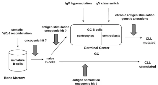
derived antigen-inexperienced naïve B-cells move to the germinal center (GC) in the peripheral lymphnodes where they undergo division and proliferation processes. In the GC, the process of immunoglobulin variable region (IgV) somatic hypermutation (SHM) modifies the antibody genes of the B-cells to generate high affinity antibodies. In the progress of normal B-cell development these B-cells with improved antigene-binding are positively selected and dif- ferentiate into memory B-cells and plasma cells. GC B-cells also undergo class switching by a somatic DNA recombination mechanism. Disruption of the reg- ulation of B-cell differentiation and activation might result in the occurrence of leukemias or lymphomas. At which step this "oncogenic hit" occurs is not clear up to now.
immature B-cells somatic V(D)J recombination
Bone Marrow
oncogenic hit ?
naive B-cells
GC B-cells
centroblasts centrocytes
antigen stimulation oncogenic hit ?
Germinal Center GC
antigen stimulation oncogenic hit ?
IgV hypermutation IgV class switch
chronic antigen stimulation genetic alterations
CLL mutated
CLL unmutated
Figure 1: Model for the cellular derivation of CLL.
Source: Own illustration
CLL cells are in general a morphologically homogenous population with a char-
acteristic immunophenotype expressing the antigens CD5, CD19 and CD23
while exhibiting low levels of surface immunoglobulin (Ig) (Caligaris-Cappio
and Hamblin, 1999). Though, on the genetic level CLL cases are hetero-
geneous with rearranged variable genes in the immunoglobulin heavy chain
(IgV
H), which can be either somatically hypermutated or unmutated (Chio-
razzi and Ferrarini, 2003; Fais et al., 1998; Oscier et al., 1997; Schroeder,
Jr. and Dighiero, 1994). Additionally, these genetically-defined subtypes are
associated with divergent clinical courses. While IgV
H-mutated CLL cases show generally a more benign clinical course, unmutated CLL cases have been demonstrated to have a less favourable prognosis. These facts at first sug- gested that there might be different tumor precursor cells for the major CLL subtypes originating from distinct stages of B-cell development. Additionally, the presence of somatically mutated antibody genes in CLL patients could be an indication for antigenic selection to be involved in the development of CLL.
This was further proofed by the finding of certain IgV
Hfamily members to be more frequently present in CLL patients, independent of their mutational status, than would be expected from their expression in the antibody reper- toire in normal B-cells (Fais et al., 1998). In several studies it was proofed that these specific IgV
Hgene repertoires in CLL patients are indeed a result of antigen selection rather than simply reflecting the aging process (Potter et al., 2002; Widhopf et al., 2004). A further pointer towards CLL cells being a result of antigen-mediated selection is the fact that unrelated CLL patients can feature almost identical B-cell receptors (BCRs) (Messmer et al., 2004;
Murray et al., 2008; Tobin et al., 2003; Tobin et al., 2004; Widhopf et al., 2004). Gene expression profiling (GEP) studies could identify a subset of genes (molecular signature), which allowed differentiation of IgV
Hunmutated and IgV
Hmutated cases. CLL cases with unmutated IgV
Hseem to express high levels of genes which are known to be activated as a result of BCR-mediated stimulation (Rosenwald et al., 2001). A physiological consequence could be antigen-mediated BCR-signaling to have impact on the clinical prognosis of CLL (Muzio et al., 2008).
Another pointer towards CLL cells being derived from antigen-stimulated B-
cells is their cell surface phenotype which resembles that of antigen-activated
B-cells (Damle et al., 2002). GEP suggested B-cells to be most closely related
to a specific B-cell subset of the CD27+ B-cells (Klein et al., 2001). Those
cells comprise a heterogeneous pool of B-cells, such as memory B-cells and
marginal zone B-cells, which are antigen-experienced. Interestingly, GEP did
not identify any correlation between CLL cells and CD5+ cells derived from
cord blood, CD27-(naïve), or GC B-cells (Klein et al., 2001; Rosenwald et al.,
2001). CD27+ cells comprise up to 40% of B-cells in the peripheral blood (PB)
of adults and are mainly found in sites of antigen entry, such as the marginal
zone and the tonsillar subepithelium (Klein et al., 1998). Normal CD27+
cells respond quickly to exogenous antigens by differentiation into antibody- secreting cells (Kindler and Zubler, 1997). These facts provide further evidence for antigen-experienced cells to be the precursor of CLL cells.
Additionally, the cytogenetic abnormalities occurring in CLL patients differ markedly from that of other B-cell malignancies and resemble most closely that of hairy cell leukemia (HCL) (Basso et al., 2004). HCL features distinct morphological and phenotypic characteristics in comparison to CLL (Harris et al., 1994), but interestingly, HCL resembles CLL in that its GEP is most closely related to that of the CD27+ cells (Basso et al., 2004).
All the mentioned evidences target on CLL being derived from one com- mon precursor, an antigen-experienced (CD5-CD27+) B-lymphocyte, which are highly presented in the lymphoid organs and the peripheral blood (appr.
40%). As virtually all CD27+ cells are CD5-, the CD5 expression in CLL cells would be a consequence of the activation phenotype of the tumor cell (Wortis et al., 1995). Besides this theory it has recently been suggested that CLL cells might arise from a small population of CD5+ B-cells detected in the lym- phoid organs (Dono et al., 2007). With this background, also the relation of monoclonal B-cell lymphocytosis, a benign clonal proliferation of CD5 B-cells, which phenotypically and genetically resemble that of the CLL cell, and CLL remains unclear (Dagklis et al., 2009; Landgren et al., 2009; Rawstron, 2004).
1.1.3.2 Genetic factors
In CLL several frequently occurring chromosomal aberrations have been de- scribed. Going along with technique advancement, the numbers of detected chromosomal abnormalities in CLL patients have increased from 50% detected by banding analysis (Carney and Wierda, 2005) to approximately 80% detected by fluorescent in vitro hybridisation (FISH), comparative genomic hybridisa- tion (CGH), single nucleotide polymorphism (SNP) and micorarrays (Pfeifer et al., 2007; Stilgenbauer et al., 2002). Genetic abnormalities in CLL are thought to be acquired after birth through chromosomal instabilities, rather then being inherited. The most common genetic defect in approximately 55%
of CLL cases is the deletion of 13q14.1 (Dohner et al., 1999). While at first
the tumor suppressor gene RB1 was suggested to be the candidate gene for
this aberration (Liu et al., 1992), this assumption was refuted as an RB1 dele- tion or mutation is only found in a small percentage of the malignant clone (Dohner et al., 1994). Instead has this region recently been described to en- code for two distinct micro RNAs (miRNA), miR15A and miR-16-1 (Calin and Croce, 2006). Up to now, there are no natural targets of those miRNAs.
An increase in T cell leukemia 1 (TCL1) and B-cell lymphoma 2 (Bcl2), both anti-apoptotic factors, was observed when these miRNAs where deleted, sug- gesting those genes to be potential targets of miR15A and miR-16-1 (Calin et al., 2005; Calin and Croce, 2006). As 13q14.1 as a sole abnormality confers a favourable prognosis (Dohner et al., 2000) the exact role of this deletion is not clear up to date. Second prevalent chromosomal defect in CLL is the deletion of 11q22 in approximately 18% of cases (Dohner et al., 1997). As a candiate gene the ATM gene has been described to be present in this region. ATM is a crucial player in cellular response to double strand DNA breaks (Stankovic et al., 2002). Besides deletion, ATM gene mutations are a frequent event in CLL (Bullrich et al., 1999; Stankovic et al., 1999). As also here, deletions in 11q22 are not always accompanied by mutations, it is thought that potentially further genes are involved (Schaffner et al., 1999). 11q22 deletion is typically associated with a poor prognosis and advanced stage disease (Dohner et al., 1997). The third most common chromosomal aberration is trisomy 12 with an incidence of approximately 16% (Oscier, 1994). The crucial segment in this region has yet to be determined. Bands 12q13-q22 include a segment that is found to be duplicated in CLL (Merup et al., 1997) with MDM2, a negative regulator of p53 as a potential oncogene, in this location. However, recently no impact of MDM2 polymophism in a small cohort (85 patients) of CLL pa- tients could be detected (Lahiri et al., 2007). Several adverse features have been associated with this abnormality such as atypical cell morohology and immunophenotype (Dewald et al., 2003; Matutes et al., 1996). Around 7%
of CLL cases exhibit a deletion on the short arm of chromosome 17 which
contains the p53 gene and is associated with short survival, rapid disease pro-
gression (Dohner et al., 1995a) and drug-resistance (Turgut et al., 2007). The
p53 protein arrests cells with damaged DNA and facilitates DNA-repair (Vo-
gelstein et al., 2000). Other than inducing DNA-repair, p53 can also promote
apoptosis to abet the destruction of the damaged cells. In this way p53 also
mediates cytotoxicity of many anticancer agents; hence it is not surprising that patients possessing a p53 deletion generally show worse response towards treatment, such as purine analogs or the anti-CD20 antibody rituximab (Byrd et al., 2003; Byrd et al., 2007; Dohner et al., 1995b).
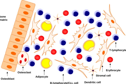
1.1.3.3 The bone marrow microenvironment
Neoplastic CLL cells are characterized by their prolonged survival due to a resistance towards apoptosis in vivo, which entails their accumulation. This feature is completely absent once CLL cells are taken out of their natural microenvironment and put under in vitro culture conditions (Collins et al., 1989), indicating the bone marrow environment to be a crucial supporter of their apoptotic resistance. Since early stages of CLL are characterized by bone marrow infiltration the bone marrow microenvironment can be considered a critical side of nurturing in the disease process. Bidirectional interactions be- tween the malignant CLL cells and the non-transformed bystander cells lead to the establishment by of an abnormal microenvironment favouring the survival of the CLL cells. In turn, this extended survival might create an intracellu- lar milieu which supports the occurrence of unfavourable genetic instabilities.
The microenvironment might also represent a niche for the CLL cell to retreat therapeutic interventions.
Several cell types are present in the bone marrow, such as stromal cell, T-cells,
follicular dendritic cells (FDCs), BM-derived endothelial cells (BMECs), um-
bilical vein endothelial cells (HUVECs), monocyte-derived nurse-like-cells and
also cells involved in bone homeostatis such as chondroclasts, osteoclasts and
osteoblasts. This heterogeneous cell population creates a dynamic microen-
vironment of direct cell-cell interactions with high concentrations of growth
factors and cytokines present.
bone matrix
B-lymphocyte/CLL cell
Erythrocyte Stromal cell Adipocyte
Dendritic cell Osteoblast
Osteoclast
T-lymphocyte
Figure 2: Bone marrow microenvironment of the CLL cell.
Source: Own illustration
Several of those accessory cells have been demonstrated to have the capacity of sustaining prolonged viability of the leukemic clone when placed together in in vitro culture (Burger et al., 2000; Ghia et al., 2005; Jewell and Yong, 1997; Lagneaux et al., 1998; Panayiotidis et al., 1996a; Pedersen et al., 2002).
FDCs stay in contact with CLL cells especially in the early phase of bone marrow involvement as well as in the lymph nodes (Chilosi et al., 1985). It was demonstrated that CLL cell survival support by FDCs involves direct cell contact dependent on CD44-ligation and subsequent upregulation of the anti- apoptotic protein Mcl1 (Pedersen et al., 2002).
The presence of T-cells in a survival supporting in vitro coculture system can be mimicked by addition of T-cell-derived cytokines, such as IL4 or soluble CD40L (Buske et al., 1997; Ranheim and Kipps, 1993). CD40 stimulation not only prevents spontaneous apoptosis, but also results in proliferation (Granziero et al., 2001; Patten et al., 2008), activation of CLL cells as well as chemokine pro- duction (Ghia et al., 2002b; Yellin et al., 1994). Activated CD40L+ T-cells are present in bone marrow-derived from CLL patients primarily in the so called
"proliferation centres" (PC) or pseudofollicles, which are the histological CLL
hallmark in lymph nodes. In PCs CLL cells are in close physical contact to
these CD40L+ T-cells providing a CD40L-stimulus (Ghia et al., 2002b). In this background it is of great interest, that lymph node and bone marrow-derived CLL cells themselves might be responsible for the presence of activated T-cells in the PCs, as they were described to express the T-cell attracting chemokines CCL17 and CCL22. Expression of these chemokines on PB-derived CLL cells could be induced upon stimulation with CD40L (Ghia et al., 2002b). Hence, CLL and T-cells in a patient’s bone marrow possess bidirectional interactions regulated by adhesion molecules and chemokines translating into a further pro- duction of cytokines by both cell types (Ghia and Caligaris-Cappio, 2000).
Besides T-cells, stromal cells possess a significant influence on CLL cells. Stro- mal cells are the key regulators of normal B-lymphopoiesis. Despite this knowledge it is up to now not completely resolved, how the precise ligand- receptor interaction between B-cells and stromal cells is controlled. Several ad- hesion molecules have been implicated, such as selectins, integrins, for example integrin β1-(CD29) and β2-(CD18), immunoglobulins, intracellular adhesion molecules ICAM-1/CD54, ICAM-2/CD102 or ICAM-3/CD50 and the CD44 family of homing receptors (Caligaris-Cappio and Hamblin, 1999; Takeuchi and Katayama, 1993). CLL cells express for example the integrins CD49d/CD11a or CD11b/CD18 which interact with CD54 and CD106 on stromal cells and have been reported to be survival-supportive for CLL cells (Plate et al., 2000).
Also other adhesion molecules, such as CD44 are highly expressed in CLL cells and are associated with an aggressive disease progression and adverse progno- sis (Eistere et al., 1996).
Besides direct cell-cell contacts also soluble factors, such as cytokines con-
tribute to CLL cell survival. Stromal cells for example produce the interleukins
IL-6, IL-7, IL-10, TGF-β, stem cell factor (SCF) and vascular endothelial
growth factor (VEGF) therewith exerting a complex regulatory function on
CLL cells (Ghia and Caligaris-Cappio, 2000). In general, CLL cells are known
to exhibit receptors for many pro-survival cytokines, suggesting a high response
potential towards these factors secreted by accessory non-tumorigenic cells in
the bone marrow and in the PB. Since CLL cells are also able to produce sev-
eral of those cytokines themselves, autocrine loops are likely to additionally
support CLL cell survival (Kay et al., 2002b).
1.1.3.4 Abberant regulation of intracellular signaling cascades Possibly reflecting the clinical heterogeneity observed in patients, various aber- rantly activated signaling cascades have been associated with the initiation and the course of CLL. A variety of humoral factors and cytokines that play a role in the deregulation of these pathways have been described.
1.1.3.4.1 B-cell receptor (BCR) signaling
A current hypothesis suggests that CLL cells are selected by some sort of antigenic pressure (Johnson et al., 1997) as evidenced by a highly restricted immunoglobulin heavy chain variable region (IgV
H) gene repertoire compared to the normal adult B-cell repertoire (Kipps et al., 1989; Meinhardt et al., 1999). Further the existence of somatic hypermutations of IgV
Hgenes (Damle et al., 2002) and expression profiling signatures (Klein et al., 2001) support the idea of BCR-mediated stimulation to be involved in the origin of the CLL cell, which was discussed earlier.
The BCR consists of membrane Igs which are associated with a CD79a/CD79b heterodimer (van Noesel et al., 1992). Signal transduction of the BCR leads to activation of non-receptor tyrosine kinases like Src and Syc (Burkhardt et al., 1991; Yamanashi et al., 1991), increased intracellular calcium levels and subsequently to cell division. This defective calcium release has been linked to changes in global tyrosine phosphorylation patterns of cytosolic phosphopro- teins.
The typical CLL cell expresses CD19, CD23 and CD5, while expression of
CD22, FMC, CD79b and surface immunoglobulins is low or absent (Zomas et
al., 1996). CD79b is usually replaced by a truncated form, which is able to in-
hibit apoptotic signaling (Alfarano et al., 1999; Cragg et al., 2002). CLL cells
of patients show a wide heterogeneity in terms of the functional response of
their BCRs to stimulation through the surface immunoglobulins (sIg). While
some cases are highly sensitive and show effective activation as detected by
increased global tyrosine phosphorylation (Lanham et al., 2003), others are
completely unresponsive. It is of interest that the BCR unresponsiveness is re-
versible in vitro and dependent on the surface levels of IgM (Mockridge et al.,
2007). High responsiveness could be correlated with unmutated IgV
Hstatus,
ZAP70 and CD38 expression (Lanham et al., 2003; Zupo et al., 1996). Nev-
ertheless, outcome seems to be highly dependent on time, strength, affinity of the binding and antigen type. It can be speculated that in the cases with more competent BCRs a constant antigenic stimulation might promote cell survival and possibly also cell growth, while in non-responsive cases an ongo- ing stimulation results in receptor desensitization an anergic state (Stevenson and Caligaris-Cappio, 2004). While CLL cells are in G0/early G1 phase of the cell cycle (Caligaris-Cappio and Hamblin, 1999), they are at the same time apparently phenotypically hyperactivated (Damle et al., 2002). The determi- nation of signaling pathways originating from the BCR have been the focus of intense research. Recently constitutive activation of the mitogen-activated protein kinase (MAPK) could be demonstrated which went along with active NF-AT transcription factor activity. Interestingly, activation of Akt was not seen in this study (Muzio et al., 2008). The combination of active MAPK and NF-AT together with inactive Akt could be correlated with an anergic state in murine B-lymphocytes (Merrell et al., 2006).
1.1.3.4.2 Apoptotic signaling pathways
Apoptosis is the process of programmed cell death and is controlled by a wide range of cell signals, such as growth factors, cytokines, hormones, or toxins.
Several apoptotic pathways converge into a common final one, which results in
the activation of the family of caspases. Caspases are cysteine proteases with
the ability to cleave a variety of substrates in the cell, subsequently resulting
in their demise. Numerous pro-death and pro-survival molecules have to be
precisely balanced in order to maintain an accurate control of cell death in-
duction and prevention. CLL is a classical example for how dysregulation of
the apoptotic pathways can lead to malignancies. In CLL the anti-apoptotic
protein Bcl2 is highly upregulated. The Bcl2 family of proteins can be consid-
ered a key regulator of programmed cell death (Reed, 1997). Bcl2 is the best
characterized member and functions via several mechanisms (Tsujimoto and
Shimizu, 2000). It prevents pro-apoptotic proteins from functioning by forming
inactivating heterodimers and it can also form channels that stabilize the mi-
tochrondrial membrane, therefore impeding the release of apoptosis-inducing
factors, such as cytochrome C. Another member of the Bcl2 family of proteins
is Mcl1. Mcl1 was recently described to function as a predictive marker in re-
gard to response to chemoimmunotherapy in CLL, where high Mcl1 levels were predictive of a poor response (Kitada et al., 1998). In CLL also the X-linked inhibitor of apoptosis (XIAP), which is a member of the family of inhibitors of apoptosis proteins (IAPs), was described to be present at high levels (Byrd et al., 2002; Schliep et al., 2004). IAPs have a direct negative influence on apop- tosis induction through inhibiting caspase activity (Deveraux et al., 1997) and their presents is therefore likely to have strong impact on the ability of a cell to undergo apoptosis.
1.1.3.4.3 Wnt/β-catenin/Lef-1 signaling pathway
The Wnt/β-catenin/Lef-1 signaling pathway is known for its crucial role dur- ing embryogenesis, while being largely downregulated or even completely shut off in the adult organism. Its aberrant activity has been associated with sev- eral cancers such as colon cancer or breast cancer. Wnt-proteins bind to a cell surface receptor complex comprised of a member of the frizzled (FZD) receptor family and its coreceptor LDL receptor related protein 5/6 (LRP5/6), lead- ing to activation of a dishevelled protein family member (DSH). Active DSH inhibits a complex consisting of glycogen synthase kinase 3β (GSK3β), Axin and adenomatous polyposis coli (APC). In its active state, this complex leads to phosphorylation of β-catenin via GSK3β, which is subsequently degraded by the proteasome. Inhibition of the GSK3β/Axin/APC complex prevents β-catenin phosphorylation and degradation. It accumulates in the cytoplasm and translocates into the nucleus, where it binds and activates a member of the T-cell factor (TCF)/lymphoid enhancer binding factor 1 (Lef-1) transcrip- tion factor family. This leads to expression of target genes, which are involved in the regulation of cellular processes, such as proliferation and differentiation (Polakis, 2000).
Reasons for aberrant activity of this pathway in cancer are variable and range
from reduced presence of natural occurring Wnt-inhibitors to constitutively
activating mutations. In CLL, the expression of several Wnt-inhibitors is re-
duced due to epigenetic silencing (Chim et al., 2008), while Wnt-proteins are
significantly overexpressed (Lu et al., 2004) and the final effector of the cascade
Lef-1 was described as one of the most overexpressed genes in CLL (Jelinek
et al., 2003). It has been demonstrated that this pathways confers a crucial
survival support to CLL cells and that it offers several option for therapeutic interventions (Gehrke I et al., 2009) Wnt-signaling inhibition by for example R-Etodolac (Lu et al., 2004) or the small molecule substances CGP049090 and PKF 115-484 (Gandhirajan RK et al., 2010) could be demonstrated to selectively induce apoptosis in CLL cells.
1.1.4 Clinical aspects
1.1.4.1 Diagnosis
Many patients are diagnosed with CLL without prior symptoms, but rather during a blood test for an unrelated health problem or a routine check up.
Usually, CLL symptoms are mainly vague and general. They include weak- ness, fatigue, weight loss, fever, night sweats and enlarged lymph notes (lym- phadenopathy). A further sign of CLL is nausea after eating small meals, which is due to an enlarged spleen (splenomegaly). Most symptoms are a consequence of the severe increase of CLL lymphocytes, which replace normal blood cells, such as healthy functional lymphocytes (leucopoenia), erythrocytes (anemia) and platelets (thrombocytopenia). The lack of these blood compo- nents results in an increased susceptibility to infections, weakness and excess bruising and bleeding. Also autoimmune effects are frequently observed, such as hematolytic anemia.
Diagnosis of CLL is further based on the revised guidelines of the national cancer institute working group (NCI-WG) (Cheson et al., 1996; Hallek et al., 2008). Several conditions have to be given in order to diagnose CLL. These are the persistence of >5*10
9mature lymphocytes of B-cell origin per litre blood in the absence of other causes. A heavy bone marrow infiltration with consecutive peripheral cytopenia compensates for lymphocyte count in blood.
The presence of <5*10
9monoclonal lymphocytes per litre blood without any other clinical symptoms has recently been termed monoclonal B lymphocytosis (MBL) (Marti et al., 2005).
Further, distinct immunophenotypic criteria apply for diagnosis of CLL: The
simultaneous existence of B-cell surface molecules, such as CD19, CD20 and
CD23, and the T-cell surface marker CD5 must be given. At the same time
no further T-cell markers should be detectable. In addition, immunoglobulins
must be light chain restricted, negative for the B-cell antigen FMC7, sur- face immunoglobulins (sIg) must be low and CD79b expression must be low or completely absent. For diagnosis of CLL special consideration has to be given to several distinct immunophenotypic and/or morphologic patterns to distinguish CLL from other hematologic malignancies with similar clinical and microscopic features, such as mantle cell lymphoma (MCL), hairy cell lym- phoma (HCL), B-cell prolymphocytic leukemia (PLL), splenic marginal zone lymphoma (SMZL) or Waldenstrom´s macroglobulinemia (Jaffe et al., 2008).
1.1.4.2 Staging
CLL patients are commonly classified by staging systems to summarize the progression of the cancer. For CLL staging two different systems exist, the Rai system and the Binet system. The former is more often used in the USA, whereas the latter is common in Europe and other parts of the world. Rai staging separates patients into five groups (O-IV) which correspond to three risk groups: low risk (stage 0), intermediate risk (stages I and II) and high risk (stages II and IV). Binet staging focuses on the number of lymphoid tissues which are involved. Enlarged lymph nodes of the neck, underarms, and groin, as well as the spleen, are each considered "one group," whether unilateral (one- sided) or bilateral (on both sides). The stages and their major clinical features are listed in the following tables.
Table 1: CLL staging after RK Rai (Rai et al., 1975).
Source: Own illustration
Table 2: CLL staging after JL Binet (Binet et al., 1981).
Source: Own illustration
1.1.4.3 Prognostic factors
CLL patients show a remarkable clinical diversity. The disease may be char- acterized by a rather indolent course with good long-term prognosis without the need of a specific therapy or it may take on an accelerated course requir- ing treatment immediately. Several prognostic factors have been established, which allow to predict time to treatment and overall survival expectancy.
The somatic hypermutational status of the rearranged variable regions of the immunoglobulin heavy chain (IgV
H) has been demonstrated to have substantial prognostic relevance in CLL by separating patients into two different groups (Hamblin et al., 1999). CLL patients who exhibit a mutated IgV
Hgene locus have a considerable better prognosis than those featuring an unmutated IgV
Hgene locus, which generally show a more aggressive disease progression, atyp- ical morphology, adverse cytogenetic features or therapy resistance (Krober et al., 2002; Oscier et al., 2002). The definition of "mutated" or "unmu- tated" is based on a defined threshold of 98% homology to the most similar germline counterparts (Dighiero, 1998; Hashimoto et al., 1995; Schroeder, Jr.
and Dighiero, 1994). Interestingly, the rearrangement of a specific variable- region gene, the V3-21 gene, has been associated with an unfavorable clinical outcome irrespective of the V
Hmutational status (Krober et al., 2002; Tobin et al., 2002).
Other prognostic markers are serum levels of the thymidinkinase (TK), β
2-
microglobulin (β
2MG) and soluble CD23 (sCD23). All these markers have
been described to positively correlate with several parameters such as disease
progression, diffuse bone marrow infiltration or rapid doubling time (Keating
et al., 2005; Wierda et al., 2005).
Also the expression of surface markers, such as CD38 and zeta-associated pro- tein (ZAP) 70, has prognostic significance in CLL. Although at first described to correlate with the IgV
Hstatus (Damle et al., 1999), CD38 was recently described to vary over time (Montillo et al., 2005) and hence, its evaluation should be independent and by its modal expression rather than by a fixed cut-off level. ZAP70 functions as a surrogate marker for the IgV
Hstatus as the majority of mutated cases are ZAP70 negative, while unmutated cases are ZAP70 positive (Rosenwald et al., 2001).
Furthermore, cytogenetic features are of prognostic value. The most com- mon genetic abberations in CLL are 13q deletion (55%), 11q deletion (10%- 32%), trisomy 12 (11%-18%) and 17p deletion (3%-27%) (Seiler et al., 2006).
Whereas patients with 17p deletion, involving p53, have generally the worst
outcome (median survival 32 months) going along with resistance towards
alkylating drugs and purine analogues (Byrd et al., 2006), patients harbor-
ing exclusively a 13q14.1 deletion are considered to have a favorable prognosis
(median survival 133months). An 11q deletion, involving the ATM gene, also
predicts poor prognosis as seen by a low median survival of 79 months (Dohner
et al., 2000; Seiler et al., 2006). Additionally, del(11q) is associated with male
gender, younger age and massive lymphadenopathy (Montillo et al., 2005).
Table 3: Prognostic factors in CLL.
Source: Own illustration
1.1.4.4 Current therapeutic strategies
CLL is a very heterogeneous disease. It progresses slowly in most cases, but can also be aggressive, developing rapidly to advanced disease stages. There- fore, the treatment strategy is highly dependent on prognosis, based on disease stage following Rai and Binet staging systems, and the general composition of prognostic markers of the individual patient. Furthermore, the appearance of symptoms guides the decision for treatment strategy (Eichhorst and Hallek, 2007).
CLL patients in early stages with slowly progressing disease do not initially require treatment. Only upon progress of the disease and the occurrence of life quality reducing effects treatment is indicated (Hallek et al., 2008). It was further demonstrated, that early treatment with alkylating agents does not prolong patient survival, confirming the "watch and wait" strategy at this disease stage (Dighiero, 1997).
In general, CLL treatment focuses on disease control and reduction of symp-
toms rather than on an outright cure of the disease. The most commonly used therapeutic strategy is conventional chemotherapy. For advanced disease stages, refractory disease or relapsed CLL bone marrow transplantation is an option (Hallek et al., 2008). Recently, also targeted therapies based on the knowledge of the biology of the CLL cell, have been gaining attention and sev- eral strategies and compounds are under evaluation. Although the disease still remains incurable, response rates and progression free survival have steadily improved over the last 10 years (Brenner et al., 2008).
1.1.4.4.1 Conventional therapy
Since the 1950´s chlorambucil was the drug of choice for treatment of CLL.
This alkylating drug was sufficient in palliation of symptoms, but overall sur- vival was not significantly affected (Sawitsky et al., 1977; Shustik et al., 1988).
After introduction of the staging systems of Rai and Binet in the 1970s and 80s, respectively (Binet et al., 1981; Rai et al., 1975), it became clear that in some instances the prognosis for CLL patients can be extremely poor and an improvement in therapy was highly required. At this point purine analogs became available. Up to now, CLL therapy is based on purine analogs alone, fludarabine, pentostatin or cladribine, or their combination with other, mainly alkylating, agents. The first line treatment is a combination of fludarabine and cyclophosphamide. The addition of the monoclonal antibody rituximab (anti-CD20) is up to now not part of the standard therapy regime, but is ap- plied frequently. For patients with insufficient kidney function either of the substance alone is the treatment of choice.
Treatment of relapsed patients is highly dependent on age and comorbid-
ity, as well as the duration of remission and how the disease was initially
treated. Long remission duration after an initial potent chemotherapy (fludara-
bin/cyclophosphamide/rituximab) suggests a repetition of the initial treat-
ment, while after short durations of remission a change in the treatment
strategy is indicated. Furthermore, the monoclonal antibody alemtuzumab
(anti-CD52) and the hybrid alkylating agent bendamustin are approved for
treatment of relapsed or fludarabine-resistant patients.
1.1.4.4.2 Hematopoietic stem cell transplantation
Hematopoietic stem cell transplantation is the intravenous infusion of hemato- poietic stem cells to re-establish hematopoietic function in patients with dam- aged or defective bone marrow or immune systems. Prior to stem cell transfu- sion the patients´ hematopoietic (neoplastic) cell population is eradicated by high dose chemotherapy. Subsequently, healthy hematopoietic stem cells are infused into the patients´ body with the aim of repopulation of the hematopoi- etic systems. Dependent on whether the infused stem cells originate from the patient himself or from a secondary healthy human leucocyte antigen (HLA)- matching donor, the transplantation is either autologous or allogeneic.
An indication for autologous or allogene stem cell transplantation is therapy- resistant disease, early relapse (within 12 months of complete remission) or 17p-abnormalities.
As a first line treatment stem cell transplantation is only indicated in young high risk patients.
1.1.4.4.3 Novel targeted therapy
Conventional chemotherapy is an unselective therapy, therefore exhibiting high levels of unwanted side effects. Additionally, treatment with cytotoxic drugs is not curative and patients invariably relapse or possess resistance towards this therapy. Hence, the development of treatment strategies aiming on selective targeting of the neoplastic cells is of high interest.
The first targeting agents in CLL therapy were, as mentioned above, ritux- imab (anti-CD20) and alemtuzumab (anti-CD52). There are several other monoclonal antibodies in clinical development, which are targeting B-cell spe- cific surface antigens, such as lumilixumab (anti-CD23) or ofatumumab (anti- CD20).
Further potential new drugs for CLL therapy are tyrosine kinase inhibitors such
as flavpiridol, which is though to act via inhibition of cyclin-kinases and sub-
sequent downregulation of anti-apoptotic proteins, immunomodulating drugs,
such as lenalidomid, antisense molecules, such as oblimersen, which targets
the mRNA of the anti-apoptotic protein Bcl2 (Pepper et al., 2001) or small
molecules, such as the pan-Bcl2-inhibitor obatoclax (O’Brien et al., 2009).
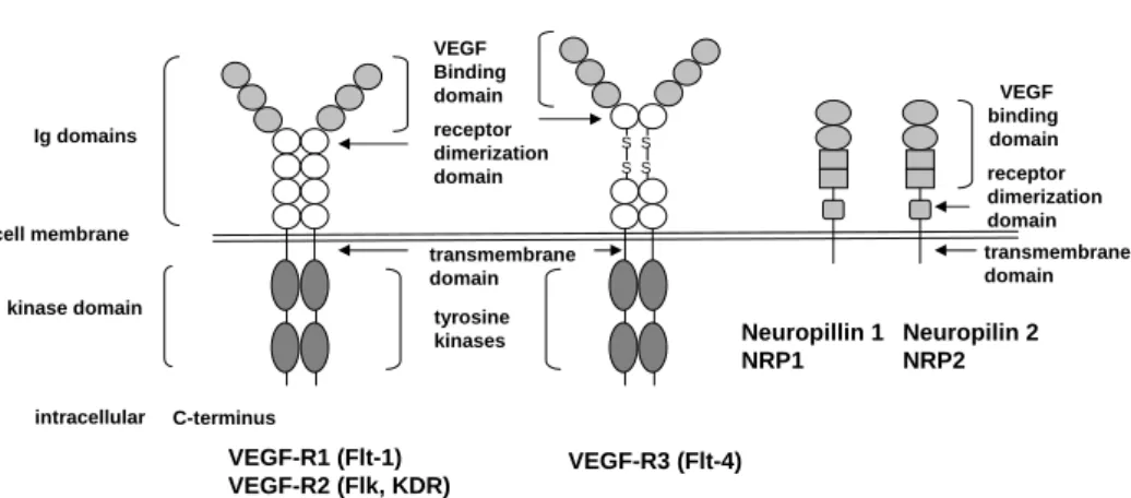
1.2 The vascular endothelial growth factor
In the late 1980´s the vascular endothelial growth factor VEGF was first de- scribed as heparin binding angiogenic growth factor with high specificity for endothelial cells (Ferrara and Henzel, 1989; Gospodarowicz et al., 1989). At around the same time, a protein promoting extravasation of proteins from tumor-associated blood vessels was characterized and named vascular perme- ability factor (VPF) (Senger et al., 1983). It turned out that VEGF and VPF are one and the same as they were derived from a single gene (Keck et al., 1989; Leung et al., 1989; Tischer et al., 1989). The term "vascular endothe- lial growth factor" has prevailed over "vascular permeability factor" and is commonly accepted.
1.2.1 The VEGF-family
VEGF denotes a family of homodimeric glycoproteins consisting of six mem- bers including VEGF-A, placental growth factor (PlGF) (Maglione et al., 1991), VEGF-B (Olofsson et al., 1996a), VEGF-C (Lee et al., 1996), VEGF-D (Achen et al., 1998; Orlandini et al., 1996) and viral homologues of VEGF, termed VEGF-E (Meyer et al., 1999; Ogawa et al., 1998; Wise et al., 1999).
All members possess a conserved central core region, the so called VEGF- homology domain, which is a central part of eight invariant cysteine residues essential for assembly of inter-and intramolecular disulfide bonds. Despite the structural similarity, all VEGF family members show distinct tissue distribu- tion and display different biological activities, mainly due to their different abilities to bind to the three VEGF-receptors.
While VEGF-A has strong mitogenic and permeability enhancing activities, PlGF has only weak potential in this regard. However, PlGF has the abil- ity to enhance VEGF-A action (Park et al., 1994). Furthermore, PlGF can form heterodimers with VEGF, which have increased potency to mediate mito- genic stimulation of endothelial cells relative to PlGF alone (Cao et al., 1996).
VEGF-B exists in two different isoforms, which are both predominantly ex-
pressed in embryonal and adult muscle tissue (myocardium and skeletal mus-
cle) and are co-expressed with VEGF in many tissues, most prominantly in
the heart (Lagercrantz et al., 1996; Olofsson et al., 1996b). VEGF-C and
VEGF-D are produced as long precursor proteins. After proteolytic process- ing several variants with different VEGF-receptor binding affinities are created (Joukov et al., 1997; Stacker et al., 1999). In midgestation embryos, VEGF-C is mainly expressed in regions where the lymphatic vessels undergo sprouting from embryonic veins (Kukk et al., 1996). In adult humans VEGF-C is pre- dominantly expressed in heart, placenta, ovary, small intestine, and the thyroid gland (Joukov et al., 1996). During embryogenesis VEGF-D was detected in high amounts in the developing mouse embryo (Stacker et al., 1999) and in human tumors (Achen et al., 2001).
VEGF-A is dimeric, disulfid-bound glycoproteins of 34-42kDa in size and the most common VEGF family member. It is generally referred to as VEGF and is the focus of this work. Due to alternative splicing six major isoforms of VEGF ranging in the sizes from 121 to 206 aminoacids exist. The primary VEGF transcript is derived from a single VEGF gene consisting of 8 exons separated by seven introns. Whereas exons 1 to 5 and exon 8 are conserved domains and present in all isoforms, alternative splicing in exons 6 and 7, which are responsible for heparan and heparin binding abilities, gives rise to the other isoforms.
3
3 3 3 3 3 3 2 2 2
2 2
2 2
1 1 1 1 1 1 1
4 4 4 4 4 4 4 5
5 5 5 5 5 5
7 6
6 6
6 6
8 8
8 8 8
8 8
7 7
7 7
5´ 3´
VEGF121 VEGF145 VEGF165
VEGF183 VEGF189 VEGF206 VEGF gene