Molecular Tools for G-Protein Coupled Receptors:
Synthesis, Pharmacological Characterization and [
3H]-Labeling of Subtype-selective Ligands for Histamine H
4and NPY Y
2Receptors
Dissertation
zur Erlangung des Doktorgrades der Naturwissenschaften (Dr. rer. nat.) an der Fakultät für Chemie und Pharmazie
der Universität Regensburg
vorgelegt von Paul Baumeister
aus Zinzenzell
2014
Die vorliegende Arbeit entstand in der Zeit von April 2010 bis April 2014 unter der Anleitung von Herrn Prof. Dr. Armin Buschauer am Institut für Pharmazie der Naturwissenschaftlichen Fakultät IV – Chemie und Pharmazie – der Universität Regensburg.
Das Promotionsgesuch wurde eingereicht im Juni 2014.
Tag der mündlichen Prüfung: 18.07.2014
Prüfungsausschuss: Prof. Dr. J. Heilmann (Vorsitzender) Prof. Dr. A. Buschauer (Erstgutachter) Prof. Dr. S. Elz (Zweitgutachter) Prof. Dr. J. Wegener (Drittprüfer)
„If you don’t turn your life into a story, you just become a part of someone else’s story.“
Terry Pratchett
I
Danksagung
An dieser Stelle möchte mich ganz herzlich bei allen bedanken, die zum Gelingen dieser Arbeit beigetragen haben und mich während der Promotionszeit begleitet haben. Besonders möchte ich danken:
Meinem Doktorvater Herrn Prof. Dr. Armin Buschauer für das Vertrauen und die Möglichkeit dieses interessante und herausfordernde Projekt zu verwirklichen, seine wissenschaftlichen Anregungen, für die mir gewährte forscherische Freiheit, seine konstruktive Kritik bei der Durchsicht der Arbeit, sein stets offenes Ohr, sowie die Mentorenschaft in der Emil-Fischer-Graduiertenschule;
Herrn Prof. Dr. Günther Bernhardt für seine stete Hilfsbereitschaft und sein Interesse am Fortschritt der Arbeit, sein Fachwissen, die Durchsicht der Arbeit, die Co-Mentorenschaft in der Emil-Fischer- Graduiertenschule, sowie für hervorragenden Sommerfeste;
Herrn Prof. Dr. Sigurd Elz für die Erstellung des Zweitgutachtens und die Teilnahme an der mündlichen Prüfung;
Herrn Prof. Dr. Joachim Wegener für die Bereitschaft als Drittprüfer an der mündlichen Prüfung teilzunehmen und die Co-Mentorenschaft in der Emil-Fischer-Graduiertenschule;
Herrn Prof. Dr. Jörg Heilmann für die Teilnahme als Vorsitzender in der mündlichen Prüfung;
Herrn Dr. Max Keller für seine fachliche und soziale Kompetenz, die hervorragende Zusammenarbeit, aufmunternde Worte, seine Motivation, die Bereitstellung von Synthesebausteinen und die gemütlichen Abende bei selbstgemachtem Wein;
Herrn Dr. Patrick Igel und Herrn Dr. Roland Geyer für die ausführliche Einführung in die Histamin- Welt, die fachlichen Tipps und die Bereitstellung von Synthesebausteinen;
Herrn Dr. Nikola Pluym für die ausführliche Einführung in die NPY-Welt, die fachlichen Diskussionen, seine soziale Kompetenz, die Bereitstellung von Synthesebausteinen und die Einführung in die Welt der Radioligandsynthese;
Herrn Dr. Thilo Spruss, Herrn Franz Wiesenmayer und Frau Petra Pistor für die Betreuung und Unterstützung bei der Durchführung der Tierversuche, sowie für das Anfertigen der Kryoschnitte von Gewebeproben und deren histologischen Färbung;
Herrn Dr. Uwe Nordemann für die Durchführung der HEK293 Zellexperimente mit [3H]JNJ7777120 und die Einweisung in die Welt der Radioligandsynthese;
Frau Nicole Kagermeier für die fachlichen Diskussionen und die Durchführung der HEK293 Zellexperimente mit [3H]UR-DE257;
Frau Maria Beer‐Krön, Frau Dita Fritsch, Frau Susanne Bollwein, Frau Elvira Schreiber und Frau Brigitte Wenzl für die tatkräftige Unterstützung bei der Durchführung vieler Assays, HPLC-Läufe und der Zellkultivierung;
Herrn Peter Richthammer für die zahlreichen netten Gespräche, seine stete Hilfsbereitschaft und Kompetenz bei allen technischen Herausforderungen und für die gute Zusammenarbeit bei der Durchführung der verschiedenen Praktika;
Frau Uta Hasselmann, Frau Karin Reindl und Frau Silvia Heinrich für die stets freundliche Unterstützung bei allen organisatorischen Angelegenheiten;
Frau Edith Bartole, Frau Shiwen Xue, Herrn Josef Auburger, Herrn Michael Schupfner und Herrn Markus Friedrich für die Durchführung von Versuche im Rahmen Ihrer Praktika;
Allen Laborkollegen, allen Mitgliedern der Histamin-Gruppe und der NPY-Gruppe für die angenehme, inspirierenden und gelegentlich auch amüsanten Atmosphäre und die sehr gute Zusammenarbeit;
Frau Edith Bartole und Frau Sabrina Biselli für die gute Zusammenarbeit im Rahmen der Betreuung Ihrer Masterarbeiten;
Allen Mitarbeitern der analytischen Abteilung der Universität Regensburg für die Aufnahme und Hilfestellung bei der Interpretation der NMR- und Massenspektren. Ein besonderer Dank geht hierbei an Herrn Fritz Kastner und Herrn Josef Kiermaier für die hilfreichen Diskussionen und die Ermittlung zahlreicher analytischer Daten;
Allen Mitgliedern der Arminia Buschauer für die tolle Zeit am Lehrstuhl, die stets gute Kollegialität, Arbeitsatmosphäre und Zusammenarbeit;
Frau Dr. Stefanie Bauer, Herrn Dr. Roland Geyer, Herrn Stefan Huber, Frau Nicole Kagermeier, Herrn Dr. Nikola Pluym, Frau Edith Bartole, Herrn Steffen Pockes und Frau Maria Beer-Kroen für die vielen netten Gespräche und schöne Zeit;
Den Mitarbeitern der FAU Erlangen, Herrn Prof. Dr. Peter Gmeiner, Herrn Prof. Dr. Markus Heinrich, Frau Dr. Nuska Tschammer, Herrn Dr. Viachaslau Bernat, Herrn Dr. Harald Hübner und Michael Fürst für die gute Zusammenarbeit;
Herrn Prof. Dr. Oliver Reiser und Dr. Julian Bodensteiner für die gute Zusammenarbeit;
Der Deutschen Forschungsgemeinschaft für die finanzielle Förderung im Rahmen des Graduiertenkollegs GRK 760;
Special thanks to my coworkers and friends Jianfei Wan and Xueke She for the great opportunity to visit their homeland China, all the hospitality, patience and unforgettable impressions. 谢谢! 干杯! Meinen Freunden und dem Trimmverein Regensburg, auf die man sich immer verlassen konnte, wenn es darauf ankam. ‚Reich sind nur die, die wahre Freunde haben‘ (Thomas Fuller);
Den Herren Dr. Daniel Bücherl, Petr Jirásek und Michel Leonhardt für die allmorgendliche Frühstücksrunde, sowie die moralische und fachliche Unterstützung;
Zuletzt all denjenigen, die das Leben lebenswert machen und mehr als nur Dank verdienen: meinen Eltern, meinen Geschwistern und meiner Freundin Marina. Ihnen ist die vorliegende Arbeit gewidmet.
III
Publications (published results prior to the submission of this thesis):
(1) Baumeister, P., Erdmann, D., Biselli, S., Kagermeier, N., Bernhardt, G., Buschauer, A.: [3H]UR- DE257: A Selective and Highly Potent Tritium-Labeled Squaramide-type Histamine H2
Receptor Antagonist. ChemMedChem 2014, in preparation.
(2) Geyer, R., Kaske, M., Baumeister, P., Buschauer, A.: Synthesis and Functional Characterization of Imbutamine Analogs as Histamine H3 and H4 Receptor Ligands. Arch.
Pharm. Chem. Life Sci. 2014, 347, 77–88.
(3) Pluym, N., Baumeister, P., Keller, M., Bernhardt, G., Buschauer, A.: [3H]UR-PLN196: A Selective Nonpeptide Radioligand and Insurmountable Antagonist for the Neuropeptide Y Y2
Receptor. ChemMedChem Comm. 2013, 8, 587-593.
(4) Bodensteiner, J., Baumeister, P., Geyer, R., Buschauer, A., Reiser, O.: Synthesis and Pharmacological Characterization of New Tetrahydrofuran Based Compounds as Conformationally Constrained Histamine Receptor Ligands. Org. Biomol. Chem. 2013, 11, 4040-4055.
(5) Bernat, V., Heinrich, M., Baumeister, P., Buschauer, A., Tschammer, N.: Synthesis and Application of the First Radioligand Targeting the Allosteric Binding Pocket of Chemokine Receptor CXCR3. ChemMedChem 2012, 7 (8), 1481-1489.
Short Lecture:
Baumeister, P.: The First Selective Tritium-labeled Nonpeptide Radioligands for the NPY Y2 Receptor.
Christmas Colloquium 2012 of the Department of Organic Chemistry, University of Regensburg, 19.12.2012.
Poster Presentations:
03/2013 Annual Meeting of the GDCh, Fachgruppe Medizinische Chemie, Frontiers in Medicinal Chemistry, München.
Baumeister, P., Pluym, N., Keller, M., Bernhardt, G., Buschauer, A.:
Subtype-selective Nonpeptide NPY Y2 Receptor Radioligands.
09/2012 XXIInd International Symposium on Medicinal Chemistry (ISMC), Berlin.
Baumeister, P., Erdmann, D., Bernhardt, G., Buschauer, A.: [3H]UR-DE257:
A New Tritium-labeled Histamine H2 Receptor Antagonist.
Bernat, V., Heinrich, M., Baumeister, P., Buschauer, A., Tschammer, N.:
Synthesis and Application of the First Small-Molecule Radioligand Targeting the Human Chemokine Receptor CXCR3. Best Poster Award
09/2012 6th Summer School in Medicinal Chemistry, Regensburg.
Geyer, R., Nordemann, U., Baumeister, P., Bernhardt, G., Buschauer, A.:
trans‐(+)‐(1S,3S)‐UR‐RG98: Synthesis, Absolute Configuration and Pharmacological Characterization of a Highly Potent and Selective Histamine H4 Receptor Agonist.
03/2011 Annual Meeting of the GDCh, Fachgruppe Medizinische Chemie, Frontiers in Medicinal Chemistry, Saarbrücken.
Baumeister, P., Buschauer, A.:
2-Arylbenzimidazoles as Potent Human Histamine H4 Receptor Agonists.
Professional Training:
04/2013 Fortbildung für Projektleiter und Beauftragter für Biologische Sicherheit (§15 und 17 Gentechniksicherheitsverordnung).
Regensburg, Germany.
06/2010 Umgang mit offenen radioaktiven Stoffen.
Regensburg, Germany.
03/2010 – 03/2012 Member of the Research Training Group (Graduiertenkolleg 760) “Medicinal Chemistry: Molecular Recognition – Ligand Receptor Interactions” of the German Research Foundation.
Regensburg, Germany.
06/2012 – 04/2014 Member of the Emil Fischer Graduate School of Pharmaceutical Sciences and Molecular Medicine.
Regensburg, Erlangen, Germany.
V
Contents
1 Introduction ... 1
1.1 G-Protein-coupled receptors ... 2
1.1.1 GPCRs as drug targets and their classification ... 2
1.1.2 G-Protein activation, ligand classification and signal transduction ... 2
1.1.3 G-Protein independent signaling, -arrestin and functional selectivity ... 5
1.2 Histamine and the histamine receptor family ... 5
1.2.1 Histamine as endogenous ligand ... 5
1.2.2 Histamine receptors and their ligands ... 6
1.2.2.1 The histamine H1 receptor ... 7
1.2.2.2 The histamine H2 receptor ... 7
1.2.2.3 The histamine H3 receptor ... 10
1.2.2.4 The histamine H4 receptor ... 11
1.3 NPY and the NPY receptor family ... 15
1.3.1 Neuropeptide Y ... 15
1.3.2 NPY receptors and their ligands ... 15
1.3.2.1 The NPY Y2 receptor and its ligands ... 15
1.3.2.2 Ligands for the NPY Y1, Y4 and Y5 receptors ... 17
1.4 Receptor–ligand binding assays ... 19
1.4.1 Radioligand binding methods ... 20
1.4.1.1 Selection of radioligands ... 20
1.4.2 Radioligands for the H2, H4 and NPY Y2 receptor ... 21
1.5 References ... 22
2 Scope and Objectives ... 37
2.1 References ... 40
3 Synthesis and Pharmacological Characterization of 2-Arylbenzimidazoles as Potent and Selective Histamine H4 Receptor Ligands ... 43
3.1 Introduction ... 44
3.2 Chemistry ... 45
3.3 Pharmacological Results and Discussion ... 52
3.3.1 Histamine receptor subtype affinities of the synthesized compounds ... 53
3.3.1.1 Variation of the substitution pattern ... 53
3.3.1.2 Structural variations of arylbenzimidazole-type hH4R ligands ... 53
3.3.1.3 Introduction of a propionyl group ... 56
3.3.1.4 Functional activities at recombinant human histamine receptor subtypes ... 59
3.3.2 Inhibition of the hH4R agonistic effect of 3.16 by standard H4R antagonists... 62
3.3.2.1 Potencies, efficacies and affinities at the mH4R ... 63
3.3.3 Muscarinic receptor subtype affinities of 3.16 on CHO-M1 and CHO-M2 cells ... 65
3.4 Summary and Outlook ... 65
3.5 Experimental Section ... 68
3.5.1 Chemistry... 68
3.5.1.1 General conditions ... 68
3.5.2 Chemistry... 69
3.5.2.1 Preparation of the imidazole 3.2 ... 69
3.5.2.2 Preparation of the benzimidazoles 3.7-3.10 ... 70
3.5.2.3 Preparation of the benzimidazolylphenyl chloroalkyl ether 3.11-3.15 ... 72
3.5.2.4 Preparation of the imidazole derivatives 3.16-3.20 ... 74
3.5.2.5 Preparation of the benzimidazolylphenoxyalkylamines 3.22-3.23 ... 77
3.5.2.6 Preparation of the guanidine 3.26 ... 79
3.5.2.7 Preparation of NG-propionyl guanidine 3.30 ... 80
3.5.2.8 Preparation of the carboxylic amides 3.31-3.37 ... 81
3.5.2.9 Preparation of the histamine homolog 3.41 ... 86
3.5.2.10 Preparation of the secondary amine 3.45 ... 87
3.5.2.11 Preparation of the primary amines 3.48-3.50 ... 88
3.5.2.12 Preparation of the primary amines 3.58-3.60 ... 90
3.5.2.13 Preparation of the amines 3.62-3.73 ... 93
VII
3.5.2.14 Preparation of the amide 3.75 ... 100
3.5.2.15 Preparation of the amide 3.76 ... 101
3.5.3 Pharmacological Methods ... 102
3.5.3.1 Competition binding experiments on membrane preparations of Sf9 insect cells ... 102
3.5.3.2 Steady-State [γ-33P]GTPase activity assay ... 103
3.5.3.3 [35S]GTPγS binding assay ... 103
3.5.3.4 Radioligand binding assay using HEK293 cells expressing the mH4R ... 104
3.5.3.5 Radioligand binding studies on hM1R or hM2R expressing CHO-K9 cells ... 104
3.5.3.6 Data analysis and pharmacological parameters... 105
3.6 References ... 107
4 Synthesis and Pharmacological Characterization of VUF 8430 Derivatives as Histamine H4 Receptor Ligands ... 111
4.1 Introduction ... 112
4.2 Chemistry ... 113
4.3 Pharmacological Results and Discussion ... 115
4.4 Summary and Outlook ... 117
4.5 Experimental Section ... 119
4.5.1 Chemistry... 119
4.5.1.1 General conditions ... 119
4.5.1.2 Preparation of the isothiourea derivatives 4.2 and 4.3 ... 119
4.5.1.3 Preparation of NG-acylated isothiourea derivatives 4.4-4.11 ... 120
4.5.1.4 Preparation of the guanidine derivatives 4.12-4.39 ... 123
4.5.1.5 Preparation of the dicarbamimidothioate 4.40 ... 134
4.5.1.6 Preparation of VUF 8430 analog 4.43 ... 135
4.5.2 Pharmacological Methods ... 136
4.5.2.1 General ... 136
4.5.2.2 Competition binding experiments on membrane preparations of Sf9 insect cells ... 136
4.6 References ... 137
5 [3H]UR-DE257: A Selective and Highly Potent Tritium-Labeled Squaramide-type Histamine H2 Receptor Antagonist ... 141
5.1 Introduction ... 142
5.2 Results and Discussion ... 143
5.2.1 Radiosynthesis ... 143
5.2.2 Determination of binding constants of [3H]UR-DE257 ... 144
5.2.3 Autoradiography ... 153
5.3 Summary ... 155
5.4 Experimental Section ... 156
5.4.1 General conditions for radiosynthesis... 156
5.4.2 Synthesis of N-[6-(3,4-Dioxo-2-{3-[3-(piperidin-1-ylmethyl)phenoxy]propyl- amino}-cyclobut-1-enylamino)hexyl]-[2,3-3H2]propionamide ([3H]UR-DE257): ... 156
5.4.3 Pharmacological methods ... 157
5.4.3.1 Histamine radioligand binding assays on membrane preparations of Sf9 insect cells ... 157
5.4.3.2 Radioligand binding assay at HEK293T CRE-Luc hH2R cells ... 158
5.4.4 Autoradiography ... 159
5.5 References ... 160
6 [3H]JNJ7777120: A Tritium-Labeled Histamine H4 Receptor Antagonist ... 163
6.1 Introduction ... 164
6.2 Chemistry ... 165
6.2.1 Optimization of the synthesis ... 165
6.2.2 Radiosynthesis of [3H]JNJ7777120 ... 166
6.3 Results and Discussion ... 168
6.3.1 Histamine receptor subtype affinities ... 168
6.3.2 Pharmacological characterization of [3H]JNJ7777120 (6.4b) on Sf9 cell membranes ... 169
6.3.2.1 Saturation binding of [3H]JNJ7777120 at the hH4R ... 169
IX
6.3.2.2 Kinetics at the hH4R ... 170
6.3.2.3 Competition binding at the hH4R ... 171
6.3.2.4 Membranes of Sf9 cells expressing the mH4R ... 172
6.3.3 Saturation binding at HEK293-SF-H4R-His6 cells ... 173
6.4 Summary and Conclusion ... 174
6.5 Experimental Section ... 176
6.5.1 Chemistry... 176
6.5.1.1 General ... 176
6.5.1.2 Preparation of the indol derivatives 6.4a and 6.5 ... 176
6.5.2 Preparation of [3H]JNJ7777120 (6.4b) ... 178
6.5.3 Radioligand binding assay for the hHxR ... 180
6.5.4 Saturation binding assay for the mH4R ... 180
6.6 References ... 181
7 Subtype-selective Nonpeptide Radioligands for the NPY Y2 Receptor ... 185
7.1 Introduction ... 186
7.2 Results and Discussion ... 186
7.2.1 Synthesis of [3H]UR-PLN208 ... 186
7.2.2 Pharmacological characterization of [3H]UR-PLN208 and ‘cold’ analogs at CHO- hY2R cells ... 188
7.2.2.1 Determination of binding constants of [3H]UR-PLN208 ... 188
7.2.2.2 Association and dissociation kinetics of [3H]UR-PLN208... 190
7.2.2.3 Competition binding experiments ... 191
7.2.2.4 Calcium assay on hY2R-expressing CHO cells ... 193
7.2.3 Pharmacological characterization of [3H]UR-PLN208 and non-labeled analogs on the hY2R at Sf9 insect cell membranes ... 194
7.2.3.1 hY2R antagonist activity of BIIE 0246 and related compounds in the steady- state [γ-33P]GTPase assay ... 194
7.2.3.2 Saturation binding of [3H]UR-PLN208 using hY2R-insect cell membrane preparations ... 196
7.2.4 Stability of argininamide-type NPY Y2R antagonists ... 197
7.3 Summary and Outlook ... 199
7.4 Experimental Section ... 200
7.4.1 General ... 200
7.4.2 Synthesis of (2S)-N-[2-(3,5-Dioxo-1,2-diphenyl-1,2,4-triazolidin-4-yl)ethyl]-N -{2- [1-({2-oxo-2-[4-(6-oxo-6,11-dihydro-5H-dibenzo[b,e]azepin-11-yl)piperazin-1- yl]ethyl})cyclopentyl]acetyl}-N -([2,3-3H2]propanoyl)argininamide ([3H]UR-PLN208) ... 201
7.4.3 Investigation of the chemical stability ... 202
7.4.4 Pharmacological methods ... 203
7.4.4.1 Cell culture ... 203
7.4.4.2 Spectrofluorimetric Ca2+ assay (Fura-2 assay) ... 203
7.4.4.3 Radioligand binding assay ... 203
7.4.4.4 Steady‐state GTPase activity assay... 204
7.4.4.5 Saturation binding of [3H]UR-PLN208 at the hY2R on membrane preparations of Sf9 insect cells ... 204
7.5 References ... 206
8 Summary ... 209
9 Appendix ... 213
9.1 Synthesis and Application of the First Radioligand Targeting the Allosteric Binding Pocket of Chemokine Receptor CXCR3 ... 214
9.1.1 Abstract ... 214
9.1.2 General conditions for radiosynthesis: ... 214
9.1.3 Radiosynthesis of [3H]N-{1-[3-(4-Ethoxyphenyl)-4-oxo-3,4-dihydropyrido[2,3- d]pyrimidin-2-yl]ethyl}-2-[4-fluoro-3-(trifluoromethyl)phenyl]-N-[(1-methylpiperidin-4- yl)methyl]acetamide (RAMX3) ... 215
9.2 Nonspecific binding of [3H]JNJ7777120 at the hH4R in Sf9 cell membranes (cf. Chapter 6) ... 219
9.3 Nonspecific binding of [3H]UR-PLN208 at CHO-hY2R cells (cf. Chapter 7)... 219
9.4 NMR-Spectra of selected compounds ... 220
9.5 HPLC Purity Data... 223
Chapter 1
1 Introduction
1.1 G-Protein-coupled receptors
1.1.1 GPCRs as drug targets and their classification
G-Protein-coupled receptors (GPCRs) constitute the largest group of integral membrane proteins that transmit a wide variety of signals across the cell membrane.1 GPCRs respond to a broad range of extracellular stimuli such as biogenic amines, purines, lipids, amino acids, peptides and proteins, odorants, pheromones, ions and even photons.2 More than 800 GPCRs are encoded in the human genome (approximately 2-3% of the human genome) including about 400 functional non-olfactory receptors.3 For roughly 120 of the latter, referred to as orphan receptors, endogenous ligands are not known to date.4 More than 50 GPCRs are targeted by approved drugs,5 which represent 30 - 40 % of all marketed drugs,6 emphasizing the current value in the treatment of human diseases, as well as the prospects for the development of GPCR ligands as future drugs. Based on structural differences, mammalian GPCRs were classified in five groups: rhodopsin, secretin, adhesion, glutamate and frizzled/taste2.7 The common structural features of the GPCR superfamily are seven membrane- spanning helices, connected by three alternating intracellular and extracellular loops and flanked by an extracellular N-terminus and an intracellular C-terminus, respectively. The rhodopsin-like family, also referred to as class A of GPCRs, is by far the largest and best studied subgroup containing receptors for odorants, small molecules such as biogenic amines, peptides and glycoprotein hormones (~700 GPCRs). The binding sites of small endogenous ligands are located within the seven transmembrane (TM) domains, whereas binding of more space filling ligands, for example peptides and glycoproteins, occurs at the amino terminus (N-terminus), extracellular loops and amino acids located at the top of the TM helices.8
1.1.2 G-Protein activation, ligand classification and signal transduction
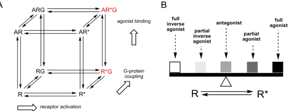
Several models have been proposed for the molecular mechanism involved in the activation of GPCRs upon interaction with appropriate ligands. Among them, the cubic ternary model9-11 is considered most suitable for explaining the pharmacodynamic activities of the majority of interacting ligands (cf. Figure 1.1). This model distinguishes between an active (R*) and an inactive (R) receptor state. These two states are in equilibrium and are allowed to isomerize independently from agonist binding. This kind of a spontaneous activation of the receptor in the absence of agonists is referred to as constitutive activity.12 Both receptor states are able to bind G‐proteins, but only the active receptor – G‐protein complex (R*G) induces GDP/GTP exchange, resulting in signal transduction.
G-Protein-coupled receptors 3
Figure 1.1 A. Two-state cubic ternary complex model of GPCR activation (R: inactive state of the receptor, R*:
active state of the receptor, G: G-Protein, A: agonist). Signaling complexes mediating GDP/GTP exchange are highlighted in red. B. Ligand classification according to their capability of shifting the equilibrium to either side of both states. According to Seifert et al.12
Ligands are classified according to their capability of shifting the equilibrium to either side of both states. Full agonists preferentially bind to the R* state, stabilizing the active conformation and thereby enhancing the functional response. On the opposite, inverse agonists particularly interact with and stabilize the inactive conformation R of the receptor and reduce the percentage of spontaneously active receptors. Neutral antagonists bind to both conformations with the same affinity without altering the equilibrium, but impairing the binding of other ligands. Partial agonists and partial inverse agonists are less effective in stabilizing the active or the inactive receptor conformation, respectively.13 However, the two-state model of GPCR activation cannot sufficiently explain all observed experimental findings. The function of GPCRs is considered much more complex in terms of ligand binding (orthosteric, allosteric), different conformational states, accessory protein interaction, phosphorylation, G-Protein coupling, oligomerization and internalization.14-17 Furthermore, there is growing evidence of several inactive and active receptor conformations,18 suggesting that structurally different ligands stabilize distinct receptor conformations, resulting in different biological responses.19 In summary, the two-state model provides a molecular basis for classical concepts of pharmacology and helps to explain the properties of drugs acting as agonist, antagonist and inverse agonist, but the real situation is not completely reflected. After activation, the majority of GPCRs is able to transduce signals into cells through G-Protein coupling.20 Agonist binding to extracellular or transmembrane domains of a GPCR (or agonist-free constitutive activity) promotes conformational changes that initiate coupling of intracellular receptor domains to a heterotrimeric G- Protein. This agonist-receptor-G-Protein complex, termed as ternary complex, triggers a G-Protein conformational change and results in the release of GDP from the Gα-subunit. Subsequently, the activated heterotrimeric G-Protein dissociates into Gα-GTP and Gβγ subunits, both of which then interact with effector proteins like enzymes or ion channels resulting in cellular biological responses.21 The intrinsic GTPase activity of the GTP-bound Gα subunit terminates the signal by the
hydrolysis of GTP to GDP, i. e. the cycle is completed by reversion of the G-Protein to the inactive heterotrimeric state (see Figure 1.2). Subsequently, the GDP-bound G -subunit re-associates with G enabling the next G-Protein cycle.22 According to the structure and signaling pathway of the G - subunits, G-Proteins are divided into four main families, termed Gs, Gi/o, Gq/11 and G12/13.23 The G s
family activates adenylyl cyclases (AC 1–9), resulting in increased cellular levels of the second messenger cAMP (3´-5´-cyclic adenosine monophosphate). By contrast, the G i family shows inverse effects, inhibiting the AC activity (AC 5 and AC 6). cAMP regulates various cellular effects such as activation of the protein kinase A (PKA) or the mitogen‐activated protein kinase (MAPK) pathway, both modulating gene expression.24 Members of the Gαq/11 family activate the phospholipases Cβ1-3 (PLCβ), which catalyze the hydrolysis of phosphatidylinositol 4,5-bisphosphate (PIP2) to 1,2- diacylglycerol (DAG) and inositol-1,4,5- trisphosphate (IP3). The latter second messenger controls calcium efflux from the endoplasmic reticulum. DAG and the released calcium control the activity of several protein kinase C (PKC) isoforms, which in turn activate a number of other proteins by phosphorylation.25,26 Finally, the Gα12 proteins interact with Ras homology GEFs (guanine-nucleotide exchange factor) (RhoGEFs) that regulate cytoskeletal assembly.27 Not only the G ‐subunit, but also the G ‐heterodimers are involved in signal transduction and regulate certain effectors such as PLC and ion channels.28
Figure 1.2 Activation of a heterotrimeric G-Protein by interaction with an agonist-occupied GPCR. The activated receptor is represented by R*, whereas the inactive form is termed R. The dissociated subunits regulate their respective effector proteins such as adenylyl cyclase (AC) and calcium channels. Further details are described in the text (modified from29).
Histamine and the histamine receptor family 5
1.1.3 G-Protein independent signaling, -arrestin and functional selectivity
Although the vast majority of GPCRs is able to transduce signals into cells via G-Protein coupling, recent work has indicated that GPCRs participate in numerous other protein-protein interactions, which generate intracellular signals independent of G-Protein activation.24 For instance, GPCR dimerization, the interaction with receptor activity‐modifying proteins (RAMPs) and the binding of various scaffolding proteins to GPCRs modulate GPCR signaling.20 Most compelling, the discovery that -arrestins (arrestin 2 and 3) function as alternative transducers of GPCR signals, has challenged the basic concept of GPCR signaling.20,30,31 Originally regarded as mediators of GPCR desensitization (through internalization into clathrin-coated pits),32,33 -arrestins are ubiquitously expressed cellular regulatory proteins that are meanwhile recognized as true adapter proteins that transduce signals to multiple effector pathways such as MAPKs (mitogen-activated protein kinase), SRC (v-src avian sarcoma (Schmidt-Ruppin A-2) viral oncogene homolog), nuclear factor B (Nf- B) and phosphatidylinositol 3-kinase (PIK3).34 An updated model of signal transduction should comprise signaling by G‐proteins and/or ‐arrestins, as well as desensitization and internalization by -arrestins.34 The selective stimulation of some, but not all, possible signaling pathways has been postulated as ‘functional selectivity’,35 also known as ‘biased agonism’36 or differential receptor- linked effector actions.37,38 Apparently, depending on the ligand, the conformational changes of GPCRs are biased, giving rise to different behavior and interactions.16 Such biased ligands are not only useful tools to investigate GPCR signaling, but might also harbor a potential as fine-tuned therapeutics.39 Besides, allosteric ligands, which could modulate the signaling cascades and biochemical responses triggered by endogenous ligands, can also impose biased agonism, showing promise in clinical pharmacology.40
1.2 Histamine and the histamine receptor family
1.2.1 Histamine as endogenous ligand
The biogenic amine histamine (2-(1H-imidazol-4-yl)ethanamine) is a local mediator, immunomodulator and neurotransmitter targeting the histaminergic system. First biological effects of histamine like vasodilatation and smooth muscle contraction have been reported more than one hundred years ago.41 Histamine contains two basic functionalities, a primary aliphatic amine and imidazole. At physiological pH, the amine group is protonated, and two different tautomers of this monocation are the predominating forms (Figure 1.3).42
In the body, histamine is synthesized from the amino acid L-histidine through decarboxylation.43,44 Nowadays, histamine is considered , an ubiquitous and multifunctional biogenic amine which is involved in various physiological and pathophysiological processes. High tissue concentrations of histamine are found in particular in the lungs, the skin, connective tissues and the gastrointestinal tract.44 It is stored in mast cells,45 basophils,46 platelets,46 enterochromaffin‐like cells (ECL) of the stomach,47 endothelial cells,48 and it is also found in neurons.49 In the brain, histaminergic neurons are involved in the sleep-wake cycle, energy and endocrine homeostasis, synaptic plasticity and learning.50 In mast cells and basophils, histamine is stored in secretory granules and released during allergic conditions, resulting
in smooth muscle contraction, vasodilatation and an increase in vascular permeability.51 After release, in response to immunological and non-immunological stimuli, histamine is degraded by two catabolic pathways. The first pathway involves methylation of histamine by histamine N-methyltransferase and the second pathway involves oxidative deamination by diamine oxidase.52 The biological effects are mediated by the interaction with currently four histamine receptor (HR) subtypes, termed H1R, H2R, H3R and H4R, all belonging to the rhodopsin-like family A of GPCRs.44,53-55
1.2.2 Histamine receptors and their ligands
In this chapter, various molecular pharmacological aspects of the four histamine receptor subtypes, including the availability of selective agonists and antagonists, will be discussed. In 1966 the term histamine H1 receptor (H1R) was introduced by Ash and Schild, who suggested the existence of a second HR subtype (non‐H1 receptor, H2R) as not all effects provoked by histamine could be antagonized by classical antihistamines.56 Activation of the H1R has long been known to be associated with allergic conditions.44,57 As a consequence, antagonists of this receptor subtype (popularly referred to as antihistamines) have been used as anti-allergic drugs since the 1940s.44 The H2R plays a pivotal role in gastric acid secretion,58 and H2R antagonists have been used as antiulcer drugs (‘H2R blockers’).57 The histamine H3R is located predominantly in the central nervous system (CNS) and acts both as a presynaptic autoreceptor,59 modulating histamine release from histaminergic neurons, and as an inhibitory heteroreceptor.60,61 H3R antagonists are being investigated as potential drugs for therapeutic applications against a variety of CNS disorders such as Alzheimer’s disease, attention- deficit/hyperactivity disorder (ADHD), epilepsy, migraine, narcolepsy, obesity, schizophrenia and depression.62 In the years 2000 and 2001, the H4R was identified and cloned independently by Figure 1.3 Tautomeric forms of the histamine monocation.
Histamine and the histamine receptor family 7
several research groups.63-69 It is considered as a new therapeutic target for the modulation of various inflammatory and immunological processes and disorders.70-73
1.2.2.1 The histamine H1 receptor
The histamine H1 receptor (H1R) was first cloned in 1993.74 The corresponding receptor protein consists of 487 amino acids.74 It is mainly expressed on smooth muscle cells, endothelial cells and in the CNS and is involved in the pathophysiology of allergy and inflammatory reactions.75 Via the H1R, histamine induces, for instance, vasodilatation, bronchoconstriction, increased vascular permeability, pain and itching upon insect stings.76 Upon agonist stimulation, the H1R predominantly couples to the pertussis-toxin insensitive Gαq/11 proteins. Its stimulation triggers the inositol phospholipid signaling system, resulting in the formation of IP3 and DAG (cf Chapter 1.1.2), which results in Ca2+-mobilization from intracellular stores and activation of protein kinase C.75,77 H1R antagonists (antihistamines) have been used for decades for the treatment of allergic disorders (e.g. allergic rhinitis, chronic urticarial and atopic dermatitis), nausea and vomiting, and for sedation.78,79 First generation antihistamines, such as mepyramine or diphenhydramine, are highly lipophilic compounds which cross the blood brain barrier, block central H1 receptors and cause sedation.44 More polar H1R antagonists such as cetirizine and fexofenadine were developed to reduce this undesired effect in the treatment of allergic diseases (‘non-sedative’ second generation of H1R blockers).57,80 Mepyramine is still the most commonly used reference H1R antagonist and radioligand ([3H]mepyramine) for pharmacological studies.81 Besides, H1R agonists such as 2-methylhistamine and supra(histaprodifen), have been used as pharmacological tools to study H1R functions in cellular systems.82 So far, betahistine is the only marketed H1R agonist; the drug is therapeutically used in the treatment of Menière`s disease (cf. Figure 1.4).83,84
1.2.2.2 The histamine H2 receptor
The histamine H2R was pharmacologically characterized by Black et al. in 1972,58 using the first H2R antagonist burimamide, which was able to block the histamine-mediated gastric acid secretion and the positive chronotropic effect on the heart. In 1991, Gantz and coworkers were able to clone the canine and human H2Rs.85,86The human H2R consists of 359 amino acids and is expressed in a variety of tissues including brain, uterus, airways, gastric parietal cells and the heart.57,87 The H2R primarily couples to the Gαs family of G-Proteins, leading to an increase in intracellular cAMP levels and the activation of PKA (cf. Section 1.1.2).85,88,89 Depending on the used cell system, the H2R may additionally trigger calcium signaling by coupling to the Gαq/11 G-Protein.90-92
Figure 1.4 Structures of selected H1R agonists and antagonists.
An essential physiological function of the H2R is the control of gastric acid secretion from parietal cells.58 Activation of cardiac H2Rs mediates positive chronotropic and inotropic effects,93 and histamine-mediated smooth muscle relaxation has been documented in airways, uterus and blood vessels.94 The first marketed H2R antagonist cimetidine revolutionized the treatment of peptic ulcer and gastro-oesophageal reflux disease.44 Following cimetidine, several other H2R antagonists ranitidine, famotidine, nizatidine and roxatidine have been successfully used in the treatment of gastric and duodenal symptoms (ulcers),95 but are nowadays mostly replaced by more effective proton pump inhibitors, such as omeprazole, and by eradication of Helicobacter pylori.44,96-98 Apart from marketed drugs, numerous structurally related compounds,87 e.g. iodoaminopotentidine,99 BMY25368,100 and tiotidine,101 are known as H2R antagonists (structures are given in Figure 1.6). For radioligand binding studies [3H]tiotidine102 and [125I]iodaminopotentidine103-105 were used. More information about available radioligands and the characterization of a new H2R radioligand is given in Chapter 5. Recently, a series of H2R antagonists was developed in our working group, replacing the cyanoguanidine group of potentidine-related piperidinomethylphenoxyalkylamines by squaramides.
Additional coupling with -aminoalkyl spacers allows for labeling reactions or bivalent ligand construction.106 Whereas H2R antagonists became standard drugs for the treatment of gastric and duodenal ulcers,107,108 H2R agonists have been mainly used as pharmacological tools to study the physiological and pathophysiological role of this histamine receptor. A first step towards a selective H2R agonist was the discovery of dimaprit and amthamine, which were found to be almost as active as histamine at the H2R, but hardly display any H1R agonism.109,110 Highly potent and selective
Histamine and the histamine receptor family 9
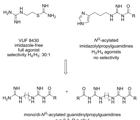
guanidine-type H2R agonists like impromidine111,112 and arpromidine113 had been developed,114 which, however, showed poor oral bioavailability.87 Drug-like properties were improved according to a bioisosteric approach, by an exchange of the guanidine by an acylguanidine moiety, resulting in NG-acylated imidazolylpropylguanidines (e.g. UR‐AK24, Figure 4.1, Chapter 4).115 Further improvement, concerning selectivity was achieved by the introduction of a 2‐amino‐4‐methylthiazol‐
5‐yl moiety as a bioisostere of the imidazole ring.116 Thus, NG-acylated aminothiazolylpropylguanidines (e.g. UR‐BIT24) combine the high selectivity for the H2R with improved pharmacokinetic properties, resulting in valuable pharmacological tools to evaluate the physiological role of H2Rs. Recently, the application of the bivalent ligand approach to acylguanidines yielded agonists, which are highly selective and up to 4000 times more potent than histamine at the guinea pig right atrium.117 Furthermore, another indication for the clinical use of histamine as H2R agonist evolved, based on the finding that histamine ameliorates the course of acute myeloid leukemia.118-120
Figure 1.5 Structures of selected H2R agonists.
Figure 1.6 Structures of selected H2R antagonists.
1.2.2.3 The histamine H3 receptor
The histamine H3R was discovered by Arrang et al. in 198359 and cloned in 1999.121 The hH3R consists of 445 amino acids122 and is mainly expressed in the CNS, where it acts as a presynaptic auto- and heteroreceptor, controlling the release of histamine and various other neurotransmitters, including dopamine,123 serotonin,124 noradrenalin46 and acetylcholine.125The H3R is suggested to be involved in various CNS functions, for instance, the regulation of locomotor activity, wakefulness and food intake, thermoregulation and memory.126 In the periphery, H3R activation was shown to occur in the cardiovascular system, the gastrointestinal tract and the airways.127-130 The activation of H3Rs leads to a decrease in intracellular cAMP levels via coupling to Gi/o proteins and inhibition of the adenylyl cyclase. Besides, activation of phospholipase A2 (PLA2), MAPKs and phosphatidyl inositol 3-kinase, inhibition of the Na+/H+exchanger and modulation of intracellular calcium was demonstrated.131,132 Antagonists for the H3R are promising agents131,133,134
in several therapeutic areas including dementia, Alzheimer`s disease, narcolepsy, deficit hyperactivity disorder, schizophrenia as well as for the treatment of myocardial ischemic arrhythmias, migraine and inflammatory and gastric acid-related diseases.62,122,135-140
Histamine and the histamine receptor family 11
Figure 1.7 Structures of selected H3R agonists and antagonists.
The first potent H3R antagonists, thioperamide141 and clobenpropit142 were derived from the structure of histamine and have an imidazole ring in common. To improve the drug‐like properties and to prevent potential drug‐drug interactions, several pharmaceutical companies developed non‐
imidazole H3R antagonists, for instance JNJ10181457 and JNJ5207852.133,143 Recently, the H3R antagonist pitolisant (tiprolisant) has been introduced as an orphan drug for the treatment of narcolepsy.144,145 Typical H3R agonists are N -methylhistamine and (R)- -methylhistamine141 as well as imetit146 and the H3R selective ligands immepip and methimepip,137 which are structurally less related to histamine (structures are shown in Figure 1.7). Almost exclusively, the application of the H3R agonists [3H]histamine, [3H]Nα-methylhistamine and [3H](R)-α-methylhistamine, as well as of the inverse agonist [125I]iodophenpropit or of the antagonist [3H]thioperamide have been described in radioligand binding experiments.107,147,148
1.2.2.4 The histamine H4 receptor
In 1975, Clark and co-workers reported on histamine induced chemotaxis of human eosinophils that was not inhibited by H1 or H2 receptor antagonists.149 Two decades later, Raible and colleagues suggested a novel HR subtype on human eosinophils. The authors observed that the histamine triggered calcium mobilization in human eosinophils could be blocked by the H3R antagonist
thioperamide. However, the potent H3R agonist (R)‐α‐methylhistamine was less potent than histamine in inducing calcium mobilization; this was not compatible with a H3R mediated effect.150,151 Finally, the human H4R was identified and cloned ‒ at that time as an orphan receptor ‒ in 2000 and 2001, independently by several research groups.63-69 Cloning of the H3R gene provided the basis for a fourth histamine receptor subtype due to their high sequence homology (about 40% overall sequence identity and about 58% sequence identity within the transmembrane domains).121 The human receptor subtype consists of 390 amino acids and, as in case of the H3R, couples to Gi/o-proteins, resulting in adenylyl cyclase inhibition,64,152 activation of MAPKs67 and calcium mobilization.153 Besides the coupling to G-Proteins, the activation of β-arrestin by several H4R ligands was recently reported.154-156 The H4R was suggested to be expressed in bone marrow and immunocytes such as mast cells, basophils, eosinophils, monocytes, T-lymphocytes and dendritic cells.157,158 Furthermore, detection of the H4R was purported on nerves from the nasal mucosa, in the enteric and, together with the other histamine receptor subtypes, in the central nervous system.152 Based on studies with H4R knockout mice, it has been suggested that the H4R plays a proinflammatory role in bronchial asthma, atopic dermatitis, allergic rhinitis, and pruritus.44,53,152,159
If so, H4R antagonists could be useful drugs for the treatment of these conditions.51,53,152,160
The H4R was also suggested as a new therapeutic target for the treatment of colitis, pain, cancer, rheumatoid arthritis and multiple sclerosis.70-72,161,162
The supposed role of the H4R in immunological responses overlaps with the function of the H1R, suggesting that combined H1- and H4-receptor ligands might be beneficial for the treatment of inflammatory diseases.51,163-166 In search for novel H4R antagonists or inverse agonists, the imidazole-containing H3R inverse agonist thioperamide has been identified as H4R inverse agonist with similar potency and has been frequently used as a reference compound (structure is given in Figure 1.7).167 A high-throughput screening campaign led to the identification of the indole carboxamide JNJ7777120 as a selective H4R antagonist.168 JNJ7777120 has been widely used as the prototypical H4R antagonist in animal models to assess the (patho)physiological role of the H4R.51. However, in vivo results of JNJ7777120 must be interpreted with caution in view of partial agonistic activity at murine H4R orthologs in vitro,169 -arrestin recruitment to the hH4R154-156 and off- target effects at higher concentrations.170 For a more detailed view at JNJ7777120 cf. Chapter 6.
Meanwhile, other highly selective and potent H4R antagonists, e.g. with quinazoline171 and pyrimidine160,172 scaffold, have been developed (cf. Figure 1.8; an overview is given by Schreeb et al.173). Some H4R ligands had entered clinical studies, e.g. UR-63325, the first H4R antagonist from which clinical data were reported,174,175 ZPL-38937887 (formerly PF-03893787)172 and JNJ39758979.160 To further investigate the pathophysiological role of the H4R, selective agonists are of particular interest as pharmacological tools.
Histamine and the histamine receptor family 13
Figure 1.8 Structures of selected H4R agonists and antagonists.
Due to the high sequence homology of the H4R with the H3R, especially in the transmembrane domains, it is not surprising that the H4R is activated by numerous compounds which were originally designed as H3R agonists, and, consequently, contain an imidazole ring, for instance (R)-α-methylhistamine, Nα-methylhistamine and imetit (Figure 1.7).167,176 Histamine and its homologs homohistamine (spacer length of three methylene groups) and imbutamine (four methylene groups) are agonists with similar hH3R and hH4R affinity, whereas the higher homolog impentamine (five methylene groups) is an almost full hH3R agonist but shows no agonistic activity at the hH4R.167,177,178
The hH3R inverse agonist clobenpropit142 turned out to be a potent partial agonist at the hH4R and one of the few compounds that activate the hH4R, but not the hH3R, rendering clobenpropit an
interesting pharmacological tool (structure is shown in Figure 1.7).167 The first ligands with improved selective H4R activation were the cyanoguanidine OUP-16, a chiral tetrahydrofurane related to imifuramine,179 and 5-methylhistamine (also referred to as 4-methylhistamine),167 which was initially reported as a selective H2R agonist.58 The dimaprit analog VUF 8430180 turned out to be an H4R agonist with about 100- and 30-fold selectivity over the other H2R and the H3R, respectively (cf.
Chapter 4).180 Similarly, NG-acylated imidazolylpropylguanidine-type H2R agonists such as UR-AK24 were shown to be more potent at the hH3R and hH4R. Truncation of the NG-acyl groups resulted in potent, nearly full, H4R agonists such as UR-PI294, which possesses improved selectivity over the hH1R and hH2R, but shows residual activity at the hH3R (cf. Chapter 4).181 Aiming at improved selectivity for the H4R, the acylguanidine moiety was replaced by a non-basic cyanoguanidine group.
Further structural optimization led to highly potent and selective cyanoguanidine-type H4R agonists such as UR-PI376 and trans-(+)-(1S,3S)-UR-RG98.182,183 Another recently reported structural class of ligands comprising selective H4R agonists are 2-arylbenzimidazoles (Lee-Dutra et al., 2006), previously developed as H4R antagonists by Johnson & Johnson.184One of these compounds, which is characterized by a histamine substructure, has sub-nanomolar hH4R affinity and is among the most potent hH4R agonists described so far.185 For a more detailed view at this class of compounds cf.
Chapter 3. Very recently, Z-configured oxime analogs of the selective H4R antagonists JNJ7777120186 and JNJ10191584 (VUF 6002)187 were reported as a new class of H4R agonists.188 All of them are selective for the hH4R and show only low (if any) affinities for the other histamine receptor subtypes.
The oxime-type compounds are of particular value as pharmacological tools for the study of the H4R in rodents. In contrast to these oxime-type ligands, numerous other H4R agonists (and antagonists) show pronounced species-dependent discrepancies regarding potencies, receptor selectivities and even opposite qualities of action.54,90,169,176,189
Thus, the predictive value of translational animals may be severely compromised due to ortholog dependent discrepancies. Except for the monkey H4R, which shows an overall amino acid homology with the human H4R of 93%,189 homologies range from 65 to 71%. Accordingly, the sequence differences between human, rat, and mouse H4R have been reported to cause significant differences in affinity for the endogenous agonist histamine.190 Hence, future prospects of H4R agonists (and antagonists) as molecular tools to study the H4R in vivo will strongly depend on balanced activities on the receptor orthologs of humans and laboratory animals.
A general overview of published radioligands for the H4R is given in Chapter 6.
NPY and the NPY receptor family 15
1.3 NPY and the NPY receptor family
1.3.1 Neuropeptide Y
The 36 amino acid peptides pancreatic polypeptide (PP), neuropeptide Y (NPY, cf. Figure 1.9) and peptide YY (PYY) are structurally related peptides, which bind to G- Protein coupled receptors of the NPY receptor family. NPY, one of the most abundant neuropeptides in the central and peripheral nervous system,192 was first isolated by
Tatemoto and coworkers from porcine brain in 1982,193 and was proven to be highly conserved in various species. For all these peptides, C-terminal amidation is essential for biological activity.194 NPY receptors are widely distributed in the central and peripheral nervous system. They are involved in the regulation of numerous physiological processes such as blood pressure, food intake, pain sensitivity, anxiety/anxiolysis, depression, obesity and hormone release.195,196
1.3.2 NPY receptors and their ligands
The diverse biological effects of NPY are mediated by the activation of different receptor subtypes which are all members of the GPCR superfamily. To date, five mammalian NPY receptor subtypes, termed Y1, Y2, Y4, Y5 and y6 receptor, have been cloned.197-204 The y6 receptor was found to be functional in mice, but non‐functional in most mammalian species.205 All NPY receptor subtypes were shown to activate pertussis toxin sensitive Gi/o proteins, mediating the inhibition of forskolin‐
stimulated cAMP accumulation.206,207
1.3.2.1 The NPY Y2 receptor and its ligands
In 1986, the Y2 receptor was identified by pharmacological studies with N‐terminally truncated analogs of NPY and PYY using vascular preparations (e.g. NPY(3‐36) and NPY(13‐36)).194 The cloned hY2R consists of 381 amino acids and has only ~30% identity to the Y1R and the Y4R, respectively. This receptor subtype turned out to be highly conserved across species with a sequence homology of 90-96%.204,208-210
The argininamide BIIE 0246,211 the first reported potent and selective Y2R antagonist, proved to be a valuable tool for the study of Y2R. Meanwhile, there is growing interest in the Y2R as a therapeutic target, not least stimulated by recently published brainpenetrant, orally available Y2R Figure 1.9 Tertiary structure of porcine NPY according to Allen et al.191 basic residues (green); acid residues (blue); tyrosine residues (cyan).
antagonists such as JNJ31020028,212 JNJ5207787213 and SF-11214, as well as structural analogs of the latter, such as CYM 9552 or CYM 9484,215 and imidazolidine-2,4-diones, such as NVP-833216 (for a recent review cf. Mittapalli et al.,217 structures are presented in Figure 1.10). Aiming at potent and subtype-selective tracers for the Y2R, a series of derivatives of argininamide-type Y2R antagonist BIIE 0246 was synthesized in our working group.218,223
Figure 1.10 Structures of selected Y2R antagonists.
Most of the resulting NG-substituted (S)-argininamides related to BIIE0246 showed Y2R antagonistic activities and binding affinities similar to those of the parent compound, corroborating the hypothesis that the guanidine–acylguanidine exchange is a promising and broadly applicable bioisosteric approach.218 Radioligands used for the study of Y2R are, for instance, [3H]NPY,219,220 [125I]PYY,221 and [125I]PYY(3–36),222,223 which are devoid of subtype selectivity. Attachment of a [2,3-3H2]propionyl group through an appropriate linker to the guanidine group of above mentioned (S)-argininamide-type NPY Y2R antagonist resulted in the first selective nonpeptide radioligand for the Y2R ([3H]UR-PLN196).224
NPY and the NPY receptor family 17
1.3.2.2 Ligands for the NPY Y1, Y4 and Y5 receptors
In the last two decades, a multitude of highly potent and selective non-peptidic Y1R antagonists with affinities in the nanomolar and subnanomolar range have been developed, including BIBP 3226225 and J-104870 (Figure 1.11).226 For a review see Brennauer et al.227 The investigation of a series of NG-substituted BIBP 3226 derivatives revealed that especially electron-withdrawing substituents such as acyl, alkoxycarbonyl and carbamoyl are tolerated. Therefore, a bioisosterism of guanidines and acylguanidines was suggested for this class of compounds.228-230 High-affinity radioligands for the characterization of the Y1R are, apart from [125I]NPY and [3H]BIBP 3226, two NG-acylated argininamides from our laboratory, [3H]UR-MK114231 and [3H]UR-MK136.232 Both compounds are highly potent and selective radiotracers for the Y1R.
The peptide VD-11, an analog of the C-terminus of NPY, was reported to act as a competitive antagonist at the Y4R.233 However, high-affinity non-peptide Y4R ligands are not known so far.
Acylguanidines such as UR-AK49 (Figure 1.11), which were designed as histamine H2 receptor ligands, proved to be weak Y4R antagonists.234,235 These compounds may serve as lead structures towards potent small molecule antagonists for the Y4R. Recently, UR-MK188, a dimeric argininamide-type neuropeptide Y receptor antagonist was reported to be the most potent Y4R antagonist known so far.236 Up to date, nonpeptidic selective radioligands for the Y4R are not described in literature.
In case of the Y5R, the situation is comparable to the Y1R. The search for new drugs for the treatment of obesity led to numerous highly potent and selective non-peptidic antagonists with broad structural diversity. Some of these compounds have entered clinical trials. Along these lines, MK-0557 (Merck & Co., Inc.) was tested in phase II trials, but was withdrawn due to lack of significant effect on body weight.237 A selection of Y5R antagonists with Ki values in the low nanomolar range is given in Figure 1.11 (for a review see Brennauer et al.227).Recently, a Y5R selective radioligand was reported as an insurmountable pseudo-irreversible non-peptide antagonist.238
Figure 1.11 Examples of nonpeptidic selective Y1R, Y4R and Y5R antagonists. [a] Rudolf et al.;225 [b] Kanatani et al.;239 [c] Ziemek et al.;235 [d] Keller et al.;236 [e] Criscione et al.;240 [f] Kanatani et al.;241 [g] Walker et al.;242 [h]
Erondu et al.243
Receptor–ligand binding assays 19
1.4 Receptor–ligand binding assays
Receptor screening methodologies make use of either the determination of a functional response (e.g. cell proliferation or changes in the concentration of second messengers such as Ca2+ or the interaction of a ligand with its receptor.244 Binding of a ligand to its receptor is the initial and indispensable step in the cascade of reactions that finally cause a pharmacological effect.245
Various assay technologies measuring receptor–ligand interactions are available and can be used in multiple ways. Firstly, they can be applied as a tool for basic research on the receptor itself by determining receptor distribution and identifying receptor subtypes. Secondly, screening of new chemical entities and the discovery of endogenous ligands is facilitated by receptor–ligand binding assays, despite the fact that receptor–ligand binding assays do not predict pharmacological activity (agonism or antagonism) of compounds.246 Finally, binding assays can be used to quantify an analyte that is present in a biological matrix with high sensitivity and reproducibility by comparing the displaced amount of labeled ligand with standard curves constructed with known concentrations of the analyte.244
Assays requiring a labeled ligand or receptor are divided in radioactive and non-radioactive assay technologies. While radioreceptor assays are fast, sensitive, easy to use and reproducible, their major disadvantages are that they are potentially hazardous to human health, produce radioactive waste, require special laboratory conditions and licenses, and there is a need for separation of bound from free ligand.247 This has led to the development of non-radioactive assays based on technologies using either colorimetry, fluorescence or (chemo-/bio-) luminescence.244 Such optical methods comprise fluorescence polarization (FP),248 fluorescence/bioluminescence resonance energy transfer (FRET/BRET),249 flow cytometry250 or total internal reflection fluorescence (TIRF).251 Furthermore, surface plasmon resonance (SPR),252 affinity selection mass spectrometry (AS-MS)253 and nuclear magnetic resonance spectroscopy (NMR)254 allow for label-free assays. In summary, a shift from radioactive to fluorescence-based or label-free detection of receptor–ligand interactions has been observed over the past years.
However, most of the new approaches are not well established and validated. Therefore radioligand binding is still an indispensable procedure in drug research, especially in the screening of compound libraries. The major advantage of radioligand binding is the robustness of the readout.244 In the following, radiochemistry-based techniques commonly applied for the investigation of ligand- receptor interactions with focus on radioligand binding experiment will be presented.255

![Table 3.3 Affinities, potencies and efficacies of selected synthesized compounds at the mH 4 R in radioligand binding studies [a] and functional [ 35 S]GTPγS assays [b]](https://thumb-eu.123doks.com/thumbv2/1library_info/5627571.1692585/82.892.111.788.153.529/affinities-potencies-efficacies-selected-synthesized-compounds-radioligand-functional.webp)
![Figure 3.6 Competition binding curves of 3.16 at the hM 1/2 R subtypes. [a] Determination of hM 1 R or hM 2 R binding data by displacement of [ 3 H]N-methylscopolamine (0.2 nM) from CHO-K9 cells expressing the hM 1 R or the hM 2 R (cf](https://thumb-eu.123doks.com/thumbv2/1library_info/5627571.1692585/83.892.123.769.569.837/figure-competition-binding-subtypes-determination-displacement-methylscopolamine-expressing.webp)
![Table 4.1 Binding data of compounds 4.12-4.40 and 4.43 at the histamine receptor subtypes H 2 , H 3 and H 4 [a] Compound hH 2 R hH 3 R hH 4 R Selectivity H 4 : H 3 K i / nM n K i / nM n K i / nM n Histamine n.d](https://thumb-eu.123doks.com/thumbv2/1library_info/5627571.1692585/134.892.103.784.134.981/binding-compounds-histamine-receptor-subtypes-compound-selectivity-histamine.webp)
![Figure 5.2 Identity and purity control of [ 3 H]UR-DE257. A: Reaction control after 20 h](https://thumb-eu.123doks.com/thumbv2/1library_info/5627571.1692585/161.892.193.696.200.413/figure-identity-purity-control-ur-de-reaction-control.webp)
![Figure 5.3 Example of a radiochromatogram of [ 3 H]UR-DE257 after storage in ethanol at -20 °C for a period of 24 months](https://thumb-eu.123doks.com/thumbv2/1library_info/5627571.1692585/162.892.247.644.107.363/figure-example-radiochromatogram-ur-storage-ethanol-period-months.webp)
![Figure 5.8 Association and dissociation kinetics of the radiolabeled H 2 R antagonist [ 3 H]UR-DE257 on hH 2 R‐G sαS](https://thumb-eu.123doks.com/thumbv2/1library_info/5627571.1692585/166.892.119.783.106.681/figure-association-dissociation-kinetics-radiolabeled-antagonist-ur-sαs.webp)
