Synthesis and Pharmacological
Characterization of Subtype-Selective Ligands, Including Radio- and
Fluorescence Labeled Ligands, for the Histamine H 2 Receptor
Dissertation
zur Erlangung des Doktorgrades der Naturwissenschaften (Dr. rer. nat.)
an der Fakultät für Chemie und Pharmazie der Universität Regensburg
vorgelegt von Sabrina Biselli aus Friedrichshafen
im Jahr 2019
für Pharmazie der Fakultät für Chemie und Pharmazie der Universität Regensburg.
Das Promotionsgesuch wurde eingereicht am: 26.07.2019 Tag der mündlichen Prüfung: 30.08.2019
Vorsitzender des Prüfungsausschusses: Prof. Dr. Dominik Horinek
Erstgutachter: Prof. Dr. Günther Bernhardt
Zweitgutachter: Prof. Dr. Sigurd Elz
Drittprüfer: Prof. Dr. Joachim Wegener
P UBLICATIONS , P OSTERS , O RAL P RESENTATIONS AND P ROFESSIONAL
T RAINING
Publications (published results prior to the submission of this thesis):
Keller, M.; Kuhn, K. K.; Einsiedel, J.; Hubner, H.; Biselli, S.; Mollereau, C.; Wifling, D.;
Svobodova, J.; Bernhardt, G.; Cabrele, C.; Vanderheyden, P. M.; Gmeiner, P.; Buschauer, A.
Mimicking of Arginine by Functionalized N(omega)-Carbamoylated Arginine as a New Broadly Applicable Approach to Labeled Bioactive Peptides: High Affinity Angiotensin, Neuropeptide Y, Neuropeptide FF, and Neurotensin Receptor Ligands As Examples. Journal of medicinal chemistry 2016, 59, 1925-1945.
Baumeister, P.; Erdmann, D.; Biselli, S.; Kagermeier, N.; Elz, S.; Bernhardt, G.; Buschauer, A.
[3H]UR-DE257: Development of a Tritium-Labeled Squaramide-Type Selective Histamine H2 Receptor Antagonist. ChemMedChem 2015, 10, 83-93.
Poster Presentations:
Biselli, S.; Alencastre, I.; Erdmann, D.; Maia, A.; Chen, M.; Lazaro, M.; Keller, M.; Bernhardt, G.;
Lamghari, M.; Buschauer, A.
Squaramide-type H2R Ligands as Molecular Tools. Emil Fischer School Research Day 2017, Erlangen, Germany.
Biselli, S.; Alencastre, I.; Erdmann, D.; Maia, A.; Chen, M.; Lazaro, M.; Keller, M.; Bernhardt, G.;
Lamghari, M.; Buschauer, A.
Histamine H2 Receptor Binding of Fluorescence Labeled Piperidinomethylphenoxypropyl Squaramide-type Ligands. 8th International Summerschool „Medicinal Chemistry“ 2016, Regensburg, Germany.
Biselli, S.; Honisch, C.; Plank, N.; Bernhardt, G.; Buschauer, A.
NG-Carbamoylated Hetarylalkylguanidines: Bioisosteric Replacement in Histamine H2 Receptor Agonists. GLISTEN Working Group Meeting 2016, Erlangen, Germany.
Biselli, S.; Baumeister, P.; Erdmann, D.; Bernhardt, G.; Buschauer, A.
Towards High Affinity Subtype-selective Antagonists as Radioligands for the Histamine H2 Receptor. 7thInternational Summerschool „Medicinal Chemistry“ 2014, Regensburg, Germany.
Biselli, S.; Baumeister, P.; Erdmann, D.; Bernhardt, G.; Buschauer, A.
Towards High Affinity Subtype-selective Antagonists as Radioligands for the Histamine H2
Receptor. EFMC-ISMC 2014 XXIII International Symposium on Medicinal Chemistry 2014, Lisbon, Portugal.
Oral Presentations:
Squaramide-type Histamine H2 Receptor Ligands as Fluorescent Molecular Tools. Emil Fischer School Research Day 2017, Erlangen, Germany.
Squaramide-type Histamine H2 Receptor Ligands as Fluorescent Molecular Tools. ChemPharm Colloquium 2017, Regensburg, Germany.
Professional Training:
12/2013 – 07/2019 Member of the Research Training Group (Graduiertenkolleg 1910)
“Medicinal Chemistry of selective GPCR Ligands”. Regensburg, Germany 12/2013 – 07/2019 Member of the Emil Fischer Graduate School of Pharmaceutical Sciences
and Molecular Medicine. Erlangen, Germany
03/2017 Gentechnikrecht: Staatlich anerkannte Fortbildungsveranstaltung zur Erlangung der Sachkunde für Projektleiter gentechnischer Arbeiten und Beauftragte für Biologische Sicherheit nach §§15 und 17 der
Gentechniksicherheitsverordnung. Regensburg, Germany
A CKNOWLEDGEMENTS AND D ECLARATION OF C OLLABORATIONS
An dieser Stelle möchte ich mich bedanken bei:
Herrn Prof. Dr. Armin Buschauer († 18.07.2017) für die Möglichkeit der Mitarbeit an diesem interessanten Projekt, seine wissenschaftlichen Anregungen und seine Förderung,
Herrn Prof. Dr. Günther Bernhardt für seine wissenschaftlichen Ratschläge, die gute Betreuung und die konstruktive Kritik bei der Durchsicht meiner Arbeit,
Herrn Dr. Max Keller für die fachliche Unterstützung , die Durchführung der Radiosynthese von [3H]UR-SB69, seine Hilfe bei der Radiosynthese von [3H]UR-DE257, seine Hilfe bei den Synthesen sowie der Charakterisierung der Fluoreszenzliganden und die Organisation des Syntheseseminars, Frau Prof. Dr. Meriem Lamghari für die Möglichkeit in ihrem Labor Fluoreszenzligand-
Bindungsstudien mit dem ImageStream X und dem IN Cell Analyzer 2000 durchzuführen, Frau Dr. Inês Alencastre für die geduldige Einarbeitung in Porto, sowie die interessanten Diskussionen, Ratschläge und Ideen, insbesondere bei Zellkultur und ImageStream X,
Herrn Dr. Andre Maia und Frau Dr. Maria Gomez Lazaro für die geduldige Einarbeitung am IN Cell Analyzer 2000 und ImageStream X und die Hilfe bei der Auswertung der Ergebnisse,
Frau Mengya Chen für die Synthese der Vorstufe 5.3, des Farbstoffes Py-5 und der Fluoreszenzliganden 5.12 und 5.13 im Rahmen ihrer Masterarbeit,
Frau Claudia Honisch für die Synthese der Vorstufen 6.13-6.18, der Carbamoylguanidine 6.47- 6.52 und die Durchführung der Stabilitätstests von 6.49, 6.50, 6.52, UR-Bit22, UR-Bit23 und UR- Bit29 im Rahmen ihrer Masterarbeit,
Frau Lisa Forster für die Durchführung der Radioligand-Bindungsexperimente an Dopamin Rezeptoren im Rahmen ihrer Doktorarbeit,
Herrn Timo Littmann für seine Hilfe bei der Durchführung der Konfokalmikroskopie-Experimente, Frau Edith Bartole für die Durchführung der Stabilitätsmessung von [3H]UR-SB69 nach 15
Monaten,
Herrn Dr. Paul Baumeister für die geduldige Einarbeitung am Lehrstuhl und vielen fachlichen Tipps,
Frau Maria Beer-Krön für die Herstellung von Membranpräparationen, sowie die tatkräftige Unterstützung bei der Durchführung von Radioligand-Bindungsexperimenten, funktionellen GTPγS Assays und Kultivierung der verschiedenen Zelllinien,
Frau Dita Fritsch und Frau Elvira Schreiber für die tatkräftige Unterstützung bei der Durchführung verschiedener Assays,
Frau Sieglinde Dechant für die Unterstützung bei der Synthese verschiedener Zwischenstufen,
Meinen Forschungspraktikanten David Konieczny, Julia Mändl, Oliver Sarosi, Josef Hartl und Niklas Rosier für die Unterstützung bei diversen Synthesen,
meiner langjährigen Laborkollegin und Freundin Edith Bartole für die unzähligen (nicht nur) wissenschaftlichen Diskussionen und ihre stete Hilfsbereitschaft und Geduld,
den Synthesechemikern Jianfei Wan, Coco (Xueke She) ,Edith Bartole, Frauke Antoni, Andrea Pegoli und Jonas Buschmann für die zahlreichen wissenschaftlichen Diskussionen und die tatkräftige Unterstützung bei Syntheseproblemen,
allen Mitgliedern des Lehrstuhls für ihre Kollegialität und das sehr gute Arbeitsklima. Mein besonderer Dank gilt Edith Bartole, Coco (Xueke She), Frauke Antoni, Jonas Buschmann, Jianfei Wan und Timo Littmann für die persönliche Unterstützung und die vielen aufmunternden Gespräche,
der Deutschen Forschungsgemeinschaft für die finanzielle Unterstützung im Rahmen des Graduiertenkollegs 1910,
und natürlich meinen Eltern, meiner Schwester Franziska und meinem Mann Attila für ihre stete Unterstützung, Liebe und unendliche Geduld, ohne die diese Arbeit niemals fertig geworden wäre.
C ONTENTS
1 G
ENERALI
NTRODUCTION... 1
1.1 THE HISTAMINE H2RECEPTOR AS A PROTOTYPIC AMINERGIC GPCR ... 2
1.2 G-PROTEIN ACTIVATION AND SIGNALING PATHWAYS ... 2
1.3 G-PROTEIN INDEPENDENT SIGNALING,LIGAND CLASSIFICATION AND FUNCTIONAL SELECTIVITY ... 4
1.4 H2RANTAGONISTS ... 4
1.5 H2RAGONISTS ... 6
1.6 RECEPTOR LIGAND BINDING ASSAYS AND LABELED MOLECULAR TOOLS FOR GPCRS... 7
1.7 REFERENCES ... 9
2 S
COPE ANDO
BJECTIVES... 15
3 G
UANIDINOTHIAZOLES: T
OWARDS THES
QUARAMIDE-T
YPEH
2R R
ADIOLIGAND[
3H]UR-SB69 ... 21
3.1 INTRODUCTION ... 22
3.2 RESULTS AND DISCUSSION ... 24
3.2.1Chemistry ... 24
3.2.2Biological Evaluation ... 28
3.2.3Chemical Stability of 3.25... 32
3.2.4Radiosynthesis ... 33
3.2.5Biological Evaluation of [3H]3.25 ... 34
3.2.6Chemical Stability of [3H]3.25 ... 37
3.3 EXPERIMENTAL SECTION ... 38
3.3.1General Procedures ... 38
3.3.2Experimental Protocols and Analytical Data ... 39
3.3.3Pharmacological Methods ... 52
3.3.4Data Analysis ... 55
3.4 SUMMARY AND CONCLUSION ... 56
3.5 REFERENCES ... 57
4 A
MINOPOTENTIDINED
ERIVATIVES ASH
IGHLYP
OTENT ANDS
ELECTIVEH
2R A
NTAGONISTS: S
YNTHESIS ANDP
HARMACOLOGICALC
HARACTERIZATION OFA
MINEP
RECURSORS AND“C
OLD” F
ORMS OFP
OTENTIALR
ADIOLIGANDS... 61
4.1 INTRODUCTION ... 62
4.2 RESULTS AND DISCUSSION ... 64
4.2.1Chemistry ... 64
4.2.2Biological Evaluation ... 69
4.3 EXPERIMENTAL SECTION ... 73
4.3.1General Procedures ... 73
4.3.2Experimental Protocols and Analytical Data ... 74
4.3.3Pharmalogical Methods ... 93
4.3.4Data Analysis ... 94
4.4 REFERENCES ... 95
5 F
LUORESCENCEL
ABELEDH
2R L
IGANDS WITHBMY25368 C
ORES
TRUCTURE:
S
YNTHESIS, C
HARACTERIZATION ANDA
PPLICATION INF
LOWC
YTOMETRY, C
ONFOCALM
ICROSCOPY ANDH
IGHC
ONTENTI
MAGING... 97
5.1 INTRODUCTION ... 98
5.2 RESULTS AND DISCUSSION ... 100
5.2.1Chemistry ... 100
5.2.2Fluorescence Properties of the Labeled Ligands... 102
5.2.3Biological Evaluation ... 104
5.3 EXPERIMENTAL SECTION ... 123
5.3.1General Procedures ... 123
5.3.2Experimental Protocols and Analytical Data ... 124
5.3.3Pharmacological Methods... 132
5.3.4Data Analysis ... 139
5.4 SUMMARY AND CONCLUSION ... 140
5.5 REFERENCES ... 141
6 C
ARBAMOYLGUANIDINE- T
YPEH
2R L
IGANDS: E
XPLORATION OFS
TABILITY ANDS
ELECTIVITYC
OMPARED TO THEA
CYLGUANIDINE- A
NALOGUES... 145
6.1 INTRODUCTION ... 146
6.2 RESULTS AND DISCUSSION ... 148
6.2.1Chemistry ... 148
6.2.2Chemical Stability of Monovalent Carbamoylguanidines Compared to Acylguanidines ... 151
6.2.3Biological Evaluation ... 154
6.3 EXPERIMENTAL SECTION ... 165
6.3.1General Procedures ... 165
6.3.2Experimental Protocols and Analytical Data ... 166
6.3.3Pharmacological Methods... 186
6.3.4Data Analysis ... 189
6.4 SUMMARY AND CONCLUSION ... 190
6.5 REFERENCES ... 191
7 S
UMMARY... 195
A
PPENDIXC HAPTER 1
G ENERAL I NTRODUCTION
1.1 T
HEH
ISTAMINEH
2R
ECEPTOR AS AP
ROTOTYPICA
MINERGICGPCR
The histamine H2 receptor (H2R) belongs to the superfamily of G-protein coupled receptors (GPCRs).1 GPCRs are integral membrane receptors and are characterized by seven hydrophobic transmembrane (TM) domains with an extracellular amino terminus and an intracellular carboxyl terminus. The extracellular regions combined with the transmembrane regions are important for ligand binding.2 The intracellular regions are substantially involved in signaling and feedback mechanisms.2 With around 30% of the most prominent approved drugs targeting these membrane receptors, GPCRs are the most important drug targets.3,4 GPCRs are mediated by numerous endogenous ligands e.g. biogenic amines (aminergic GPCRs), amino acids, peptides, proteins, purins and lipids, to name only a few.1,5,6
The H2R is one of currently four histamine receptor subtypes (H1R, H3R and H4R), which are all activated by binding the endogenous ligand histamine and therefore are aminergic GPCRs.7-10 All histamine receptors belong to the rhodopsin family of GPCRs.1 The H2R is primarily located on parietal cells in the stomach,11 in mammalian brain,12,13 on neutrophiles and eosinophiles14 as well as on smooth muscle cells15 (e.g. in the heart, airways and uterus). An essential physiological function of the H2R is the control of the gastric acid secretion.8,11 Furthermore, activation of H2R results in smooth muscle relaxation and positive inotropic and chronotropic effects.16
The H2R species isoforms (e.g. human (hH2R), guinea pig (gpH2R), rat (rH2R), mouse (mH2R) and dog (cH2R)), like many GPCRs, interact similarily with their endogenous ligand, but quite differently with most synthetic ligands.17,18 The pharmacological differences between the hH2R and the gpH2R mainly concern agonists and not antagonists, which was very fortunate as the first potent antagonists for the treatment of gastroduodenal ulcers were developed relying on animal models.17 The cH2R exhibits an increased constitutive activity compared to hH2R and rH2R.18 These findings show that for the development of highly potent and selective agonists it is crucial to study hH2R and not the orthologs.
1.2 G-P
ROTEINA
CTIVATION ANDS
IGNALINGP
ATHWAYSIn the classical model the active receptor conformation (either stabilized by agonist binding or constitutively active) is functioning as a guanosine nucleotide exchange factor (GEF) on the Gα subunit of the heterotrimeric G-protein (Figure 1.1).5 The binding of the G-protein complex to the active receptor leads to conformational changes which result in the release of GDP from its binding site at the Gα subunit and the formation of the ternary complex.19 Subsequently, GTP is bound and the ternary complex dissociates into the Gα–GTP subunit, the Gβγ complex and the free receptor.19 Both subunits can interact with effector proteins resulting, through an increase or a decrease in the concentration of second messangers, in various cellular responses.19 After a certain period of time, the intrinsic GTPase activity of the Gα subunit converts GTP to GDP and phosphate.19 The Gα–GDP subunit re-associates with the Gβγ complex to the inactive heterotrimeric G-protein.19
Based on their structures and signaling pathways, G-proteins are grouped in four families according to their Gα subunit: Gαi/o, Gαs, Gαq/11, Gα12/13.20,21 The H2R predominantly couples to
Gαs proteins, resulting in an increase of the second messenger cAMP by stimulation of the isoforms of the effector protein adenylyl cyclase (Figure 1.1).22,23 By contrast, the H3R and the H4R signal mainly via Gαi/o proteins, which inhibit the adenylyl cyclase.24 The H1R preferentially couples to Gαq/11 leading to the activation of phospholipase C (PLC) and subsequent release of IP3 and DAG.24,25
Figure 1.1. Activation of the heterotrimeric G-protein by the agonist occupied receptor using the H2R as an example. R represents the inactive receptor conformation and R* the active receptor conformation. The dissociated subunits (Gαs
and Gβγ complex) regulate effector proteins such adenylyl cyclase (AC), which is activated by Gαs. Modified from Rasmussen et al.26
For analyzing GPCR-mediated guanine nucleotide exchange at G-proteins, a widely employed method is the [35S]GTPγS binding assay.27 This assay utilizes, like the closely related steady-state GTPase assay, the intrinsic GTPase activity of the Gα subunit. An advantage of the GTPγS binding assay (and GTPase assay) is that it assesses coupling at a proximal level, avoiding potential bias introduced by downstream events.27 For the H2R the usage of membranes of Sf9 insect cells, which are expressing mammalian H2R-Gsα fusion proteins is well established.17,28,29 GPCR-Gsα fusion proteins ensure a defined 1:1 stoichiometry of the signaling partners and efficient coupling.17,29,30 Therefore, the ternary complex formation is more efficient compared to the coexpression of H2R plus Gsα.28 In our workgroup, the H2R-Gsα fusion protein system is routinely employed for analyzing new ligands for the H2R in radioligand binding and functional studies (GTPγS binding assay and GTPase assay).31-36
1.3 G-P
ROTEINI
NDEPENDENTS
IGNALING, L
IGANDC
LASSIFICATION ANDF
UNCTIONALS
ELECTIVITYBesides the signal transduction cascades mediated by G-proteins, GPCRs are reported to participate in numerous other protein-protein-interactions which initiate signaling pathways independent from G-protein activation.19,37,38 Most intriguing is the interaction with β-arrestins, which are mainly involved in receptor desensitization and internalization, but also act as alternative signal transducers.19,37 β-Arrestin recruitment is initiated by phosphorylation of the active conformation of the GPCR by G-protein coupled receptor kinases (GRK).38,39 The β-arrestin binds to the cytosolic surface of the phosphorylated receptor and sterically hinders an interaction with the G-proteins.40 Furthermore, β-arrestins were reported to be involved in the degradation of second messengers.41,42 These two effects effectively lead to the deactivation of the G-protein mediated signal transduction. Beyond desensitization, the bound β-arrestin also mediates internalization via clathrin-coated pits.38
A classical “two state” model, which is often suitable for explaining the pharmacodynamic activity of ligands is the cubic ternary complex model.43-45 This model distinguishes between an active (R*) and inactive (R) receptor state, which are in equilibrium and are able to isomerize without agonist binding. This spontaneous activation of the receptor in the absence of an agonist is referred to as constitutive activity.46 The G-protein is able to bind to both states, albeit only the G-protein-active-receptor-complex (R*G) activates intra cellular signaling via a GDP-GTP exchange. Ligand binding can shift the equilibrium of the receptor state. Agonists bind with high affinitiy to R* and stabilize the active conformation. Inverse agonists prefer to bind to R and stabilize the inactive conformation. Neutral antagonists bind with the same affinity to both conformations and therefore do not alter the equilibrium. With regard to β-arrestin mediated signaling, along site with other mechanism such as phosphorylisation, internalization and oligomerisation, there is growing evidence that there are multiple active and inactive receptor conformations.47,48 Structurally different ligands stabilize distinct receptor conformations leading to an activation of only a subset of cellular effectors 48 This selective activation of only some of all possible signaling pathways has been referred to as ´functional selectivity´,49 ´biased agonism´50 or ´differential receptor-linked effector actions´51.
Recently, several monomeric and dimeric H2R ligands were investigated for biased agonism regarding G-protein activation and β-arrestin recruitment.52 The β-arrestin recruitment was measured by an enzyme fragment complementation assay using split luciferase fragments from P. termitilluminans, developed by Misawa et al.53 While all antagonists were unbiased, the investigated acyl- and carbamoyl guanidine agonists revealed varying degrees of G-Protein bias.52
1.4 H
2R A
NTAGONISTSThe classical H2R antagonists can be devided into two groups depicted in Figure 1.2: compounds comprising a flexible chain (group I) and compounds containing diaryl moiety (goup II).54 The antagonists consist of an aromatic system, which is linked to a polar, planar group (urea equivalent) by either a flexible chain (group I) or by a second aromatic system (group II). The
classification of the antagonists is made according to the aromatic system. Most H2R antagonists belong to one of four major structural classes: imidazole-, guanidinothiazole-, aminomethylfurane- and piperidinomethylphenoxy-containing compounds.
Figure 1.2. Selected H2R antagonists and their classification into two groups: compounds with a flexible chain and compounds with a diaryl moiety.54
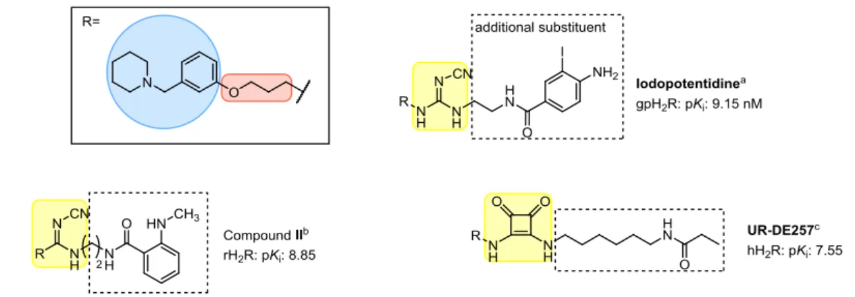
The aminopotentidine derivatives as well as fluorescent ligands (e.g. compound II) showed that within the piperidinomethylphenoxy-containing compounds additional substituents at the urea equivalent are well tolerated or provided additional H2R binding affinity (Figure 1.3).55-57 Up to date, iodoaminopotentidine, a piperidinomethylphenoxy-containing cyanoguanidine (Figure 1.3), which was also synthesized in a radiolabeled form ([125I]iodoaminopotentidine), showed the highest affinity.13 This radioligand was used to map the H2R densities in human and mammalian brain.12,13 Recently, a series of piperidinomethylphenoxyalkylamine-containing ligands, coupled with various polar groups (“urea equivalents”) such as cyanoguanidine, nitroethenediamine, amide or squaric amide moieties, and a terminal amino group, connected via a linker of different length, was developed by our group.35 The squaramides, which also tolerated propionylation at the terminal amino-group showed the highest affinities. UR-DE257, which showed a high affinity (pKi value: 7.55), was also synthesized in radiolabeled form ([3H]UR-DE257) and is frequently used in competition binding experiments.35,36
Figure 1.3. Structures of exemplary piperidinomethylphenoxy-containing ligands. a: Hirschfeld et al and Ruat et al13,57; b: Malan et al58; c: Baumeister et al35.
Whereas in the late 1970s to 1980s, the H2R antagonists (H2 blockers) revolutionized the treatment of peptic ulcers, former blockbuster drugs like cimetidine, ranitidine or famotidine are outdated. They were largely superseded by the more effective proton pump inhibitors (e.g.
omeprazole). Nevertheless, H2R antagonists are valuable molecular tools to study the H2R, especially it´s role in the brain, which is still far from being completely understood.
1.5 H
2R A
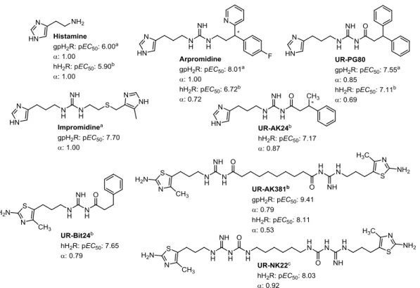
GONISTSThe early agonists for the H2R were derived from histamine and consististed of an imidazole pharmakophore coupled to a guanidine by a flexible linker (e.g. impromidine and arpromidine, see Figure 1.4).59,60 Arpromidine and related compounds showed up to 400 times potency of histamine at the spontaneously beating guinea pig right atrium, but the strongly basic guanidine moiety led to poor oral bioavailability and CNS penetration.59 The bioisosteric replacement of the guanidine (pKa ~13) with an acylguanidine (pKa ~8) resulted in ligands with either retained or even increased agonistic potency (e.g. UR-PG80 and UR-AK24, see Figure 1.4).31,32 Modification of these NG-acylated imidazolylpropylguanidines, which lacked subtype selectivity (H3R and H4R), to NG-acylated aminothiazolylpropylguanidines led to highly potent and selective H2R agonists.34 Surprisingly, the H2R potency was increased up to 4000-fold the potency of histamine by linking two acylguanidine moieties (e.g. UR-AK381, see Figure 1.4).33 The aminothiazole dimeric ligands are the most potent and selective H2R agonists known so far. Traditionally, dimeric (bivalent) ligands consist of two pharmacophoric moieties linked through a spacer and are designed to bridge two neighboring receptor protomers.61 Porthogese et al suggested a distance of about 22- 27 Å between the two orthosteric binding sites of a receptor dimer.62 Interestingly, the most active dimeric ligands have spacer of lengths insufficient to bridge the protomers of putative H2R dimers.33 The enormous gain in potency is speculated to result from an interaction with the orthosteric and an accessory binding site at the same protomer.33 Recently, it was shown that bioisosteric replacement of the acylguanidines with the more stable carbamoylguanidine led to dimeric ligands with retained potency and intrinsic activity (e.g. UR-NK22, see Figure 1.4).36 So far, there is no H2R agonist for therapeutic use on the market, but H2R agonists are valuable molecular tools to study the H2R. Nonetheless, there are numerous possible indications e.g. as positive inotropic vasodilators for the treatment of congestive heart failure or as differentiation- introducing agents for treatment of acute myeloid leukemia (AML). For the later, the endogenous
agonist histamine is used as an orphan drug in combination with interleukin 2. Histamine promotes the activation of T cells and natural killer cells by interleukin 2, which results in the killing of cancer cells.63 Given the effect of histamine is mediated via H2R, the application of highly selective H2R agonists might be beneficial in regard to potency and a reduction of adverse effects.
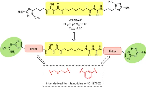
Recently, investigation of the dimeric NG-carbamoylated aminothiazolylpropylguanidines on human monocytes revealed a high H2R agonist potency, suggesting that this class of compounds is a promising starting point for the development of H2R agonists for the treatment of AML.36
Figure 1.4. Structures of selected H2R agonists. Agonism measured on aguinea pig right atrium32, bsteady-state GTPase assay31,33,34 or cGTPγS binding assay36.
1.6 R
ECEPTORL
IGANDB
INDINGA
SSAYS ANDL
ABELEDM
OLECULART
OOLS FORGPCR
SThe initial step in every signaling cascade, that causes a receptor-mediated biological response, is the binding of a ligand to the receptor. There are multiple ways to utilize receptor-ligand- interactions in research, e.g. as a tool for determining receptor distribution, for identification of receptor subtypes and for screening of new compounds.64
The classical approach for the determination of ligand affinity is the radioligand binding assay, which has been unchallenged for a long time regarding sensity and reproducibility.64,65 Radioligand binding experiments can be divided in three basic types: Saturation binding experiments are used to determine the affinity of the radioligand and the number of specific binding sites. In kinetic experiments, the rate constants of association and dissoviation of a radioligand can be determined. Competition binding experiments are widely used to identify unlabeled compounds, which bind to the receptor in question by displacement of a radioligand.
The major disadvantages include that radioligands are potentially hazardous to human health,
produce high costs in production and waste disposal, require special licences and laboratory equippment, and separation of bound from unbound ligand is necessary.
Today, new highly sensitive fluorescence and bioluminescence methods such as fluorescence polarization (FP),66 total internal reflection fluorescence (TIRF),67 fluorescence/bioluminescence resonance energy transfer (FRET/BRET),68 fluorescence recovery after photobleaching (FRAP),69 high content imaging70 and flow cytometry71 became promising alternatives. Like radioligands, fluorescent ligands can be used in the basic types of binding experiments. Several peptidic and non-peptidic fluorescent ligands were identified for GPCRs, including NPY,72,73 muscarinic74 and histamine55,56,75 receptors. In general, a fluorescent ligand comprises of a pharmacophore, a linker and the fluorophore. A major challenge in the development of small-molecule fluorescent ligands is to retain affinity, when a, compared to the ligand, bulky fluorophore is attached. In comparison, a radiolabel, especially tritium, does not alter the affinity of the ligand.
When selecting a radio- or fluorescent labeled ligand for binding experiments, several aspects have to be considered.65,76,77 Firstly, the ligand should be selective and bind with high affinity to the respective receptor. Secondly, high (radiochemical) purity and high specific activity (radioligand) or quantum yield (fluorescent ligand) is required. Thirdly, the labeled ligand should be chemically stable under assay conditions for at least the duration of the experiment performed. Furthermore, unspecific binding has to be considered, the choice of radio- or fluorescent label and, whether an agonist or an antagonist is desired as labeled ligand. Under unspecific binding all binding sites other than the receptor of interest are summarized. A competition binding assay, were only 50% of total radioligand binding is specific is considered adequate, 70% is good and 90% is excellent.76 Tritium is often considered as the radioisotope of choice. Compared to 125I or the occasionally used 32/33P or 35S, tritium has a longer half-life (14-87 days vs. 12.3 years) and the tritiated compounds are more convenient in handeling with respect to safety precautions.76 When choosing a fluorophore, excitation and emission wavelengths, stoke shifts and quantum yields have to be considered. Generally, red-emitting (λem: > 600 nm) fluorophores with a long stoke shift and a high quantum yield are preferred. 77 Agonists label only an active conformation of the receptor and therefore, only a fraction of the total active receptor population.78 By contrast, antagonists bind to all receptor states with the same affinity according to the classical model described above.78
1.7 R
EFERENCES1. Fredriksson, R.; Lagerstrom, M. C.; Lundin, L. G.; Schioth, H. B. The G-protein-coupled receptors in the human genome form five main families. Phylogenetic analysis, paralogon groups, and fingerprints. Molecular pharmacology 2003, 63, 1256-1272.
2. Luttrell, L. M. Reviews in molecular biology and biotechnology: transmembrane signaling by G protein-coupled receptors. Mol Biotechnol 2008, 39, 239-264.
3. Wise, A.; Gearing, K.; Rees, S. Target validation of G-protein coupled receptors. Drug Discov Today 2002, 7, 235-246.
4. Stevens, R. C.; Cherezov, V.; Katritch, V.; Abagyan, R.; Kuhn, P.; Rosen, H.; Wuthrich, K.
The GPCR Network: a large-scale collaboration to determine human GPCR structure and function.
Nat Rev Drug Discov 2013, 12, 25-34.
5. Pierce, K. L.; Premont, R. T.; Lefkowitz, R. J. Seven-transmembrane receptors. Nat Rev Mol Cell Biol 2002, 3, 639-650.
6. Vassilatis, D. K.; Hohmann, J. G.; Zeng, H.; Li, F.; Ranchalis, J. E.; Mortrud, M. T.; Brown, A.;
Rodriguez, S. S.; Weller, J. R.; Wright, A. C.; Bergmann, J. E.; Gaitanaris, G. A. The G protein- coupled receptor repertoires of human and mouse. Proc Natl Acad Sci U S A 2003, 100, 4903- 4908.
7. Ash, A. S.; Schild, H. O. Receptors mediating some actions of histamine. Br J Pharmacol Chemother 1966, 27, 427-439.
8. Black, J. W.; Duncan, W. A.; Durant, C. J.; Ganellin, C. R.; Parsons, E. M. Definition and antagonism of histamine H 2 -receptors. Nature 1972, 236, 385-390.
9. Arrang, J. M.; Garbarg, M.; Schwartz, J. C. Auto-inhibition of brain histamine release mediated by a novel class (H3) of histamine receptor. Nature 1983, 302, 832-837.
10. Oda, T.; Morikawa, N.; Saito, Y.; Masuho, Y.; Matsumoto, S. Molecular cloning and characterization of a novel type of histamine receptor preferentially expressed in leukocytes. J Biol Chem 2000, 275, 36781-36786.
11. Domschke, W.; Domschke, S.; Classen, M.; Demling, L. Histamine and cyclic 3',5'-AMP in gastric acid secretion. Nature 1973, 241, 454-455.
12. Traiffort, E.; Pollard, H.; Moreau, J.; Ruat, M.; Schwartz, J. C.; Martinez-Mir, M. I.;
Palacios, J. M. Pharmacological characterization and autoradiographic localization of histamine H2 receptors in human brain identified with [125I]iodoaminopotentidine. Journal of neurochemistry 1992, 59, 290-299.
13. Ruat, M.; Traiffort, E.; Bouthenet, M. L.; Schwartz, J. C.; Hirschfeld, J.; Buschauer, A.;
Schunack, W. Reversible and Irreversible Labeling and Autoradiographic Localization of the Cerebral Histamine H-2-Receptor Using [I-125] Iodinated Probes. P Natl Acad Sci USA 1990, 87, 1658-1662.
14. Reher, T. M.; Brunskole, I.; Neumann, D.; Seifert, R. Evidence for ligand-specific conformations of the histamine H(2)-receptor in human eosinophils and neutrophils. Biochem Pharmacol 2012, 84, 1174-1185.
15. Mitznegg, P.; Schubert, E.; Fuchs, W. Relations between the effects of histamine, pheniramin and metiamide on spontaneous motility and the formation of cyclic AMP in the isolated rat uterus. Naunyn Schmiedebergs Arch Pharmacol 1975, 287, 321-327.
16. Reinhardt, D.; Schmidt, U.; Brodde, O. E.; Schumann, H. J. H1 - and H2-receptor mediated responses to histamine on contractility and cyclic AMP of atrial and papillary muscles from guinea-pig hearts. Agents Actions 1977, 7, 1-12.
17. Kelley, M. T.; Burckstummer, T.; Wenzel-Seifert, K.; Dove, S.; Buschauer, A.; Seifert, R.
Distinct interaction of human and guinea pig histamine H2-receptor with guanidine-type agonists.
Molecular pharmacology 2001, 60, 1210-1225.
18. Preuss, H.; Ghorai, P.; Kraus, A.; Dove, S.; Buschauer, A.; Seifert, R. Constitutive activity and ligand selectivity of human, guinea pig, rat, and canine histamine H2 receptors. The Journal of pharmacology and experimental therapeutics 2007, 321, 983-995.
19. Hilger, D.; Masureel, M.; Kobilka, B. K. Structure and dynamics of GPCR signaling complexes. Nat Struct Mol Biol 2018, 25, 4-12.
20. Downes, G. B.; Gautam, N. The G protein subunit gene families. Genomics 1999, 62, 544- 552.
21. Simon, M. I.; Strathmann, M. P.; Gautam, N. Diversity of G proteins in signal transduction.
Science 1991, 252, 802-808.
22. Bristow, M. R.; Cubicciotti, R.; Ginsburg, R.; Stinson, E. B.; Johnson, C. Histamine- mediated adenylate cyclase stimulation in human myocardium. Molecular pharmacology 1982, 21, 671-679.
23. Leurs, R.; Smit, M. J.; Menge, W. M.; Timmerman, H. Pharmacological characterization of the human histamine H2 receptor stably expressed in Chinese hamster ovary cells. British journal of pharmacology 1994, 112, 847-854.
24. Hamm, H. E. The many faces of G protein signaling. J Biol Chem 1998, 273, 669-672.
25. Lieb, S.; Littmann, T.; Plank, N.; Felixberger, J.; Tanaka, M.; Schafer, T.; Krief, S.; Elz, S.;
Friedland, K.; Bernhardt, G.; Wegener, J.; Ozawa, T.; Buschauer, A. Label-free versus conventional cellular assays: Functional investigations on the human histamine H1 receptor. Pharmacological research 2016, 114, 13-26.
26. Rasmussen, S. G.; DeVree, B. T.; Zou, Y.; Kruse, A. C.; Chung, K. Y.; Kobilka, T. S.; Thian, F.
S.; Chae, P. S.; Pardon, E.; Calinski, D.; Mathiesen, J. M.; Shah, S. T.; Lyons, J. A.; Caffrey, M.;
Gellman, S. H.; Steyaert, J.; Skiniotis, G.; Weis, W. I.; Sunahara, R. K.; Kobilka, B. K. Crystal structure of the beta2 adrenergic receptor-Gs protein complex. Nature 2011, 477, 549-555.
27. Harrison, C.; Traynor, J. R. The [35S]GTPgammaS binding assay: approaches and applications in pharmacology. Life sciences 2003, 74, 489-508.
28. Houston, C.; Wenzel-Seifert, K.; Burckstummer, T.; Seifert, R. The human histamine H2- receptor couples more efficiently to Sf9 insect cell Gs-proteins than to insect cell Gq-proteins:
limitations of Sf9 cells for the analysis of receptor/Gq-protein coupling. Journal of neurochemistry 2002, 80, 678-696.
29. Milligan, G. Insights into ligand pharmacology using receptor-G-protein fusion proteins.
Trends in pharmacological sciences 2000, 21, 24-28.
30. Seifert, R.; Wenzel-Seifert, K.; Kobilka, B. K. GPCR-Galpha fusion proteins: molecular analysis of receptor-G-protein coupling. Trends in pharmacological sciences 1999, 20, 383-389.
31. Xie, S. X.; Ghorai, P.; Ye, Q. Z.; Buschauer, A.; Seifert, R. Probing ligand-specific histamine H1- and H2-receptor conformations with NG-acylated Imidazolylpropylguanidines. The Journal of pharmacology and experimental therapeutics 2006, 317, 139-146.
32. Ghorai, P.; Kraus, A.; Keller, M.; Gotte, C.; Igel, P.; Schneider, E.; Schnell, D.; Bernhardt, G.;
Dove, S.; Zabel, M.; Elz, S.; Seifert, R.; Buschauer, A. Acylguanidines as bioisosteres of guanidines:
NG-acylated imidazolylpropylguanidines, a new class of histamine H2 receptor agonists. Journal of medicinal chemistry 2008, 51, 7193-7204.
33. Birnkammer, T.; Spickenreither, A.; Brunskole, I.; Lopuch, M.; Kagermeier, N.; Bernhardt, G.; Dove, S.; Seifert, R.; Elz, S.; Buschauer, A. The Bivalent Ligand Approach Leads to Highly Potent and Selective Acylguanidine-Type Histamine H-2 Receptor Agonists. Journal of medicinal chemistry 2012, 55, 1147-1160.
34. Kraus, A.; Ghorai, P.; Birnkammer, T.; Schnell, D.; Elz, S.; Seifert, R.; Dove, S.; Bernhardt, G.; Buschauer, A. N-G-Acylated Aminothiazolylpropylguanidines as Potent and Selective Histamine H-2 Receptor Agonists. ChemMedChem 2009, 4, 232-240.
35. Baumeister, P.; Erdmann, D.; Biselli, S.; Kagermeier, N.; Elz, S.; Bernhardt, G.; Buschauer, A. [3H]UR-DE257: Development of a Tritium-Labeled Squaramide-Type Selective Histamine H2 Receptor Antagonist. ChemMedChem 2015, 10, 83-93.
36. Kagermeier, N.; Werner, K.; Keller, M.; Baumeister, P.; Bernhardt, G.; Seifert, R.;
Buschauer, A. Dimeric carbamoylguanidine-type histamine H receptor ligands: A new class of potent and selective agonists. Bioorganic & medicinal chemistry 2015.
37. Gurevich, V. V.; Gurevich, E. V. Overview of different mechanisms of arrestin-mediated signaling. Curr Protoc Pharmacol 2014, 67, Unit 2 10 11-19.
38. Peterson, Y. K.; Luttrell, L. M. The Diverse Roles of Arrestin Scaffolds in G Protein-Coupled Receptor Signaling. Pharmacological reviews 2017, 69, 256-297.
39. Gurevich, V. V.; Gurevich, E. V. Extensive shape shifting underlies functional versatility of arrestins. Curr Opin Cell Biol 2014, 27, 1-9.
40. Shenoy, S. K.; Lefkowitz, R. J. beta-Arrestin-mediated receptor trafficking and signal transduction. Trends in pharmacological sciences 2011, 32, 521-533.
41. Perry, S. J.; Baillie, G. S.; Kohout, T. A.; McPhee, I.; Magiera, M. M.; Ang, K. L.; Miller, W.
E.; McLean, A. J.; Conti, M.; Houslay, M. D.; Lefkowitz, R. J. Targeting of cyclic AMP degradation to beta 2-adrenergic receptors by beta-arrestins. Science 2002, 298, 834-836.
42. Nelson, C. D.; Perry, S. J.; Regier, D. S.; Prescott, S. M.; Topham, M. K.; Lefkowitz, R. J.
Targeting of diacylglycerol degradation to M1 muscarinic receptors by beta-arrestins. Science 2007, 315, 663-666.
43. Weiss, J. M.; Morgan, P. H.; Lutz, M. W.; Kenakin, T. P. The cubic ternary complex receptor-occupancy model. III. resurrecting efficacy. J Theor Biol 1996, 181, 381-397.
44. Weiss, J. M.; Morgan, P. H.; Lutz, M. W.; Kenakin, T. P. The Cubic Ternary Complex Receptor–Occupancy Model I. Model Description. Journal of Theoretical Biology 1996, 178, 151- 167.
45. Weiss, J. M.; Morgan, P. H.; Lutz, M. W.; Kenakin, T. P. The Cubic Ternary Complex Receptor–Occupancy Model II. Understanding Apparent Affinity. Journal of Theoretical Biology 1996, 178, 169-182.
46. Seifert, R.; Wenzel-Seifert, K. Constitutive activity of G-protein-coupled receptors: cause of disease and common property of wild-type receptors. Naunyn Schmiedebergs Arch Pharmacol 2002, 366, 381-416.
47. Perez, D. M.; Karnik, S. S. Multiple signaling states of G-protein-coupled receptors.
Pharmacological reviews 2005, 57, 147-161.
48. Kenakin, T. Ligand-selective receptor conformations revisited: the promise and the problem. Trends in pharmacological sciences 2003, 24, 346-354.
49. Urban, J. D.; Clarke, W. P.; von Zastrow, M.; Nichols, D. E.; Kobilka, B.; Weinstein, H.;
Javitch, J. A.; Roth, B. L.; Christopoulos, A.; Sexton, P. M.; Miller, K. J.; Spedding, M.; Mailman, R.
B. Functional selectivity and classical concepts of quantitative pharmacology. The Journal of pharmacology and experimental therapeutics 2007, 320, 1-13.
50. Jarpe, M. B.; Knall, C.; Mitchell, F. M.; Buhl, A. M.; Duzic, E.; Johnson, G. L. [D-Arg(1),D- Phe(5),D-Trp(7,9),Leu(11)]substance P acts as a biased agonist toward neuropeptide and chemokine receptors. Journal of Biological Chemistry 1998, 273, 3097-3104.
51. Roth, B. L. Modulation of phosphatidylinositol-4,5-bisphosphate hydrolysis in rat aorta by guanine nucleotides, calcium and magnesium. Life sciences 1987, 41, 629-634.
52. Felixberger, J. Luciferase complementation for the determination of arrestin recruitment:
Investigations at histamine and NPY receptors. University of Regensburg, Regensburg, 2014.
https://epub.uni-regensburg.de/31292/.
53. Misawa, N.; Kafi, A. K.; Hattori, M.; Miura, K.; Masuda, K.; Ozawa, T. Rapid and high- sensitivity cell-based assays of protein-protein interactions using split click beetle luciferase complementation: an approach to the study of G-protein-coupled receptors. Anal Chem 2010, 82, 2552-2560.
54. Williams, M.; Glennon, R. A.; Timmermans, P. B. M. W. M. (ed.). Receptor Pharmacology and Function. Marcel Dekker Inc: 1989; Vol. 13, p 808.
55. Li, L.; Kracht, J.; Peng, S.; Bernhardt, G.; Elz, S.; Buschauer, A. Synthesis and pharmacological activity of fluorescent histamine H2 receptor antagonists related to potentidine.
Bioorganic & medicinal chemistry letters 2003, 13, 1717-1720.
56. Xie, S. X.; Petrache, G.; Schneider, E.; Ye, Q. Z.; Bernhardt, G.; Seifert, R.; Buschauer, A.
Synthesis and pharmacological characterization of novel fluorescent histamine H2-receptor ligands derived from aminopotentidine. Bioorganic & medicinal chemistry letters 2006, 16, 3886- 3890.
57. Hirschfeld, J.; Buschauer, A.; Elz, S.; Schunack, W.; Ruat, M.; Traiffort, E.; Schwartz, J. C.
Iodoaminopotentidine and Related-Compounds - a New Class of Ligands with High-Affinity and Selectivity for the Histamine-H2-Receptor. Journal of medicinal chemistry 1992, 35, 2231-2238.
58. Malan, S. F.; van Marle, A.; Menge, W. M.; Zuliani, V.; Hoffman, M.; Timmerman, H.;
Leurs, R. Fluorescent ligands for the histamine H2 receptor: synthesis and preliminary characterization. Bioorganic & medicinal chemistry 2004, 12, 6495-6503.
59. Buschauer, A. Synthesis and in vitro pharmacology of arpromidine and related phenyl(pyridylalkyl)guanidines, a potential new class of positive inotropic drugs. Journal of medicinal chemistry 1989, 32, 1963-1970.
60. Durant, G. J.; Duncan, W. A.; Ganellin, C. R.; Parsons, M. E.; Blakemore, R. C.; Rasmussen, A. C. Impromidine (SK&F 92676) is a very potent and specific agonist for histamine H2 receptors.
Nature 1978, 276, 403-405.
61. Portoghese, P. S. Bivalent Ligands and the Message-Address Concept in the Design of Selective Opioid Receptor Antagonists. Trends in pharmacological sciences 1989, 10, 230-235.
62. Portoghese, P. S. From models to molecules: opioid receptor dimers, bivalent ligands, and selective opioid receptor probes. Journal of medicinal chemistry 2001, 44, 2259-2269.
63. Yang, L. P.; Perry, C. M. Spotlight on histamine dihydrochloride in acute myeloid leukaemia. Drugs Aging 2011, 28, 325-329.
64. Kenakin, T. A Pharmacology Primer. 3 ed.; Elsevier Academic Press: Burlington, 2009; p 389.
65. Crevat-Pisano, P.; Hariton, C.; Rolland, P. H.; Cano, J. P. Fundamental aspects of radioreceptor assays. J Pharm Biomed Anal 1986, 4, 697-716.
66. Jameson, D. M.; Ross, J. A. Fluorescence polarization/anisotropy in diagnostics and imaging. Chem Rev 2010, 110, 2685-2708.
67. Fish, K. N. Total internal reflection fluorescence (TIRF) microscopy. Curr Protoc Cytom 2009, Chapter 12, Unit12 18.
68. Lohse, M. J.; Nuber, S.; Hoffmann, C. Fluorescence/bioluminescence resonance energy transfer techniques to study G-protein-coupled receptor activation and signaling.
Pharmacological reviews 2012, 64, 299-336.
69. Deschout, H.; Raemdonck, K.; Demeester, J.; De Smedt, S. C.; Braeckmans, K. FRAP in pharmaceutical research: practical guidelines and applications in drug delivery. Pharm Res 2014, 31, 255-270.
70. Zanella, F.; Lorens, J. B.; Link, W. High content screening: seeing is believing. Trends Biotechnol 2010, 28, 237-245.
71. Black, C. B.; Duensing, T. D.; Trinkle, L. S.; Dunlay, R. T. Cell-based screening using high- throughput flow cytometry. Assay Drug Dev Technol 2011, 9, 13-20.
72. Keller, M.; Erdmann, D.; Pop, N.; Pluym, N.; Teng, S.; Bernhardt, G.; Buschauer, A. Red- fluorescent argininamide-type NPY Y1 receptor antagonists as pharmacological tools. Bioorganic
& medicinal chemistry 2011, 19, 2859-2878.
73. Dukorn, S.; Littmann, T.; Keller, M.; Kuhn, K.; Cabrele, C.; Baumeister, P.; Bernhardt, G.;
Buschauer, A. Fluorescence- and Radiolabeling of [Lys4,Nle17,30]hPP Yields Molecular Tools for the NPY Y4 Receptor. Bioconjugate chemistry 2017, 28, 1291-1304.
74. Bonnet, D.; Ilien, B.; Galzi, J. L.; Riche, S.; Antheaune, C.; Hibert, M. A rapid and versatile method to label receptor ligands using "click" chemistry: Validation with the muscarinic M1 antagonist pirenzepine. Bioconjugate chemistry 2006, 17, 1618-1623.
75. Li, L.; Kracht, J.; Peng, S.; Bernhardt, G.; Buschauer, A. Synthesis and pharmacological activity of fluorescent histamine H1 receptor antagonists related to mepyramine. Bioorganic &
medicinal chemistry letters 2003, 13, 1245-1248.
76. Keen, M. The problems and pitfalls of radioligand binding. Methods in molecular biology 1995, 41, 1-16.
77. Kuder, K. J.; Kiec-Kononowicz, K. Fluorescent GPCR Ligands as New Tools in Pharmacology-Update, Years 2008-Early 2014. Curr Med Chem 2014, 21, 3962-3975.
78. Leff, P. The two-state model of receptor activation. Trends in pharmacological sciences 1995, 16, 89-97.

![Figure 3.4. Displacement of the respective radioligand by amine precursor 3.22 (A) or ligand 3.25 (B) from membrane preparations of Sf9 insect cells co-expressing the hH 1 R-G sαS fusion protein and RGS4 (radioligand: [ 3 H]mepyramine, c = 5 nM, K d =](https://thumb-eu.123doks.com/thumbv2/1library_info/3735722.1509013/39.892.132.791.110.687/displacement-respective-radioligand-precursor-preparations-expressing-radioligand-mepyramine.webp)

![Figure 3.7. RP-HPLC analysis of the radioligand [ 3 H]3.25 (A) before and (B) after purification by RP-HPLC (conditions, see Experimental Section)](https://thumb-eu.123doks.com/thumbv2/1library_info/3735722.1509013/44.892.111.776.109.321/figure-hplc-analysis-radioligand-purification-conditions-experimental-section.webp)
![Figure 3.9. Association (A) and dissociation (B) kinetics of [ 3 H]3.25 determined at membrane preparations from Sf9 insect cells expressing the hH 2 R-G sαS fusion protein at room temperature](https://thumb-eu.123doks.com/thumbv2/1library_info/3735722.1509013/46.892.113.761.109.389/association-dissociation-kinetics-determined-membrane-preparations-expressing-temperature.webp)
![Figure 4.1. Displacement of the radioligand [ 3 H]UR-DE257 (c = 20 nM, K d = 12.2 nM) by (A) compounds 3.35-3.38 and (B) compounds 4.44, 4.46, 4.47, 4.50 and antagonism of (C) compounds 4.35, 4.37, 4.38 and (D) compounds 4.40, 4.41,4.47](https://thumb-eu.123doks.com/thumbv2/1library_info/3735722.1509013/81.892.138.783.109.686/figure-displacement-radioligand-compounds-compounds-antagonism-compounds-compounds.webp)
![Figure 5.2. Chemical structures of the H 2 R radioligand [ 3 H]UR-DE257, the parent compound BMY 2536, as well as of the pyridinium labeled H 2 R antagonist UR-DE229 and the cyanine labeled H 2 R antagonist UR-DE56](https://thumb-eu.123doks.com/thumbv2/1library_info/3735722.1509013/109.892.170.746.392.583/figure-chemical-structures-radioligand-compound-pyridinium-antagonist-antagonist.webp)
