Pharmacological tools for the NPY receptors: [ 35 S]GTPγS binding assays,
luciferase gene reporter assays and labeled peptides
Dissertation
zur Erlangung des Doktorgrades der Naturwissenschaften (Dr. rer. nat.) an der Fakultät für Chemie und Pharmazie
der Universität Regensburg
vorgelegt von Stefanie Dukorn
aus Straubing
2017
Die vorliegende Arbeit entstand in der Zeit von Januar 2013 bis März 2017 unter der Leitung von Herrn Prof. Dr. Armin Buschauer am Institut für Pharmazie der Naturwissenschaftlichen Fakultät IV - Chemie und Pharmazie - der Universität Regensburg.
Das Promotionsgesuch wurde eingereicht im März 2017.
Tag der mündlichen Prüfung: 21. April 2017 Prüfungsausschuss:
Prof. Dr. S. Elz (Vorsitzender)
Prof. Dr. A. Buschauer (Erstgutachter)
Prof. Dr. G. Bernhardt (Zweitgutachter)
Prof. Dr. J. Wegener (Drittprüfer)
Danksagungen
An dieser Stelle möchte ich mich herzlich bedanken bei:
Herrn Prof. Dr. Armin Buschauer, dass ich die Möglichkeit für dieses vielseitige Projekt erhalten habe, seine wissenschaftlichen Anregungen sowie Förderungen und seine konstruktive Kritik bei der Durchsicht der Arbeit,
Herrn Prof. Dr. Günther Bernhardt für seine Hilfsbereitschaft und sein Interesse am Fortschritt der Arbeit, sein Fachwissen und die Durchsicht der Arbeit,
Herrn Prof. Dr. Oliver Zerbe und Dr. Jacopo Marino für die Kooperation zu Beginn meiner Arbeit, mit Unterstützung von Dr. Oliver Biehlmaier der Imaging Core Facility in Basel, Herrn Dr. Paul Baumeister, der zu Beginn und im weiteren Verlauf meiner Doktorarbeit immer ein offenes Ohr für jegliche Angelegenheiten hatte und entsprechend seinen fachlichen Ratschlägen,
Herrn Dr. Max Keller für die wertvollen fachlichen Tipps, die hervorragende Zusammenarbeit, das Bereitstellen von verschiedenen Substanzen, sowie für die fachlichen Diskussionen und Ratschläge,
Herrn Dr. Kilian Kuhn für die sehr gute Zusammenarbeit, NPY hat uns beide während dieser Zeit begleitet und entsprechend beschäftigt,
Herrn Timo Littmann für die Durchführung der Arrestin Assays, Frau Prof. Dr. Chiara Cabrele für die Bereitstellung der Peptide,
Frau Dr. Birgit Kraus für die Unterstützung bei der Fluoreszenzmikroskopie, Frau Dr. Andrea Bleckmann für die Bereitstellung des Konfokalmikroskops,
Frau Elvira Schreiber für die Durchführung der zahlreichen pharmakologischen Bestimmungen am FACS und Durchführung von Calcium Assays,
Frau Brigitte Wenzl für die Unterstützung v.a. zu Beginn meiner praktischen Laborarbeit und für die gute Zusammenarbeit während der Praktikumsbetreuung,
Frau Susanne Bollwein und Frau Dita Fritsch für die Durchführung von HPLC-Läufen und Experimenten,
Frau Maria Beer- Krön für ihre Unterstützung und der Durchführung von [
35S]GTPγS Assays und Radioligand-Bindungsexperimenten,
meiner Praktikantin Lisa Schindler für die Durchführung von Calcium Assays,
Herrn Josef Kiermaier und Herrn Wolfgang Söllner für die Anfertigung der MS- Untersuchungen,
the office 14.2.08 for the great collegiality, especially Timo Littmann, Dr. Nicole Plank, Andrea Pegoli and Frauke Antoni,
meinem ehemaligen Kollegen Herrn Dr. Uwe Nordemann für die Betreuung während meines Forschungspraktikums,
Frau Karin Reindl, Frau Uta Hasselmann, Frau Silvia Heinrich und Herrn Peter Richthammer für die Unterstützung bei technischen und organisatorischen Angelegenheiten,
allen Mitgliedern des Lehrstuhls für ihre Kollegialität, Hilfsbereitschaft und ein gutes Arbeitsklima,
meinen Freunden für die gemeinsame Zeit und das Interesse am Fortgang meiner Arbeit,
„Freundschaft fließt aus vielen Quellen, am reinsten aus Vertrauen und Respekt.“
Zuletzt all denjenigen, die mehr als nur Dank verdienen:
Meiner Familie. Ihnen ist die vorliegende Arbeit gewidmet.
Publications and Poster Presentations
Publications (published results prior to the submission of the thesis):
Kuhn K., Ertl T., Dukorn S., Keller M., Bernhardt G., Reiser O., Buschauer A., High Affinity Agonists of the Neuropeptide Y (NPY) Y
4Receptor Derived from the C-terminal Pentapeptide of Human Pancreatic Polypeptide (hPP): Synthesis, Stereochemical Discrimination and Radiolabeling, Journal of Medicinal Chemistry 2016, 59 (13), S. 6045-6058
Keller M., Weiss S., Hutzler C., Kuhn K., Mollereau C., Dukorn S., Schindler L., Bernhardt G., König B., Buschauer A., N(omega)-carbamoylation of the argininamide moiety: an avenue to insurmountable NPY Y
1receptor antagonists and a radiolabeled selective high affinity molecular tool ([
3H]UR-MK299) with extended residence time, Journal of Medicinal Chemistry 2015, 58 (22), S. 8834-8849
Poster Presentations:
Dukorn, S., Keller, M., Bernhardt, G., Buschauer, A., Investigations on the effect of sodium on hY
4R binding using fluorescent and radiolabelled [Lys
4,Nle
17,30]hPP as new molecular tools, 8th Summer School Medicinal Chemistry, Regensburg, September 2016.
Dukorn S., Keller M., Kuhn K., Cabrele C., Bernhardt G., Buschauer A., Molecular tools for the NPY Y
4receptor: Fluorescence- and radiolabelled [Lys
4,Nle
17,30]hPP, 11
thInternational NPY-PYY-PP Meeting, Leipzig, August 2015.
Dukorn S., Marino J., Zerbe O., Bernhardt G. and Buschauer A., [
3H]Propionyl-K
4hPP: A novel radioligand for the neuropeptide Y (NPY) Y
4receptor, 7th Summer School Medicinal Chemistry, Regensburg, September 2014.
Dukorn, S., Marino, J., Zerbe, O., Bernhardt, G., Buschauer, A., A valuable tool for the NPY
Y
4receptor: [
3H]Propionyl - K
4hPP, Annual Meeting “Frontiers in Medicinal Chemistry”,
Tübingen, March 2014.
I
Contents
Abbreviations ... IV
1 Introduction ... 1
1.1 Neuropeptide Y ... 2
1.2 NPY receptors and their ligands ... 2
1.2.1 The NPY Y
1, Y
2and Y
5receptors and their ligands ... 4
1.2.2 The NPY Y
4receptor and its ligands ... 8
1.3 Sodium sensitivity of the NPY receptors ... 10
1.4 Tritium- and fluorescence-labeled NPY receptor ligands ... 11
1.4.1 Tritiated radioligands ... 11
1.4.2 Fluorescent ligands for cellular investigations ... 11
1.5 References ... 12
2 Scope and objectives ... 19
3 Development of a [ 35 S]GTPγS binding assay for hY 2 and hY 4 receptors in Sf9 membranes ... 21
3.1 Introduction ... 22
3.2 Material and methods ... 23
3.2.1 Materials ... 23
3.2.2 Cell culture, generation of recombinant baculoviruses, Sf9 cell membrane preparation ... 24
3.2.3 [
35S]GTPγS binding assay at the hY
2R and the hY
4R ... 24
3.2.4 Radioligand binding at hY
4R Sf9 membranes ... 25
3.3 Results and discussion ... 25
3.3.1 [
35S]GTPγS binding assay at the hY
2R ... 25
3.3.2 [
35S]GTPγS binding assay at the hY
4R ... 27
3.4 Summary and conclusion ... 31
3.5 References ... 31
4 Development of luminescence based reporter gene assays for the human neuropeptide Y 2 and Y 4 receptor ... 33
4.1 Introduction ... 34
4.2 Materials and methods ... 36
4.2.1 Stable transfection of HEK293T-hY
2-CRE Luc and HEK293T-hY
4-CRE Luc cells with the pGL4.29[luc2P/CRE/Hygro] vector ... 36
4.2.2 Cell culture ... 37
4.2.3 Saturation binding on HEK293T-CRE-Luc hY
2R and HEK293T-CRE-Luc
hY
4R cells... 37
II
4.2.4 Luciferase reporter gene assay ... 38
4.2.4.1 Preparation of stock solutions and dilution series ... 38
4.2.4.2 Preparation of the cells and determination of bioluminescence ... 38
4.2.4.3 Effect of the solvent on luciferase activity ... 39
4.2.4.4 Determination of the optimal forskolin concentration ... 39
4.2.4.5 Effects of different forskolin concentrations on the concentration-response curves of pNPY and [Lys
4, Nle
17,30]hPP ... 39
4.2.4.6 Effect of bacitracin on the stability of pNPY and [Lys
4, Nle
17,30]hPP ... 40
4.2.4.7 Monitoring the time course of luciferase expression ... 40
4.2.4.8 Control experiments using HEK293-CRE-Luc cells devoid of hY
2R and hY
4R ... 40
4.3 Results and discussion ... 40
4.3.1 Reporter gene assay for the human neuropeptide Y
2receptor ... 40
4.3.1.1 Saturation binding assay using HEK293T-hY
2-CRE Luc cells ... 40
4.3.1.2 Effects of the solvent on the luciferase activity ... 41
4.3.1.3 Optimization of stimulation with forskolin ... 41
4.3.1.4 Optimization of the period of incubation ... 42
4.3.1.5 Effects of different forskolin concentrations on pNPY concentration-response curves ... 43
4.3.1.6 Effect of bacitracin on the stability of pNPY ... 43
4.3.1.7 Control experiments on cells devoid of the hY
2R ... 44
4.3.1.8 Functional activities of selected ligands at the hY
2R ... 44
4.3.2 Reporter gene assay for the human neuropeptide Y
4receptor ... 46
4.3.2.1 Saturation binding assay using HEK293T-hY
4-CRE Luc cells ... 46
4.3.2.2 Effect of the solvent on the luciferase activity ... 47
4.3.2.3 Optimization of stimulation with forskolin ... 47
4.3.2.4 Optimization of the incubation time ... 48
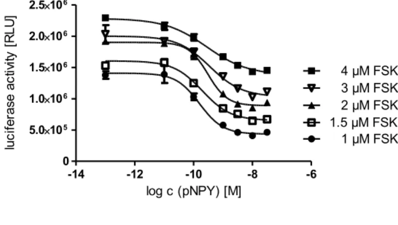
4.3.2.5 Effects of different forskolin concentrations on the concentration-response curve of [Lys
4, Nle
17,30]hPP ... 48
4.3.2.6 Effect of bacitracin on the stability of [Lys
4, Nle
17,30]hPP ... 49
4.3.2.7 Control experiments on cells devoid of the hY
4R ... 49
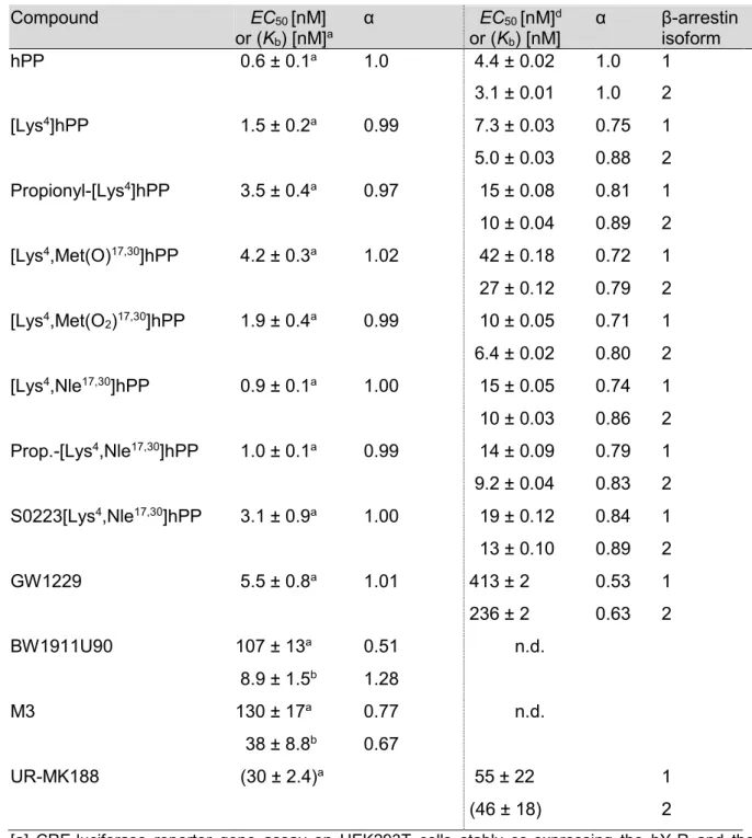
4.3.2.8 Functional activities of selected ligands at the hY
4R ... 50
4.4 Summary and conclusion ... 53
4.5 References ... 53
5 Fluorescence- and radio-labeling of [Lys 4 ,Nle 17,30 ]hPP yields molecular tools for the NPY Y 4 receptor ... 57
5.1 Introduction ... 58
5.2 Materials and methods ... 60
5.2.1 General experimental conditions ... 60
5.2.2 Chemistry: experimental protocols and analytical data ... 62
5.2.3 Fluorescence spectroscopy and determination of quantum yields ... 64
5.2.4 Investigation of the chemical stability of the peptides in buffer ... 64
5.2.5 Cell culture ... 64
5.2.6 Buffers used in binding and functional experiments ... 65
5.2.7 Radioligand binding assay... 65
5.2.8 Flow cytometric binding assay at the Y
4R ... 68
5.2.9 Luciferase assay ... 68
5.2.10 Arrestin recruitment assay ... 68
5.2.11 Confocal microscopy ... 69
5.2.12 Data analysis... 69
III
5.3 Results and discussion ... 70
5.3.1 Synthesis of the tritiated and fluorescently labeled peptides ... 70
5.3.2 Functional studies at the hY
4R ... 72
5.3.3 Y
4R binding of the radiolabeled and fluorescence labeled compounds 8, 12 and 13 ... 73
5.3.4 Confocal microscopy of CHO hY
4R cells incubated with the fluorescent ligand 13 ... 77
5.3.5 Y
4R competition binding ... 78
5.3.6 NPY receptor subtype selectivity ... 80
5.3.7 Effect of Na
+and osmolarity on the binding of [
3H]propionyl-pNPY to the Y
1, Y
2and Y
5R and binding of [³H]8, [³H]12 and 13 to the Y
5R ... 80
5.4 Conclusions ... 82
5.5 Supporting Information ... 83
5.5.1 Synthesis of the ligands [
3H]propionyl-pNPY, 8 and [
3H]8... 83
5.5.2 Synthesis of [Lys
4,Nle
17,30]hPP (11) ... 85
5.5.3 Purity, identity and long-term stability of [³H]8 determined by HPLC ... 86
5.5.4 Chemical stability of the peptides hPP and 7-11 ... 87
5.5.5 Fluorescence spectra and quantum yields of peptide 13 ... 90
5.5.6 Osmolality of the buffers used in NPY receptor binding studies ... 91
5.5.7 Saturation binding with [³H]8 at the hY
4R ... 91
5.5.8 Kinetic experiments with [³H]8 at the hY
4R ... 91
5.5.9 Saturation binding with [³H]12 at the hY
4R... 92
5.5.10 Binding characteristics of the fluorescent ligand 13 in buffer I containing glycine at the hY
4R ... 93
5.5.11 Morphology and volumes of CHO-hY
4R cells under hypotonic and isotonic conditions ... 94
5.5.12 Scattergrams of CHO-hY
4R cells in the presence of 13 in different buffers ... 95
5.5.13 Competition binding with [³H]8 at the hY
4R ... 96
5.5.14 Saturation binding with [³H]propionyl-pNPY at the hY
1R, hY
2R, and hY
5R ... 96
5.5.15 Saturation binding with [³H]8 at the hY
5R ... 98
5.5.16 Saturation binding with [³H]12 at the hY
5R... 98
5.5.17 Saturation binding with 13 at the hY
5R ... 98
5.6 References ... 99
6 Summary ...103
7 Appendix ...107
Eidesstattliche Erklärung ... 109
IV
Abbreviations
α intrinsic activity or selectivity factor
AC adenylyl cyclase
Ala alanine
AMP adenosine monophosphate
aq. aqueous
Arg arginine
Asp aspartate (-acid) a
sspecific activity
atm atmosphere
ATP adenosine 5’-triphosphate a
vactivity concentration
B
maxmaximum number of binding sites
Bq becquerel
BSA bovine serum albumin
c concentration
cAMP cyclic 3’,5’-adenosine monophosphate calcd. calculated
[Ca
2+]
iintracellular calcium ion concentration
cDNA complementary DNA
CHO chinese hamster ovary
Ci curie
CLSM confocal laser scanning microscopy CNS central nervous system
cpm counts per minute
CRE cAMP response element
CREB cAMP response element binding protein
Cys cysteine
DCC N,N´-dicyclohexylcarbodiimide
DCM dichloromethane
DIPEA N,N´-diisopropyl-ethylamine
DMEM Dulbecco’s modified eagle medium
DMF dimethylformamide
DMSO dimethylsulfoxide
DNA deoxyribonucleic acid
dpm disintegrations per minute
V DSMZ Deutsche Sammlung von Mikroorganismen und Zellkulturen
DTT dithiothreitole
EC
50agonist concentration which induces 50 % of maximum response ECL extracellular loop
EDTA ethylenediaminetetraacetic acid EGF epidermal growth factor
EGTA ethyleneglycol-O,O’-bis(2-aminoethyl)-N,N,N’,N’-tetraacetic acid
eq equivalent(s)
E
maxmaximal response relative to the endogenous ligand (1.00)
EtOH ethanol
EMEM Eagle’s minimum essential medium FACS fluorescence activated cell sorter
FCS fetal calf serum
Fl-4 fluorescence channel of the flow cytometer FLIPR fluorescence imaging plate reader
FRET fluorescence-resonance energy transfer
FSK forskolin
G418 geneticin
Gα
i2α-subunit of the G-protein that inhibit adenylyl cyclase Gβγ βγ-subunits of a heterotrimeric G-protein
GF/C glass microfiber, grade c (fine)
GDP guanosine diphosphate
Gln glutamine
Glu glutamate
Gly glycine
Gly-Gly glycyl-glycine
GPCR G-protein coupled receptor
GRK G-protein coupled receptor kinase GTP guanosine triphosphate
GTPγS guanosine 5'-thiotriphosphate
h hour(s)/human (in context with receptor subtypes) HEC-1B human endometrial carcinoma
HEL human erythroleukemia
HEPES 2-(4-(2-hydroxyethyl)-1-piperazinyl)ethansulfonic acid HEK293 cells human embryonic kidney cells
HPLC high-pressure liquid chromatography
HR-MS high resolution mass spectrometry
VI
HTS high throughput screening
Hz hertz
IC
50antagonist concentration which suppresses 50 % of an agonist induced effect or displaces 50 % of a labeled ligand from the binding site
ICER induceable cAMP early repressor ICL intracellular loop
Ile isoleucine
IP
3inositol-1,4,5-trisphosphate
IR infrared
k retention (capacity) factor
K
bdissociation constant derived from a functional assay
K
ddissociation constant derived from a saturation experiment or kinetics K
idissociation constant derived from a competition binding assay k
obsobserved/macroscopic association rate constant
k
offdissociation rate constant k
onassociation rate constant
L liter
Leu leucine
Lys lysine
m milli or mouse (in context with a receptor name)
M molar (mol/L) or mega
mAU milli absorbance units
MCF-7 cells human breast adenocarcinoma cells MeCN acetonitrile
MeOH methanol
Met methionine
min minute(s)
µ micro
MS mass spectrometry
mtAEQ mitochondrially targeted apoaequorin
MW molecular weight
n nano or amount of substance NaOH sodium hydroxide
n.d. not determined
N
Gguanidino-nitrogen
NHS N-hydroxysuccinimide
Nle norleucine
VII
nm nanometer
NMR nuclear magnetic resonance
NPY neuropeptide Y
p porcine
PBS phosphate buffered saline
pEC
50negative decade logarithm of EC
50PEI polyethyleneimine
Phe phenylalanine
PKA, PKC protein kinase A or C, respectively pK
inegative decade logarithm of K
iPP Pancreatic polypeptide
ppm parts per million
Pro proline
PYY peptide YY
QY quantum yield
ref reference
RGS regulators of G-protein signaling RLU relative luminescence units
RP reversed phase
rpm revolutions per minute
rt room temperature
s second(s)
SEM standard error of the mean
Ser serine
Sf9 Spodoptera frugiperda (an insect cell line)
t
0hold-up time
t
Rretention time
TFA trifluoroacetic acid
THF tetrahydrofurane
TM transmembrane domain
Tris tris(hydroxymethyl)aminomethane
UV ultraviolet
Vis visible
w/v weight per volume
v/v volume per volume
Y
nNPY receptor subtypes, n = 1, 2, 4, 5, 6
1
Chapter 1
1 Introduction
2 1.1 Neuropeptide Y
1.1 Neuropeptide Y
Neuropeptide Y (NPY), a highly conserved 36-amino acid peptide neurotransmitter, was first isolated by Tatemoto et al. from porcine brain in 1982 (Tatemoto et al., 1982). It is one of the most abundant neuropeptides in the mammalian brain and structurally and functionally related to pancreatic peptide (PYY) and pancreatic polypeptide (PP) (Figure 1.1) (Michel et al., 1998). For all these peptides, C-terminal amidation is essential for biological activity (Wahlestedt et al., 1986). Between mammals, 22 positions are identical in all NPY sequences known (for reviews cf. (Merten and Beck-Sickinger, 2006; Mörl and Beck- Sickinger, 2015)).
Figure 1.1. Amino acid sequences of hNPY, pNPY hPYY and hPP. Constant positions in all species for the peptides are underlined. The seven constant residues within the NPY-family are indicated (boxed) (Larhammar, 1996).
In the periphery NPY is abundant in sympathetic neurons, where it is co-stored and co- released with noradrenaline, and it was also found in the parasympathetic nervous system (Sundler et al., 1993; von Hörsten et al., 2004). In the central nervous system (CNS) NPY was found in numerous brain regions including basal ganglia, hypothalamus, amygdala, hippocampus, locus coeruleus, nucleus accumbens, and the cerebral cortex (Chronwall, 1985; Fetissov et al., 2004; Heilig and Widerlov, 1995). The NPY receptors are involved in the regulation of numerous physiological processes such as blood pressure, food intake, pain sensitivity, anxiety/anxiolysis, depression and hormone release (Brothers and Wahlestedt, 2010; Yulyaningsih et al., 2011). Recent reviews give an overview of the role of NPY (Gotzsche and Woldbye, 2016; Reichmann and Holzer, 2016; Tasan et al., 2016).
1.2 NPY receptors and their ligands
The physiological functions of NPY family of peptides are mediated by Y receptors which belong to the rhodopsin-like G
icoupled G protein-coupled receptor (GPCR) family. Four functional NPY receptor subtypes, designated Y
1R, Y
2R, Y
4R, and Y
5R, have been identified in humans (Blomqvist and Herzog, 1997; Bromee et al., 2006; Salaneck et al., 2008). Among the NPY receptors, the Y
4R is unique, as it prefers PP over NPY and PYY as the endogenous ligand. The y
6R is functional expressed in rabbit and mouse, while no y
6gene was detected in rat (Burkhoff et al., 1998). In humans and other primates, the receptor is non-functional because of a frameshift mutation (single base deletion) in the third intracellular
YPSKPDNPGEDAPAEDMARYYSALRHYINLITRQRY-NH
2hNPY (1)
YPSKPDNPGEDAPAEDLARYYSALRHYINLITRQRY-NH
2pNPY (2)
YPIKPEAPGEDASPEELNRYYASLRHYLNLVTRQRY-NH
2hPYY
APLEPVYPGDNATPEQMAQYAADLRRYINMLTRPRY-NH
2hPP (3)
Chapter 1 3 loop leading to a truncated receptor protein after the 6
thtransmembrane region (Rose et al., 1997). The main signal transduction pathway of NPY receptors results in an inhibition of adenylyl cyclase mediated cAMP formation (Holliday et al., 2004; Michel et al., 1998).
Additionally, elevation of the intracellular calcium concentration after NPY receptor stimulation has been shown in cells natively expressing (Michel et al., 1998; Müller et al., 1997) as well as in cells heterologously expressing NPY receptors (Bard et al., 1995; Gerald et al., 1995; Grouzmann et al., 2001; Selbie et al., 1995). Nevertheless, the Ca
2+response upon NPY receptor activation depends on the cell type (Wahlestedt et al., 1986). An overview of the most important characteristics of NPY receptors is given in Table 1.1 (for NPY receptor ligands see sections 1.2.1 and 1.2.2).
Table 1.1. Characteristics of the mammalian NPY receptor subtypes (adapted from (Merten and Beck-Sickinger, 2006)).
Y
1R
Receptor properties 384 amino acids (AA), highly conserved, 90–96% overall identity across mammals
Expression pattern Cerebral cortex, vascular smooth muscle cells, colon, human adipocytes Physiological functions Analgesia, anxiolysis, circadian rhythm regulation, endocrine regulation,
increase in feeding, sedative, vasoconstriction
Ligand binding profile NPY ≈ PYY ≈ [Leu
31,Pro
34]NPY > NPY2-36 ≈ NPY3-36 ≥ PP ≈ NPY13-36 Y
2R
Receptor properties 381 AA, highly conserved across species (sequence homology > 90%;
~ 80% identity mammalian compared to chicken Y
2R) Expression pattern Nerve fibers, hippocampus, intestine, blood vessels
Physiological functions Angiogenesis, anxiogenesis, enhanced memory, decreased neurotransmitter secretion, decrease feeding, anticonvulsant
Ligand binding profile PYY > PYY3-36 ≈ NPY3-36 ≈ NPY2-36 ≈ NPY13-36 >>[Leu
31,Pro
34]NPY Y
4R
Receptor properties 375 AA, 74–86% sequence homology across species
Expression pattern Hypothalamus, skeletal muscle, thyroid gland, stomach, small intestine, colon
Physiological functions Pancreatic secretion, gall bladder contraction, LH secretion, decrease feeding
Ligand binding profile PP ≥ GW1229 > PYY ≥ NPY > NPY2-36
4 1.2 NPY receptors and their ligands
Y
5R
Receptor properties 445 AA, 82–95% overall identity between different mammals
Expression pattern Hypothalamus, cerebral cortex, intestine, ovary, spleen, pancreas, skeletal muscle
Physiological functions Circadian rhythm regulation, increase in feeding, anticonvulsant, reproduction
Ligand binding profile NPY ≈ PYY ≈ NPY2-36 ≈ [Leu
31,Pro
34]NPY > hPP >[D-Trp
32]NPY
> NPY13-36 > rPP
1.2.1 The NPY Y 1 , Y 2 and Y 5 receptors and their ligands
The Y
1receptor was the first “PP-fold peptide” binding receptor to be cloned. Across all
species, the Y
1receptor displays more than 95% amino acid sequence identity in the
transmembrane regions (Larhammar et al., 2001). High affinity for NPY and PYY and a low
affinity for PP are characteristic of the Y
1receptor (Cabrele and Beck-Sickinger, 2000). In the
last years, a library of selective and highly potent non-peptidic Y
1R antagonists with affinities
in the nanomolar and subnanomolar range have been developed, including the
argininamides BIBP3226 and BIBO3304 (Rudolf et al., 1994; Wieland et al., 1998) (for a
recent review on Y
1antagonists cf. (Moreno-Herrera et al., 2014). Bioisosteric replacement of
the guanidine group in BIBP3226 by an acyl- or carbamoylguanidine moiety afforded ligands
with increased Y
1R affinity, including the radiolabeled Y
1R antagonists [
3H]UR-MK114 (Keller
et al., 2008) and [
3H]UR-MK136 (Keller et al., 2011). Both compounds turned out to be high-
affinity selective radioligands for the Y
1R. Recently, focussing on N
ω-carbamoylated
argininamides, [
3H]UR-MK299 (Keller et al., 2015b) proved to be by far superior to the
homolog [
3H]UR-MK136 regarding affinity and target residence time. Structural modifications
of the argininamide-type Y
1R antagonist BIBO3304 were tolerated as well and led to highly
selective antagonists with affinities in the single-digit nanomolar range, suggesting the
preparation of radiolabeled analogs (Keller et al., 2015a). A selection of non‐peptidic Y
1R
antagonists is shown in Figure 1.2.
Chapter 1 5
Figure 1.2. Examples of non-peptidic selective Y
1R ligands.
a(Rudolf et al., 1994),
b(Wieland et al., 1998),
c(Keller et al., 2008),
d(Keller et al., 2011),
e(Keller et al., 2015b),
f(Kanatani et al., 1999),
g
(Kanatani et al., 2001)
Originally, the Y
2receptor was postulated based on pharmacological studies with amino terminally truncated fragments of NPY and PYY, such as NPY3-36 and NPY13-36, using vascular preparations (Wahlestedt et al., 1986). In contrast to the Y
1receptor, the truncated peptides were full agonists with similar potency as the native peptides at the postulated Y
2receptor. A detailed structural model of NPY bound to the Y
2R suggested that larger peptide
ligands also share the proposed common ligand binding cradle of rhodopsin-like GPCRs
(Venkatakrishnan et al., 2013), even if they are not expected to bind deep in the
transmembrane bundle. It was concluded from NMR studies that changes in the C-terminal
amino acids can easily disturb receptor binding or switch receptor selectivity (Kaiser et al.,
2015; Pedragosa Badia et al., 2013). Human cells endogenously expressing the Y
2R, for
example SMS-KAN (Shigeri and Fujimoto, 1994), LN319 (Beck-Sickinger et al., 1992), CHP-
234 (Lynch et al., 1994) and MHH‐NB‐11 cells (Hofliger et al., 2003), have a broad
application for investigations on Y
2R mediated cellular reponses. Very recently, a molecular
6 1.2 NPY receptors and their ligands
basis was established to elucidate the actions of chicken Y
2, Y
5and Y
7receptors and their ligands in birds which may be helpful to uncover the conserved roles of these ligand-receptor pairs in vertebrates (He et al., 2016).
In 1999 the argininamide BIIE0246 was described as the first potent and selective Y
2R
antagonist (Doods et al., 1999; Dumont et al., 2000). In the last decade, the Y
2R was put into
focus as a potential therapeutic target, not least by brain penetrant, orally available Y
2R
antagonists such as JNJ-31020028 (Shoblock et al., 2010), JNJ-5207787 (Bonaventure et
al., 2004), SF-11 (Brothers et al., 2010) and analogs (cf. review (Mittapalli and Roberts,
2014). In our workgroup, a library of derivatives of the argininamide BIIE0246, including
radiolabeled and fluorescence-labeled ligands, was synthesized and characterized at CHO
cells stably expressing the hY
2R (Pluym et al., 2011). Most compounds showed Y
2R
antagonistic activities and binding affinities similar to those of BIIE0246, confirming that the
guanidine–acylguanidine replacement is an effective non-conventional bioisosteric approach
(Pluym et al., 2011). Acylation with succinimidyl [
3H]propionate resulted in selective non-
peptide radioligands [
3H]UR-PLN196 and [
3H]UR-PLN208 as pharmacological tools
(Baumeister, 2014; Pluym et al., 2013). Very recently, docking studies showed that binding
sites of BIIE0246 and SF-11 derivatives overlap with that of the endogenous agonist NPY. It
is suggested that these antagonists share the same deep, hydrophobic binding pocket (L
4.60,
L
5.46, L
6.51) and interact with TM2 and TM7 (Y
2.64, F
7.35). The interaction with D
6.59is only
observed in case of BIIE0246 (Burkert et al., 2016). A selection of Y
2R ligands is depicted in
Figure 1.3.
Chapter 1 7
Figure 1.3. Structures of selected Y
2R antagonists.
a(Doods et al., 1999),
b(Pluym et al., 2013),
c
(Baumeister, 2014),
d(Shoblock et al., 2010),
e(Bonaventure et al., 2004),
f(Brothers and Wahlestedt, 2010)
In 1992, the Y
5receptor was first proposed as a variant of the Y
1R based on the observation
that NPY and NPY2-36 produced a strong increase in food intake after
intracerebroventricular administration in rats (Stanley et al., 1992). The first Y
5selective
agonist [Ala
31,Aib
32]NPY (Aib = aminoisobutyric acid) was active in a cAMP assay and was
also able to induce feeding in rats (Cabrele et al., 2000). In 1997, the first selective Y
5R
antagonist was reported (Criscione L, 1997). CGP 71683A showed more than 1000-fold
affinity to the Y
5R compared to the Y
1, Y
2and Y
4subtypes (Criscione et al., 1998). Due to off-
target effects CGP 71683A is not an ideal tool to investigate the role of the Y
5receptor in the
regulation of food consumption in vivo (Della Zuana et al., 2001). However, the discovery of
this compound stimulated the search for Y
5R antagonists as potential drugs for the treatment
of obesity (Moreno-Herrera et al., 2014). Meanwhile, this approach is no longer considered
promising. Very recently, a correlation between high serum NPY and metastases was
reported. Increased Y
5R expression in angioinvasive neuroblastoma implicates that the
8 1.2 NPY receptors and their ligands
NPY/Y
5R axis as a metastatic pathway and patients with disseminated disease could be identified as a candidate for anti-NPY therapy. Due to high systemic NPY levels with disease relapse, the clinical utility as a minimally invasive biomarker for monitoring neuroblastoma progression could be important in future therapy (Galli et al., 2016). A selection of Y
5R antagonists with K
ivalues in the low nanomolar range is given in Figure 1.4.
Figure 1.4. Selection of Y
5R antagonists.
a(Criscione et al., 1998),
b(Kanatani et al., 2000),
c(Erondu et al., 2006),
d(Walker et al., 2009)
1.2.2 The NPY Y 4 receptor and its ligands
The Y
4R shares only low sequence identity with the other NPY receptor subtypes and is an exception as it prefers PP as endogenous ligand (Lundell et al., 1995). First insights into the complex binding pocket of the hY
4R system, derived from a combination of modelling and mutagenesis, were recently described. According to this study the top of trans-membrane helix 2 (TM2) and the top of transmembrane helices 6 and 7 (TM6-TM7) form the core of the peptide binding pocket (Pedragosa-Badia et al., 2014).
The Y
4R shares the highest sequence identity (42%) with the Y
1R. A similarity between both receptors is also reflected by the Y
4R affinity of several ligands, which were initially designed as Y
1R antagonists, for example, the ‘dimeric ligands’ GW1229 (4) (Parker et al., 1998), a Y
1antagonist/Y
4agonist, and the non-peptidic Y
1antagonist/Y
4antagonist UR-MK188 (Keller et
al., 2013). This suggests that Y
4R antagonists may be identified among or developed from
known Y
1R ligands. Another example of a highly potent Y
4receptor agonist is D/L-2,7-
diaminooctanedioyl-bis(YRLRY-NH
2) (BVD-74D), a diastereomeric mixture of D/L-(2,7)-BVD-
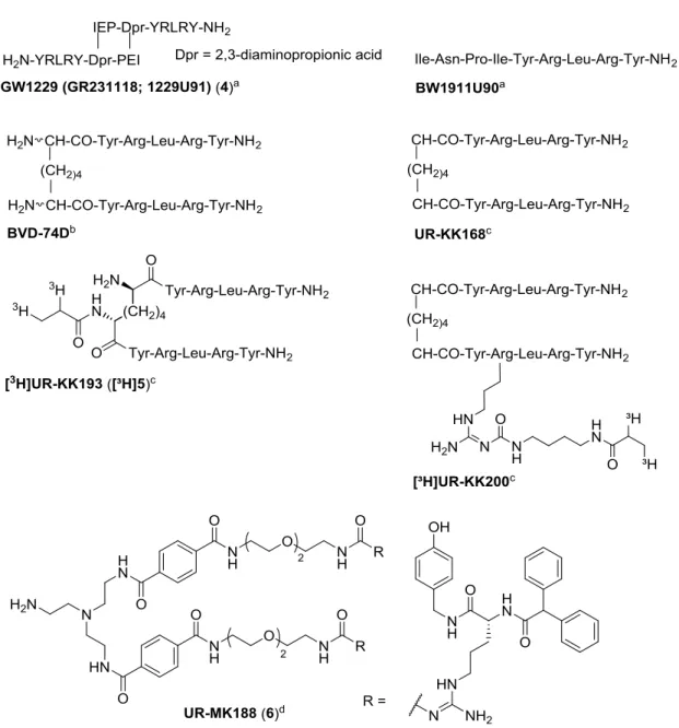
Chapter 1 9 74D (Balasubramaniam et al., 2006). Derived from the C-terminal pentapeptide of hPP, the respective pure diastereomers and a series of homo- and heterodimeric analogs were synthesized in our working group. In binding and functional cellular assays, (2R,7R)-BVD- 74D was superior to (2S,7S)-BVD-74D, and two analogs gave access to the corresponding tritiated high affinity Y
4R radioligands [³H]UR-KK193 (5) and [³H]UR-KK200 (Kuhn et al., 2016). Recently, a fluorescently labeled analog of BVD-74D, (sulfo-Cy5) labeled (R,R)- analog (mono-sCy5-(2R,7R)-sub(YRLRY-NH
2)
2) was reported as a novel Y
4R agonist with nanomolar affinity (Liu et al., 2016).
With regard to the potential therapeutic use of Y
4R agonists, peptides with increased stability in plasma were developed. Lipidation and PEGylation of PP not only prolonged plasma half‐
life but also inhibited arrestin recruitment and receptor internalization (Mäde et al., 2014).
Behavioural experiments with mice upon peripheral injection of a novel PEGylated derivative of PP, the selective Y
4R agonist [K
30(PEG2)]hPP2‐36, suggest that these derivatives penetrate across the blood-brain barrier (Verma et al., 2016).
Truncated hPP analogs such as [Nle
30]hPP25−36 and [Leu
34]pNPY25−36 were found to be
Y
4R selective partial agonists (Berlicki et al., 2013). [Ala
27]hPP and [Leu
27]hPP were used to
localize specific interactions between hPP and the hY
4R (Pedragosa-Badia et al., 2014). Full-
length PP/NPY chimeras were designed and characterized by Cabrele et al. (Cabrele et al.,
2001). Aiming at labeled hPP derivatives using amino-reactive reagents the amino acid in
position 4 was replaced by lysine ([Lys
4]hPP, 7). This gave access to the fluorescent
peptides S0586- and cy5-[Lys
4]hPP, which turned out to be valuable pharmacological tools
for the investigation of Y
4R ligands, e. g. by flow cytometry or confocal microscopy (Ziemek,
2006; Ziemek et al., 2007). A selection of Y
4R ligands is given in Figure 1.5.
10 1.3 Sodium sensitivity of the NPY receptors
Figure 1.5. Structures of selected peptidic agonists and the non-peptidic antagonist UR-MK188.
a


![Figure 3.3. Concentration-response curves of agonists (A) and antagonists (B) investigated in the [ 35 S]GTPγS assay at the hY 2 R](https://thumb-eu.123doks.com/thumbv2/1library_info/3939837.1533104/43.892.140.728.109.321/figure-concentration-response-curves-agonists-antagonists-investigated-gtpγs.webp)
![Figure 5.2. Purity, identity and long-term stability of the radiolabeled peptide [ 3 H]12 and the fluorescent ligand 13 determined by HPLC](https://thumb-eu.123doks.com/thumbv2/1library_info/3939837.1533104/87.892.127.761.226.576/figure-purity-identity-stability-radiolabeled-peptide-fluorescent-determined.webp)
