Speed-dependent interaction of sensory signals and local, pattern-generating activity during walking in Drosophila
Inaugural-Dissertation zur
Erlangung des Doktorgrades
der Mathematisch-Naturwissenschaftlichen Fakultät der Universität zu Köln
vorgelegt von
Volker Berendes
aus Bensberg
Köln Mai 2016
Berichterstatter: Prof Dr. Ansgar Büschges
(Gutachter) PD Dr. Silvia Daun (ehem. Gruhn) Tag der mündlichen Prüfung: 08.07.2016
Table of Contents
Table of Contents
Abstract ... III Zusammenfassung ... V
1. Introduction ... 1
1.1 Central pattern generators (CPGs) and coordination ... 2
1.2 Inter-leg coordination at different walking speeds ... 3
1.3 Amputation experiments and the control mechanisms of walking ... 5
1.4 Studying walking in Drosophila ... 5
1.5 The present thesis ... 7
2. Material and Methods... 9
Animals ... 9
2.1 Experiments on leg kinematics and inter-leg Coordination ... 9
Experimental setup ... 9
Video acquisition... 12
Data Analysis ... 14
Software ... 18
2.2 Walking Speed and Activity Assay ... 19
Experimental setup ... 19
Data analysis ... 20
Software ... 21
3. Results ... 25
3.1 Experiments on leg kinematics and interleg coordination ... 25
3.1.1 Experiments on the side-view setup ... 25
Intact flies ... 25
R1 amputees: ... 29
R2 amputees ... 32
R3 amputees ... 36
3.1.2 Experiments on the top-view setup ... 42
Intact flies ... 44
R1 amputees ... 46
R2 amputees ... 51
R3 amputees ... 54
3.2 Animal Speed and Activity Assay ... 58
Intact animals ... 59
Table of Contents
R1 amputees ... 61
R2 amputees ... 62
R3 amputees ... 64
4. Discussion ... 66
4.1 Rhythmic Motor Activity of Leg Stumps in Walking Drosophila ... 67
4.2 Descending Control of Walking Speed in Drosophila ... 69
4.3 Speed-dependence of coordination strength... 70
4.4 Differences between the ball setups ... 71
4.5 Differences between free and tethered walking ... 72
4.6 New insights and open questions ... 75
Bibliography ... 77
Appendix ... 88
List of Figures ... 108
Danksagung ... 111
Mein Besonderer Dank gilt: ... 111
Teilpublikationen ... 112
Conference abstracts ... 112
Erklärung ... 113
Curriculum vitae ... 114
Ausbildung ... 114
Praktika ... 115
Abstract
Abstract
Locomotion in six legged insects requires effective mechanisms for inter-leg coordination. Such mechanisms could be realized by mechanical coupling of the legs via the ground during stance phase, by direct connections between the rhythm generating networks in the ventral nerve cord (VNC) of these animals, or they could rely on an intersegmental exchange of phasic sensory feedback. This thesis investigates the role of local pattern-generating networks and inter-leg sensory influences for the generation of rhythmic motor activity during walking at different speeds in Drosophila. For this purpose, a series of already existing techniques was used in combination for the first time in the model organism Drosophila melanogaster. Single leg amputation was used to reduce sensory feedback of one leg allowing the residual stump to move freely and thereby providing insight into the rhythmic activity of motor pattern-generating networks in the VNC. This approach has already been used to investigate the control mechanisms of walking in several animals, including stick insects (e.g.
Wendler, 1964) and cockroach (e.g. Delcomyn, 1988). In the present thesis, oscillation periods, phases, and absolute inter-segmental intervals of movements in the intact legs and single leg stumps were quantified in tethered flies walking on top of an air-cushioned ball. A similar setup has previously been used for several other animals such as cockroaches (Spirito and Mushrush, 1979) and also Drosophila (Seelig and Jayaraman, 2013; Seelig et al., 2010). High-speed video analysis of the walking behavior was performed manually (Strauss and Heisenberg, 1990; Wosnitza et al., 2013) as well as in semi automatic fashion (Branson et al., 2009; Mendes et al., 2013). The nan[36a] mutant (Kim et al., 2003), which has defective chordotonal organs, was used to investigate the influence of sensory feedback from chordotonal organs in the intact legs on movements of the stump.
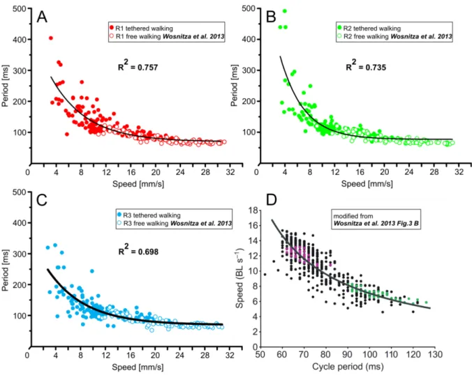
Consistent with findings in cockroaches and stick insects rhythmic oscillatory movements were found for stumps of single front, middle and hind legs during tethered walking in Drosophila. The stumps oscillated with a frequency of approximately 10 Hz that was largely consistent for the whole range of recorded walking speeds. Intact legs showed step periods of 100 ms only during relatively fast walking, thus, during slow walking sequences multiple stump oscillations were found for one step period of the intact legs. Consequently, the phase relation between stumps and intact legs was very variable at low walking speeds. Nevertheless, preferred absolute time intervals were found between intact leg liftoff and subsequent levation or depression onset in the stump, even if the frequency of stump oscillations was much higher than the step frequency of intact legs. With increasing walking speed the stump oscillations became highly coordinated with respect to the intact legs. Interestingly, the transition range to strong coordination occurred at the point where the stepping period in intact legs becomes very similar to the base frequency of the stump oscillations.
Single middle leg stumps of nan[36a] mutant flies showed the same high frequency oscillations that were found during experiments with wild type flies. The stumps oscillated almost independently of
Abstract walking speed with a movement period of about 100 ms. In contrast to wild type flies stump oscillations in the mutant flies failed to entrain to the stepping behavior of the intact legs at high walking speeds and the absolute time intervals between liftoff events in intact legs and subsequent onset of levation or depression in the stump were more variable.
These results lead to the following four conclusions: First, a putative descending control of walking speed does not target the rhythm generating networks directly but it probably has an indirect influence by changing the gain factor of sensory signals, for instance. Second, if the relatively high frequency of stump oscillations reflects a high natural frequency of the investigated pattern generating networks this would facilitate the coordination at high walking speeds, where precise coordination is very important. Fourth, coordinating signals from the intact legs influences the stump movements even during slow walking, but it is probably more effective during fast walking where the stump shows a cycle to cycle coupling to the intact legs. This indicates a stronger inter-leg coordination at high walking speeds. Fourth, the chordotonal organs in the intact legs play an important role for this coordination.
During the second part of this thesis the speed range and activity pattern of intact Drosophilae and single leg amputees were studied during voluntary untethered walking. For this purpose, a behavioral paradigm was created that allowed for the study of walking behavior in nine individual flies in separate petridish enclosures. Additionally, software was developed to provide a semi- automatic analysis of the recorded videos. It was found that compared to intact animals the occurrence of walking speeds above 5 mm/s was strongly reduced in amputees. During voluntary untethered walking hindleg amputees showed the highest speed range of all amputees. A reduced level of walking activity was found in frontleg and hindleg amputees, whereas middleleg amputees showed the same probability to walk as the intact animals. The flies walked in short bouts of mostly less than two seconds. As previously shown in the literature (Martin, 2004; Valente et al., 2007) the probability for fast walking was higher in the center of the walking arena compared to the area close to the wall of the enclosures, where the flies spend most of the time. In any case walking activity was only found for a maximum 30 % of the recorded time (in R2 amputees).
Zusammenfassung
Zusammenfassung
Die Fortbewegung von sechsbeinigen Insekten erfordert effektive Mechanismen um die Bewegung der Einzelbeine miteinander zu koordinieren. Solche Mechanismen könnte realisiert sein, durch mechanische Kopplung der Einzelbeine über den Boden während der Stemmphase, durch direkte Verbindungen zwischen den Rhythmus erzeugenden Netzwerken im ventralen Nervensystem dieser Tiere oder durch intersegmentalen Austausch von phasischer sensorischer Information. Die vorliegende Arbeit untersucht die Rolle von lokalen Rhythmus generierenden Netzwerken und dem Austausch von sensorischer Information zwischen den Beinen auf die Entstehung rhythmischer motorischer Aktivität beim Laufen mit verschiedenen Geschwindigkeiten bei Drosophila. Zu diesem Zweck wurde eine Reihe von bereits existierenden Techniken das erste Mal in Kombination am Modellorganismus Drosophila verwendet. Amputationen einzelner Beine wurden durchgeführt um die sensorische Rückmeldung von einem Beines zu reduzieren, der Stumpf kann sich dabei frei bewegen und erlaubt dadurch Einblicke in die rhythmische Aktivität der mustergenerierenden Netzwerke im ventralen Nervensystem. Diese Methode wurde bereits bei verschiedenen Tieren eingesetzt um die Kontrollmechanismen des Laufens zu untersuchen dazu gehören Stabheuschrecken (z.B. Wendler, 1964) und Schaben (z.B. Delcomyn, 1988). In der vorliegenden Arbeit wurden Oszillationsperiode, Phasenlage und absolute intersegmentale Zeitintervalle bei der Bewegung von intakten Beinen und Stümpfen quantifiziert während die Tiere gehaltert auf einem Ball laufen, der auf einem Luftkissen schwebt. Ein ähnlicher experimenteller Aufbau wurde bereits bei verschiedenen anderen Tieren wie Schaben (Spirito and Mushrush, 1979) und auch Drosophila (Seelig and Jayaraman, 2013; Seelig et al., 2010) benutzt. Hochgeschwindigkeitsvideos des Laufverhaltens wurden manuell (Strauss and Heisenberg, 1990; Wosnitza et al., 2013) und halbautomatisch (Branson et al., 2009; Mendes et al., 2013) ausgewertet. Die nan[36a] Mutante, welche defekte Chordotonalorgane hat, wurde benutzt um den Einfluss sensorischer Information von den Chordotonalorganen in den intakten Beinen auf die Bewegungen des Stumpfes zu untersuchen.
In Übereinstimmung mit Ergebnissen von Schaben und Stabheuschrecken wurden rhythmische oszillatorische Bewegungen der Einzelbeinstümpfe von Vorder-Mittel und Hinterbein beim gehalterten Laufen von Drosophila gefunden. Die Stümpfe oszillierten mit einer Frequenz von etwa 10Hz und das relativ konstant über den gesamten Bereich der aufgezeichneten Laufgeschwindigkeiten.
Intakte Beine zeigten eine Schrittdauer von 100 ms nur bei relativ schnellem Laufen, so dass bei langsamen Laufsequenzen häufig mehrere Oszillationen des Stumpfes während einer Schrittperiode der intakten Beine gefunden wurden. Als Konsequenz war die Phasenlage zwischen Stümpfen und intakten Beinen bei langsamen Läufen sehr variabel. Nichtsdestotrotz gab es vorherrschende Zeitintervalle zwischen dem Abheben der intakten Beine und direkt darauffolgenden Levationen oder Depressionen des Stumpfes, selbst wenn die Frequenz der Stumpfoszillationen viel höher war als die
Zusammenfassung Schrittfrequenz der intakten Beine. Mit ansteigender Laufgeschwindigkeit wurden die Stumpfoszillationen im Bezug auf die intakten Beine sehr koordiniert. Interessanterweise lag der Übergangsbereich zu hoher Koordinationsstärke in dem Geschwindigkeitsbereich in dem die Schrittperiode der intakten Beine sich der Grundfrequenz der Stümpfe näherte. Einzelne Mittelbeinstümpfe der nan[36a] Mutante zeigten die gleichen hochfrequenten Oszillationen die auch während der Experimente mit wildtypischen Fliegen gefunden wurden. Die Stümpfe oszillierten fast unabhängig von der Laufgeschwindigkeit mit einer Periodendauer von um die 100 ms. Im Gegensatz zu wildtypischen Fliegen gab es keine Ankopplung der Stumpfoszillationen an die Schreitbewegungen der intakten Beine bei hohen Laufgeschwindigkeiten und die die absoluten Zeitintervalle zwischen dem Abheben der intakten Beine und direkt darauffolgenden Levationen oder Depressionen des Stumpfes waren variabler.
Diese Ergebnisse führen zu den folgenden vier Annahmen: Erstens, eine mögliche zentrale Kontrolle der Laufgeschwindigkeit beeinflusst die CPGs nicht direkt sondern hat einen indirekten Einfluss, zum Beispiel auf die Verstärkungsfaktoren von sensorischen Signalen. Zweitens, wenn der relativ hohen Frequenz der Stumpfoszillationen eine hohe natürliche Frequenz der mustergenerierenden Netzwerke zugrunde liegt dann würde das die Koordination bei schnellem Laufen erleichtern, da dort eine präzise Koordination sehr wichtig ist. Drittens, ein koordinierendes Signal von den intakten Beinen beeinflusst die Bewegungen des Stumpfes auch beim langsamen Laufen, aber es ist effektiver beim schnellen Laufen, wo der Stumpf von Zyklus zu Zyklus an die intakten Beine gekoppelt ist. Viertens, die Chordotonalorgane in den intakten Beinen sind eine wichtige Quelle für diese koordinierenden Signale.
Für den zweiten Teil dieser Arbeit wurde der Geschwindigkeitsbereich und die Häufigkeit des Laufverhaltens von intakten und Einzelbein amputierten Tieren während spontanen ungehaltertem Laufens untersucht. Zu diesem Zweck wurde ein Verhaltensparadigma entwickelt das es erlaubte das Laufverhalten von 9 Fliegen in separaten Petrischalen zu untersuchen. Zusätzlich wurde Software entwickelt die eine teilautomatisierte Auswertung der aufgezeichneten Videos erlaubte. Es wurde herausgefunden, dass im Vergleich zu intakten Fliegen das Auftreten von Laufgeschwindigkeiten über 5mm/s, bei Fliegen mit Einzelbeinamputationen stark reduziert war. Bei spontanem ungehaltertem Laufen zeigen hinterbeinamputierte Tiere den größten Geschwindigkeitsbereich aller amputierten Tiere. Vorder und hinterbeinamputierte Tiere zeigten eine reduzierte Laufaktivität währenddessen mittelbeinamputierte Tiere mit der gleichen Wahrscheinlichkeit Laufen wie intakte Tiere. Die Fliegen laufen in kurzen Bouts von meist weniger als zwei Sekunden. Wie bereits in der Literatur gezeigt (z.B.
Martin, 2004; Valente et al., 2007) war die Wahrscheinlichkeit für schnelle Läufe in der Mitte der Laufarena höher verglichen mit dem Rand, wo die Fliegen sich die meiste Zeit aufhalten. In jedem Fall wurde Laufaktivität nur in maximal 30% der aufgezeichneten Zeit beobachtet.
1. Introduction
1. Introduction
Parts of this section are included in a paper draft with the working title: "Speed-dependent interplay between local, pattern generating activity and sensory feedback during walking in Drosophila"
that has recently been submitted for publication in Journal of Experimental Biology.
Locomotion is important in the animal kingdom to explore the environment, find food, escape predators and fulfill the requirements for reproduction (Dickinson et al., 2000). Walking is an important form of locomotion for many animals and therefore walking behavior has been studied in a vast variety of species (for review see Büschges, 2005; Orlovsky, 1999; Pearson, 1993).
Walking animals use body appendages known as limbs or legs to produce a cyclic behavior consisting of two phases: A stance phase during which the legs have contact to the ground and generate propulsion and a swing phase that is used to return the appendages back to their starting position in order to generate the next stance phase (for review see Büschges and Gruhn, 2007; Graham, 1985a).
For neuroscientists, walking is of particular interest because it poses a series of rather complicated tasks to the neural circuitry controlling this behavior. There are several levels of increasing complexity. On the lowest level it requires motor output to be generated for each of the leg´s joints that results in coordinated swing and stance phases (intra-leg coordination). On an intermediate level, all legs of an animal have to be coordinated with each other (inter-leg coordination). On the highest level, the walking pattern has to be adaptable with regard to walking speed or direction.
Insects such as cockroaches, locusts and stick insects have proven to be particularly useful for locomotor studies, because compared to most vertebrates they are small in size and their nervous system is relatively simple and easily accessible (for review see Büschges and Bässler, 1998;
Büschges, 2005; Pearson, 2004).
1. Introduction
1.1 Central pattern generators (CPGs) and coordination
For insects (e.g. stick insect: Büschges et al., 1995; hawk moth: Johnston and Levine, 1996a) and several other animals for example leech (Nusbaum et al., 1987), turtle (Robertson et al., 1985), lamprey (Wallén and Williams, 1984) and other vertebrates (Brown, 1911; for review see Grillner, 1985) the existence of rhythmic central neural networks (also called CPGs) has been demonstrated in the past. CPGs are relatively small and autonomous neural networks that are able to provide the timing for motoneuron discharge, without rhythmic central or sensory input, albeit sensory information may be essential to shape the CPG output for behavioral needs of the animal (for review see Marder and Calabrese, 1996).
During walking CPGs are believed to provide rhythmic activity that is modulated on a cycle to cycle basis by sensory afferents (e.g. stick insect: Borgmann et al., 2009; cockroach: Fuchs et al., 2012) to generate the magnitude and timing of the motor output (for review see Büschges, 2005). The coupling between different segments of a leg (intra-leg coupling) is probably achieved indirectly by sensory signals that connect the CPGs controlling individual leg joints (stick insect: Akay et al., 2001; 2004;
Chung et al., 2015; Daun-Gruhn and Toth, 2011; Hess and Büschges, 1999; crayfish: Bacqué- Cazenave et al., 2015).
In this regard the system that controls stepping in insects can be seen as a set of oscillators that are weakly and indirectly coupled via sensory feedback (locust: Laurent and Burrows, 1989; stick insect:
Ludwar, 2004; cockroach: Pearson and Iles, 1973). The nature of this coupling is not entirely clear and it is possible that an additional and more direct coupling of the CPGs via interneurons exists.
Apart from intra-leg coordination walking also requires coordination between the different legs of an animal (inter-leg coordination). Studies in cockroaches (Delcomyn, 1989; Delcomyn, 1991a; 1991c) and stick insects (Foth and Bässler, 1985a; 1985b) suggest a set of behavioral rules for inter-leg coordination (Cruse, 1990; Dürr et al., 2004) that are active locally between neighboring legs. The rules are numbered from 1-6 (Cruse et al., 1995).
1. If a posterior leg is in swing phase, it inhibits the start of a swing phase in its anterior neighboring leg.
2. If a posterior leg starts its stance phase it excites the start of a swing phase in the anterior leg.
3. If a leg has reached an increasingly posterior position during its stance phase it excites the start of a swing phase in its posterior neighbor.
4. During swing phase the posterior leg targets the resting position of the anterior legs tarsus.
5. An increased resistance increases force during stance (co-activation) and an increased load prolongs the stance phase.
6. If a leg steps on the tarsus of its neighboring leg the trod-on leg tells the one on top to step off.
(Dürr et al., 2004)
1. Introduction
Fig. 1:" Leg modules and their connection via coordination rules (from Dürr et al. 2004). L1, L2, L3 left front, middle, and hind leg, respectively. R1, R2, and R3 stand for the corresponding right legs. The question mark indicates that there are ambiguous data concerning this influence." (Figure and caption literally quoted from Schilling et al., 2013 Fig. 3)
Similar to intra-leg coordination these rules are based on sensory afferents providing feedback about the current state of the locomotor system (Borgmann et al., 2009; 2012; Daun-Gruhn et al., 2012;
Ludwar, 2004). Additionally, all legs of an animal that are simultaneously in stance phase are mechanically coupled trough the ground, which may have a strong influence on inter-leg coordination (Zill et al., 2009).
General activation of the CPGs and the control of locomotion frequency is believed to be provided by a descending tonic excitatory input whose magnitude can vary in order to allow for control of walking speed (Gal and Libersat, 2006; Ridgel and Ritzmann, 2005; Roeder, 1937).
The origin of such a tonic excitation in insects is believed to be the gnathal (former: sub-esophageal) ganglion (Graham, 1979; Kien and Altman, 1984; Kien and Williams, 1983). How these signals work in detail and if they target CPGs directly or indirectly is so far unknown, but there is evidence that they influence the way sensory feedback is processed (Gabriel and Büschges, 2007; Sauer et al., 1996).
1.2 Inter-leg coordination at different walking speeds
Walking speed has an influence on temporal coordination between the legs of an insect, which leads to different coordination patterns for different speed ranges (Graham, 1972; Gruhn et al., 2009; Mendes et al., 2013; Wendler, 1964; Wosnitza et al., 2013).
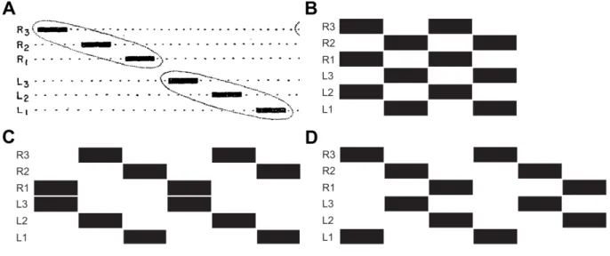
1. Introduction During slow walking many insects use a coordination pattern called wave gait (Hughes, 1952), where only one of the six legs is in swing phase at a given time (Fig. 2 A). When the walking speed is increased a coordination pattern called tetrapod gait (Burns, 1973; Graham, 1972) was described where four legs of the animal are in swing phase at the same time (Fig. 2 C and D). Fast walking insects mostly use the so called tripod gait (Delcomyn, 1971; Graham, 1972; Mendes et al., 2013;
Strauss and Heisenberg, 1990; Wahl et al., 2015; Wosnitza et al., 2013), where the middle leg on one body side and the front and hind leg on the contralateral side are simultaneously in swing phase (Fig. 2 B). However, the aforementioned patterns should not be regarded as strict states of the locomotor system, as studies indicate that they form a speed dependent continuum with rather seamless transitions between them (Spirito et al., 1979; Wendler, 1965; for review see Graham, 1985; Wilson, 1966).
Fig. 2: Stereotypic footfall patterns of hexapedal animals. Black bars indicate swing phases of the legs. (A) wave gait:
Only one of the six legs of the animal is in swing phase at a given time. (A modified from Wilson, 1966 Fig. 1 A). (B) tripod gait: middle leg on the ipsilateral side swings in synchrony with front and hind leg on the contralateral side (C, D) Tetrapod gait: two diagonal legs swing synchronously. (B,C and D modified from Grabowska et al., 2012 Fig. 1)
An increase in stepping frequency and walking speed in insects is achieved by a decrease in stance duration (retraction time), while the swing duration (protraction time of the legs) of the legs is kept almost constant (Burns, 1973; Mendes et al., 2013; Wendler, 1964; Wosnitza et al., 2013; for review see Wilson, 1966; Büschges, 2012). Intracellular recordings in stick insects (Gabriel and Büschges, 2007) provide evidence that stepping velocity is controlled by synaptic inputs that act on stance phase motor neurons exclusively during stance. As stance phase motor neurons are influenced by sensory feedback signaling ground contact (Berendes et al., 2013; Schmitz et al., 2015; for review see Zill et al., 2004) it is possible that modifications in the gain of these sensory feedback loops contribute to the changes in stance duration and thereby cycle period and speed .
Behavioral observations show that two neighboring legs are never in swing phase at the same time (equivalent to Cruse rules 1 to 3, Cruse, 1990; for review see Dürr et al., 2004) consequently the occurrence of swing phases has to be timed precisely at high walking speeds to coincide with the
1. Introduction comparatively short stance phases of the neighboring legs. This implies a rather strict inter-leg coordination at high walking speeds.
1.3 Amputation experiments and the control mechanisms of walking
One of the oldest methods to investigate the control mechanisms of walking behavior is leg amputation (e.g. Buddenbrock, 1921). A leg stump that cannot contact the walking substrate is mechanically uncoupled from the intact walking legs. Furthermore, sensory feedback from all leg segments distal to the lesion is gone and load sensors proximal to the lesion cannot provide a ground contact signal.
Previous amputation studies in cockroaches (Delcomyn, 1988; Delcomyn, 1991b; Delcomyn, 1991c;
Hughes, 1957; Noah et al., 2004) and stick insects (Graham, 1977; Wendler, 1964) revealed that the animals still show coordinated walking behavior with their remaining intact legs, while the stumps of single legs show rhythmic patterns of motor activity, despite the lack of sensory input created by the amputation. However, many of these studies report multiple burst of motor activity in the stump during single steps of the adjacent intact legs, especially during slow walking (Delcomyn, 1988; Noah et al., 2004). These findings provide important hints that sensory feedback mediating ground contact might be crucial to maintain inter-leg coordination. Delcomyn (1988) compared the patterns of motor activity in the stump with those that he found during searching (Delcomyn, 1987). He concluded that the activity of the stump is probably still driven by the interneurons that control the intact leg during walking and that therefore an analysis of the stump motor pattern might reveal important features of the locomotor system.
1.4 Studying walking in Drosophila
Since in 1910 Thomas Hunt Morgan started his genetic studies on Drosophila melanogaster MEIGEN, 1830, fruit flies have become an important model organism, with a large set of genetic tools available (for review see Jennings, 2011; Rubin, 1988). These genetic tools allow experimenters to find and specifically activate (e.g. Hamada et al., 2008; Klapoetke et al., 2014; Zhang et al., 2007) or inhibit (e.g. Chuong et al., 2014) sense organs (e.g. Kim et al., 2003) and neurons (e.g. Bidaye et al., 2014) that are necessary to control or maintain walking behavior. Albeit electrophysiology is difficult to perform in Drosophila, mainly due to its small body size, genetically encoded calcium indicators can be used to study the firing patterns of neurons (e.g. Berlin et al., 2015). High-speed video recording can be used in concert with computer based tracking and classification algorithms to quantify many behavioral parameters (e.g. Branson et al., 2009; Kain et al., 2013; Mendes et al., 2013; Valente et al., 2007). As Drosophila is a vigorous walker with a large range of different walking speeds, all those
1. Introduction tools taken together could provide new insights for still unanswered questions regarding walking behavior in insects.
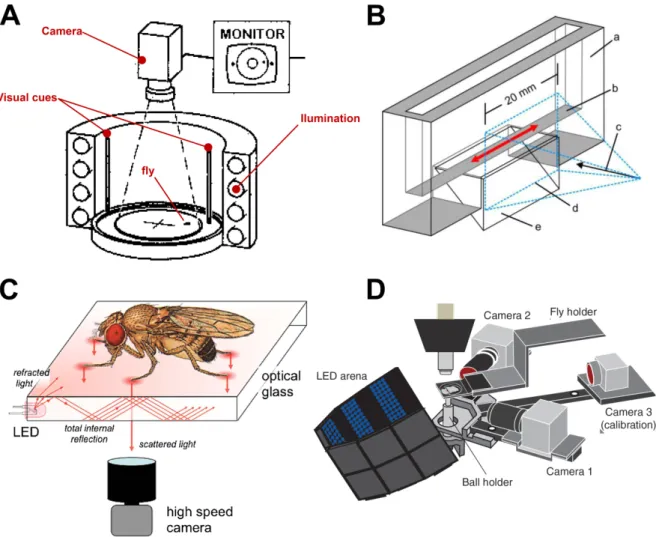
Fig. 3: Setups used to study the walking behavior in fruit flies. (A) Schematic diagram of Buridan´s paradigm, a circular luminous arena where a wingless fly is placed on disc surrounded by water. The fly patrols between two dark optical cues placed opposite from each other. Videos of the flies' behavior are taken by a camera mounted above the setup. (modfied from Bülthoff et al., 1982 Fig. 2) (B) Schematic diagram of the tunnel setup. Walking behavior of the flies on a walkway (red arrows) was recorded with a high-speed camera through a 20 mm wide window simultaneously from one side and from below (a: acrylic glass, coated on the inside with a layer of Fluon to prevent the flies from scaling the glass; b: 5 mm wide transparent walkway; c: camera viewpoint; d: camera field of view, free of Fluon; e: glass prism, providing a ventral view of the walkway) (modified from Wosnitza et al., 2013 Fig. 1A).
(C) Schematic overview of the fTIR setup. The flies walk on a glass surface that has LEDs attached at the edges. The light propagates within the optical glass via total internal reflection. Tarsal contact causes Light scattering, which is observed by a high-speed camera mounted below the fly. (figure modified from Mendes et al., 2013 Fig. 1A)
(D) Modified schematic overview of the setup used by Seelig and colleagues (2010) for two-photon imaging from the brain of head-fixed flies walking on a ball. The ball is floating on air. Movement and velocity around each of the ball three rotational axes was recorded by two optical sensors (called camera 1 and camera 2 in the scheme).
However, so far only few studies exist that investigate the kinematics and coordination of the legs during walking behavior in the fruit fly Drosophila melanogaster (Mendes et al., 2013 and 2014;
Strauss and Heisenberg, 1990a; Wosnitza et al., 2013). Strauss and Heisenberg 1990 studied voluntary walking of untethered Drosophila in the Buridans paradigm, a uniform white arena were the flies patrolled between two inaccessible black landmarks (Fig. 3 A). Using high-speed video analysis they found that flies walk in alternating tripod coordination during fastest walks and that the strictness of this tripod coordination depends on walking speed. Wosnitza and collegues 2013 analyzed high-speed videos of flies walking in a tunnel setup (Fig. 3 B) and found that flies also use wave-gait like and
1. Introduction tetrapod walking patterns at low speeds. These findings suggest that inter-leg coordination in Drosophila is flexible and dependent on walking speed. During some experiments the authors amputated a single hind leg of the flies and found that they show immediate adaptations in body posture, leg kinematics and inter-leg coordination, and were still able to walk coordinately. They conclude that the neural system that controls walking behavior in Drosophila allows for a modular control of single leg stepping, thus that individual legs of fruit flies are largely independent from each other and only loosely coupled. Mendes and collegues (2013) used a setup were light travels through a glass plate on which the flies walk (Fig. 3 C). While the tarsi of the flies contacted the glass surface during stance phase of walking they light up due to a phenomenon called frustrated Total Internal Reflection (fTIR). This was used to automatically detect tarsi position and stance phase timing with a computer based approach. A tripod coordination pattern was found for most walking speeds with gradually diminishing frequencies for slower flies, abrupt gait transitions, known for example from horses (Hoyt and Taylor, 1981), were not found in Drosophila. Furthermore, Mendes and collegues (2013) found that genetically modified flies, which lack most of the proprioceptive feedback from sensory structures in the leg, including the chordotonal organs, hair plates, campaniform sensilla and mechanosensory bristles, have a deficient step precision, but are still able to walk in tripod coordination pattern. On the other hand a more recent study of the same group showed that functional chordotonal organs are important for the flies to respond kinematically to changes in walking conditions, such as carrying an additional weight (Mendes et al., 2014).
This poses the question to which extend Drosophila relies on sensory feedback during walking, especially in the light of results from Chung and collegues (2001), who showed that flies cannot walk at all if they lack sensory feedback from both their chordotonal organs and campaniform sensilla (Chung et al., 2001 Fig. 3 B).
Currently the role of sensory feedback during walking in Drosophila is far from being fully understood. Further investigations are necessary to disentangle the influence of rhythmic neural networks and sensory influence on the motor output during walking.
1.5 The present thesis
For the present thesis single leg amputations were used as a method to investigate the role of local pattern generating networks and inter-leg sensory influences on the generation of rhythmic motor activity during walking in Drosophila. As indicated by previous experiments, fruit flies are still able to walk coordinately with only five legs, although inter-leg coordination becomes more variable (Wosnitza et al., 2013). In the first part of this thesis high-speed video analysis was used to investigate the movements of single front, middle and hind leg stumps during tethered walking on an air-
1. Introduction cushioned ball. The ball setup was adapted from the literature (Fig. 3 D) and allowed to infer the walking path of the fly from the movements of the ball around its three rotational axes (Seelig et al., 2010). Coordination among the remaining intact legs was analyzed as well as the coordination of the stump movements with respect to these intact legs. A special focus was drawn here on changes that occur with increasing walking speed. If the stump movements largely reflect the activity of central pattern generating networks and if a central descending drive directly influences these networks to modulate a change in walking speed this influence should affect stump movements when speed increases. As the stump is not mechanically coupled to the intact walking legs a possible coupling between its movements and the stepping behavior of the intact legs should be a result of inter-leg coordinating signals. Previous studies in intact flies and hind leg amputees found that inter-leg coordination becomes stricter with an increase in walking speed (Wosnitza et al., 2013). If this increase in coordination is not mediated by mechanical coupling of the legs via the ground, it should be reflected in the stump movements. A putative neural inter-leg coupling might be realized via interneurons that directly connect the rhythm generating networks responsible for the legs movements.
Alternatively, inter-leg coupling might be achieved by an exchange of sensory information between the legs (Borgmann et al., 2009). Both motor activity and, consequently, sensory information are correlated with the rhythmic activity of the pattern generating network. In order to distinguish between these two alternatives some experiments of the current thesis were conducted with nan[36a] mutant flies that lack sensory feedback from chordotonal organs (Kim et al., 2003). As previous studies indicate chordotonal organs might play an important role for leg coordination during walking in fruit flies (Chung et al., 2001; Mendes et al., 2014). The mass of the air-cushioned ball (see materials and methods) that has to be moved by the fly might reduce the range of recorded walking speeds. These issues were addressed in the second part of this thesis, which uses a custom made behavioral paradigm and a semi automatic computer based approach to analyze the speed range and activity frequency of intact and single leg amputated Drosophila during voluntary untethered walking. The goal of these experiments was to unravel possible differences between the speed range and activity level that flies exhibit during tethered walking on a ball, compared to untethered voluntary walking in an arena.
Concerning the amputees such a comparison is particularly useful because tethered flies do not have to carry their own body weight therefore amputation of a leg does not affect the stability of an animal during walking. This situation is quite different during untethered walking where the impact of a single leg amputation on the stability of an animal during walking might differ depending on the amputated leg. Several previous studies (Mendes et al., 2013; Strauss and Heisenberg, 1990; Wosnitza et al., 2013) have shown that Drosophila uses the tripod coordination pattern during fast walking, therefore one might assume that an amputation of the middle leg has a strong impact on the ability of the animal to walk.
2. Material and Methods
2. Material and Methods
Animals
For all experiments 4 to 6 day old males of the Drosophila melanogaster wild-type strain Canton-S (wtCS) or the mutant strain nan [36a] were used. The flies were raised on standard medium with cornmeal, yeast, agar and molasses (Zoological Institute, Univ. of Cologne, Cologne, Germany). Flies were kept at 23-25°C and 60% humidity on a 12h light /12h dark cycle.
2.1 Experiments on leg kinematics and inter-leg Coordination
Parts of this section are included in a paper draft with the working title: "Speed-dependent interplay between local, pattern generating activity and sensory feedback during walking in Drosophila"
that has recently been submitted for publication in Journal of Experimental Biology.
Experimental setup
Flies were cold anesthetized and isolated from each other 1-2 hours prior to an experiment. Under anesthesia the flies were transferred to separate enclosures, where they were kept at room temperature for the next 1-2h. Each enclosure contained a wet filter paper to provide water without food. The starvation was done to increase the activity level of the flies by inducing food searching behavior (Knoppien et al., 2000). In the cases were amputations were made, leg or leg parts were amputated with micro scissors during cold anesthesia.
2. Material and Methods
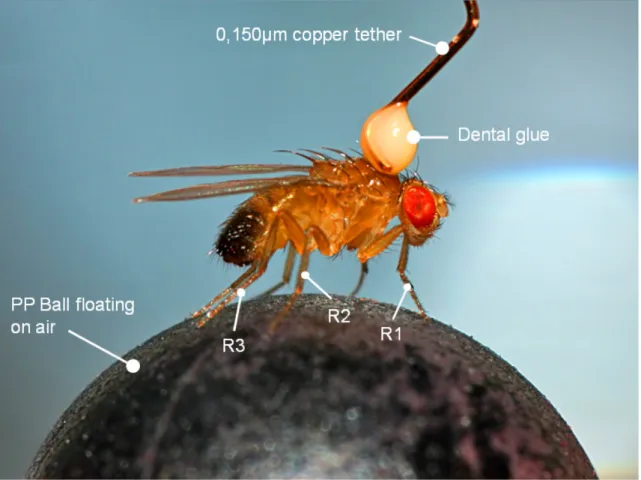
Fig. 4: Exemplary image of a male Drosophila tethered with dental glue and a copper wire on top of a 6 mm polypropylene (PP) ball.
The flies that were used for the experiments had either all six legs intact (Fig. 5A) or one leg amputated (front, middle, or hind leg) at the proximal half of the femur so that only a stump remained (see Fig. 5 B, C and D).
For all experiments with tethered animals the flies were cold anesthetized again immediately before the experiment and placed ventral side down in the groove of a custom build aluminum block (Electronics workshop, Zoological Institute, Univ. of Cologne, Cologne, Germany). A copper wire with a diameter of 0.150 mm was glued with a light curing composite dentist glue (ESPE Sinfony, 3M ESPE AG, Seefeld, Germany) on the thorax of the animal under a dissecting microscope. The glue was cured with a curing light (wavelength 420 nm to 480 nm).
The tethered flies were then mounted on top of an air-suspended 6 mm Polypropylene (PP) ball (Fig.
4), with a mass of approximately 100 mg. Movements of the ball around all three rotational axes were monitored by two sensors derived from optical computer mice with a method similar to the one described by Seelig and colleagues (2010). The original setup can be seen in the Introduction (Fig. 3 D).
2. Material and Methods
Fig. 5: Flies used had either all legs intact or one leg amputated. (A) Intact animal on the ball, lateral view showing the right body side. (B) Fly with amputated right front leg (red arrow). (C) Fly with amputated right middle leg (green arrow). (D) Fly with amputated right hind leg (blue arrow).
Two synchronized optical sensors measured (ADNS-9500, Avago Technologies, San José, CA, USA) optic flow on the ball's surface. Each sensor measured the horizontal and vertical movement velocity of a small patch of surface at the equator of the ball with a temporal resolution of 50 Hz. This patch was illuminated by a red diode laser (LG series, 1 mW, 660 nm, Lasertechs, Aschaffenburg, Germany).The four sensor values were used to reconstruct the movement velocities of the ball's three rotational axes. Assuming that all movements of the ball were caused by the fly walking atop it was possible to reconstruct the fly's virtual walking trajectories and walking speed. Acquisition of ball motion data was synchronized with high-speed video acquisition via a TTL trigger in a 1:10 ration: for every ten video frames one motion measurement of the ball was acquired. Low-level control of the optical sensors and synchronization to the high-speed camera was implemented with custom-made hardware (Electronics workshop, Zoological Institute, University of Cologne); high-level control was implemented by Dr. Till Bockemühl (Zoological Institute, University of Cologne, Cologne, Germany) with custom-written software in MATLAB 2011b (The Mathworks, Inc., Natick, MA).
2. Material and Methods Video acquisition
Two versions of the ball setup were used during this study:
The first setup had the camera mounted on the animal’s right body side, which allowed capturing videos of the protraction, retraction, levation, and depression movements of ipsilateral legs or stumps.
Video resolution was 320x240 pixels. This setup will be referred to as the side-view setup in the following text (Fig. 6).
Fig. 6: Schematic overview of the side-view setup (courtesy of Dr Till Bockemühl). Flies were tethered and placed atop an air-suspended 6 mm PP ball. Flies were recorded with a high-speed video camera at 500fps from the right body side. A custom-built pulsed IR-LED ring around the camera objective was used to illuminate the scene. Movements of the ball around all three rotational axes were monitored by two sensors derived from optical computer mice with a method similar to the one described by Seelig and colleagues (2010). The ball surface was illuminated for the motion sensor by red diode lasers (660 nm).
For the second setup the camera was mounted above the fly. Additionally, slanted mirrors were attached to the ball holder on both sides of the animal. The angle of 25° to the main camera axis allowed capturing the protraction and retraction movements of all 6 legs without occlusion by the wings or the body (Fig. 7). Videos were recorded at a resolution of 1280x700 pixels in this setup in order to cover the top-view of the fly and the two mirror views (Fig. 9). The second setup will be called the top-view setup in the following text.
2. Material and Methods
Fig. 7: Schematic overview of the top-view setup (courtesy of Dr Till Bockemühl). The setup was technically identical to the side-view setup but the camera was mounted above the ball. Additionally, two mirrors were attached above the ball holder. The mirrors were slanted 25° with regard to the main camera optical axis providing the camera with a view of the right and the left body side of the walking fly additional to the top-view.
For both setups a high-speed camera (AOS S-PRI High-Speed Color 5.2, AOS Technologies AG, Baden Daettwil, Switzerland) was used to record videos of the walking fly at 500 fps with a shutter time of 200 µs. A custom-built IR-LED ring (λ=880 nm, Siemens AG, Munich, Germany) around the camera objective was used to illuminate the scene (Electronics workshop, Zoological Institute, University of Cologne). Infrared light of 880 nm is invisible to the flies (Yamaguchi et al., 2010). The LEDs were synchronized with the camera shutter and were active only during actual image acquisition (shutter time: 200 µs). As videos were recorded at a frame rate of 500 fps the maximum accuracy for determining time points in the analyses is 2 ms.
2. Material and Methods Data Analysis
With regard to the walking behavior of the animals the following terms are used:
AEP (Anterior Extreme Position): The touchdown of a leg during walking.
PEP (Posterior Extreme Position): The lift-off of a leg during walking.
VEP (Ventral Extreme Position): The most depressed position of the stump during its oscillation.
DEP (Dorsal Extreme Position): The most elevated position of the stump during its oscillation.
Swing duration: The time between PEP and the following AEP of the same leg.
Stance duration: The time between AEP and the following PEP of the same leg.
Leg period: The time between two consecutive PEP events of the same leg.
Stump oscillations period: The time interval between two DEPs.
DF (Duty Factor): Stance duration divided by cycle period for intact legs or the time between DEP and subsequent VEP divided by stump oscillation period for stumps.
R1: right front leg.
R2: right middle leg.
R3: right hind leg.
Z-scores: (value-mean)/Standard Deviation
Fig. 8: Scheme illustrating the spatial conventions used in the text. The picture shows a single video frame obtained from a single walk of a hind leg amputee. Trajectories of intact front leg (red) and hind leg stump (blue) were superimposed on the video frame.
2. Material and Methods For the intact ipsilateral legs (right body side) the movements of the tarsi were tracked manually for each video frame recorded in the side-view setup. In the top-view setup, the same was done in a semi- automatic fashion for all intact legs of the animal (right and left body side). Additionally, the points in time of touch-down (anterior extreme position, AEP) and lift-off (posterior extreme position, PEP) were determined. The AEP was defined as the first video frame during a stance period that showed the tarsus touching the ball. The PEP was defined as the last video frame of a given stance period in which the tarsus still touched the ground. For leg stumps the movement of the stump tip was tracked manually in both setups.
In the side-view setup protraction and retraction as well as levation and depression movements of the stump were visible in the videos. The data evaluation for the side-view experiments in this thesis was focused on levation and depression movements of the stump as they were larger and easier to track compared to the stumps’ protraction and retraction. For all experiments performed in this setup the ventral and dorsal extreme positions (VEP and DEP) for each complete stump oscillation were determined as the minima (VEP) and maxima (DEP) of the levation and depression movement of the stump.
In the top-view setup only protraction and retraction movements of the stump were visible in the videos. For all experiments performed in this setup the last video frame before the stump switched from retraction to protraction was determined. This event will be called sPEP in the current text to distinguish it from a PEP in an intact leg, which is defined differently (see above).
For all analysis regarding leg coordination either AEP or PEP was used as a reference point in time for the intact legs. For leg stumps either VEP or DEP (side-view setup) or sPEP (top-view setup) events were used as a reference.
The ball-tracking system delivered the data on the fly's speed and position in mm, whereas the tracking information obtained for the leg movements from the high-speed video was in pixel.
Therefore, the ball radius served as a calibration to convert pixels to mm. With this calibration, it was possible to calculate the body length (BL, head to abdomen) of each individual fly and then convert the corresponding data on speed in from mms/s to BL/s. This was done to obtain a normalized speed value that accounts for possible differences in body size and therewith step length of the flies.
2. Material and Methods
Fig. 9: Single video frame of an intact fly walking on a 6 mm polypropylene ball in the top-view setup. The camera is mounted above the fly. The direct top-view on the fly can be seen in the center of the frame. Reflections of the fly in the two slanted mirrors, which were attached to the ballholder, can be seen on the left and on the right side of the center.
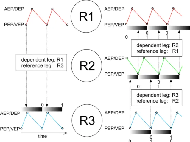
As a measure for coordination and coordinateness the time between the occurrences of an event in an investigated leg relative to the time that elapsed between two consecutive events in a reference leg was calculated. Technically, it is the percentage of the reference cycle at which the event in the investigated leg occurred. This value is called phase relation throughout the present study (Fig. 10). A phase of 0 would mean that an event would be in-phase with the reference. A phase relation value of 0.5 would mean that the events are in exact anti-phase.
The phase values were calculated for each experimental condition. In order to show general trends, circular mean values for the dataset were calculated using a gliding window with a size of 15 values and a step size of 1 (Fig. 11). The mean resultant vector (r-vector) was used to quantify the circular spread of the phase data; it was calculated with the same gliding window approach that has been used for the phase values.
2. Material and Methods
Fig. 10: Schematic drawing to illustrate the calculation of phase relations in between legs. R1, R2 and R3 refers to the right front-, middle- and hind leg. The colored traces illustrate movements of the legs (red: movement of R1, green movement of R2 and blue movement of R3). Black circles in the traces indicate the occurrence of extreme positions that could be used as a reference- or dependent event for the calculation of phases. Phase calculation works as follows:
The time between two adjacent reference events in a given dependent leg is separated in values increasing from 0 to 1, each time a control event in the dependent leg occurs the current value between 0 and 1 of the reference leg is chosen.
This value called the phase between the reference leg and the dependent leg.
To further investigate if the occurrence of the reference events AEP and PEP in intact legs has an influence on the stump movement, traces of levation and depression movements in the stump was superimposed for a time window of 100 ms before and after a PEP event in an intact reference leg (Fig. 24).
Additionally, bee swarm plots of the absolute time intervals between events in the stump (DEP or VEP) and subsequent reference events in intact legs (PEP) and vice versa were created (Fig. 23 and Appendix Fig. 44). To investigate the effect of walking speed on these intervals the data were divided into two subsets; the first subset refers to time intervals at walking speeds lower or equal to 5 BL/s the second refers to time intervals at walking speeds higher than 5 BL/s. The investigated time intervals are named in the present text according to the form X>Y, where X refers to a PEP in a given intact leg and Y to a VEP or DEP in a given stump. For instance, R2>VEP refers to the time that elapsed between a PEP in the middle leg (R2) and the next VEP in the front leg stump.
2. Material and Methods
Fig. 11: Scheme illustrating the gliding window approach used to calculate mean phase values for the dataset. Gray dots represent the phase relation values that were sorted for increasing speed. Each red dot represents a linear mean value for the speed and a circular mean value for the phase relation of 15 gray dots. This window of 15 values was shifted with a step size of 1 towards increasing speed.
Software
AOS imaging studio v3 (AOS Technologies AG, Baden Daettwil, Switzerland) was used to operate the AOS S-PRI High-Speed Color 5.2 camera, and to download videos to the computer in proprietary .raw3 format. The videos were later converted to AVI files with either AOS Imaging studio v3 or a MATLAB function called RAW3Dir2AVI written by Dr. Till Bockemühl.
For all experiments, conducted with the side-view setup tracking was done manually on a frame by frame basis using ProAnalyst 3D Professional edition 1.5.4.0 Software (XCitex, Inc., Cambridge, MA, USA). The tracking information was exported to Excel 2007 (Microsoft Corporation, Redmond, WA, USA) and then imported to MATLAB 2011b (MathWorks, Inc., Natick, MA, USA) to be further analyzed with custom written functions. All functions that are mentioned by name during the given text are available in digital form on a CD, which is part of the Supplementary Material.
For all experiments, conducted on the top-view setup tracking was done semi-automatically with a custom written MATLAB function called FlyMoCap written by Dr. Till Bockemühl. In this case the data were already accessible in MATLAB.
For the detection of maxima and minima the MATLAB function peakfinder (Nathanael C. Yoder 2011) available on the Matlab File Exchange was used.
2. Material and Methods
Due to their circular nature all phase data were processed with the CircStat Toolbox (Berens, 2009), which was downloaded from the Matlab File Exchange.
Figures and graphs were created in MATLAB 2011b, Origin 8.5G (OriginLab Coporation, Northampton, MA, USA), and Corel Draw X4 (Corel Corporation, Ottawa, ON, Canada).
2.2 Walking Speed and Activity Assay
Experimental setup
The animals were handled in the same way as described in section 2.1 but without a second cold anesthesia because they were transferred from the separate enclosures to the experimental setup with an aspirator (Dhooria, 2008). The experimental setup for behavioral analysis consisted of walking arenas for single flies. The arenas were made from a 290 x 290 x 8 mm (length x with x height) Plexiglas board which had 3 x 3 milled circles on it (Fig. 12). The circles had an inner diameter of 53 mm and a depth of 2 mm. The space between neighboring circles was 3 mm. The lids of petri dishes with an outer diameter of 54 mm tightly fitted into them, forming a round enclosure with a height of 4 mm and a diameter of 53 mm. The setup was illuminated from below with an IR spotlight consisting of a 96 LED Matrix (wavelength 850 nm). Additional light was provided by the fluorescent lamps of the ambient light in the laboratory.
A Marlin F 145B2 Camera (Allied Vision Technologies, Stadtroda, Germany) was mounted above the setup. Videos of the flies’ behavior inside the petri dishes were taken for about 30 min at a frame rate of 10 fps during each experimental trial. To ensure a stable frame rate the camera was externally triggered with square pulses (interval 100 ms, 5 V) from a pulse generator (Model MS501, Electronics workshop, Zoological Institute, Cologne). Videos were recorded at a resolution of 940x940 Pixels and a shutter time of 50 ms.
2. Material and Methods
Fig. 12: Schematic overview of the setup for activity and speed monitoring during voluntary walking. The setup consists of a 290 x 290 mm Plexiglas board with an array of 3 x 3 lids of 54 mm petri dishes on it. Single flies were placed in the petri dishes. Videos of the flies’ behavior inside the petri dishes were taken at 10 fps. The setup was illuminated from below by a spotlight of 96 IR LEDs (880 nm) which was constantly on.
Data analysis
The datasets for a particular experimental situation, e.g. intact Canton-S flies were pooled and histograms, which showed the probability of occurrence for several parameters, were created. Those parameters were speed during walking (Fig. 38 and Fig. 39), length of single walking bouts (Fig. 40 and Fig. 41) and walking activity during the 30 min of the recording (Fig. 42 and Fig. 43). The activity was displayed in the following way: If an animal showed walking activity during a video frame in any of its recorded trials the activity was counted as 1. If all animals would show walking activity in at least 1 frame of a video the histogram bin of that frame would have a value that equals the number of animals, if no animal would be active at that time point the value would be 0.
2. Material and Methods Software
Custom made MATLAB software called AVTRecorder (Dr. Till Bockemühl) was used to operate the F 145B2 Camera and to record videos to the PC.
The position of the flies was detected automatically for each video frame with a custom written MATLAB function called flytrackingparadigm. This function was developed during this study using background subtraction to detect the fly as a moving object in front of a static background (for review see Emile et al., 2008).
The first step during image processing was to fit a grid on the video image with a separate rectangle for each enclosure that contained a fly to be tracked (Fig. 13 A). An average frame was calculated from all frames in the video or a subsample of them using a custom written MATLAB function called avgFrame 2 (modified from Dr. Till Bockemühl). This average frame only shows objects that were static throughout the video (Fig. 13), i.e. the enclosures without the flies. For further processing the gray values of the average frame were inverted. If a fly did not move in most of the video frames it would also appear in the average frame (see magenta arrow in Fig. 13B). The average frame could be exported as a portable network graphics (.png), thus that the fly could be removed by cloning the neighborhood surrounding pixels in the flies’ location with the help of the image processing software GIMP Portable 2.8 (open source program obtained from portableapps.com). The modified average frame was then re-imported to the MATLAB function.
Next, the video was calibrated to pixels per mm to convert the walking speed from pixels/s to mm/s later. As all petri dishes used on the setup had a radius of 27 mm, calibration was achieved by fitting a circle to one of the petri dishes with a MATLAB function called Circle Fit by Pratt (Matlab File Exchange). A pixel per mm value was obtained by dividing the radius of that circle in pixel by the radius of the petri dishes in mm.
For all further processing steps the original video image and average frame were cropped according to the rectangles that were selected in the first step (Fig. 13C). Each rectangle should contain an enclosure with just one fly. The program processed the rectangles one by one.
Isolating the fly in every video frame was done by subtraction of the cropped average frame from each video frame. This method led to a deletion of the background from each video frame, leaving only the fly. As both average and video frame had been inverted before subtraction the fly appears as a white dot in the resulting image (Fig. 13D). The image was then converted to monochrome (Fig. 13E), which means that all luminance values were converted to either black or white depending on whether they were below or above a threshold value which could be chosen in the program interface (Fig. 14).
The whole image was now a matrix consisting of one's (white) and zeros (black).The fly's position
2. Material and Methods detected by the program was the center of the white dots (the ones) within the black background (the zeros) (see red circle Fig. 13 E).
Fig. 13: Figure showing the image processing steps involved during background subtraction. (A) Example of a single image captured by the camera during video recording. The image is overlaid with a grid (black rectangles) used to divide the video in smaller images showing one fly enclosure each. From this step on all post processing is done on single enclosures. The red square indicates the fly enclosure shown in C, D, and E. (B) Average frame calculated from all frames of the video shown with inverted gray values. The average frame shows the static background of the recorded video. All enclosures containing moving objects (flies) should be empty in this picture. The magenta arrow indicates a fly that has not moved for most of the video frames and therefore appears in the average frame. (C) Single frame of the video cropped to the red rectangle indicated in A. Gray values of the image have been inverted. The fly appears white in this image. (D) Image obtained by subtraction of the cropped average frame from the video frame shown in C. The whole static background was deleted by this method leaving only the fly behind. (E) Same image as shown in D but with gray values converted to a monochrome picture. Threshold used for conversion: 0.2. The fly position calculated by the program is indicated as a red circle in the image.
The program determined the position of the flies in each video frame. Furthermore, the Euclidean distance of the fly's position between consecutive video frames was calculated and saved as well as the speed of the fly, which was given by the Euclidean distance divided by the time interval between the video frames.