of cation/proton antiport systems in Corynebacterium glutamicum
Inaugural-Dissertation zur
Erlangung des Doktorgrades
der Mathematisch-Naturwissenschaftlichen Fakultät der Universität zu Köln
vorgelegt von
Anna Margarida Bartsch aus Stuttgart
Köln
2015
Prof. Dr. Ulf-Ingo Flügge
Tag der mündlichen Prüfung: 26.11.2014
etwas, in das man viel Zeit und Arbeit gesteckt hat, wieder wegzuwerfen."
Albert Einstein
Corynebacterium glutamicum is a Gram-positive soil bacterium of the actinomycetes sub- order Corynebacterineae and was first described in 1958 as a glutamic acid secreting bacterium. C. glutamicum is one of the major industrial production organisms for amino acids and related compounds. In addition, C. glutamicum serves as a model organism for closely related, dangerous human pathogens like Mycobacterium tuberculosis and Corynebacterium diphtheriae.
This work aimes to get a better insight into Na
+- and K
+-homeostasis in C. glutamicum whereas the main focus was placed on the export of the cations. Good candidates for this function are cation/proton antiporters which exchange internal Na
+and/or K
+with external protons. Four putative cation/proton antiporter candidates were found in the genome of C. glutamicum: Mrp1, Mrp2, ChaA and NhaP. Their function as Na
+/H
+- and/or K
+/H
+- antiporters was proven on a physiological and on a biochemical level.
The growth phenotype of C.glutamicum antiporter deficient mutants in presence of high NaCl or KCl concentrations revealed that Mrp1 and Mrp2 are the cation/proton antiporters with highest relevance during salt stress. The sole absence of Mrp1 lead to a Na
+sensitive phenotype whereas the lack of Mrp2 caused K
+sensitivity. ChaA and NhaP were of minor physiological importance. Antiporter deficient mutants also showed increased inter- nal sodium concentrations ([Na
+]
i). Interestingly, the increase of [Na
+]
icaused the reduction of the internal potassium concentration ([K
+]
i). It could be shown that this fact is not caused by an altered membrane potential so that the cells probably actively downregulate [K
+]
i. Com- plementation studies with a C. glutamicum antiporter quadruple mutant (AQM) revealed that Mrp1 and ChaA are involved in Na
+ion export over a wide pH range of pH 6.5 to pH 9.0. The expression of mrp2 and nhaP at pH 9.0 also improved growth of the mutant. In addition, Mrp1, ChaA and NhaP were able to reduce the internal Na
+concentration in the C. glutamicum AQM background.
The acridine orange fluorescence assay was used for the biochemical characterization of the
putative antiporters. The corresponding C. glutamicum genes were expressed in the antiporter-
deficient Escherichia coli strain KNabc and everted membrane vesicles were used to determine
cation/proton antiport activities. All four antiporters were able to mediate Na
+/H
+- and K
+/H
+-
antiport whereas Mrp1 showed a preference for sodium and NhaP seemed to prefer potassium.
Corynebacterium glutamicum ist ein Gram-positives Bodenbakterium aus der Actino- bakterienunterordnung Corynebacterineae und wurde zum ersten Mal 1958 als ein Glutamat sezernierendes Bakterium beschrieben. In der Biotechnologie wird C. glutamicum zur Produktion von Aminosäuren und verwandten Produkten verwendet. Außerdem dient C. glutamicum als wichtiger Modellorganismus für seine human-pathogenen Verwandten wie zum Beispiel M. tuberculosis und C. diphtheriae.
Das Ziel dieser Arbeit ist die Untersuchung der Na
+- und K
+-Homöostase in C. glutamicum, wobei das Hauptaugenmerk auf dem Export dieser Kationen lag. Hervoragende Kandidaten für diese Funktion sind Kationen/Protonen-Antiporter, die intrazelluläre Na
+- und/oder K
+- Ionen gegen externe Protonen tauschen. Im Genom von C. glutamicum wurden vier putative Kationen/Protonen-Antiporter gefunden: Mrp1, Mrp2, ChaA and NhaP. Ihre Funktion als Na
+/H
+- und/oder K
+/H
+-Antiporter wurde physiologisch und biochemisch untersucht.
Wachstumsexperimente mit Antiportermutanten in Gegenwart von hohen NaCl oder KCl Konzentrationen zeigten, dass Mrp1 und Mrp2 die wichtigsten Kationen/Protonen-Antiporter unter Salzstress sind. Die alleinige Abwesenheit von Mrp1 bzw. Mrp2 führte bereits zu Na
+- bzw. K
+-Sensitivität. ChaA und NhaP spielten eine untergeordnete Rolle. Außerdem zeigten die Antiportermutanten eine erhöhte interne Na
+-Konzentration. Ein Anstieg dieser führte zu einer Abnahme des internen Kaliumgehaltes. Dies konnte nicht auf ein verändertes Membranpotential zurückgeführt werden, sodass die Zellen die interne Kaliumkonzentration aktiv runter regulieren. Komplementationsstudien mit einer C. glutamicum Mutante, der alle Antiporter fehlen, konnten zeigen, dass Mrp1 und ChaA am Export von Na
+-Ionen in einem pH-Bereich von 6.5-9.0 beteiligt sind. Bei pH 9.0 konnte auch die Expression von mrp2 und nhaP das Wachstum der Mutante verbessern. Außerdem konnten Mrp1, ChaA und NhaP die interne Na
+-Konzentration der Mutante verringern.
Die biochemische Charakterisierung der Antiporter erfolgte in invertierten Membranvesikeln
aus E. coli KNabc. Die entsprechenden Gene aus C. glutamicum wurden in diesem Stamm
exprimiert und anschließend die Aktivität der Kationen/Protonen-Antiporter mit Hilfe eines
Acridinorangefluoreszenz-basierten Tests bestimmt. Alle vier Antiporter waren in der Lage
Na
+/H
+- und K
+/H
+-Austausch zu vermitteln, wobei Mrp1 eine Präferenz für Na
+-Ionen und
NhaP für K
+-Ionen zeigte.
Abbreviations 1
1 Introduction 2
1.1 Corynebacterium glutamicum . . . . 2
1.2 The need for ion homeostasis . . . . 2
1.3 Importance of cations for bacterial cells . . . . 3
1.4 Cation transporting systems . . . . 4
1.4.1 Respiratory chain and ATP synthase . . . . 5
1.4.2 The Na
+cycle . . . . 6
1.4.3 Potassium transport systems . . . . 6
1.4.4 Cation/proton antiporters . . . . 7
2 Thesis objectives 10 3 Materials & Methods 11 3.1 Bacterial strains, oligonucleotides and plasmids . . . . 11
3.2 Media and cultivation of E. coli and C. glutamicum . . . . 16
3.2.1 Media preparation . . . . 16
3.2.2 Media and cultivation of E. coli . . . . 16
3.2.3 Media and cultivation of C. glutamicum . . . . 16
3.2.4 Cultivation of C. glutamicum in microtiter plates . . . . 17
3.3 Molecular biology methods . . . . 18
3.3.1 DNA purification, restriction digest and ligation . . . . 18
3.3.2 Polymyerase chain reaction . . . . 18
3.3.3 Agarose gel electrophoresis and isolation of DNA from agarose gels . 18 3.3.4 Competent E. coli cells and transformation . . . . 18
3.3.5 Competent C. glutamicum cells and transformation . . . . 19
3.3.6 Gene deletion in C. glutamicum . . . . 19
3.4 Biochemical methods . . . . 19
3.4.1 Preparation of everted membrane vesicles from E. coli . . . . 19
3.4.2 Determination of protein concentration . . . . 20
3.4.3 Acridine orange fluorescence assay . . . . 20
3.4.4 Determination of intracellular cation concentrations . . . . 20
3.4.5 Determination of osmolality . . . . 21
3.4.6 Radioactive measurement of membrane potential . . . . 21
4.2 Construction of cation/proton antiporter deficient C. glutamicum . . . . 24
4.3 Conversion of OD
595measured in a plate reader to OD
600measured in a photometer . . . . 26
4.4 Investigation of C. glutamicum cation/proton antiporter single mutants . . . . 27
4.4.1 The absence of Mrp1 leads to Na
+-sensitivity, a lack of Mrp2 causes K
+-sensitivity . . . . 27
4.4.2 The presence of Mrp1 and Mrp2 masks ChaA and NhaP activity . . . 29
4.4.3 A completely antiporter deficient C. glutamicum is still able to grow . 31 4.5 Comparative investigation of different cation/proton antiporter deficient C. glutamicum strains . . . . 32
4.5.1 Mrp1 and Mrp2 are the most important cation/proton antiporters in C. glutamicum . . . . 32
4.5.2 The presence of Mrp1 is crucial for good growth in complex medium 35 4.5.3 An increase of internal [Na
+] causes the reduction of internal [K
+] . . 37
4.5.4 Glucose as carbon source reduces the sodium uptake via Na
+/solute symporters . . . . 40
4.6 Complementation of C. glutamicum AQM . . . . 41
4.6.1 Mrp1 and ChaA function as Na
+/H
+antiporters within a wide pH range 41 4.6.2 Mrp1, ChaA and NhaP are able to reduce the intracellular Na
+con- centration of C. glutamicum AQM . . . . 43
4.7 Biochemical characterization of cation/proton antiporters from C. glutamicum . . . . 45
4.7.1 Establishment of the acridine orange fluorescence measurement . . . 45
4.7.2 C. glutamicum antiporters accept Na
+and K
+ions with different pref- erences . . . . 46
4.7.3 Mrp1 is a Na
+(K
+)/H
+antiporter with low K
mvalues . . . . 48
4.7.4 NhaP is a K
+(Na
+)/H
+antiporter with clear preference for potassium 50 5 Discussion 51 5.1 Ion homeostasis is crucial for all living cells . . . . 51
5.1.1 Importance of ion homeostasis for bacterial stress response . . . . 51
5.1.2 Cation/proton antiport is an important pathogenicity factor . . . . 52
5.2 Does C. glutamicum need all its cation/proton antiporters? . . . . 54
5.3 C. glutamicum not only tolerates high [K
+]
ibut also high [Na
+]
i. . . . 56
+ +
the lowest ever reported . . . . 63 5.7 Concluding remarks . . . . 66
References 67
[K
+]
i. . . intracellular K
+concentration [Na
+]
i. . . intracellular Na
+concentration
AO . . . acridine orange, N,N,N’,N’-Tetramethylacridine-3,6-diamine
AQM . . . antiporter quadruple mutant, C. glutamicum ∆mrp1∆mrp2∆chaA∆nhaP BLAST . . . basic local alignment search tool
BTP . . . bis-tris-propane, buffer
CHES . . . N-cyclohexyl-2-aminoethanesulfonic acid, buffer DNA . . . deoxyribonucleic acid
HCl . . . hydrochloric acid
HEPPS . . . 3-[4-(2-hydroxyethyl)-1-piperazinyl]propanesulfonic acid, buffer KOH . . . potassium hydroxide
MES . . . 2-(N-morpholino)ethanesulfonic acid, buffer MOPS . . . 3-(N-morpholino)propanesulfonic acid, buffer NaOH . . . sodium hydroxide
o/n . . . over night
OD
600. . . optical density at 600 nm ORF . . . open reading frame PCR . . . polyermase chain reaction pmf . . . proton motive force rpm . . . revolutions per minute smf . . . sodium motive force TAE . . . tris-acetate-EDTA
Tris . . . tris(hydroxymethyl)aminomethane
WT . . . wild type
1 Introduction
1.1 Corynebacterium glutamicum
Corynebacterium glutamicum is a Gram-positive soil bacterium which was first described in 1958 as a glutamic acid secreting bacterium [68]. As a member of the actinomycetes sub- order Corynebacterineae this bacterium has an outer membrane-like cell wall structure with mycolic acid esters [32]. Together with Escherichia coli, C. glutamicum is one of the major industrial production organisms for amino acids and related compounds [7]. With an annual market of 2.5 and 1.5 million tons of L-glutamate and L-lysine, respectively, are the most important amino acids that are produced fermentatively with C. glutamicum [7, 32]. L-glutamate serves as a flavor enhancer whereas L-lysine is widely used in animal nutrition. In addition, the fermentation of nucleotides became of great commerical interest because the purine ribonucleoside 5’-monophosphates guanylic acid (GMP), inosinic acid (IMP) and xanthylic acid (XMP) were found to act as strong flavor enhancers [21, 121]. Again C. glutamicum is the organism of choice to produce these compounds [22, 23].
Besides its importance in biotechnology, C. glutamicum also serves as an important model organism. Dangerous human pathogens like Mycobacterium tuberculosis, Mycobacterium leprae and Corynebacterium diphtheriae also belong to the Corynebacterineae and thus they are close relatives [32, 115].
C. glutamicum fulfills some important requirements. First of all this bacterium is non- pathogenic and thus easy to handle. Furthermore, it grows fast to high optical densities and is able to use a variety of different carbon sources in parallel [39, 51, 87]. In addition, a big toolbox for genetic engineering is available [69]. All this is helpful for the improvement of productions strains as well as for the identification of putative drug targets, which are, for example, transporters involved in ion homeostasis.
1.2 The need for ion homeostasis
All living cells show an unequal distribution of ions across their cell surface membrane. Ion homeostasis describes the maintenance of these ion gradients which are crucial for survial.
Almost all enzymes have a very narrow pH optimum for optimal function and many of them
also need ion cofactors. Furthermore, the structure folding of proteins and also protein- protein interactions are only stable within a narrow range of ionic strength and pH. For these reasons it is important for the cell to regulate the intracellular pH, osmotic strength of the cytoplasm and the overall charge balance by means of ion homeostasis. The cell’s challenge is to discriminate between the ions that are present in the cytoplasm. Those with essential roles have to be accumulated to an appropriate concentration whereas useless or toxic ions need to be excluded from the cell. This process also has to function under varying extracellular conditions, for example pH- or salt stress. In addition, the selective ion transport processes also lead to the formation of charge differences between the two sides of the membrane with an excess of negatively charged ions on the inside, known as membrane potential. The energy that is stored in the electrochemical potential of the ion gradients can be used to facilitate important processes. For example, using the electrochemical proton potential, ATP is synthesized by the ATP synthase. Furthermore, the existing ion gradients and electro- chemical potentials are used for secondary transport, such as the import of nutrients or the export of toxic metabolites, against their concentration gradient, either by a symport or antiport mechanism [58, 78, 138]. Besides the maintenance of electrochemical ion potentials, pH homeostasis is of major importance. Microbes are able to colonize many different environ- ments whose pH values range from below pH 0 (acid mine waters) up to pH 13 (soda lakes) [63, 111, 120]. In all cases, the intracellular pH has to be kept in a much narrower range and for this reason efficient pH homeostasis mechanism are required.
1.3 Importance of cations for bacterial cells
H
+, Na
+and K
+are the major and most important monovalent cations for bacterial cells. Like other living cells, bacteria tend to create gradients of these ions between the cytoplasm and the environment. Normally the intracellular sodium concentration is much lower than that of the surrounding medium and vice versa for potassium [25].
As mentioned earlier, a electrochemical proton potential is the driving force for several
important cellular processes such as ATP synthesis and transport of toxic ions or
metabolites. Furthermore, the intracellular H
+concentration is the pH of the cytoplasm and
thus is equally important to provide an appropriate intracellular environment.
Sodium ions play a very important physiological role because a number of endergonic and exergonic processes are connected to the cycling of Na
+ions across the membrane [58].
Especially for cells living in extreme environments, e.g. marine, halophilic and alkalophilic bacteria, sodium cycling displays an essential physiological function [25, 65]. Sodium ions can not only be used for sodium-coupled energy conservation and energy transduction but also for pH homeostasis and activation of enzymes [129]. Furthermore, alkalophilic Bacillus spp. as well as Vibrio alginolyticus strains can use a sodium gradient to drive flagellar movement [25]. Often the essential role of sodium ions for growth of bacterial cells is ascribed to the function in solute uptake by Na
+/solute-cotransport. For example, E. coli uses Na
+to transport proline, panthothenate and melobiose [25, 65]. The electrochemical sodium potential needed for all these processes is established by the action of Na
+/H
+antiporters which use the electrochemical H
+potential or by primary Na
+pumps such as decarboxylases, ATPases or Na
+-dependent electron transport complexes [25, 65].
Although, or perhaps in particular because, sodium is the most abundant cation in natural environments, potassium represents the most abundant cation in the prokaryotic cytosol [20, 89]. K
+ions are involved in a large variety of cellular processes. Besides its role in the control of the plasma membrane potential, potassium is required for the regulation of the internal pH value and the activation of intracellular enzymes [11, 20, 36, 44]. In addition, for C. glutamicum and E. coli it was shown that potassium is essential for growth at low external pH values [44]. Moreover, potassium can act as an osmotic solute and upon hyperosmotic stress many bacteria rapidly accumulate K
+ions to restore their turgor pressure [26, 36, 106, 124].
1.4 Cation transporting systems
The described fundamental physiological functions of cations are largely dependent on the
presence of specific uptake- and extrusion systems. C. glutamicum harbours several transport
systems mediating efflux and influx of the cations H
+, K
+and Na
+. The transport systems that
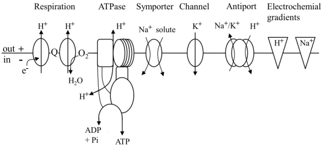
are relevant for cation homeostasis in C. glutamicum are shown in figure 1.
out
in Q O
2Respiration
H2O
ADP
+cPi ATP
ATPase
H+ H+ H+
H+
Na+ solute
Symporter
K+
Channel Antiport
+ -
e
-Na+/K+ H+
Na+ H+
Electrochemial gradients
Figure 1: The major transporters that are relevant for cation homeostasis inC. glutamicum
The respiratory chain establishes an electrochemical proton potential at the plasma membrane. This in turn can be used to energize the synthesis of ATP by the F1FO-ATPase or the export of Na+ and K+ions by antiporters.
The main entry route for sodium is the sodium-solute symport whereas the only known way for potassium to enterC. glutamicumcells is a channel. In addition, the direction of the electrochemical potentials of H+and Na+ ions is shown. Figure modified from [125].
1.4.1 Respiratory chain and ATP synthase
The respiratory chain plays a crucial role for bacteria. It translocates protons across the membrane and thereby establishes the primary electrochemical potential which is the driving force for many important transport-associated processes. C. glutamicum contains a so-called branched electron transport chain. The reducing equivalents are first transferred to menaquinone and then either to a supercomplex comprising the cytochrome bc1 complex and the cytochrome aa3 oxidase or to the cytochrome bd oxidase [12]. When using oxygen as terminal electron acceptor these three enzymes couple electron transfer to the export of protons and thus to the generation of the electrochemical proton potential. In detail, the cytochrome bc1-aa3 supercomplex exports six protons per two electrons while the bd oxidase only translocates two H
+per electron pair.
As many other aerobic respiring bacteria, C. glutamicum not only gains ATP by substrate level
phoysphorylation but also by oxidative phosphorylation [73]. The F
1F
O-ATPase is
essential for oxidative phosphorylation and uses the electrochemical proton potential, which
was established by the respiratory chain, for the generation of ATP. By fulfilling its function the
ATPase transports protons along their electrochemical potential into the cell. It is thought that
three to four protons are needed to form one ATP [95]. The ATPase can also work in reverse
1.4.2 The Na
+cycle
As mentioned earlier, the essential role of sodium ions for bacterial cells is often ascribed to its function in solute uptake. Na
+-substrate cotransport is a common way which is used for sub- strate uptake in all living cells [138]. The prerequisite for this type of transport is the presence of an electrochemical sodium potential that provides energy for the transport of substrates against their concentration gradient. Na
+ions that enter the cell are exported by Na
+/H
+antiporters in exchange with extracellular protons. This exchange helps to maintain the sodium gradient but it also driven by the electrochemical proton potential which again has to be maintained by the H
+ATPase as described above (Fig. 1).
1.4.3 Potassium transport systems
Bacteria are able to accumulate potassium by a number of different uptake systems. With the help of these uptake systems the intracellular potassium concentration of most bacteria can reach values of several hundred millimolar, even if the external K
+concentration is very low [36, 84]. The adaptation to different extracellular potassium availabilities is reflected in differences in kinetics, regulation and energetic coupling [36, 123].
Potassium channels are the evolutionary oldest form of potassium uptake systems from which active carrier systems developed [28]. The transport mediated by channels is passive, which means that K
+ions rapidly diffuse across the membrane down their electrochemical potential [27, 55, 62]. In addition to passive transport systems, there are also active potassium uptake systems present in most bacteria. One example for a primary active system is the E. coli Kdp system. This transporter is an inducible P-type ATPase which is only expressed when the cell needs K
+and the uptake by other systems is not sufficient [36]. However, the stimulus for this transporter is largely unknown. Secondary active transporters are, for example, the Trk and Kup systems which are K
+/H
+symporters [36, 124, 128]. Some bacteria also have an additional type of potassium uptake system, the secondary, Na
+-dependent Ktr [94].
In most species two or more independent saturable K
+-transport systems are present [36].
However, so far it is unknown why some strains harbour a variety of potassium uptake systems
while others only have simple channels. One explanation for this difference might be the
adaptation to different environments and pathogenicity [123].
Although there were some studies on potassium export, it is still only rudimentarily under- stood. The most completely described potassium efflux system is the E. coli Kef system.
This system is a glutathione-gated potassium/proton-antiporter that exchanges cytoplasmic potassium with extracellular protons and protects the cell against the toxicity of electrophilic compounds [91, 113]. K
+/H
+antiporters are ideal candidates to mediate the efflux of K
+ions [36]. Indeed, some cation/proton-antiporters already have been found with this function, for example the Vibrio cholerae Mrp systems or the antiporter ChaA from E. coli [29, 41].
Besides Helicobacter pylori, C. glutamicum also belongs to the apparently exceptional bacteria that harbour a potassium channel as the only functional uptake system [44, 123]. The function of CglK mediated K
+uptake is the internal accumulation of high potassium concentrations which is particular important under conditions of acidic and osmotic stress.
This also includes the adjustment of the membrane potential and the internal pH [44, 99]. The genome of C. glutamicum also contains an ORF (cg0187) putatively encoding a protein which is similar to the E. coli Kup systems. Under all conditions tested so far, no potassium uptake function could be ascribed to this transporter [44]. There have not yet been any investigations on the export of potassium in C. glutamicum.
1.4.4 Cation/proton antiporters
Cation/proton antiporters play crucial roles in bacteria and their need was first postulated by Mitchell in 1961 [92]. Na
+/H
+- and K
+/H
+-antiporters are described to be involved in cytoplasmic pH homeostasis, alkali tolerance, and resistance to elevated temperature and osmolality fluctuations [101, 120]. Concomitantly they mediate resistance to toxic levels of the transported cation. As secondary active transporters, antiporters use the electrochemical proton potential across the membrane as energy source. Thus, in the case of electrogenic Na
+/H
+antiporters that transport more protons than sodium ions, the primary electro- chemical proton potential is converted into an electrochemical sodium potential which in turn can be used for sodium-substrate costransport. The genomes of most non-marine bacteria contain five to nine genes that are predicted to encode putative Na
+/H
+- or K
+/H
+-antiporters [83]. Generally it is thought that bacteria exceedingly exposed to many different types of stress harbour a higher number of antiporters than pathogenic bacteria that live inside host cells.
Based on sequence similarity, monovalent cation/proton antiporters were classified into at least
family, sub-divided into CPA1, CPA2 and CPA3, and the Nha family, containing NhaA, NhaB, NhaC and NhaD proteins. Another family contains calcium/proton (CaCA) antiporters of which some are also able to transport Na
+and K
+ions. The majority of the antiporters are single gene products which in some cases form homooligomers to function [83].
On the structural level the CPA3 antiporters, also called Mrp (multiple resistance and pH-related), are the most complex type of antiporters. They are composed of six to seven different proteins that are thought to form hetero-oligomeric complexes in the membrane in which all members are required for full functionality. The according genes are organized in operons. mrp operons from different organisms were also named according to their function, for example pha (pH adaptation), sha (sodiumhydrogen antiporter) and mnh (multi-subunit Na
+/H
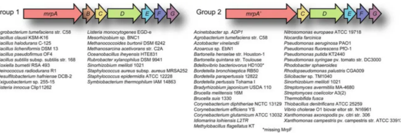
+antiporter) [125]. The mrp operons can be divided into two groups: group 1 and group 2 operons (Fig. 2). Most experimental data published so far were obtained with group 1 Mrp systems. These antiporters are encoded by seven genes ordered from mrpA to mrpG, and are found in Gram-positive as well as Gram-negative bacteria. For group 2 operons there is no separate mrpB gene but a larger so-called mrpA’ gene. This type of operon is dominantly found in Gram-negative bacteria. However, there are some groups of bacteria and archaea, such as enteric bacteria, lactobacilli and streptococci, that do not possess any Mrp antiporter systems [125].
Figure 2: Gene arrangements of group 1 and group 2mrpoperons.
Group 1 operons include seven genes namedmrpA-Gand are found in Gram-positive and Gram-negative bacteria.
Group 2 operons are dominantly found in Gram-negative bacteria and consist of six genes: the largermrpA’and mrpC-G. Figure taken from [125].
Cytoplasmic pH homeostasis at alkaline external pH of alkaliphilic Bacillus halodurans was the first role described for a Na
+/H
+antiporter from the Mrp family [54]. The import of protons supports the cytoplasmic pH homeostasis at external alkaline pH values. A Bacillus halodurans Mrp mutant did not grow at alkaline pH because the intracellular pH could not be retained lower than that of the medium. In addition, the Mrp system contributed to resistance to cytotoxic Na
+[71]. Meanwhile important physiologic roles in alkali-, Na
+- and K
+-resistance have been ascribed to other Mrp systems which were found to mediate Na
+(Li
+)/H
+- and/or K
+/H
+-antiport [10, 54, 60, 76, 77, 105, 141].
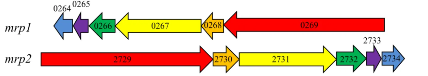
The genome of C. glutamicum contains genes for two CPA3 antiporters (Mrp1, Mrp2), one CPA1 antiporter (NhaP) and one CaCA antiporter (ChaA). Interestingly, genes for antiporters of the Nha family are missing [103]. Both mrp antiporters belong to the Mrp group 2, which means that both are encoded by an operon of six genes (Fig. 3)[125]. The precise function of these systems in C. glutamicum is not understood. The assumption that they might be Na
+/H
+antiporters is based on the observation that insertion mutants of both show a sodium- sensitive phenotype [43]. Interestingly, the Vibrio cholerae Mrp system, which also belongs to group 2, is able to transport potassium ions in addition to sodium and lithium ions [29].
Whether this is true for Mrp1 and/or Mrp2 from C. glutamicum has to be investigated. A nhaP insertion mutant was only briefly investigated and did not show a pH- or Na
+-sensitive phenotype [43]. A chaA insertion mutant was only investigated in regard to its involvement in potassium transport which could not be confirmed under the conditions tested [8]. Thus, for all four putative cation/proton antiporters from C. glutamicum there is not any proof for their function as cation/proton antiport activity so far.
mrp1 mrp2
0264 0265
0266 0267 0268 0269
2729 2730 2731 2732 2734
2733
Figure 3: Themrp-like operonsmrp1andmrp2fromC. glutamicum
Mrp1 is encoded by the genes cgl0269-cgl0264 and Mrp2 includes cgl2729-cgl2734 whereas cgl0269 and cgl2729correspond tomrpA’.
2 Thesis objectives
This work aims to get a better insight into cation homeostasis in Corynebacterium glutamicum.
Particular attention is directed on sodium and potassium ions. To do so it is necessary to understand the two ion fluxes that are involved in this process: import and export.
The main entry route for sodium ions into bacterial cells is represented by Na
+-solute symporters which were not investigated in this work. The only way for potassium to enter C.glutamicum is the potassium channel CglK. A basic characterization of CglK was already done before [44, 99]. As the entry routes for Na
+and K
+ions were already known, the main focus was directed on the export of these ions. Good candidates for this function are cation/proton antiporters which exchange internal Na
+and/or K
+with external protons.
There were already four putative candidates known: the two complex cation/proton-antiporters Mrp1 and Mrp2 and two simple antiporters ChaA and NhaP. Whether theses candidates function as Na
+/H
+and/or K
+/H
+antiporters should be proven on a physiological and on a biochemical level.
The physiological characterization should be performed with the native host C.glutamicum.
The investigation of single and multiple deletion mutants as well as a completely cation/proton antiporter deficient strain represents the main part of this project. Their growth behavior in presence of elevated Na
+or K
+concentrations should give information about the transported cation. As mutants probably accumulate one or the other cation, the measurement of the intracellular cation concentrations is required to support the physiological information. In addition, the influence of the external pH value has to be considered. At high external pH values less protons are available and cells lacking one or more antiporters might have problems with pH homeostasis and/or cation export.
As physiological experiments can only give hints on the transported cation, a biochemical
approach has to be used to prove Na
+/H
+and/or K
+/H
+antiport function. A system for this
has previously been established and uses everted E.coli membrane vesicles containing the
antiporter to be investigated. Addition of acridine orange to the vesicles and establishment of
a pH gradient (acid inside) leads to quenching of fluorescence. The addition of a transported
cation leads to dequenching of fluorescence and reflects cation-dependent proton movement
out of the everted membrane vesicles [126]. This approach allows the determination of the
substrate specificities as well as transport kinetics.
3 Materials & Methods
3.1 Bacterial strains, oligonucleotides and plasmids
All strains, oligonucleotides and plasmids used in this work are listed in table 1, table 2 and table 3, respectively.
Table 1:Strains used in this work
Cells were stored at -80◦C using Roti®-Store cryo vials as recommended by the manufacturer (Carl Roth GmbH
& Co. KG, Karlsruhe, Germany).
E. coli Description Reference
DH5αMCR F-endA1 supE44 thi-1λ-recA1 gyrA96 relA1 deoR∆(lacZYA-argF) [50]
U169 80ΦdlacZ∆M15mcrA∆(mrr hsdRMS mcrBC)
KNabc TG1 (∆nhaA∆nhaB∆chaA) [97]
KNabc_pGEM3Zf(+) KNabc carrying pGEM3Zf(+)empty plasmid This study
KNabc_pGEM3Zf(+)_Bpmrp KNabc carrying pGEM3Zf(+)_Bpmrp This study
KNabc_pGM36 KNabc carrying pGM36 This study
KNabc_pGEM3Zf(+)_PronhaAmrp1 KNabc carrying pGEM3Zf(+)_PronhaAmrp1 This study
KNabc_pGEM3Zf(+)_mrp2 KNabc carrying pGEM3Zf(+)_mrp2 This study
KNabc_pGEM3Zf(+)_chaA KNabc carrying pGEM3Zf(+)_chaA This study
KNabc_pGEM3Zf(+)_nhaP KNabc carrying pGEM3Zf(+)_nhaP This study
C. glutamicum Description Reference
ATCC13032 wild type [1]
ATCC13032_pEKEx2 ATCC13032 carrying pEKEx2 empty plasmid This study
∆mrp1 ATCC13032 with deletion of themrp1operon (cgl0269-cgl0264) This study
∆mrp2 ATCC13032 with deletion of themrp2operon (cgl2729-cgl2734) This study
∆chaA ATCC13032 with deletion of the gene coding for ChaA (cgl1082) This study I:nhaP ATCC13032 with insertion of pDRIVE in the gene coding for NhaP (cgl1436) [64]
∆mrp1∆mrp2∆chaA ATCC13032 with deletion ofmrp1,mrp2andcgl1082 This study
∆mrp1∆mrp2∆nhaP ATCC13032 with deletion ofmrp1,mrp2andcgl1436 This study AQM ATCC13032 with deletion ofmrp1,mrp2,cgl1082andcgl1436 This study
AQM_pEKEx2 AQM carrying pEKEx2 empty plasmid This study
AQM_pEKEx2_mrp1-His AQM carrying pEKEx2_mrp1-His This study
AQM_pEKEx2_mrp2-His AQM carrying pEKEx2_mrp2-His This study
AQM_pEKEx2_chaA-His AQM carrying pEKEx2_chaA-His This study
AQM_pEKEx2_nhaP-His AQM carrying pEKEx2_nhaP-His This study
∆mrp1∆mrp2∆chaA∆nhaP∆cg3038 ATCC13032 with deletion ofmrp1,mrp2,cgl1082,cgl1436andcg3038 This study
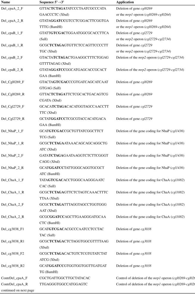
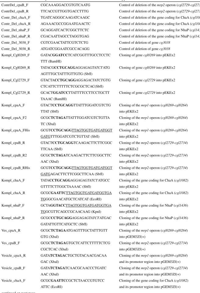
Table 2:Oligonucleotides used in this work.
Ribosome binding sites are in italic and the sequence for the 6xHis-tag is underlined. Restriction sites are in bold and the corresponding restriction endonucleases are named in brackets. All primers were purchased from Operon (Cologne, Germany) and solved in water to obtain a concentration of 100 nmol/ml.
Name Sequence 5’→3’ Application
Del_cpaA_1_F GTATAGTCGACGTCTTGCATTGCCGCTGC Deletion of themrp1operon (cgl0269-cgl0264) TGAT (SalI)
Del_cpaA_1_R GTACTATCTAGAGCTTTTAGAAAAAAAGG Deletion of themrp1operon (cgl0269-cgl0264) GGCGTGCC (XbaI)
Table 2 - continued from previous page
Name Sequence 5’→3’ Application
Del_cpaA_2_F GTTACTCTAGATATCCCTAATCGCCCATA Deletion of genecgl0269
GAACCCTC (XbaI) or themrp1operon (cgl0269-cgl0264) Del_cpaA_2_R GTATAGGATCCGTCCTCGGACTTCGGTGA Deletion of genecgl0269
TTTG (BamHI) or themrp1operon (cgl0269-cgl0264) Del_cpaB_1_F GTATTGTCGACTGGAATGGCGCACCTTCA Deletion of genecgl2729
(SalI) or themrp2operon (cgl2729-cgl2734)
Del_cpaB_1_R GCGCTCTAGAGTGTTCTCCAGTTCCCCTT Deletion of genecgl2729
TGC (XbaI) or themrp2operon (cgl2729-cgl2734)
Del_cpaB_2_F GTACTATCTAGACTGAAGGCTTTCTGGAG Deletion of themrp2operon (cgl2729-cgl2734) GTTTTAGAG (XbaI)
Del_cpaB_2_R GTATAGGATCCGGCATGAGCACCGCACT Deletion of themrp2operon (cgl2729-cgl2734) GAA (BamHI)
Del_Cgl0269_F GTACTAGTCGACCCGTGATCAGCATCAAT Deletion of genecgl0269 GTGAG (SalI)
Del_Cgl0269_R GTTACTCTAGATTCTCGCACTGACAGTCG Deletion of genecgl0269 CGATA (XbaI)
Del_Cgl2729_F GCACATCTAGACACATGGTAGCCAACCTT Deletion of genecgl2729 TTC (XbaI)
Del_Cgl2729_R GCTATGGATCCTCGCGTACCACATGACA Deletion of genecgl2729 GAA (BamHI)
Del_NhaP_1_F GCATGTCGACCGCTGTTATCGGCTTCT Deletion of the gene coding for NhaP (cgl1436) TCG (SalI)
Del_NhaP_1_R GCGCTCTAGAATAAACAGCAGCAGGCTG Deletion of the gene coding for NhaP (cgl1436) ATC (XbaI)
Del_NhaP_2_F GATATCTAGAGAATAAGGTCTCTTCGGGT Deletion of the gene coding for NhaP (cgl1436) CAGG (XbaI)
Del_NhaP_2_R GCATGGATCCTATTGGGCAGGTGCGCT Deletion of the gene coding for NhaP (cgl1436) ATC (BamHI)
Del_ChaA_1_F TATAGTCGACACCTGGGCAAGGGAATC Deletion of the gene coding for ChaA (cgl1082) CAC (SalI)
Del_ChaA_1_R GCGCTCTAGAGTTCTCTAGTCAAACTTTC Deletion of the gene coding for ChaA (cgl1082) TTAA (XbaI)
Del_ChaA_2_F GCGCTCTAGATTTAGGTAGCCTGGTGGG Deletion of the gene coding for ChaA (cgl1082) AAT (XbaI)
Del_ChaA_2_R GCGCGGATCCAGCTTGAAGGGATGCAA Deletion of the gene coding for ChaA (cgl1082) CTC (BamHI)
Del_cg3038_F1 GCATGTCGACACGCCCAATCCTCCTAC Deletion of genecg3038 TAC (SalI)
Del_cg3038_R1 GCGCTCTAGACTCTAGGTGGCGTTTTAAG Deletion of genecg3038 (XbaI)
Del_cg3038_F2 GCGCTCTAGACACTGTCTCGTGTATCTAT Deletion of genecg3038 ATCG (XbaI)
Del_cg3038_R2 GCATGGATCCGTGGTGGTGGTTGATGAT Deletion of genecg3038 TG (BamHI)
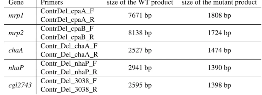
ContrDel_cpaA_F CGCTGATTGGCTTGCTATACAC Control of deletion of themrp1operon (cgl0269-cgl0264) ContrDel_cpaA_R TTGAGGGTGGCCATGGAGTC Control of deletion of themrp1operon (cgl0269-cgl0264) continued on next page
Table 2 - continued from previous page
Name Sequence 5’→3’ Application
ContrDel_cpaB_F CGCAAAGGACCGTGTCAATG Control of deletion of themrp2operon (cgl2729-cgl2734) ContrDel_cpaB_R TTCACCGTTGGTGACCTTTG Control of deletion of themrp2operon (cgl2729-cgl2734) Contr_Del_chaA_F TGATCAGGGCAAGATCAAGC Control of deletion of the gene coding for ChaA (cgl1082) Contr_Del_chaA_R AGAAACGCCGGAATGAACTC Control of deletion of the gene coding for ChaA (cgl1082) Contr_Del_nhaP_F GCAGGATCACTCGGCTTCTC Control of deletion of the gene coding for NhaP (cgl1436) Contr_Del_nhaP_R CGACAATTAGCCTAGGTGAG Control of deletion of the gene coding for NhaP (cgl1436) Contr_Del_3038_F CGTCGAACTATTCGTCTCTG Control of deletion of genecg3038
Contr_Del_3038_R ATGATCGGAATCGCCACAGG Control of deletion of genecg3038 Kompl_Cgl0269_F GATACGGATCCTCATCGGTTTGCCTCCTC Cloning of genecgl0269into pEKEx2
TTT (BamHI)
Kompl_Cgl0269_R TATACGCCTGCAGGAGGAGAGTATCTATG Cloning of genecgl0269into pEKEx2 AGTTTGCTATTTGTTGTG (SbfI)
Kompl_Cgl2729_F GTACTACCTGCAGGAGGAGACTATCTGTG Cloning of genecgl2729into pEKEx2 CTCATTCTTTTTCTCGCGCTCAC(SbfI)
Kompl_Cgl2729_R GCACTGGATCCTTATTTTCCTTCCTGCTT Cloning of genecgl2729into pEKEx2 TAAAC (BamHI)
Kompl_cpaA_F GTACTCCTGCAGGTTATTTGGATCGTCTG Cloning of themrp1operon (cgl0269-cgl0264)
TTAT (SbfI) into pEKEx2
Kompl_cpaA_F2 GCGCTCTAGATTATTTGGATCGTCTGTTA Cloning of themrp1operon (cgl0269-cgl0264)
TC (XbaI) into pEKEx2
Kompl_cpaA_FHis GCGTCCTGCAGGTTAGTGGTGATGATGGT Cloning of themrp1operon (cgl0269-cgl0264) GATGTTTGGATCGTCTGTTAT (SbfI) into pEKEx2
Kompl_cpaB_R GTACTCCTGCAGGTCAAGACTTCTTCGGC Cloning of themrp2operon (cgl2729-cgl2734)
TTCAA (SbfI) into pEKEx2
Kompl_cpaB_R2 GCGCTCTAGATCAAGACTTCTTCGGCTTC Cloning of themrp2operon (cgl2729-cgl2734)
AAC (XbaI) into pEKEx2
Kompl_cpaB_RHis GCGTCCTGCAGGTTAGTGGTGATGATGGT Cloning of themrp2operon (cgl2729-cgl2734) GATGAGACTTCTTCGGCTTCAA (SbfI) into pEKEx2
Kompl_chaA_F TATACCTGCAGGAGGAGAGTATCTATGCC Cloning of the gene coding for ChaA (cgl1082) GTTTTCTTGGCTAAAAC (SbfI) into pEKEx2
Kompl_chaA_R GCGCGAATTCTTAGTGGTGATGATGGTGA Cloning of the gene coding for ChaA (cgl1082) TGGGCGAACATGCTCATCAT (EcoRI) into pEKEx2
Kompl_nhaP_F GCTAGGTACCTTAGTGGTGATGATGGTGA Cloning of the gene coding for NhaP (cgl1436) TGGCGTTCAGCCGCAACAAG (KpnI) into pEKEx2
Kompl_nhaP_R GCGCCCTGCAGGAGGAGAGTATCTATGAC Cloning of the gene coding for NhaP (cgl1436)
GATATTGTTCATGCTC (SbfI) into pEKEx2
Ves_cpaA_R GCGCTCTAGAATGAGTTTGCTATTTGTT Cloning of themrp1operon (cgl0269-cgl0264)
GTG (XbaI) into pGEM3Zf(+)
Ves_cpaB_F GCGCTCTAGAGTGCTCATTCTTTTTCTCG Cloning of themrp2operon (cgl2729-cgl2734)
CGCTCAC (XbaI) into pGEM3Zf(+)
Vesicle_cpaA_R GATATCTAGACTGCTGTACAACGACAA Cloning of themrp1operon (cgl0269-cgl0264)
GAC (XbaI) and its promotor region into pGEM3Zf(+)
Vesicle_cpaB_F GATATCTAGATCAACGCAACCCTGATC Cloning of themrp2operon (cgl2729-cgl2734)
AAC (XbaI) and its promotor region into pGEM3Zf(+)
Vesicle_chaA_F GCGCGAATTCCGCTCTAACCGTGTCC Cloning of the gene coding for ChaA (cgl1082) ATTC (EcoRI) and its promotor region into pGEM3Zf(+)
Table 2 - continued from previous page
Name Sequence 5’→3’ Application
Vesicle_chaA_R GCGCCCTGCAGGTCAGGCGAACATGCTCA Cloning of the gene coding for ChaA (cgl1082) TCAT (SbfI) and its promotor region into pGEM3Zf(+) Vesicle_nhaP_F GCTACCTGCAGGCTAGCGTTCAGCCGCAA Cloning of the gene coding for NhaP (cgl1436)
CAAG (SbfI) and its promotor region into pGEM3Zf(+) Vesicle_nhaP_R GCTAGGTACCATTGGGCAGGTGCGCTA Cloning of the gene coding for NhaP (cgl1436)
TCC (KpnI) and its promotor region into pGEM3Zf(+)
mrp1_R ATGAGTTTGCTATTTGTTGTG Cloning ofmrp1(cgl0269-cgl0264) and thenhaAE.coli ormrpB. pseudofirmusOF4promotor into pGEM3Zf(+) mrp2_F ATGCTCATTCTTTTTCTCGCGCTCAC Cloning ofmrp2(cgl2729-cgl2734) and thenhaAE.coli
ormrpB. pseudofirmusOF4promotor into pGEM3Zf(+) Pro_Bpmrp_F GCGCTCTAGACGAACTTGACCTAAGCC Cloning of themrp1/mrp2operon and thenhaAE.coli
(XbaI) ormrpB. pseudofirmusOF4promotor into pGEM3Zf(+) Pro_nhaA_F GCGCTCTAGAGAAAAGAAGAACTAACTCG Cloning of themrp1/mrp2operon and thenhaAE.coli
(XbaI) promotor into pGEM3Zf(+)
Pro_Bpmrp_R_mrp1 AACAAATAGCAAACTCATATCACATACCT Cloning of themrp1operon (cgl0269-cgl0264) and the CCTTAAATGG mrpB .pseudofirmusOF4promotor into pGEM3Zf(+) Pro_nhaA_R_mrp1 AACAAATAGCAAACTCATTTTTTATTTCT Cloning of themrp1operon (cgl0269-cgl0264) and the
CTTTCAGGTGAATAG nhaAE. colipromotor into pGEM3Zf(+)
Pro_Bpmrp_R_mrp2 CGAGAAAAAGAATGAGCATATCACATACC Cloning of themrp2operon (cgl2729-cgl2734) and the TCCTTAAATGG mrpB. pseudofirmusOF4promotor into pGEM3Zf(+) Pro_nhaA_R_mrp2 CGAGAAAAAGAATGAGCATTTTTTATTTC Cloning of themrp2operon (cgl2729-cgl2734) and the
TCTTTCAGGTGAATAG nhaAE. colipromotor into pGEM3Zf(+)
Seq_0269_2_R GTGAACGGCATCTTCTTGAC Control PCRs
Seq_0269_2_F GCATTTGGGTTGGATCGTAG Control PCRs
Seq_0269_R AGAGCTCGCCCAACGTATCC Control PCRs
Seq_0269_F CGCATCAAGCCCAGCAGAAG Control PCRs
Seq_0267_R GCCACGCCACCGACATAAAG Control PCRs
Seq_0267_F GTGTGGCGTGAAGTCTTCTG Control PCRs
Seq_2731_F CCACTGCCCTGTTGGTTTCC Control PCRs
Seq_2731_2_F CCATGCTGACGGTGAGTTCC Control PCRs
Seq_2731_R CAAGGTGAGCAGCACGTATG Control PCRs
Seq_2729_F CGGCCGTATCTTGGGAACTG Control PCRs
Seq_2729_R AACCGATGACGATCAGGTAG Control PCRs
Seq_2729_2_F GCTGGATACCGTGTTGAATG Control PCRs
Seq_2729_2_R GTGCGATCCACAGCTTAAAC Control PCRs
Table 3:Plasmids used in this study
Plasmid Relevant characteristic Reference
pJET Cloning vector. AmpR Thermo Scientific ,USA
pGEM3Zf(+) Cloning vector. AmpR Promega
pGEM3Zf(+)_Bpmrp pGEM3Zf(+) derivative; contains the fullmrpoperon [126]
fromB. pseudofirmusOF4
pGM36 pBR322 derivative; contains the fullnhaAgene fromE.coli [48]
pGEM3Zf(+)_mrp1 pGEM3Zf(+) derivative; contains the fullmrp1operon (cgl0269-cgl0264) This study fromC. glutamicum
pGEM3Zf(+)_mrp2 pGEM3Zf(+) derivative; contains the fullmrp2operon (cgl2729-cgl2734) This study fromC. glutamicum
pGEM3Zf(+)_chaA pGEM3Zf(+) derivative; contains the full gene coding for ChaA (cgl1082) This study fromC. glutamicum
pGEM3Zf(+)_nhaP pGEM3Zf(+) derivative; contains the full gene coding for NhaP (cgl1436) This study fromC. glutamicum
pGEM3Zf(+)_PronhaAmrp1 pGEM3Zf(+) derivative; contains themrp1operon (cgl0269-cgl0264) This study fromC. glutamicumunder the control of thenhaAE. colipromotor
pEKEx2 Expression vector; KmR,lacIQ,tacP,oriof pBL1, pUC18 mcs [33]
pEKEx2_mrp1-His pEKEx2 derivative; contains themrp1operon (cgl0269-cgl0264) This study fromC. glutamicumfollowed by the sequence for a 6xHis-tag
pEKEx2_mrp2-His pEKEx2 derivative; contains themrp2operon (cgl2729-cgl2734) This study fromC. glutamicumfollowed by the sequence for a 6xHis-tag
pEKEx2_chaA-His pEKEx2 derivative; contains the gene coding for ChaA (cgl1082) This study fromC. glutamicumfollowed by the sequence for a 6xHis-tag
pEKEx2_nhaP-His pEKEx2 derivative; contains the gene coding for NhaP (cgl1436) This study fromC. glutamicumfollowed by the sequence for a 6xHis-tag
pK19msB Gene deletion plasmid forC. glutamicum. KanR, oriVE.coli, oriT, mob, sacB [117]
pK19msB_0269 pK19msB derivative; contains the flanking regions of genecgl0269 This study fromC. glutamicum
pK19msB_2729 pK19msB derivative; contains the flanking regions of genecgl2729 This study fromC. glutamicum
pK19msB_cpaA pK19msB derivative; contains the flanking regions of This study themrp1operon (cgl0269-cgl0264) fromC. glutamicum
pK19msB_cpaB pK19msB derivative; contains the flanking regions of This study themrp2operon (cgl2729-cgl2734) fromC. glutamicum
pK19msB_chaA pK19msB derivative; contains the flanking regions of the gene coding This study for ChaA (cgl1082) fromC. glutamicum
pK19msb_nhaP pK19msB derivative; contains the flanking regions of the gene coding This study for NhaP (cgl1436) fromC. glutamicum
pK19msB_3038 pK19msB derivative; contains the flanking regions of This study genecg3038fromC. glutamicum
3.2 Media and cultivation of E. coli and C. glutamicum
3.2.1 Media preparation
All media and solutions were sterilized by autoklaving or passing through a membrane syringe filter (pore size 0.22 µ m). For plates, agar (15 g/l) was added to the liquid medium before sterilization.
3.2.2 Media and cultivation of E. coli
E. coli was grown in LB, LBK or L
0(see below). If appropriate, 25 µg/ml kanamycin and/or 100 µ g/ml carbenicillin were added. Incubation occured at 37
◦C and 125 rpm.
LB LBK L
0Tryptone 10 g/l 10 g/l 10 g/l
Yeast extract 5 g/l 5 g/l 5 g/l
NaCl 10 g/l - -
KCl - 6 g/l -
pH 7.0 by NaOH 7.0 by KOH 7.0 by KOH
Precultivation was performed in a volume of 5 ml in glass test tubes. For growth experiments, a volume of 10 ml was used in 100 ml baffled shaking flasks. For membrane vesicle preparation, cultivation was carried out with 500 ml medium in 2 l baffled shaking flasks.
3.2.3 Media and cultivation of C. glutamicum
As complex medium for C. glutamicum LB, L
0and L
0S (L
0with 400 mM sorbitol, 7 mM KCl, 100 mM BTP, pH 7.5 by HCl) were used. If appropriate, 25 µ g/ml kanamycin and/or 1% glucose were added.
Minimal media used were MMI and CgXII (see below).
MMI CgXII (NH
4)
2SO
45 g/l 20 g/l
urea 5 g/l 5 g/l
KH
2PO
47.3 mM 1 g/l K
2HPO
45.7 mM 1 g/l NaH
2PO
47.3 mM - Na
2HPO
45.7 mM -
CgXII was supplemented with 250 mg/l MgSO
4* 7 H
2O, 10 mg/l CaCl
2, 1 mg/l FeSO
4* 7 H
2O, 1.375 mg/l MnSO
4* 4 H
2O, 0.1 mg ZnSO
4* 7 H
2O, 0.031 mg CuSO
4* 5 H
2O, 2 µg NiCl
2* 6 H
2O, 30 µ g/ml protocatechuic acid and 200 µ g/l biotin. MOPS (0.2 M), HEPPS (0.2 M), MES (0.2 M), CHES (0.2 M) or BTP (0.1 M) were used to buffer CgXII. The pH was adjusted with NaOH, KOH or HCl. If appropriate, 25 µ g/ml kanamycin was added.
Incubation occured at 30
◦C and 125 rpm.
The first preculture was grown in a volume of 5 ml in glass test tubes, followed by a second preculture in 10 ml medium in 100 ml baffled shaking flasks. Growth experiments were carried out in 10 ml medium in 100 ml baffled shaking flasks or in 200 µ l medium in 96-well microtiter plates.
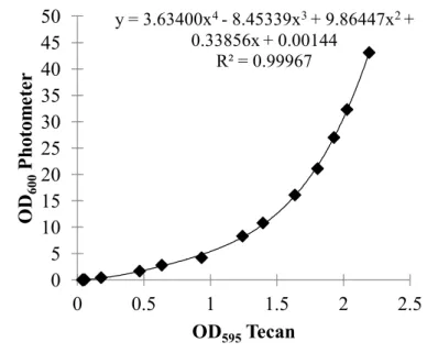
3.2.4 Cultivation of C. glutamicum in microtiter plates
C. glutamicum cells from an overnight preculture were washed once in CgXII minimal medium (w/o glucose) and resuspended to an OD
600of 20. A volume of 5 µ l was added to 195 µl medium in a microtiter plate well. Plates were sealed with a Breathe-Easy
®sealing membrane (Diversified Biotech, Dedham, USA). Incubation occured at 30
◦C and 1200 rpm on a special microtiter plate shaker. The optical density was measured at 595 nm in a plate reader (Tecan infinite
®F200 Pro, Tecan Group Ltd., Männedorf, Germany). For the determination of growth rates the measured values were converted into OD
600using equation 1. The corresponding curve is shown in figure 4 (p. 26).
OD600=3.634(OD595)4−8.45339(OD595)3+9.86447(OD595)2+0.33856(OD595) +0.00144 (1)
3.3 Molecular biology methods
3.3.1 DNA purification, restriction digest and ligation
Plasmid DNA from E. coli was isolated with the High Pure Plasmid Isolation Kit (Roche, Switzerland). DNA concentration was determined with the Ultrospec 2100 Pro spectrophotometer (GE Healthcare, Freiburg, Germany). FastDigest
®restriction enzymes were used for restriction digest, ligation was performed with T4 DNA Ligase (Thermo Scientific, USA).
3.3.2 Polymyerase chain reaction
Polymerase chain reaction (PCR [93]) was performed in an analytikjena FlexCycler (Analytik Jena, Jena, Germany). For preparative PCR Phusion
®High-Fidelity DNA Polymerase (Thermo Scientific, USA) was used with chromosomal or plasmid DNA as a template. PCR fragments were either digested and directly ligated into the destination plasmid or sub-cloned into pJET using the CloneJet PCR Cloning Kit (Thermo Scientific, USA). For analytic purposes colony PCRs were performed with EconoTaq
®PLUS GREEN 2X Master Mix (Lucigen, USA). An E. coli or C. glutamicum colony was resuspended in 50 µ l water and 1 µ l was used as template.
3.3.3 Agarose gel electrophoresis and isolation of DNA from agarose gels
PCR products and digested plasmid DNA were seperated by gel electrophoresis on 0.8%
agarose gels (Agarose NEEO Ultra-Quality, Roth, Karlsruhe, Germany) in 1x TAE buffer at 85 V [116]. Afterwards DNA was stained by incubating the gel in 1 µ g/ml ethidium bromide solution. Visualization was performed with the UVP BioDoc-It
™Imaging system (Analytik Jena, Jena, Germany). The High Pure PCR Product Purification Kit (Roche, Switzerland) was used to isolate DNA from agarose gels.
3.3.4 Competent E. coli cells and transformation
Competent E. coli DH5α were prepared as described previouly and stored at -80
◦[116].
Chemical competent E. coli KNabc were prepared using the CaCl
2method. 100 µ l of a L
0over-night culture were used to inoculate 10 ml of the same medium in a Corning
®50 ml
centrifuge tube (Sigma Aldrich, USA). Cells were grown until an OD
600of about 0.4-0.6 was reached. 1 ml per reaction was transferred to a reaction tube and centrifuged for one minute at 11,000 rpm. Cells were resuspended in 500 µ l ice-cold CaCl
2(0.05 M) by vortexing and incubated on ice for 10 minutes. Subsequently cells were centrifuged as before, resuspended in 300 µ l ice-cold CaCl
2(0.05 M) by pipetting and incubated on ice for 30 minutes.
The transformation procedure was the same for both E. coli strains. 10 µ l of ligation mixture or 0.25 µl of purified plasmid was added to the competent cells. After an incubation on ice for 30 minutes, a heat shock was performed (42
◦C, 2 minutes). 600 µl of LB or L
0was added to the cells, followed by incubation at 37
◦C and 125 rpm for one hour. Afterwards cells were centrifuged (11,000 rpm; 1 min) and resuspended in 100 µ l of fresh medium. An appropriate amount was plated on agar plates which were incubated at 37
◦C over night.
3.3.5 Competent C. glutamicum cells and transformation
Preparation of competent C. glutamicum cells and transformation were performed as described previously with following exceptions [131]. Cells were washed only once with 50 ml ice-cold 10% (v/v) glycerol, frozen in 55 µl aliquots and stored at -80
◦C.
3.3.6 Gene deletion in C. glutamicum
Gene deletion in C. glutamicum was performed as described before [117, 32]. DNA sequences of about 500 bp homologous to the up- and downstream regions of the target gene were amplified by PCR and directly ligated into pK19mobsacB. PCR with primers flanking the target sequence was applied to verify successful target gene deletion.
3.4 Biochemical methods
3.4.1 Preparation of everted membrane vesicles from E. coli
A single colony was used to inoculate 5 ml LBK medium. After 8 hours 2.5 ml of the
preculture was used to inoculate 500 ml LBK medium, this culture was incubated for 16
hours. If appropriate 50-100 mM NaCl were added to both cultures. Cells were harvested
(5,000 rpm, 15 min), washed with 50 ml TCDG buffer (10 mM Tris (pH 8.0 by HCl), 140 mM
choline-Cl, 5 mM MgCl
2, 10% (v/v) glycerol, 1 mM DTT (fresh)) and resuspended in 20 ml of the same buffer supplemented with DNase and protein inhibitor cocktail. Vesicles were made by single passage through a French®Pressure Cell Press at 1,100 psi. Cell debris was removed by centrifugation (4,000 rpm, 20 min). Supernatants were centrifuged twice at 12,00 rpm for 10 minutes. Vesicles were collected by ultracentrifugation at 40,000 rpm for 90 minutes and resuspended in 1 ml of fresh TCDG buffer. All steps were performed at 4
◦C. Samples were stored at -20
◦C or -80
◦C.
3.4.2 Determination of protein concentration
Protein concentrations of everted membrane vesicle preparations were determined with a modified Lowry method [104]. BSA was used as standard and the assay volume was down-scaled to 1/10.
3.4.3 Acridine orange fluorescence assay
To measure ∆pH-dependent antiport activity aliquots of vesicles (66 µ g) were added to 2 ml of an assay buffer (50 mM Bis-Tris-Propane, adjusted to the indicated pH, 140 mM choline-Cl, 2.5 mM MgCl
2and 1 µ M acridine orange (AO)) and incubated for 30 minutes at room temperature to equilibrate. The measurement occured in a thermostated cuvette (23
◦C).
Respiration-dependent formation of the ∆pH was initiated by the addition of 2.5 mM Tris- succinate (pH 8.0), and the resulting quenching of AO fluorescence was monitored with an Aminco-Bowman spectrofluorometer (excitation at 493 nm and emission at 528 nm). Antiport activity was assessed as the percent of dequenching. NaCl or KCl were added at 2.5 mM in the pH-profile determinations and at 0.2-5 mM for the determinations of halfmaximal effective concentrations (apparent K
M). Finally, 10 mM NH
4Cl was added to the assay buffer to abolish any ∆pH.
3.4.4 Determination of intracellular cation concentrations
A volume of 1.8 ml cell suspension was centrifuged for 2 minutes at 11,000 rpm. The supernatant was removed and stored at -20
◦C. The pellet was resuspended in 1.8 ml of dis- tilled water and cells were broken by incubation in an ultrasonic bath at 85
◦C for 45 minutes.
Cell debris was removed by centrifugation at 20,000 rpm for 30 minutes. The supernatant was
removed and used to determine cation concentrations with a flame photometer (ELEX 6361, Eppendorf, Germany). Calibration was performed with solutions of 20-150 µ M NaCl or 50- 300 µ M KCl. For calculations of the intracellular cation concentrations a cytoplasmic volume of 1.6 µl per mg of cell dry matter (CDM) was assumed and equation 2 was used to calculate the CDM. The contamination by external medium in the pellet was assumed to be 1.5 times the cytoplasmic volume.
CDM[mg] =0.36∗OD600∗cellvolume[ml] (2)
3.4.5 Determination of osmolality
The osmolality of media was measured with a cryoscopic osmometer (Osmomat
®030, Gonotec GmbH, Berlin, Germany). Calibration was performed with solutions of 0.1-2.5 osmol/kg and samples were analysed as recommended by the manufacturer.
3.4.6 Radioactive measurement of membrane potential
The membrane potential was essentially determined from the distribution of the membrane permeable, lipophilic cation [
14C]-Tetraphenylphosphoniumbromid (TPP
+) as described be- fore [114, 66]. The accumulation of TPP
+inside the cell depends on the electrical potential across the membrane. In equilibrium the electrical membrane potential is equal to the chemical potential of TPP
+. Therewith it is possible to calculate the electrical potential when knowing the intra- and extracellular concentration of TPP
+using equation 3.
∆Ψ= (−2.303R∗T/F)∗log([T PP+]in/[T PP+]ex) (3)
with F = faraday constant, R = universal gas constant, T = asolute temperature.
A volume of 2 µ l/(ml cell suspension) TPP
+working solution (final concentration 10 µ M,
specific activity 2,21 x 10
9D/min mmol) were added to the cells. After 30 minutes 200 µl
cell suspension were subjected to silicone oil centrifugation (70 µ l 1.09 g/cm
3silicone oil,
30 µ l 20% perchloric acid) for the determination of the extracellular TPP
+concentration.
The radioactivity of 150 µl of the supernatant was measured by liquid scintillation counting.
To determine the intracellular TPP
+concentration 150 µ l of the cell suspension was directly measured and the concentrations were calculated with equation 4.
[T PP+]in= [T PP+]total−[T PP+]ex (4)