Report
Limitation of TCA Cycle Intermediates Represents an Oxygen-Independent Nutritional Antibacterial Effector Mechanism of Macrophages
Graphical Abstract
Highlights
d
Hypoxia curtails C. burnetii replication in macrophages without reducing bacterial viability
d
Hypoxia induces robust HIF1 a accumulation in infected macrophages
d
HIF1 a reduces STAT3 activation and citrate availability in infected hypoxic macrophages
d
Low levels of TCA metabolites impede C. burnetii replication in macrophages
Authors
Inaya Hayek, Fabian Fischer,
Jan Schulze-Luehrmann, ..., Stefan Wirtz, Jonathan Jantsch, Anja L€ uhrmann
Correspondence
jonathan.jantsch@ukr.de (J.J.), anja.luehrmann@uk-erlangen.de (A.L.)
In Brief
The mechanisms that control bacterial infection under hypoxic conditions are only partially understood. Hayek et al.
show that hypoxia-mediated stabilization of HIF1 a results in the inhibition of STAT3 activation and the reduction of TCA metabolite levels, including citrate, in macrophages. This prevents C. burnetii replication without reducing bacterial viability.
Hayek et al., 2019, Cell Reports 26 , 3502–3510 March 26, 2019 ª 2019 The Author(s).
https://doi.org/10.1016/j.celrep.2019.02.103
Cell Reports
Report
Limitation of TCA Cycle Intermediates
Represents an Oxygen-Independent Nutritional Antibacterial Effector Mechanism of Macrophages
Inaya Hayek,
1,7Fabian Fischer,
1,2,7Jan Schulze-Luehrmann,
1,7Katja Dettmer,
3Katharina Sobotta,
4Valentin Schatz,
5Lisa Kohl,
1Katharina Boden,
4Roland Lang,
1Peter J. Oefner,
3Stefan Wirtz,
6Jonathan Jantsch,
5,* and Anja L€ uhrmann
1,8,*
1
Mikrobiologisches Institut, Universita¨tsklinikum Erlangen, Friedrich-Alexander-Universita¨t (FAU) Erlangen-N€ urnberg, 91054 Erlangen, Germany
2
Klinik f€ ur Innere Medizin I, Universita¨tsklinikum Regensburg, 93053 Regensburg, Germany
3
Institut f€ ur Funktionelle Genomik, Universita¨t Regensburg, 93053 Regensburg, Germany
4
Institut f€ ur Medizinischen Mikrobiologie, Universita¨tsklinikum Jena, 07743 Jena, Germany
5
Institut f€ ur Klinische Mikrobiologie und Hygiene, Universita¨tsklinikum Regensburg, Universita¨t Regensburg, 93053 Regensburg, Germany
6
Medizinische Klinik 1, Universita¨tsklinikum Erlangen, Friedrich-Alexander-Universita¨t (FAU) Erlangen-N€ urnberg, 91052 Erlangen, Germany
7
These authors contributed equally
8
Lead Contact
*Correspondence: jonathan.jantsch@ukr.de (J.J.), anja.luehrmann@uk-erlangen.de (A.L.) https://doi.org/10.1016/j.celrep.2019.02.103
SUMMARY
In hypoxic and inflamed tissues, oxygen (O
2)-depen- dent antimicrobial defenses are impaired due to a shortage of O
2. To gain insight into the mechanisms that control bacterial infection under hypoxic condi- tions, we infected macrophages with the obligate intracellular pathogen Coxiella burnetii, the causative agent of Q fever. Our experiments revealed that hyp- oxia impeded C. burnetii replication in a hypoxia- inducible factor (HIF) 1 a -dependent manner. Mecha- nistically, under hypoxia, HIF1 a impaired the activity of STAT3, which in turn reduced the intracellular level of TCA cycle intermediates, including citrate, and impeded C. burnetii replication in macrophages.
However, bacterial viability was maintained, allowing the persistence of C. burnetii, which is a prerequisite for the development of chronic Q fever. This knowl- edge will open future research avenues on the path- ogenesis of chronic Q fever. In addition, the regula- tion of TCA cycle metabolites by HIF1a represents a previously unappreciated mechanism of host de- fense against intracellular pathogens.
INTRODUCTION
O
2levels of <1%, which can be found in inflamed and infected tissues, are known to incapacitate key antimicrobial and immu- nomodulating effector enzymes such as indoleamine 2,3-diox- ygenase, phagocyte oxidase, and type 2 nitric oxide (NO) syn- thase (NOS2), which depend on oxygen (O
2) as a substrate (Jantsch and Scho¨del, 2015). Nevertheless, bacterial infections can be controlled in the absence of O
2-dependent antimicrobial defense (Campbell et al., 2014). Enhanced expression of anti- microbial peptides (Peyssonnaux et al., 2005) may fulfill this
task in part and restrain the replication of pathogens, until tissue O
2levels normalize and O
2-dependent antimicrobial effectors such as nicotinamide adenine dinucleotide phos- phate-positive (NADPH) oxidase (PHOX) or NOS2 kick in (Jantsch and Scho¨del, 2015). However, additional defense strategies are likely to contribute to the control of infection un- der hypoxic conditions. We hypothesized that the limitation of metabolites that are essential for the proliferation of pathogens may function as an antimicrobial effector mechanism in in- flamed hypoxic tissue.
To test this hypothesis, we resorted to Coxiella burnetii, an obligate intracellular pathogen and the causative agent of the zoonotic disease Q fever (Maurin and Raoult, 1999). It is able to withstand hypoxic conditions, and its replication in macro- phages is only in part controlled by O
2-dependent defense mechanisms, as NOS2 and PHOX contribute to the control of C. burnetii replication in macrophages, but macrophage lacking NOS2 and PHOX are still able to control C. burnetii infection (Brennan et al., 2004; Zamboni and Rabinovitch, 2003). In addi- tion, C. burnetii requires hypoxic conditions for axenic replication (Omsland et al., 2009). However, whether hypoxia is also required for intracellular replication within the C. burnetii-con- taining vacuole (CCV) is unknown.
Acute Q fever often presents as a mild flu-like illness, but inter- stitial pneumonia or hepatitis can also be seen. The infection can become chronic months or years after primary infection. Chronic Q fever mainly manifests as endocarditis and is potentially fatal (Maurin and Raoult, 1999). Interleukin 10 (IL-10) is overproduced by the monocytes of patients with chronic Q fever (Honstettre et al., 2003) and impairs the killing of C. burnetii in human mac- rophages (Ghigo et al., 2001), indicating a role for IL-10 in chronic Q fever. The immunomodulatory cytokine IL-10 deactivates macrophages through signal transducer and activator of tran- scription 3 (STAT3)-dependent signaling. However, the role of STAT3 during C. burnetii infection has not been studied yet.
Here, we report hypoxia-induced hypoxia-inducible factor
1 a (HIF1 a ) reduces the intracellular availability of Krebs cycle
intermediates citrate, succinate, and itaconate in macrophages in a STAT3-dependent manner, thereby impairing the growth but not the survival of C. burnetii in macrophages.
RESULTS
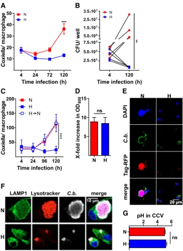
Hypoxia Prevents C. burnetii Replication
To test whether hypoxia may induce O
2-independent antimicro- bial control measures, we infected macrophages with C. burnetii Nine Mile phase II (NMII) under normoxia or hypoxia and deter- mined the number of bacteria per macrophage by qRT-PCR of the C. burnetii dotA and the mouse Alb1 genes. Replication of NMII was observed only under normoxia (Figure 1A), suggesting that hypoxia impeded bacterial replication. Human monocyte- derived macrophages (hMDM) also allowed NMII replication only under normoxic conditions (Figure 1B). To evaluate whether NMII is killed by hypoxic macrophages, we reoxygenated in- fected hypoxic macrophages, which led to the renewed replica- tion of NMII (Figure 1C). Next, we lysed infected macrophage at 120 h post-infection to isolate and cultivate NMII in acidified cit- rate cysteine medium-2 (ACCM-2). As shown in Figure 1D, NMII replicated equally well, regardless of whether it was isolated from normoxic or hypoxic macrophages, indicating that hypoxic macrophages restricted NMII replication without killing the path- ogen. To verify this assumption, we used NMII carrying a plasmid with isopropyl b -
D-1-thiogalactopyranoside (IPTG)-inducible Tag-red fluorescent protein (RFP). Macrophages infected for 120 h with NMII were treated with IPTG, and the expression of Tag-RFP was visualized by confocal microscopy. Under both normoxia and hypoxia, NMII was able to synthesize Tag-RFP (Figure 1E), thus confirming that NMII remained viable in hypoxic macrophages. These results were surprising, as C. burnetii re- quires hypoxic conditions for axenic replication, indicating that not O
2per se, but O
2-mediated alterations, are essential for the observed phenotype.
Hypoxia Does Not Prevent Maturation of the CCV C. burnetii requires a phagolysosomal-like compartment for replication (Howe et al., 2010; Schulze-Luehrmann et al., 2016).
This prompted us to characterize the CCV. By using lysosomal- associated membrane protein 1 (LAMP-1) and LysoTracker Red to label lysosomal compartments, we found CCVs positive for LAMP1 and LysoTracker Red under both culture conditions.
However, the CCVs in hypoxic macrophages were much smaller (Figure 1F). Similarly, regardless of the available O
2, CCVs were positive for DQ Red BSA, a dye that produces bright fluores- cence only upon hydrolysis by proteases, indicating that the CCV is a lysosomal-like compartment with degradative activity under both hypoxic and normoxic conditions (Figures S1A and S1B). Since replication of C. burnetii requires a pH of 4.0–5.0 (Hackstadt and Williams, 1981), we determined the pH of the CCVs. At 48 h post-infection, it was 5.0 under both normoxia and hypoxia (Figure 1G). Thus, the control of bacterial replication in hypoxic macrophages is not mediated by an altered phago- some maturation process.
Reduced IL-10 Secretion under Hypoxia Does Not Explain the Inhibition of C. burnetii Replication
Given that the cytokine IL-10 is known to suppress important antimicrobial effector functions and to facilitate C. burnetii repli- cation in macrophages (Bogdan et al., 1991; Meghari et al., 2008), we analyzed IL-10 in the supernatant of NMII-infected A
D
G E
F C
B
Figure 1. Hypoxia Impairs C. burnetii Replication in Macrophages (A) Macrophages were infected with NMII for the times indicated under nor- moxia (N) or hypoxia (H). Bacterial load was assessed by qPCR. Means ± SEMs, n = 26, two-way ANOVA with Bonferroni post-test.
(B) Human MDM from four different donors was infected with NMII for 4 and 120 h under N or H. Bacterial numbers were determined by colony-forming unit (CFU) counts. n = 4, Friedman test with Dunn’s post-test.
(C) Macrophages were infected with NMII for the times indicated under N, H, or re-oxygenation after 48 h of H (H / N). Bacterial load was assessed by qPCR. Means ± SEMs, n = 4, two-way ANOVA with Bonferroni post- test.
(D) After 120 h, bacteria were purified and cultured in ACCM-2 for 72 h. Bac- terial replication was assessed by optical density 600 (OD
600) measurement.
Means ± SEMs, n = 5, Mann-Whitney U test.
(E) Macrophages were infected with NMII harboring an IPTG-inducible RFP plasmid. At 120 h post-infection, IPTG was added for 8 h and the cells were fixed and stained with DAPI and an antibody against C. burnetii. One of 3 experiments with similar results is shown.
(F) At 120 h post-infection, LysoTracker Red was added, and the cells were stained with DAPI and with antibodies against LAMP1 andC. burnetii. One of 3 experiments with similar results is shown.
(G) At 48 h post-infection, LysoSensor dye was added, and the vacuolar pH was measured in at least 40 CCVs in two independent experiments. Means ± SEMs, t test.
*p < 0.05, **p < 0.01, ***p < 0.001, and ns, p > 0.05.
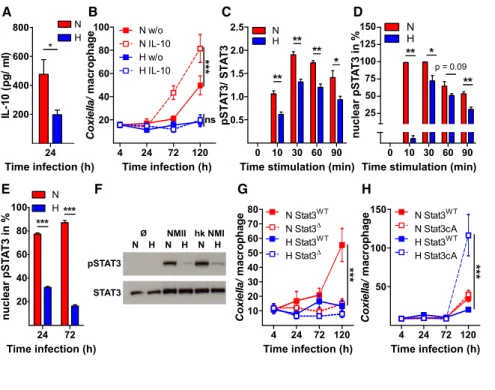
macrophages. Secretion of IL-10 by infected hypoxic macro- phages was reduced (Figure 2A). However, the addition of IL-10 failed to restore the replication of NMII in hypoxic macro- phages, while it boosted the growth of NMII in normoxic macro- phages (Figure 2B). Thus, the hypoxia-induced reduction of IL-10 secretion only partially explains the inhibition of C. burnetii repli- cation. Therefore, we hypothesized that hypoxia may specifically interfere with IL-10 signaling.
Activation of STAT3 Is Crucial for C. burnetii Replication IL-10 signals via the Janus kinase 1 (JAK1)/STAT3 pathway (Mur- ray, 2006). The activation of this pathway leads to STAT3 phos- phorylation and translocation into the nucleus (Yu et al., 2014).
The addition of IL-10 to normoxic macrophages induced the phosphorylation of STAT3 within 10 min. Under hypoxia, the phosphorylation of STAT3 was reduced (Figures 2C and S1C).
This difference was even more pronounced for the subcellular localization of phosphorylated STAT3 (pSTAT3). Ten minutes after the addition of IL-10 to normoxic macrophages, pSTAT3 was detectable in nearly 100% of the nuclei, while hardly any hypoxic macrophages showed nuclear pSTAT3 (Figures 2D and S1D). Similarly, the nuclear localization of pSTAT3 was reduced in NMII-infected macrophages under hypoxia (Fig- ure 2E). NMII, a biosafety level 2 organism, is avirulent in immu- nocompetent mice. It was established in embryonated eggs by the serial passage of phase I C. burnetii NM, a virulent pathogen and a biosafety level 3 bacteria, which resulted in severely trun- cated lipopolysaccharides (LPSs) (Howe et al., 2010). Infection with heat-killed NMI resulted in the reduced phosphorylation of
STAT3 under hypoxia (Figures 2F and S1E), suggesting that the observed phenotype may also be applicable to NMI and does not depend on LPS length.
Infection of hMDM with NMII only induced potent phosphory- lation of STAT3 under normoxia (Figure S1F). These data sug- gested that hypoxia impaired the activation of STAT3. Therefore, we analyzed the role of STAT3 in C. burnetii replication by study- ing the outcome of C. burnetii infection in STAT3-deficient (STAT3
D) or constitutively active STAT3 (STAT3cA) macro- phages and their respective wild-type controls (STAT3
WT). Under normoxia, C. burnetii was only able to replicate in WT and in STAT3cA macrophages, but not in STAT3
Dmacrophages (Fig- ures 2G and 2H), demonstrating that STAT3 activity is essential for C. burnetii replication. Of note, in STAT3cA macrophages, there was a pronounced C. burnetii replication under hypoxia (Figure 2H). These data suggest that the hypoxia-induced impairment of STAT3 activation and not the lack of O
2itself pre- vents the proliferation of C. burnetii in hypoxic macrophages.
HIF1 a and STAT3 Differently Regulate C. burnetii Replication
HIF1 a , the key regulator of cellular hypoxic responses, is known to interfere with STAT3 signaling (Jiang et al., 2013). In addition, HIF1 a has been recognized to control key microbicidal functions of immune cells, as it is involved in phagocytosis and bacterial killing (Palazon et al., 2014). Therefore, we analyzed HIF1 a levels.
At 24 h post-infection, neither uninfected nor NMII or heat- killed NMI-infected macrophages showed robust stabilization of HIF1 a in the presence of ample O
2. However, hypoxia induced
A B C D
E F G H
Figure 2. STAT3 Signaling Is Essential for C. burnetii Replication in Macrophages (A) At 24 h post-infection, IL-10 levels were analyzed by ELISA. Means ± SEMs, n = 14, t test.
(B) Infected macrophages were stimulated with 20 ng/mL IL-10. Bacterial load was analyzed by qPCR. Means ± SEMs, n = 10.
(C and D) Uninfected cells were stimulated with (C) 0.5 ng/mL or (D) 20 ng/mL IL-10. (C) Ratio of phosphorylated STAT3 (pSTAT3) to total STAT3 was analyzed by immunoblot. Means ± SEMs, n = 3, t test. (D) Nuclear translocation of pSTAT3 was assessed by confocal microscopy. 200 cells from 3 independent experiments were analyzed.
Means ± SEMs, n = 5, t test or Mann-Whitney U test.
(E) In at least 100 infected macrophages nuclear pSTAT3 was assessed by confocal microscopy.
Means ± SEMs, n = 3, t test.
(F) Macrophages not infected or infected with NMII or heat-killed NMI for 24 h were subjected to immunoblot analysis using antibodies against pSTAT3 and STAT3. One representative immu- noblot from three independent experiments is shown.
(G) STAT3-deficient (STAT3
D) and wild-type (STAT3
WT) macrophages were infected. Bacterial load was assessed by qPCR. Means ± SEMs, n = 6.
(H) Macrophages with constitutively active STAT3 (STAT3cA) and wild-type (STAT3
WT) macrophages were infected. Bacterial load was assessed by qPCR. Means ± SEMs, n = 8.
(B, G, and H) Two-way ANOVA with Bonferroni post hoc test.
*p < 0.05, **p < 0.01, ***p < 0.001, and ns, p > 0.05.
prominent HIF1 a accumulation, which was further augmented upon infection (Figures 3A and S2A). Similar results were ob- tained from NMII-infected hMDM (Figure S2B). We hypothesized that hypoxia-induced HIF1 a accumulation may account for the impaired activation of STAT3 and the lack of C. burnetii replica- tion. To address this, we analyzed the nuclear localization of pSTAT3 in HIF1 a -deficient (HIF1 a
D) and controls (HIF1 a
WT) mac- rophages upon stimulation with IL-10. Under normoxia, pSTAT3 was translocated into the nucleus within 10 min, regardless of the HIF1 a status (Figure 3B). In contrast, hypoxia delayed and reduced the nuclear localization of pSTAT3 in WT macrophages (Figures 3B and S2C). In HIF1 a
Dmacrophages, this hypoxia- induced impairment of pSTAT3 translocation was abrogated (Figures 3B and S2C). Similarly, the hypoxia-induced impairment of pSTAT3 translocation was partially abolished in NMII-infected HIF1 a
Dmacrophages exposed to hypoxic conditions (Figure 3C).
These data suggest that HIF1 a hampered the activation of STAT3. To confirm this, we used pharmacological agents to alter HIF1 a or STAT3 activity. Treatment with dimethyloxallyl glycine (DMOG) under normoxia led to HIF1 a stabilization and reduced STAT3 activation. In contrast, stattic inhibited the phosphoryla- tion of STAT3 under normoxia without influencing HIF1 a stabili- zation (Figures 3D, 3E, and S2D).
From these findings, we predicted that NMII would replicate in macrophages lacking HIF1 a under hypoxic conditions. As shown in Figure 3F, this is indeed the case. Thus, our data sug- gest that hypoxia suppresses STAT3 activation via HIF1 a and thereby prevents the replication of NMII. NMII replication under hypoxia in HIF1 a
Dand STAT3cA macrophages exceeds the
replication under normoxia. To test whether this may be due to the impaired activity of O
2-dependent defense mechanisms, we treated NMII-infected macrophages with diphenyleneiodo- nium (DPI), which is an inhibitor of the O
2-dependent antimicro- bial effector enzymes NOS2 and PHOX. Treatment with DPI increased the replication rates of NMII under normoxia (Fig- ure 3G). This conforms to findings that NOS2 and PHOX are involved in the control of C. burnetii (Brennan et al., 2004; Zam- boni and Rabinovitch, 2003) and supports our assumption that the increased C. burnetii replication under hypoxia in HIF1 a
Dand STAT3cA macrophages may be due to the shortage of O
2, which represents a critical substrate for the antimicrobial effector enzymes NOS2 and PHOX.
HIF1 a and STAT3 Differently Regulate the Intracellular Citrate Level
Recently, it was shown that STAT3 elevates the intracellular citrate level (Li et al., 2017) by facilitating Slc13a5-dependent cit- rate uptake (von Loeffelholz et al., 2017) and by promoting the expression of citrate synthase (MacPherson et al., 2017), which suggests that STAT3 may shift metabolism toward oxidative phosphorylation. In contrast, the lack of O
2and the consequent stabilization of HIF1 a caused a switch from oxidative phosphor- ylation to anaerobic glycolysis (Kelly and O’Neill, 2015). Hypoxia led to a metabolic shift toward anaerobic glycolysis during C. burnetii infection, as indicated by the increased levels of lactate and the reduced amounts of the Krebs cycle intermedi- ates citrate, succinate, and cis-aconitate-derived itaconate (Figures 4A and S3). However, in both HIF1 a
Dmacrophages
A B C
D E F G
Figure 3. HIF1 a Regulates STAT3 Activity and NMII Regulation under Hypoxia (A) Ratio of HIF1 a to actin was analyzed by immunoblot. Means ± SEMs, n = 3, one-sample t test or one-way ANOVA with Tukey post-test.
(B) Uninfected wild-type (HIF1 a
WT) macrophages or macrophages deficient for HIF1 a (HIF1 a
D) were stimulated with 20 ng/mL IL-10 under normoxia and hypoxia. A total of 200 cells were analyzed per experiment for the nuclear localization of pSTAT3.
Means ± SEMs, n = 4, t test.
(C) Nuclear localization of pSTAT3 was analyzed in 100 NMII-infected macrophages from 3 indepen- dent experiments in HIF1a
WTor HIF1a
Dmacro- phages; t test.
(D and E) Macrophages infected with NMII were treated with either 3 m M stattic, 0.1 mM DMOG, or DMSO, as a control. At 24 h post-treatment STAT3 activity and HIF1a stability was analyzed by immunoblot analysis.
(D) The ratio of pSTAT3 to STAT3 is displayed.
Means ± SEMs, n = 3, t test.
(E) Ratio of HIF1 a to actin is displayed. Means ± SEMs, n = 3, one sample t test or t test.
(F) Macrophages deficient for HIF1a (HIF1a
D) or from littermate controls (HIF1a
WT) were infected and bacterial load was assessed by qPCR. Means ± SEMs, n = 6, two-way ANOVA with Bonferroni post hoc test.
(G) Macrophages were infected with NMII under N with or without the addition of DPI at the times indicated. Bacterial load was assessed at 120 h post-infection by qPCR. Means ± SEMs, n = 3, one-way ANOVA with Tukey post-test.
*p < 0.05, **p < 0.01, ***p < 0.001, and ns, p > 0.05.
(Figure 4B) and STAT3cA macrophages (Figure S2E), but not in STAT3
Dmacrophages (Figure S2F), hypoxia failed to reduce citrate levels, indicating that HIF1 a antagonized STAT3-induced Krebs cycle intermediate availability in infected macrophages under hypoxia. Under normoxia, neither HIF1 a deficiency nor STAT3 deficiency resulted in altered citrate levels (Figures 4B and S2F), suggesting the neither HIF1 a nor STAT3 controls metabolic processes in the presence of ample O
2.
In the following, we concentrated on the investigation of the role of citrate as one example of an HIF1 a - and STAT3-regulated metabolite. Thus, to identify a possible underlying mechanism for the decreased availability of citrate under hypoxia, we analyzed the mRNA expression level of the murine citrate syn- thase (Cs). We detected an infection-dependent upregulation of Cs. However, under hypoxia, the expression of Cs was reduced (Figure 4C), supporting our conclusion that HIF1 a antagonizes STAT3-induced activities. The upregulation of Cs may cause the observed increase in citrate levels during infec- tion (Figure S3).
The axenic growth medium, which allows extracellular C. burnetii replication, contains high levels of citrate. If impaired replication of C. burnetii under hypoxia were due to a reduction in citrate levels, then pharmacological inhibition of the mitochon- drial citrate transport protein (CTP) should abolish bacterial repli- cation. The addition of CTP inhibitor prevented NMII replication under normoxia (Figure 4D), suggesting that mitochondrial func- tion and mitochondrial-derived metabolites play an important role in C. burnetii replication. Next, we asked whether supple- mentation of hypoxic NMII-infected macrophages with citrate abrogates the inhibition of replication. Thus, NMII-infected hyp-
oxic macrophages were treated with trimethylated citrate (TMC), a membrane-permeable form of citrate. Treatment with TMC led to the strong replication of NMII under hypoxia (Figures 4E and 4F). Whether this is a direct or indirect effect of TMC must be determined. These data indicate that hypoxia impedes STAT3 activation via HIF1 a and thereby reduces the intracellular avail- ability of citrate and containment of C. burnetii.
In addition to the increased expression of the host cell citrate synthase during C. burnetii infection (Figure 4C), we observed that the bacterium adjusts the transcription of its own genes involved in citrate metabolism. Thus, the expression of proteins consuming citrate was downregulated under hypoxia (Figures 4G and 4H), while the expression of proteins participating in the production of citrate was upregulated (Figure 4I).
The HIF1 a -STAT3-Citrate Axis Is Functional in Legionella pneumophila-Infected Macrophages
Next, we analyzed whether the HIF1 a -STAT3-citrate axis is also operational during infection with L. pneumophila, the etiological agent of Legionnaires’ pneumonia. We used L. pneumophila D flaA, a flagellin-deficient strain, which does not induce Toll- like receptor 5 (TLR5) and MyD88 signaling (Bartfeld et al., 2009). Hypoxia prevents the replication of L. pneumophila D flaA (Figures S4A and S4B), indicating that hypoxia-induced in- hibition of bacterial replication is not C. burnetii specific. The infection of macrophages with L. pneumophila also induced potent phosphorylation of STAT3 under normoxia (Figure S4C).
In addition, L. pneumophila-infected macrophages did not show robust stabilization of HIF1 a in the presence of ample O
2. However, hypoxia triggered prominent HIF1 a stabilization
A B C D
E F G H I
Figure 4. The HIF1 a and STAT3 Contrari- wise Regulate Intracellular Citrate Levels, and Thereby NMII Replication
(A) Citrate was extracted from infected cells and determined by liquid chromatography-tandem mass spectrometry (LC-MS/MS). Means ± SEMs, n = 3, t test.
(B) Intracellular citrate levels in macrophages deficient for HIF1a (HIF1a
D) or wild-type controls (HIF1a
WT). Means ± SEMs, n = 3, t test.
(C) Gene expression of murine citrate synthase was determined by qRT-PCR. The data are shown as means ± SEMs of DD CT values (using hypo- xanthine phosphoribosyltransferase [HPRT] as a calibrator), two-way ANOVA with Bonferroni post- test. n = 3.
(D) Infected macrophages were treated with 0.1 m M CTP inhibitor (CTP-I) or left untreated. The bacterial load was assessed by qPCR. Means ± SEMs, n = 6, two-way ANOVA with Bonferroni post hoc test.
(E and F) Infected macrophages were treated with 1 mM trimethylated citrate (TMC). (E) The bacterial load was assessed by qPCR or (F) cells were stained with DAPI (blue) and an antibody against C. burnetii (red). Means ± SEMs, n = 6, two-way ANOVA with Bonferroni post hoc test.
(G–I) Gene expression of C. burnetii 2-methylcitrate dehydratase (prpD) (G), isocitrate dehydrogenase (icd) (H), and citrate synthase (gltA) (I) was determined by qRT-PCR. Means ± SEMs of DDCT values (using IS1111 as a calibrator), one-sample t test. n = 3.
*p < 0.05, **p < 0.01, ***p < 0.001, and ns, p > 0.05.
that was augmented by infection (Figure S4D). Furthermore, infection with L. pneumophila resulted in the upregulation of cit- rate only in the presence of ample O
2(Figure S4E). In contrast to the situation during C. burnetii infection, neither genetic ablation of HIF1 a nor treatment with CTP inhibitor (CTP-I) or TMC rescued L. pneumophila replication (Figures S4F and S4G).
These experiments demonstrate that although the HIF1 a - STAT3-citrate axis is operational during L. pneumophila infec- tion, neither HIF1 a nor citrate is decisive for L. pneumophila intracellular replication.
DISCUSSION
O
2availability in the microenvironment has a critical impact on immune responses (Jantsch and Scho¨del, 2015; Taylor and Col- gan, 2017). The key transcription factor regulating O
2homeosta- sis is HIF1 a (Fuhrmann and Br€ une, 2017). Here, we provide evidence that hypoxia restrains the intracellular growth of C. burnetii by limiting intracellular citrate levels (Figures 1A, 1B, 4A, 4D, and 4E). However, suppression of bacterial replication by the reduction of tricarboxylic acid (TCA) cycle intermediates did not lead to the elimination of C. burnetii in macrophages.
Instead, the bacteria persisting in hypoxic macrophages re- mained fully viable (Figures 1C–1E). Our discovery that this state of persistence was elicited by hypoxia via the induction of HIF1 a (Figure 3F), the suppression of STAT3 (Figures 2G and 2H), and the restriction of TCA cycle metabolites (Figures 4A and S3) establishes a hitherto unknown link between the tissue microen- vironment and the host cell metabolism, which is of principal relevance for the understanding of pathogen control and evasion. Our data suggest that citrate depletion is critically involved in this state of affairs. However, it is unclear whether cit- rate depletion on its own or alterations in host and/or pathogen metabolism induced by citrate limitation mediate the restriction of C. burnetii replication.
Information about the trigger(s) and site(s) of C. burnetii persis- tence is rare. Our results suggest that C. burnetii, although contained, may persist in hypoxic tissues. Previous reports suggested that C. burnetii may hide either in the bone marrow (BM) or in adipose tissue (Bechah et al., 2014; Harris et al., 2000). As O
2levels in the BM of rodents range from 0.6% to 2.8% O
2(Spencer et al., 2014), the BM may provide a niche that facilitates the persistence of C. burnetii. Similarly, Mycobac- teria tuberculosis survives within granulomas, in which hypoxia induces a state of dormancy (Belton et al., 2016; Wayne and Hayes, 1996).
Infection with various pathogens and macrophage stimulation with pathogen-associated molecular patterns (PAMPs) results in the accumulation of HIF1 a , even in the presence of ample O
2. Normoxic HIF1 a stabilization requires nuclear factor (NF)- k B activation (Rius et al., 2008) and involves transcriptional and posttranslational signaling events (Tannahill et al., 2013). Several studies have demonstrated that the accumulation of HIF1 a is required to promote innate antimicrobial defenses (Palazon et al., 2014).
However, under normoxic conditions, C. burnetii fails to induce this inflammatory HIF1 a activation (Figures 3A, S2A, and S2B), suggesting that C. burnetii has evolved strategies
to prevent hypoxia-independent HIF1 a activation. In contrast, M. tuberculosis infection leads to an increase in HIF1 a protein level and to a switch toward aerobic glycolysis under normoxia (Shi et al., 2015). Similarly, infection with Chlamydia trachoma- tis and Anaplasma phagocytophilum results in a metabolic shift toward aerobic glycolysis, which is linked to HIF1 a (Cabezas- Cruz et al., 2017; Rother et al., 2018). In agreement with the above-mentioned ability of C. burnetii to prevent HIF1 a stabi- lization on its own in the presence of O
2, we only detect a switch toward glycolysis during NMII infection under hypoxia (Figure S3).
STAT3 signaling is essential for the anti-inflammatory response (Murray, 2006). Recent evidence demonstrates that STAT3 is also involved in metabolism, as it controls the expression of the citrate transporter Slc13a5 (von Loeffelholz et al., 2017) and citrate synthase (MacPherson et al., 2017). In line with these findings, we found that the increased expression of citrate syn- thase correlates with increased STAT3 activity (Figure 4C).
HIF1 a , in contrast, facilitates the switch from oxidative phosphor- ylation to glycolysis and inhibits the Krebs cycle (Kelly and O’Neill, 2015). This leads to reduced levels of acetyl coenzyme A (CoA), which cannot be processed to citrate any longer (Kim et al., 2006). Overall, both STAT3 and HIF1 a regulate the intracel- lular citrate level, but in opposite directions. Accordingly, we found that during C. burnetii and L. pneumophila infection, STAT3 activity increased the intracellular citrate level, while HIF1 a activity prevented the elevation of citrate levels. This regu- latory system is only operational under hypoxia, but not under normoxia.
Citrate, the first intermediate of the Krebs cycle, plays an important role in immunity. For instance, citrate is required for fatty acid biosynthesis, which is involved in mounting an appropriate immune response (Moon et al., 2015). Moreover, citrate can be converted via cis-aconitate to itaconate, which possesses antimicrobial and anti-inflammatory potential (Mi- chelucci et al., 2013). Thus, citrate is thought to form a building block for the production of inflammatory mediators and antimi- crobial molecules. However, high levels of citrate may also entertain the proliferation of C. burnetii (Omsland et al., 2008), which is likely to counteract the positive effects of cit- rate on microbial defense. Citrate is not only important in im- munity but also in cancer, as it provides an important source of carbon (Hatzivassiliou et al., 2005; Mycielska et al., 2018).
Of note, several cancers are characterized by HIF1 a stabiliza- tion, which impedes citrate production through oxidative meta- bolism. Thus, there are remarkable parallels in the regulation of cellular metabolism in immunity and cancer with respect to citrate limitation. However, tumor cells may escape citrate lim- itation by the use of the reductive carboxylation pathway (Wise et al., 2011); C. burnetii is apparently unable to compensate for this limitation.
Not only the infection with C. burnetii and L. pneumophila
(Figure S3) but also the infection with Chlamydia trachomatis
caused an increase in citrate levels (Rother et al., 2018), sug-
gesting that bacterial sensing by the host cell may induce a
switch in metabolism to fight against invading pathogens. How-
ever, in contrast to L. pneumophila, C. burnetii may require
citrate for replication, suggesting that this host cell defense
mechanism is counterproductive during C. burnetii infection.
How C. burnetii benefits from citrate is unknown at present. It was shown that Staphylococcus aureus is able to sense host cell citrate and subsequently to adjust the expression of virulence genes in addition to metabolism (Ding et al., 2014). It remains to be elucidated whether host cell levels of citrate do indeed not only affect the expression of metabolic genes (Fig- ures 4G–4I) but also of virulence factors.
In conclusion, we have demonstrated that under hypoxia, HIF1 a prevents the activation of STAT3 in macrophages, which in turn results in the reduced availability of the Krebs cycle in- termediates succinate, itaconate, and citrate. The lack of these TCA cycle metabolites is linked to the shutdown of C. burnetii replication, while it simultaneously induces a state of persis- tence, which may be important for the establishment of chronic Q fever. These findings demonstrate that the regula- tion of TCA cycle metabolites by innate HIF1 a signaling repre- sents a principle of nutritional pathogen containment under hypoxia.
STAR + METHODS
Detailed methods are provided in the online version of this paper and include the following:
d
KEY RESOURCES TABLE
d
CONTACT FOR REAGENT AND RESOURCE SHARING
d
EXPERIMENTAL MODEL AND SUBJECT DETAILS B Mice
B Murine bone marrow derived macrophages B Human peripheral blood derived macrophages B Coxiella burnetii
B Legionella pneumophila
d
METHOD DETAILS B Cell culture B Infection
B Treatment with chemicals B DNA purification
B Quantification of C. burnetii load B Colony-forming units
B Analysis of IL-10 B RNA
B STAT3 immunoblot B HIF1 a immunoblot B Immunofluorescence B TagRFP assay
B Ratiometric pH measurements B GC-MS
d