The Contribution of Macrophages to Inflammatory Corneal Lymphangiogenesis
Inaugural Dissertation zur
Erlangung des Doktorgrades Dr. nat. med.
der Medizinischen Fakultät und
der Mathematisch-Naturwissenschaftlichen Fakultät der Universität zu Köln
vorgelegt von Deniz Hos aus Nürnberg
Köln, 2016
Berichterstatter/Berichterstatterin: Prof. Dr. Hamid Kashkar
Prof. Dr. Matthias Hammerschmidt
Tag der letzten mündlichen Prüfung: 20.12.2016
Table of Contents
Table of Contents ………...……...
Summary ……….……...…..…
Zusammenfassung ……….……...…….
1. Introduction ………..……….………..…….
1.1. The cornea ………..………….……...…….
1.2. Corneal (lymph)angiogenic privilege ………..………….……...….…
1.3. Pathological corneal (lymph)angiogenesis ..………..….….…..
1.4. Corneal (lymph)angiogenesis as a risk factor for corneal graft rejection ...….
1.5. The role of corneal lymphangiogenesis in dry eye disease ………
1.6. The contribution of macrophages to corneal (lymph)angiogenesis …..….…..
1.7. Aims of the thesis ………..
2. Results ………..…………...……
2.1. Suppression of inflammatory corneal lymphangiogenesis by application of topical corticosteroids. ……….………..…….
2.2. Blockade of insulin receptor substrate-1 inhibits corneal
lymphangiogenesis.……….……….
2.3. IL-10 indirectly regulates corneal lymphangiogenesis and resolution of inflammation via macrophages. ……….………
3. Discussion ……….…...…….
3.1. Main findings of the three presented studies in summary ………….……...….
3.2. Novel anti-(lymph)angiogenic treatment strategies for corneal
neovascular diseases ……….……….
3.3. An anti-inflammatory role of lymphatic vessels at the cornea? ...
3.4. Concluding remarks ……….………..….……….
References ……….……….….
Curriculum Vitae .……….………..
Acknowledgements ……….……….……….
Erklärung .……….………
Publications .……….………..
1 2 4 6 6 7 9 12 15 15 18 20
20
23
25 28 28
29 32 34 37 41 46 47 49
Summary
The cornea is physiologically devoid of blood and lymphatic vessels.
However, severe inflammation can result in a secondary ingrowth of both vessel types, a process termed “corneal neovascularization”. Macrophages seem to play an important role in this process, although the underlying mechanisms are not well defined. Corneal blood vessels lead to reduced visual acuity when they grow into the optical center, and lymphatic vessels contribute to corneal transplant rejection, dry eye disease, or ocular allergy. In this regard, lymphatic vessels facilitate antigen presenting cell trafficking to the lymph nodes, where accelerated sensitization against allo- or autoantigens occurs. Thus, persistent corneal neovascularization is considered as harmful, and anti(lymph)angiogenic therapy has recently emerged as a novel approach in the treatment of several inflammatory corneal diseases.
One aim of this work was to identify targets for anti(lymph)angiogenic therapy at the cornea and to identify effective anti(lymph)angiogenic compounds, which might be used to treat corneal neovascular diseases. Using a mouse model of sterile corneal inflammation, we found that glucocorticosteroids are strong inhibitors of corneal (lymph)angiogenesis. Glucocorticosteroids suppressed macrophage infiltration into the cornea and inhibited pro-inflammatory cytokine expression in macrophages. Furthermore, we identified insulin receptor substrate-1 (IRS-1) as a mediator of inflammatory corneal (lymph)angiogenesis. IRS-1 was expressed by corneal macrophages, and inhibition of IRS-1 reduced vascular endothelial growth factor (VEGF)-A, VEGF-C and VEGF-D expression in these cells. Consistently, treatment of inflamed corneas with GS-101, an antisense oligonucleotide directed against IRS-1, strongly inhibited inflammatory corneal (lymph)angiogenesis.
Another aim of this study was to analyze whether corneal lymphangiogenesis might also have beneficial functions, as studies in extraocular tissues had demonstrated that lymphatic vessels are also important to terminate inflammatory responses. However, studies showing similar functions for corneal lymphatic vessels were missing and the mediators of this putative anti-inflammatory process were unknown. Here, we analyzed the role of Interleukin-10 (IL-10), a primarily anti- inflammatory cytokine, in the regulation of inflammatory corneal lymphangiogenesis.
IL-10 was expressed in inflamed corneas by infiltrating macrophages. Furthermore, macrophages treated with IL-10 upregulated pro-lymphangiogenic VEGF-C expression, which is known to induce lymphatic vessel growth. IL-10 deficiency or conditional ablation of IL-10 signaling specifically in myeloid cells lead to reduced inflammatory corneal lymphangiogenesis and prolonged corneal inflammation, whereas treatment with IL-10 promoted lymphangiogenesis and faster egress of macrophages from inflamed corneas. These results collectively indicate that IL-10 indirectly regulates corneal lymphangiogenesis and resolution of corneal inflammation via macrophages, which is the first report of a beneficial function of corneal lymphangiogenesis in (sterile) corneal inflammation.
Taken together, we have identified novel anti(lymph)angiogenic compounds, which mainly affect the contribution of macrophages to inflammatory corneal lymphangiogenesis. Furthermore, we have described a novel anti-inflammatory role for corneal lymphangiogenesis, which is mediated by macrophages. Our work highlights the importance of macrophages for corneal lymphangiogenesis, and might contribute to future immunomodulatory therapeutic strategies promoting corneal repair or preventing disease.
Zusammenfassung
Die Hornhaut enthält physiologisch keine Blut- und Lymphgefäße. Schwere Entzündungen können allerdings zu einem sekundären Einwachsen von Blut- und Lymphgefäßen in die Hornhaut führen. Korneale Blutgefäße reduzieren die Transparenz der Hornhaut, wenn sie in das optische Zentrum einwachsen, und korneale Lymphgefäße tragen zu Abstoßungsreaktionen nach Hornhaut- transplantation, dem trockenen Auge oder der okulären Allergie bei. Hierbei erleichtern Lymphgefäße die Migration von Antigen-präsentierenden Zellen in die Lymphknoten, wo es zu einer Sensibilisierung gegenüber Allo- oder Autoantigenen kommt. Daher werden korneale Blut- und Lymphgefäße in der Regel als nachteilig angesehen, und anti(lymph)angiogene Therapie scheint ein neuer Ansatz bei der Behandlung verschiedener entzündlicher Hornhauterkrankungen zu sein.
Ein Ziel dieser Arbeit war, Zielstrukturen für eine anti(lymph)angiogene Therapie und wirksame Therapeutika zu identifizieren, die zur Therapie von neovaskulären Hornhauterkrankungen genutzt werden könnten. Wir konnten zeigen, dass Glukokortikosteroide potente (Lymph)angiogenese-Inhibitoren sind.
Glukokortikosteroide unterdrücken die Rekrutierung von Makrophagen und hemmen die pro-inflammatorische Zytokin-Expression dieser Zellen. Darüber hinaus konnten wir zeigen, dass insulin receptor substrate-1 (IRS-1) eine wichtige Rolle bei der kornealen (Lymph)angiogenese spielt. IRS-1 wird u.a. von Makrophagen exprimiert, und eine Hemmung von IRS-1 reduziert die Expression von vascular endothelial growth factor (VEGF)-A, VEGF-C und VEGF-D in diesen Zellen. Eine Behandlung entzündeter Hornhäute mit GS-101, einem Antisense-Oligonukleotid der gegen IRS- 1 gerichtet ist, inhibiert die entzündliche (Lymph)angiogenese an der Hornhaut.
Weiteres Ziel dieser Arbeit war es zu analysieren, ob korneale Lymphgefäße auch positive Funktionen haben können, wie es in Arbeiten an extraokulären Geweben bereits gezeigt wurde. Dabei scheinen Lymphgefäße wichtig für das Beenden von Entzündungsreaktionen zu sein. Studien, die ähnliche Funktionen an der Hornhaut zeigen, fehlten bisher und die Mediatoren dieser vermeintlich anti- entzündlichen Funktion waren unbekannt. Wir untersuchten daher die Rolle des anti-inflammatorischen Zytokins Interleukin-10 (IL-10). Wir konnten zeigen, dass IL- 10 während einer Entzündungsreaktion durch infiltrierende Makrophagen exprimiert wird, welche nach Stimulation mit IL-10 die Expression von VEGF-C steigern. Das Fehlen von IL-10 oder eine Blockade des IL-10 Signalweges in myeloischen Zellen führt zu einer reduzierten Lymphangiogenese und einer persistierenden Entzündungsreaktion, während eine Stimulation mit IL-10 Lymphangiogenese induziert und zu einem schnelleren Austritt von Makrophagen aus der Hornhaut führt. Somit konnten wir zeigen, dass IL-10 indirekt über Makrophagen die korneale Lymphangiogenese und die Auflösung von Entzündungsprozessen reguliert. Dies ist der erste Nachweis einer positiven Funktion kornealer Lymphgefäßen.
Zusammenfassend haben wir neue anti(lymph)angiogene Therapeutika identifiziert, die Ihren Effekt überwiegend durch Modulation von Makrophagen erzielen. Darüber hinaus haben wir eine neue, anti-entzündliche Rolle für korneale Lymphgefäße aufgezeigt, die durch Makrophagen vermittelt wird. Unsere Arbeit betont die Bedeutung von Makrophagen für die korneale Lymphangiogenese und kann dazu beitragen, künftige immunmodulatorische Therapiestrategien zur Förderung von Reparaturprozessen oder zur Verhütung von Krankheiten an der Hornhaut zu entwickeln.
1. Introduction
1.1. The cornea
The cornea is the outermost part and the major refractive element of the eye.
It is physiologically transparent, about 550 µm thick and consists of five layers: 1) a multi-layered squamous non-keratinized epithelium at the outside; 2) the subjacent basement membrane-like Bowman's layer; 3) a middle stromal layer which is sparsely populated with resident tissue cells and mainly consists of collagen fibrils;
and the inner layers of 4) Descemet´s membrane and 5) corneal endothelium (Figure 1). The corneal epithelium is covered by the tear film, which contains an aqueous phase secreted by the lacrimal glands, a mucinous phase built by the globlet cells of the conjunctiva, and a lipid phase secreted by the meibomian glands at the lid margin. The tear film and the corneal epithelium form an important barrier to the outside environment and guard the eye from microbial invasion, chemical and toxic damage and foreign bodies, to which the cornea is constantly exposed due to its anatomical position. The corneal stroma, which underlies the epithelium and comprises approximately 80 to 90% of the cornea´s total thickness, shows a highly periodical distribution of its collagen fibrils to minimize light scattering. In addition, continuous dehydration of the stroma by the carbonic anhydrase activity of the corneal endothelial cells results in compactly packed collagen lamellae that ensures transparency.
Figure 1: Eye anatomy and corneal histology. A. Schematic diagram of the human eye. B.
Histology section of H&E stained human cornea; EP: epithelium, BM: Bowman's layer, ST: stroma, DM: Descemet´s membrane, EN: endothelium (modified from (Leal and Pearlman 2012))
1.2. Corneal (lymph)angiogenic privilege
The cornea is one of the very few tissues of the organism that is devoid of blood and lymphatic vessels (Cursiefen 2007). This absence of blood and lymphatic vessels, termed “corneal (lymph)angiogenic privilege” is not a passive condition but is rather actively maintained. Recently, several mechanisms that contribute to this privilege have been identified. In this regard, the corneal epithelium plays a pivotal role in maintaining corneal avascularity, as it expresses soluble forms of the vascular endothelial growth factor (VEGF) receptors (sVEGFR-1, sVEGFR-2, sVEGFR-3), which function as decoy receptors for the key (lymph)angiogenic growth factors VEGF-A, VEGF-C and VEGF-D (Albuquerque et al. 2009; Ambati et al. 2006; Singh et al. 2013). In addition, the corneal epithelium expresses (non- vascular) membrane-bound VEGFR-3 (mVEGFR-3), which is also able to trap VEGF-C and VEGF-D (Cursiefen et al. 2006). Further potent anti(lymph)angiogenic molecules expressed in the cornea are angiostatin, endostatin, pigment epithelium-
derived factor, thrombospondin-1, and thrombospondin-2, which all exhibit multiple inhibitory functions such as direct inhibition of vascular endothelial cell migration and proliferation as well as indirect interference with growth factor mobilization and binding (Armstrong and Bornstein 2003; Cursiefen et al. 2011; Cursiefen et al. 2004;
Lin et al. 2001). Especially thrombospondin-1 is a crucial molecule for corneal alymphaticity, as it has been shown to inhibit the expression of VEGF-C by corneal inflammatory cells. Accordingly, thrombospondin-1 deficient mice show increased corneal VEGF-C expression and also higher inflammatory cell numbers, which result in spontaneous and isolated ingrowth of lymphatic vessels into the cornea (Cursiefen et al. 2011). Another antiangiogenic mechanism of the cornea is the expression of the inhibitory PAS domain protein (IPAS), which negatively regulates hypoxia-induced upregulation of VEGF and thereby maintains corneal avascularity even under hypoxic conditions (Makino et al. 2001).
Despite of its avascular and alymphatic nature, the cornea also relies on blood components to remain healthy. Delivery of nutrients and subsequent clearance of metabolites are carried out by the tear film from the corneal surface, the aqueous humour from the anterior chamber and at the limbus from the lateral margin. The limbus is a transition zone where the cornea fades into the opaque and vascularized sclera and overlying conjunctiva. In contrast to the cornea, the limbus is physiologically vascularized: limbal blood vessel arterioles originate from the anterior ciliary arteries, sprout towards the corneal border and form small pericorneal arcades. The lymphatic vasculature of the limbus also consists of a ring shaped network which is connected to the conjunctival lymphatic vessels. Limbal lymphatic vessels do not form arcades but rather consist of a main circumferential
lymphatic vessel with blind ending extensions directed towards the cornea (Figure 2).
Figure 2: Vascular anatomy of the cornea. Whole mounts of healthy murine corneas stained with CD31 (blood vessels; left panel) or LYVE-1 (lymphatic vessels, right panel; arrowheads indicate centrally oriented vascular extensions from the main limbal lymphatic vessel). Blood and lymphatic vessels terminate at the limbal border (modified from (Hos et al. 2013)).
1.3. Pathological corneal (lymph)angiogenesis
Corneal avascularity and alymphaticity are not invulnerable. Whereas minor inflammatory vascular stimuli are buffered by the corneal (lymph)angiogenic privilege and do not induce an angiogenic response, severe tissue damage and inflammation can result in a strong upregulation of pro(lymph)angiogenic growth factors, which might “overwhelm” the cornea’s anti(lymph)angiogenic mechanisms and lead to an ingrowth of blood and clinically invisible lymphatic vessels from the limbus into the cornea (corneal [lymph]angiogenesis) (Cursiefen 2007). Diseases that can result in corneal (lymph)angiogenesis include infectious (e.g. bacterial, viral, or fungal keratitis), inflammatory (e.g. ocular pemphigoid, Lyell-Syndrome, Stevens-Johnson Syndrome, Graft-versus-Host Disease, corneal graft rejection),
hypoxic (e.g. extended wear of contact lenses), and traumatic/toxic diseases (e.g.
chemical or physical burns). Diseases that lead to a loss of limbal barrier function (e.g. limbal stem cell deficiency) are also frequently accompanied by corneal (lymph)angiogenesis. Blood and lymphatic vessels mostly grow in parallel into the cornea and it seems that the degree of corneal hemangiogenesis generally correlates with the degree of corneal lymphangiogenesis (Cursiefen et al. 2006;
Cursiefen et al. 2002).
Corneal (lymph)angiogenesis after tissue damage possibly has several important functions including supply of components of cellular and humoral immunity to combat infections, supply of additive oxygen and growth factors to promote the healing response, and drainage and removal of debris and cells from the injured site. However, this process might also cause loss of transparency in case of blood vessel growth into the optical zone, which can lead to light scattering or obscuration. Furthermore, also secondary effects such as fluid and lipid deposition through immature capillaries and exuberant influx of inflammatory cells might contribute to corneal edema, loss of corneal compactness and opacification (Cursiefen 2007). Therefore, after the repair response is completed and barrier function has been achieved, corneal blood vessels must resolve rapidly to restore corneal transparency and thereby functionality. However, under certain pathologic conditions and chronic inflammatory diseases, corneal blood vessels regress only very slowly or may even persist and permanently impair vision (Figure 3). Whereas it is well accepted that persisting pathological corneal blood vessels contribute to loss of corneal transparency, the contribution of clinically invisible lymphatic vessels to corneal diseases was less apparent. Recently, however, several experimental
studies provided evidence for the contribution of corneal lymphatic vessels to ocular pathologies, such as transplant rejection and dry eye disease (Dietrich et al. 2010;
Goyal et al. 2010). To date, corneal lymphangiogenesis is therefore considered as pathological and undesirable. However, it is well known that in extraocular tissues, lymphatic vessels exert important physiological functions during the inflammatory response. Lymphatic vessels regulate tissue pressure and allow the drainage of debris and egress of inflammatory cells from the injured site (Oliver and Detmar 2002). In particular, it has been shown that lymphatic vessels seem to be important for the termination of inflammatory responses and recent studies show that the inhibition of lymphangiogenesis might lead to chronic inflammation and edema, whereas the specific activation of lymphatic vessels can ameliorate these conditions (Huggenberger et al. 2011; Huggenberger et al. 2010). However, studies demonstrating similar beneficial functions for lymphatic vessels at the cornea are still missing.
Figure 3: Corneal neovascularization.
Clinical picture of a patient with pathological corneal blood vessels secondary to recurrent herpetic ulcerative keratitis. Blood vessels originate from the limbus and grow centrally towards the cornea. Note secondary central corneal scar (from (Hos et al. 2016)).
1.4. Corneal (lymph)angiogenesis as a risk factor for corneal graft rejection Corneal transplantation is the oldest, most common and the most successful form of (tissue) transplantation (Streilein et al. 1999). The reason for this extraordinary success is that the cornea is considered to be an immune-privileged anatomical structure, as it: 1) contains only very low numbers of MHC class II positive antigen presenting cells (APCs) in the corneal centre; 2) shows generally reduced expression of MHC class I; and 3) expresses Fas ligand (CD95L) and programmed death ligand 1 (PD-L1), which inhibit T cell responses. Another important mechanism responsible for the corneal immune privilege is the phenomenon of anterior chamber-associated immune deviation (ACAID), which causes antigen-specific systemic immune tolerance characterized by downregulation of delayed-type hypersensitivity and cytotoxic T cells responses (Niederkorn 2010; Niederkorn 1999; Streilein et al. 1999). Interestingly, it has been shown that the corneal immune privilege depends, at least partially, on its intact (lymph)angiogenic privilege (Cursiefen 2007).
When corneal transplantations are performed because of non-inflammatory and non-vascular diseases with intact (lymph)angiogenic and immune privilege, e.g.
because of corneal (endothelial) dystrophies, more than 75% of corneal grafts survive for longer than 5 years, even without previous HLA-matching and without any systemic immunosuppressive therapy (Williams and Coster 2007). However, when severe inflammatory processes have overcome the cornea´s (lymph)angiogenic and immune privilege and donor corneas are grafted into prevascularized recipients, transplant survival rates significantly decrease, even under aggressive systemic immunosuppression. Therefore, vascularization of the
cornea prior to transplantation is considered as one of the most important risk factors for immunological graft rejection (Dana and Streilein 1996; Sano et al. 1995).
Preclinical evidence suggests that the reason for decreased graft survival rates in prevascularized corneas is due to an enhanced facilitation of allogenic immune responses: via preexisting corneal lymphatic vessels, donor and antigen-loaded recipient APCs have direct and immediate access to the regional lymph nodes, where increased and accelerated allosensitization occurs (Cursiefen et al. 2003;
Hos and Cursiefen 2014). Subsequently, via preexisting corneal blood vessels, immune effector cells, such as alloreactive T cells, can easily reach and subsequently reject the graft (Figure 4).
Figure 4: The role of blood and lymphatic vessels in corneal transplant rejection. Schematic drawing illustrating the role of blood and lymphatic vessels in corneal transplantations performed in pathologically prevascularized recipients. (1) Magnification of the host-graft interface: blood (red) and lymphatic vessels (green) reach the host-graft interface. Antigen-presenting cells (APCs) and antigen (Ag) can leave the cornea via corneal lymphatic vessels (2) and reach the regional lymph nodes (3);
after stimulation of alloreactive T cells, these and other effector cells can reach the graft via corneal blood vessels (4) (from (Cursiefen et al. 2003)
As persistent pathological blood and lymphatic vessels play such an important role in the development of corneal graft rejection, several groups have analyzed the impact of a pharmacological inhibition of corneal (lymph)angiogenesis in the context of corneal transplantation. Importantly, blockade of corneal hem- and lymphangiogenesis significantly promoted corneal graft survival in the experimental setting (Cursiefen et al. 2004; Hos et al. 2008). Moreover, using specific pharmacological drugs or adenoviral vectors that modulate lymphangiogenesis without affecting hemangiogenesis, it was demonstrated that lymphatic vessels - which ease the connection between the graft and the secondary lymphatic organs - play a superior role in the mediation of graft rejection when compared to blood vessels (Albuquerque et al. 2009; Dietrich et al. 2010) (Figure 5). Thus, antilymphangiogenic therapy at the cornea has recently emerged as a novel therapeutic concept to reduce corneal graft rejection, and recent efforts aim to identify effective anti(lymph)angiogenic compounds which might potentially be used in the clinic in the near future (Bock et al. 2013).
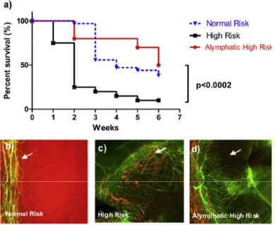
Figure 5: Lymphatic vessels define the high risk status of transplants in prevascularized corneal hosts.
(a) Kaplan-Meier survival curve showing that absence of lymphatic vessels in the recipient cornea prior to transplantation improves graft survival in the experimental setting;(b to d) corneal whole mounts stained with CD31 (blood vessel; green) and LYVE-1 (lymphatic vessels, red) in different recipient beds (from (Hos et al. 2014)).
1.5. The role of corneal lymphangiogenesis in dry eye disease
Dry eye disease is one of the most frequent diseases in ophthalmology. It is generally accepted that it is not only a condition with reduced tear quantity, but rather a complex, multifactorial disorder of the ocular surface where chronic inflammation plays an important role and where influx and activation of inflammatory cells results in disturbed tear production, abnormal tear composition and subsequent damage of the ocular surface (Barabino et al. 2012; De Paiva et al.
2009). Recent evidence suggests that also the adaptive immune system is involved in maintaining ocular surface inflammation in dry eye disease (Stern et al. 2013;
Stevenson et al. 2012). Interestingly, it has been shown in several mouse models that in this disease, an isolated ingrowth of lymphatic but not blood vessels into the cornea can be observed (Cursiefen et al. 2011; Goyal et al. 2010). These corneal lymphatic vessels seem to serve as conduits that enable easier autoantigen- transport and accelerated trafficking of activated APCs from to the ocular surface to the lymph nodes (Goyal et al. 2010; Stevenson et al. 2012). Furthermore, it has been shown that inhibition of corneal lymphangiogenesis significantly improves the clinical course of dry eye, at least in the experimental setting (Goyal et al. 2012).
Therefore, antilymphangiogenic therapy might offer a promising approach not only to prevent graft rejection after corneal transplantation, but also to treat dry eye disease.
1.6. The contribution of macrophages to corneal (lymph)angiogenesis
Macrophages are derived from blood monocytes, which originate in the bone marrow and are released into blood circulation. Thereafter, pro-inflammatory stimuli
can cause the recruitment of monocytes to injured peripheral sites, where differentiation into (inflammatory) macrophages occurs. Recent evidence suggests that there is also another distinct population of macrophages, which seem to develop independently from the bone marrow but rather from progenitor cells generated in the yolk sac (Schulz et al. 2012). These macrophages represent the resident tissue macrophage population and are important for maintaining tussue homeostasis during “steady-state”. However, to which extent this also accounts for corneal tissue macrophages is so far undetermined.
Substantial preclinical evidence indicates that macrophages are essential mediators of corneal (lymph)angiogenesis after injury (Chung et al. 2009; Cursiefen et al. 2004). It is known that macrophages are able to secrete VEGF-A, VEGF-C und VEGF-D and thereby induce vascular endothelial cell proliferation and migration (Cursiefen et al. 2004). In addition, macrophages also express the respective receptors (VEGFR-1 and VEGFR-3), which mediate chemotactic effects and thereby amplify the inflammatory (lymph)angiogenic response (Cursiefen et al.
2004). Furthermore, under certain, so far still poorly understood conditions, macrophages are able to express lymphendothelial markers and integrate into newly formed corneal lymphatic vessels or even generate primitive lymphatic vessel-like structures de novo (Maruyama et al. 2005). Moreover, macrophages are frequently localized in close proximity to already formed blood and lymphatic vessels, suggesting a continuing interaction with these (Maruyama et al. 2005). The crucial role of macrophages in mediating corneal (lymph)angiogenesis is further supported by the fact that macrophage depletion, e.g., by clodronate liposomes,
almost completely prevents inflammatory corneal (lymph)angiogenesis (Cursiefen et al. 2004).
Macrophages are a heterogeneous, highly plastic cell population and various functional phenotypes have been described (Gordon and Taylor 2005; Sica and Mantovani 2012). One current conceptual model classifies macrophages into at least two subpopulations (paradigm of M1/M2 polarization). In this model,
“classically activated” (also called M1-polarized) macrophages are considered to exert pro-inflammatory activities, promote type I immune responses and are involved in the eradication of invading microorganisms. In contrast, “alternatively activated” (also called M2-polarized) macrophages, which are hyporesponsive to pro-inflammatory stimuli, are mainly involved in debris scavenging, tissue remodeling and the resolution of inflammatory responses (Gordon and Martinez 2010). Classical macrophage activation is mediated by pro-inflammatory stimuli, such as IFN-γ or TNF-α, followed by a microbial trigger (e.g. lipopolysaccharide).
Mediators of alternative macrophage activation are IL-4 and IL-13, or IL-10 (Gordon and Taylor 2005). By now, however, it is becoming clear that the paradigm of M1/M2 polarization is an oversimplification and reflects two extremes of macrophage polarization, and that in tissues a broad spectrum of activation states exists in parallel (Martinez and Gordon 2014). Although macrophage biology is an extensively studied research field, there are still numerous unaddressed questions.
In the context of corneal inflammation for instance, it is so far unknown whether specific macrophage subpopulations occur and which macrophage subpopulations mediate corneal (lymph)angiogenesis. It is also unclear whether macrophages exert
a different (lymph)angiogenic potential during the various stages of corneal inflammation.
Taken together, macrophages are known to be importantly involved in inflammatory corneal (lymph)angiogenesis. However, the specific mechanisms are not yet identified. Thus, more detailed analysis of corneal macrophage activation and polarization is required, before macrophages can be considered as a potential tool to control corneal physiology or disease. On the one hand, inhibition of pro- (lymph)angiogenic macrophage subsets would be an interesting therapeutic option to modulate corneal (lymph)angiogenesis, e.g. in the context of cornea transplantation or dry eye disease. On the other hand, if corneal lymphangiogenesis and pro-lymphangiogenic macrophages would be involved in physiological wound healing responses in the cornea, specific activation of these would be of great interest to promote corneal repair.
1.7. Aims of the thesis
Persistent corneal lymphatic vessels contribute to the development of detrimental corneal pathologies, such as corneal graft rejection or dry eye disease.
Antilymphangiogenic therapy at the cornea has emerged as a promising approach to treat these inflammatory diseases. Therefore, this study aimed to identify effective antilymphangiogenic compounds, which might potentially be used to treat patients at risk for corneal graft rejection or with dry eye disease. In addition, this work aimed to find novel potential targets for antilymphangiogenic therapy at the cornea.
In the first part of this thesis, we analyzed the impact of glucocorticosteroids on inflammatory corneal lymphangiogenesis and investigated the potential
underlying mechanisms. This was of particular interest, as glucocorticosteroids are widely used as the standard anti-inflammatory treatment at the cornea and have also been shown to reduce the risk of corneal graft rejection and to ameliorate dry eye disease (Nguyen et al. 2007). However, little was known about the ability of these drugs to suppress corneal lymphangiogenesis.
In the second part of this work, we analyzed whether inflammatory corneal lymphangiogenesis is regulated by insulin receptor substrate-1 (IRS-1). IRS-1 is a cytosolic scaffolding protein that interacts with the VEGFR complex, and it was recently shown that IRS-1 plays a role in blood vessel development (Andrieu-Soler et al. 2005; Miele et al. 2000). However, it was unclear whether IRS-1 is also involved in lymphatic vessel development and whether IRS-1 is a potential target to treat corneal lymphangiogenesis.
In contrast to the role of corneal lymphatic vessels under pathological conditions, a physiological function for corneal lymphatic vessels has not been described so far. Studies in extraocular tissues have demonstrated that lymphangiogenesis might also be important to terminate inflammatory responses (Huggenberger et al. 2011). However, studies showing similar beneficial functions for corneal lymphatic vessels are missing and the mediators of this putative anti- inflammatory lymphangiogenesis are unknown.
Therefore, in the third part of this thesis, we analyzed the role of Interleukin- 10 (IL-10), a primarily anti-inflammatory cytokine, in the regulation of inflammatory corneal lymphangiogenesis. We determined the impact of IL-10 on macrophages, inflammatory corneal lymphangiogenesis and the resolution of corneal inflammation.
2. Results
2.1. Suppression of inflammatory corneal lymphangiogenesis by application of topical corticosteroids.
Deniz Hos, Daniel R. Saban, Felix Bock, Birgit Regenfuss, Jasmine Onderka, Sharmila Masli, Claus Cursiefen
Objectives: To analyze whether topical application of corticosteroids inhibits inflammatory corneal lymphangiogenesis and to study the potential underlying antilymphangiogenic mechanisms.
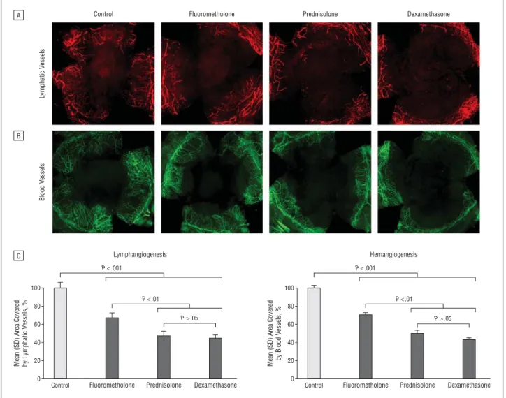
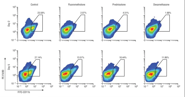
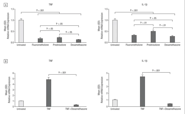
Methods: Inflammatory corneal neovascularization was induced by suture placement, and the corneas were then treated with topical fluorometholone, prednisolone acetate, or dexamethasone sodium phosphate. After 1 week, the corneas were stained with lymphatic vessel endothelial hyaluronan receptor 1 for detection of pathological corneal lymphangiogenesis. The effect of these corticosteroids on macrophage recruitment was assessed via fluorescence- activated cell sorting analysis. The effect of these corticosteroids on proinflammatory cytokine expression by peritoneal exudate cells was tested via real- time polymerase chain reaction. Furthermore, the effect of steroid treatment on the proliferation of lymphatic endothelial cells was assessed via enzyme-linked immunosorbent assay.
Results: Treatment with corticosteroids resulted in a significant reduction of inflammatory corneal lymphangiogenesis. The antilymphangiogenic effect of fluorometholone was significantly weaker than that of prednisolone and dexamethasone. Corneal macrophage recruitment was also significantly inhibited by
the application of topical steroids. Treatment of peritoneal exudate cells with corticosteroids led to a significant downregulation of the RNA expression levels of tumor necrosis factor and interleukin 1β. Additionally, proliferation of lymphatic endothelial cells was also inhibited.
Conclusions: Corticosteroids are strong inhibitors of inflammatory corneal lymphangiogenesis, with significant differences between various corticosteroids in terms of their antilymphangiogenic potency. The main mechanism of the antilymphangiogenic effect seems to be through the suppression of macrophage infiltration, proinflammatory cytokine expression, and direct inhibition of proliferation of lymphatic endothelial cells.
Clinical relevance: Steroids block corneal lymphangiogenesis, the main risk factor for immune rejections after corneal transplantation. The different antilymphangiogenic potency of these drugs should be taken into account when using steroids in clinical practice (e.g., after keratoplasty).
Own contribution to publication 1:
I performed all in vivo inflammatory corneal neovascularization assays, treated all animals with the respective drugs and subsequently performed all whole mount immunohistochemical stainings and vessel analyses (Figure 1). I also performed the in vivo experiments and treatments for the FACS analysis and assisted in FACS data acquisition and interpretation (Figure 2). I further isolated the peritoneal macrophages, carried out all in vitro treatments and performed and analyzed the quantitative real-time PCRs (Figure 3). In addition, I performed and analyzed the lymphatic endothelial proliferation assays (Figure 4). All images
illustrating my experimental data were generated by me. I wrote the manuscript and handled the revision phase of this project.
Contribution of co-authors to publication 1:
Dr. Daniel R. Saban established, performed and analyzed the FACS experiments (Figure 2). Dr. Felix Bock and Dr. Birgit Regenfuss provided help in analyzing the in vivo angiogenesis data (Figure 1), provided valuable general suggestions and critically read the manuscript. Jasmine Onderka assisted and provided support in lymphatic endothelial culture and in vitro proliferation assays (Figure 4). Dr. Sharmila Masli provided help in isolation and culture of peritoneal macrophages (Figure 3). Prof. Claus Cursiefen conceptualized and coordinated the project and wrote the manuscript.
2.2. Blockade of insulin receptor substrate-1 inhibits corneal lymphangiogenesis.
Deniz Hos, Birgit Regenfuss, Felix Bock, Jasmine Onderka, Claus Cursiefen
Purpose: To analyze whether insulin receptor substrate (IRS-1) is involved in lymphatic vessel development and whether IRS-1 blockade can inhibit lymphangiogenesis in vivo.
Methods: The impact of IRS-1 blockade by GS-101 (Aganirsen), an antisense oligonucleotide against IRS-1, on lymphatic endothelial cell (LEC) proliferation was assessed by ELISA. Furthermore, the effect of IRS-1 blockade on prolymphangiogenic growth factor expression by LECs and macrophages (peritoneal exudate cells) was tested by real-time PCR. The mouse model of inflammatory corneal neovascularization was used to analyze the effect of IRS-1 blockade in vivo: after corneal suture placement, mice were treated with GS-101 eye drops (twice daily afterwards for 1 week, 5 µL per drop; 50, 100, or 200 µM).
Afterward, corneal wholemounts were prepared and stained for blood and lymphatic vessels.
Results: Blockade of IRS-1 by GS-101 inhibited LEC proliferation dose dependently.
GS-101 led to decreased VEGF-A expression levels in LECs, whereas VEGF-C, VEGF-D, and VEGFR3 showed no significant change. In macrophages, VEGF-A expression levels were also inhibited by IRS-1 blockade. Additionally, GS-101 strongly inhibited macrophage-derived VEGF-C, VEGF-D, and VEGFR3 expression.
In vivo, corneal hemangiogenesis was significantly inhibited when used at a concentration of 200 µM (by 17%; P < 0.01). Corneal lymphangiogenesis was significantly inhibited when used at a dose of 100 µM (by 21%; P < 0.01), and the
highest used dose (200 µM) showed an even stronger inhibition (by 28%; P <
0.001).
Conclusions: Blockade of IRS-1 inhibits not only hemangiogenesis but also lymphangiogenesis. To the authors' knowledge, this is the first evidence that IRS-1 is involved in the molecular pathway leading to lymphangiogenesis.
Own contribution to publication 2:
I performed and analyzed all lymphatic endothelial proliferation assays (Figure 1). Furthermore, I performed and analyzed all real-time PCRs with lymphatic endothelial cells (Figure 2). I also isolated the peritoneal macrophages, carried out all in vitro treatments and performed and analyzed the real-time PCRs (Figure 3).
The in vivo inflammatory corneal neovascularization assays, all treatments and whole mount immunohistochemical stainings with subsequent vessel analyses were carried out by me (Figure 4). In addition, I performed the corneal macrophage stainings (Figure 5). All images illustrating my experimental data were generated by me. I wrote the manuscript and handled the revision phase of this project.
Contribution of co-authors to publication 2:
Dr. Birgit Regenfuss provided help in establishing the real-time PCR protocols (Figure 2 and 3). In addition, Dr. Birgit Regenfuss and Dr. Felix Bock supported me in analyzing the in vivo angiogenesis data (Figure 4), provided general suggestions and corrected the manuscript. Jasmine Onderka assisted in lymphatic endothelial culture, in vitro proliferation assays (Figure 1) and histological stainings (Figure 5). Prof. Claus Cursiefen conceptualized and coordinated the project.
2.3. IL-10 Indirectly Regulates Corneal Lymphangiogenesis and Resolution of Inflammation via Macrophages.
Deniz Hos, Franziska Bucher, Birgit Regenfuss, Marie-Luise Dreisow, Felix Bock, Ludwig M. Heindl, Sabine A. Eming, Claus Cursiefen
The role of IL-10, a primarily anti-inflammatory cytokine, in the regulation of inflammatory lymphangiogenesis is undetermined. Herein, we show that IL-10 modulates corneal lymphangiogenesis and resolution of inflammation. IL-10 was not expressed in healthy corneas but was up-regulated in inflamed corneas by infiltrating macrophages. Macrophages up-regulated the expression of prolymphangiogenic vascular endothelial growth factor-C upon stimulation with IL- 10. Consistently, corneal inflammation resulted in reduced expression of vascular endothelial growth factor-C and decreased corneal lymphangiogenesis in IL-10- deficient mice (IL-10(-/-)). The effect of IL-10 on lymphangiogenesis was indirect via macrophages, because IL-10 did not directly affect lymphatic endothelial cells. The expression of proinflammatory cytokines and the numbers of infiltrating macrophages increased and remained elevated in inflamed corneas of IL-10(-/-) mice, indicating that IL-10 deficiency led to more severe and prolonged inflammation. The corneal phenotype of IL-10 deficient mice was mimicked in mice with conditional deletion of Stat3 in myeloid cells (lysozyme M Cre mice Stat3(fl/fl) mice), corroborating the critical role of macrophages in the regulation of lymphangiogenesis. Furthermore, local treatment with IL-10 promoted lymphangiogenesis and faster egress of macrophages from inflamed corneas.
Taken together, we demonstrate that IL-10 indirectly regulates inflammatory corneal
lymphangiogenesis via macrophages. Reduced lymphangiogenesis in IL-10(-/-) and lysozyme M Cre Stat3(fl/fl) mice is associated with more severe inflammatory responses, whereas IL-10 treatment results in faster resolution of inflammation. IL- 10 might be used therapeutically to terminate pathological inflammation.
Own contribution to publication 3:
I conceptualized, performed and analyzed all experiments. In particular, I performed all real-time PCR experiments (Figure 1A, Figure 2, Figure 4A and 4B, Figure 5A and 5B, Figure 6A and 6B, Figure S1B and S1C). I performed the immunhistochemical stainings on cryosectioned mouse corneas (Figure 1B; Figure 3). I performed the lymphatic endothelial proliferation assay (Figure S1A). Protein isolation of inflamed murine corneas and the subsequent VEGF-C protein ELISA was carried out by me (Figure 4C). In addition, I performed the subconjunctival injections of recombinant IL-10 (Figure 7). Furthermore, the in vivo angiogenesis assays and whole mount immunohistochemical stainings with subsequent cell and vessel analyses were carried out by me (Figure 4D to 4I, Figure 5C to 5I, Figure 6C to 6H, Figure 7). All images illustrating my experimental data were generated by me.
I wrote the manuscript and handled the revision phase of this project.
Contribution of co-authors to publication 3:
Dr. Franziska Bucher helped in performing parts of the in vivo assays (Figure 4D to 4H, Figure 5C to 5H) and critically read the manuscript. Dr. Birgit Regenfuss provided help in establishing parts of the real-time PCR protocols (Figure 3). In addition, Dr. Birgit Regenfuss analyzed parts of the in vivo angiogenesis data (Figure 4D to 4I), provided valuable general suggestions and critically read the
manuscript. Marie-Luise Dreisow helped in mice genotyping and cryosectioned the corneas for immunhistochemical stainings (Figure 1B, Figure 3). Dr. Felix Bock analyzed parts of the in vivo angiogenesis data (Figure 4D to 4I), provided valuable general suggestions and critically read the manuscript. Prof. Ludwig M. Heindl provided suggestions and critically read the manuscript. Prof. Sabine A. Eming provided the Lysozyme M Cre and floxed Stat3 mice (Figure 6), provided valuable general suggestions and critically read the manuscript. Prof. Claus Cursiefen conceptualized and coordinated the project and wrote the manuscript.
3. Discussion
3.1. Main findings of the three presented studies in summary
1. Glucocorticosteroids are not only inhibitors of inflammatory corneal hem-, but also lymphangiogenesis. Glucocorticosteroids strongly suppress macrophage infiltration into the inflamed cornea and inhibit the expression of proinflammatory cytokines in macrophages. In addition, glucocorticosteroids also directly reduce lymphatic endothelial cell proliferation.
2. Corneal macrophages express insulin receptor substrate -1 (IRS-1), and inhibition of IRS-1 reduces macrophage-derived expression of pro(lymph)angiogenic VEGF-A, VEGF-C and VEGF-D. Topical application of GS- 101 (Aganirsen), an antisense oligonucleotide directed against IRS-1, strongly inhibits inflammatory corneal hem- and lymphangiogenesis.
3. IL-10 is expressed in inflamed corneas by infiltrating macrophages.
Stimulation of macrophages with IL-10 upregulates the expression of pro- lymphangiogenic VEGF-C. IL-10 deficiency results in less corneal lymphangiogenesis and prolonged corneal inflammation. Local treatment with IL-10 promotes corneal lymphangiogenesis and faster egress of macrophages from inflamed corneas, leading to the resolution of inflammation.
3.2. Novel anti-(lymph)angiogenic treatment strategies for corneal neovascular diseases
A variety of clinical indications exist for anti(lymph)angiogenic treatment at the cornea, e.g., to reduce sight-threatening neovascularization after inflammation, to improve graft survival after corneal transplantation or to treat dry eye disease (Bock et al. 2013).To date, glucocorticosteroid therapy is the standard anti- inflammatory treatment for these diseases and glucocorticosteroids have been shown to reduce the risk of corneal graft rejection and to ameliorate dry eye disease (Marsh and Pflugfelder 1999; Nguyen et al. 2007). This may largely be attributable to the fact that glucocorticosteroids are very potent anti-inflammatory substances.
However, whether these drugs are also able to suppress corneal lymphangiogenesis, which crucially contributes to corneal graft rejection and dry eye disease, was unknown. We demonstrated that glucocorticosteroids are strong inhibitors of inflammatory corneal hem- and lymphangiogenesis in vivo. The fact that glucocorticosteroids are also able to inhibit inflammatory corneal lymphangiogenesis may lead one to conclude that both the anti-inflammatory effects and the anti- lymphangiogenic properties contribute to the beneficial effects of these substances in the treatment of corneal graft rejection or dry eye disease.
Corneal flow cytometry analyses revealed that glucocorticosteroids strongly suppress macrophage infiltration into the inflamed cornea. Furthermore, glucocorticosteroids also inhibit the expression of pro-inflammatory cytokines, such as TNF-α and IL-1β in macrophages. We tested several glucocorticosteroids, namely fluorometholone, prednisolone and dexamethasone, and observed that the anti-inflammatory effects correlated with the anti-lymphangiogenic effects. This
supports the fact that macrophages are very important mediators of inflammatory corneal lymphangiogenesis. However, our work does not fully allow the conclusion that the impact of glucocorticosteroids on corneal lymphangiogenesis is solely mediated via the modulation of macrophages, as we demonstrated that glucocorticosteroids also directly reduce lymphatic endothelial cell proliferation.
Nevertheless, the strong correlation of the anti-inflammatory and anti- lymphangiogenic effects of glucocorticosteroids suggests a very close interrelation between corneal inflammation, macrophages and (lymph)angiogenesis.
The use of glucocorticosteroids in the management of neovascular corneal diseases remains controversial because of the adverse effects associated with this type of therapy. Although glucocorticosteroids are very potent and useful compounds, the (prolonged) use of these drugs might lead to delayed epithelial wound healing, elevated intraocular pressure, cataract, or increased risk of infections (Becker 1964). Furthermore, although glucocorticosteroids suppress the formation of new vessels in progressive corneal neovascular diseases, clinical experience shows that these drugs are less effective in regressing already present, mature vessels. Furthermore, especially in highly inflamed settings, glucocorticosteroids are not sufficient to fully block corneal neovascularization, even when used at high dosage (Cursiefen et al. 2001). Thus, there is still need for alternative, more specific anti(lymph)angiogenic therapeutic approaches at the cornea.
Recently, several specific angiogenesis inhibitors have been approved by the US Food and Drug Administration for the treatment of pathologic neovascularization at the posterior part of the eye. Ranibizumab (Lucentis), an antibody fragment
directed against VEGF-A, and Aflibercept (Eylea), which targets VEGF-A and placenta growth factor (PlGF), are both approved for the treatment of age related macular degeneration, diabetic macular edema, and macular edema following retinal vein occlusion. Bevacizumab (Avastin), a humanized antibody directed against VEGF-A, is approved for the treatment of several cancer entities and is also widely used off-label to treat vaso-proliferative retinopathies. In contrast to the posterior part of the eye, however, there is no specific angiogenesis inhibitor, which is used to treat neovascularization at the anterior segment of the eye. Therefore, recent efforts try to identify novel targets for anti(lymph)angiogenic therapy at the cornea and test potential (lymph)angiogenesis inhibitors in preclinical models. One promising candidate is insulin receptor substrate-1 (IRS-1), a cytosolic adapter protein that has been shown to interact with the VEGF-receptor complex (Miele et al. 2000). Although no study had analyzed the contribution of IRS-1 to lymphatic vessel growth, it was previously shown that hypoxia regulates IRS-1 expression in endothelial cells and that hypoxic retinal blood vessel growth is reduced in IRS-1 deficient mice (Jiang et al. 2003). Furthermore, it was demonstrated that IRS-1 is also expressed in the cornea, pointing to a potential role of IRS-1 signaling also in corneal neovascularization (Andrieu-Soler et al. 2005). Here, we demonstrate that IRS-1 is expressed by corneal macrophages, and that treatment with GS-101 (Aganirsen, Gene-Signal), an antisense oligonucleotide that blocks the expression of IRS-1, leads to reduced VEGF-A, VEGF-C and VEGF-D levels in these cells.
Consistently, topical application of Aganirsen eye drops significantly reduced inflammatory corneal hem- and lymphangiogenesis in treated mice. The anti- (lymph)angiogenic polarization of macrophages by Aganirsen and the fact that it
was previously shown that this compound also decreases the number of macrophage recruitment to inflamed sites (Andrieu-Soler et al. 2005) further supports the fact that macrophages are essentially involved in the process leading to corneal lymphangiogenesis. However, it is known that IRS-1 is not only expressed by macrophages, and additional effects of Aganirsen on non- inflammatory cells might also contribute to the anti(lymph)angiogenic effect of this inhibitor. Indeed, we also observed a direct modulation of lymphatic endothelial cell proliferation by inhibition of IRS-1 signaling. Importantly, our study provides the first evidence that IRS-1 signaling modulates macrophage-derived (lymph)angiogenic growth factor expression and contributes to lymphatic vessel growth. The blockade of IRS-1 by Aganirsen is therefore a promising approach to treat corneal neovascular diseases, and Aganirsen has now been introduced in the clinical setting and is currently tested as eye drops in phase II and phase III trials. Initial results show that Aganirsen eye drops are able to inhibit progressive corneal neovascularization (Cursiefen et al. 2009). Furthermore, Aganirsen eye drops reduce the need for corneal transplantations in patients with herpetic keratitis- associated corneal neovascularization (Cursiefen et al. 2014). Aganirsen might be the first anti(lymph)angiogenic drug that will be approved for the topical treatment of corneal neovascularization.
3.3. An anti-inflammatory role of lymphatic vessels at the cornea?
It is generally accepted that corneal lymphatic vessels play a critical role in the induction and maintenance of various inflammatory diseases at the ocular surface, such as corneal graft rejection, dry eye disease, and, as we could recently
demonstrate, also in ocular allergy (Dietrich et al. 2010; Goyal et al. 2010; Lee et al.
2015). Therefore, corneal lymphangiogenesis is mostly considered as pathological, and the majority of corneal immunology and lymphvascular research is directed towards blocking lymphatic vessels (Bock et al. 2013; Hos et al. 2014). In contrast to this unfavorable role of corneal lymphatic vessels, a physiological, potentially beneficial function for corneal lymphatic vessels was not been described so far.
Several studies in extraocular tissues had demonstrated that lymphatic vessels also exert important physiological functions during inflammatory reactions, because lymphatic vessels regulate tissue pressure and allow the drainage of debris and egress of inflammatory cells from the inflamed site (Oliver and Detmar 2002). In this context, lymphatic vessels have also been shown to contribute to the termination of ongoing inflammatory responses, and studies in the skin for instance indicate that the blockade of lymphangiogenesis might result in increased inflammatory edema formation and inflammatory cell accumulation, whereas the specific activation of lymphatic vessels might limit acute inflammation under certain circumstances (Huggenberger et al. 2011; Huggenberger et al. 2010). Here, we show for the first time that similar functions exist also for corneal lymphatic vessels, and that IL-10 is an important mediator of this putative anti-inflammatory lymphangiogenesis. Our results indicate that mainly anti-inflammatory polarized, IL-10 expressing macrophages display pro-lymphangiogenic properties. In addition, we have further demonstrated that inflammatory corneal lymphangiogenesis was reduced in IL-10 deficient mice and mice with conditional deletion of Stat3 in myeloid cells, which further supports our hypothesis that IL-10 regulates corneal lymphangiogenesis via macrophages. The pro-lymphangiogenic polarization of anti-inflammatory