Biochemical Analysis of the Immunity-Related GTPase Irga6 In Vivo and In Vitro;
The Role of the Myristoyl Group
Inaugural-Dissertation zur
Erlangung des Doktorgrades
der Mathematisch-Naturwissenschaftlichen Fakultät der Universität zu Köln
vorgelegt von Nataša Papić aus Belgrad, Serbien
Köln 2007
Berichterstatter: Prof. Dr. Jonathan C. Howard Prof. Dr. Thomas Langer Tag der mündlichen Prüfung: 15. 01. 2008
To my family
Table of contents
1. Introduction... 1
1.1 GTPases - general mechanism of GTP hydrolysis ... 1
1.1.1. Nucleotide binding domain of GTPases... 2
1.2. Regulation of GTPase activity ... 3
1.2.1. Guanine-nucleotide dissociation inhibitors (GDI)... 4
1.2.2. Guanine-nucleotide exchange factors (GEF)... 4
1.2.3. GTPase-activating proteins (GAP)... 5
1.3. Dynamins... 5
1.3.1. Dynamin as GTPase... 6
1.4. IFN-inducible GTPases... 7
1.4.1. Mx proteins ... 7
1.4.2. The guanylate-binding proteins (GBPs)... 9
1.4.3. Very large inducible GTPase (VLIG)... 11
1.4.4. Immunity-related GTPases (IRGs)... 11
1.4.4.1. Genomic structure of IRG genes ... 12
1.4.4.2. Localisation of IRG proteins... 12
1.4.4.3. The involvement of IRG proteins in resistance to intracellular pathogens ... 13
1.4.4.4. The involvement of IRG proteins in cell-autonomous resistance to intracellular pathogens ... 14
1.4.4.5. Biochemical properties of IRG proteins ... 15
1.5 Lipid modifications and membrane-binding properties of GTPases... 17
1.5.1 S-acylation... 18
1.5.2. Prenylation... 18
1.5.3. Myristoylation... 19
1.6. The aim of this study... 21
2. Material and Methods ... 23
2.1 Reagents and Cells ... 23
2.1.1 Chemicals, Reagents and Accessories ... 23
2.1.2 Equipment ... 23
2.1.3 Materials ... 24
2.1.4 Enzymes/ Proteins... 24
2.1.5 Kits... 24
2.1.6 Vectors ... 24
2.1.7 Cell lines ... 25
2.1.8 Media... 25
2.1.9 Bacterial and protozoan strains ... 26
2.1.10. Serological reagent... 26
2.1.10.1. Primary antibodies and sera ... 26
2.1.10.2. Secondary antibodies and antisera... 26
2.2 Molecular Biology ... 27
2.2.1 Agarose gel electrophoresis ... 27
2.2.2 Generation of Irga6 expression constructs ... 27
2.2.3 Cloning of PCR amplification products ... 29
2.2.4 Purification of DNA fragments from agarose gels... 30
2.2.5 Ligation ... 30
2.2.6 Preparation of competent cells ... 30
2.2.7 Transformation of competent bacteria ... 31
2.2.8 Plasmid isolation... 31
2.2.9 Determination of the concentration of DNA ... 32
2.2.10 Site directed mutagenesis ... 32
2.2.11 DNA Sequencing ... 33
2.2.12. Transduction of insect Sf9 cells ... 33
2.3. Expression and purification of recombinant proteins ... 34
2.3.1. Expression and purification of Irga6 proteins from E. coli... 34
2.3.2. Purification of Irga6 proteins from Sf9 cells ... 34
2.4. Biochemical methods... 35
2.4.1. Dynamic light scattering... 35
2.4.2. GTP hydrolysis assay ... 36
2.5. Cell biology ... 36
2.5.1. Transfection... 36
2.5.2.Induction with IFNγ... 36
2.5.3. Hypotonic lysis... 37
2.5.4. Triton X-114 partitioning assay ... 37
2.5.5. Cross-linking... 38
2.5.6. Treatment with aluminium fluoride... 38
2.5.7. Antibody purification ... 38
2.5.8. Papain digestion ... 39
2.5.9. Size Exclusion Chromatography... 40
2.5.9.1. Size Exclusion Chromatography of cell lysates ... 40
2.5.9.2. Size exclusion chromatography of 10E7 and 10D7 fragments ... 40
2.5.10. Immunoprecipitation ... 40
2.5.11. Pull-down... 41
2.5.12. Immunofluorescence ... 42
2.5.12.1. Immunofluorescence with antibody fragments... 43
2.5.13. Western blotting... 43
2.5.13.1. Western blotting with antibody fragments ... 43
2.5.14. Colloidal Coomassie staining... 44
2.5.15. Silver staining (method modified according to Blum) ... 44
3. Results... 45
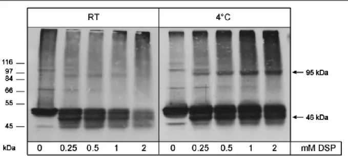
3.1. Chemical cross-linking of IFNγ-induced Irga6 ... 45
3.1.1. Irga6 is found in higher molecular weight complex upon chemical cross-linking ... 45
3.1.2. Analysis of cross-linking conditions ... 47
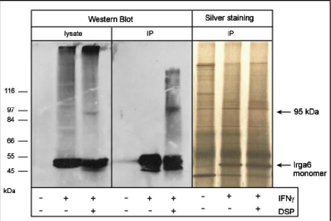
3.1.3. Immunoprecipitation of cross-linked Irga6 ... 48
3.2 Size Exclusion Chromatography... 49
3.2.1. Size Exclusion Chromatography of Irga6 proteins in Thesit ... 49
3.2.2. Size Exclusion Chromatography of IFNγ-induced Irga6 in Octyl-β-D glucopyranoside ... 51
3.2.3. Size Exclusion Chromatography of IRG proteins in Thesit ... 52
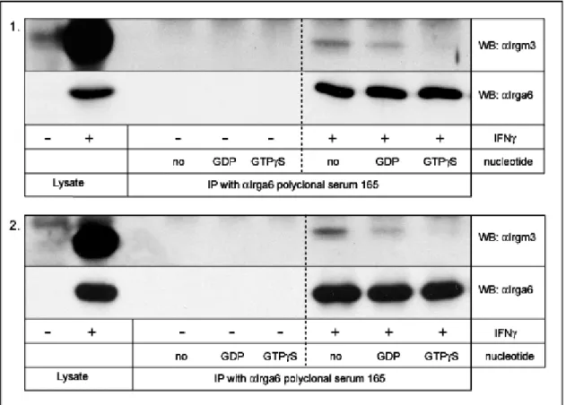
3.3. Irga6-Irgm3 interactions... 53
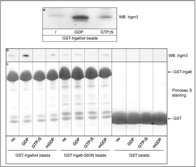
3.3.1. Co-immunoprecipitation of Irgm3 with Irga6 (in collaboration with Julia Hunn)53 3.3.2. Pull-down of Irgm3 with GST-Irga6 fusion proteins (in collaboration with Julia Hunn)... 55
3.4. Irga6 nucleotide-dependent self-interactions ... 57
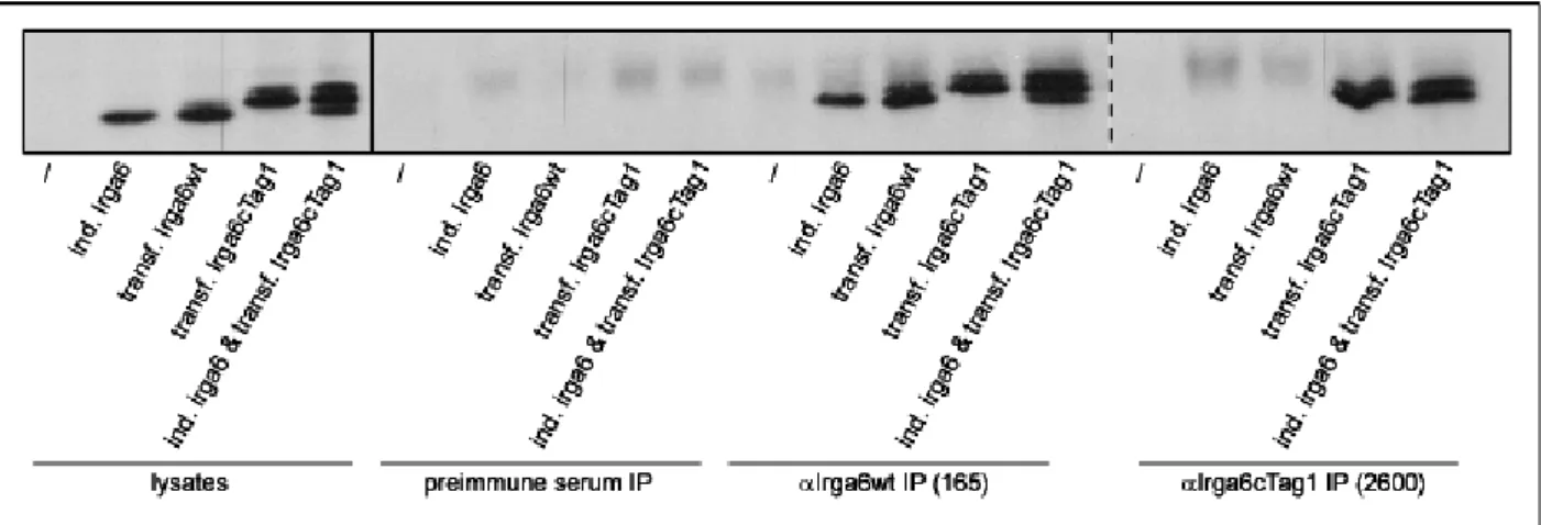
3.4.1. αcTag1 immunoprecipitation... 57
3.4.2. Co-immunoprecipitation of IFNγ-induced Irga6 with transfected Irga6cTag1 . 59 3.4.3. Effect of IFNγ induction on co-immunoprecipitation of Irga6 proteins... 61
3.4.4. Effect of mutations on co-immunoprecipitation of Irga6 proteins ... 63
3.5. IFNγ-induced factors are necessary for proper localisation of Irga6... 65
3.6. 10D7 antibody in immunofluorescence recognises transfected Irga6 and Irga6 on
T. gondii PV but not Irga6 that is relocalised to the ER ... 66
3.7. 10D7 antibody affinity determination ... 69
3.7.1. Purification of 10D7 and 10E7 Fab and Fc fragments ... 69
3.7.2. Determination of affinity of 10D7 and 10E7 fragments in Western blot... 74
3.7.3. Estimation of the relative affinities of 10D7 antibody and 10D7 C3 fragments in immunofluorescence ... 76
3.8. The 10D7 epitope is located between amino acids 20-25 of Irga6 ... 77
3.9. 10D7 immunoprecipitations ... 82
3.9.1. 10D7 precipitates Irga6 proteins in the presence of OGP but not in the presence of Thesit ... 82
3.9.2. Effect of nucleotide in Irga6 immunoprecipitation by 10D7 ... 84
3.9.3. Effects of detergents on binding of Irga6 to 10D7... 85
3.9.4. Effects of mutations in Irga6 on binding of Irga6 to 10D7 ... 86
3.9.5. Immunofluorescence analysis of Irga6 mutants... 88
3.10. Biochemical analysis of recombinant myristoylated Irga6... 95
3.10.1. Purification of myristoylated Irga6... 95
3.10.2. Triton X-114 partitioning assay ... 97
3.10.3. Effect of the myristoyl group on running behaviour of recombinant Irga6 proteins in Size Exclusion Chromatography ... 99
3.10.4. Dynamic light scattering of Ins-Irga6 proteins...100
3.10.5. 10D7 immunoprecipitation of Ins-Irga6 proteins ...102
3.10.6. Hydrolysis properties of myristoylated Irga6 ...104
4. Discussion ...109
4.1. Irga6 interacts with Irgm3 in a GDP-dependent manner...109
4.2. Irga6 forms GTP-dependent homooligomers in vivo in the absence of IFNγ- induced factors ...112
4.3. Possible Irga6 homooligomers in cells infected with Toxoplasma gondii...114
4.4. Analysis of the binding affinity of the αIrga6 monoclonal 10D7 antibody ...116
4.5. 10D7 epitope is located in the first N-terminal α helix of Irga6 but excludes its myristoyl group and the GTP-binding domain...119
4.6. Active form of Irga6 is recognised by 10D7 antibody...119
4.7. Model of conformational change of Irga6 induced by GTP binding ...121
4.8. Effects of the nucleotide state and the myristoyl group on Irga6 relocalisation to
the parasitophorous vacuole membrane ...123
4.9. Effect of the myristoyl group on biochemical properties of Irga6 protein ...125
4.10. Myristoylated Ins Irgs6wt hydrolyses GTP to GDP and GMP in vitro...128
4.11. Model of Irga6 regulation in vivo...129
5. References ...135
6. Summary ...156
7. Zusammenfassung...157
8. Acknowledgement ...159
9. Erklärung...160
10. Lebenslauf...161
1. Introduction
1.1 GTPases - general mechanism of GTP hydrolysis
An estimated 10–18% of all known gene products contain the mononucleotide-binding fold, which forms part of the P-loop domain (G1 motif) of both guanosine triphosphatases (GTPases) and adenosine triphosphatases (ATPases) (Leipe et al., 2002). However, the biological roles of GTPases and ATPases are very different. ATP hydrolysis liberates the energy needed to move motor proteins and accelerate metabolic reactions. Instead, GTP hydrolysis appears to have evolved exclusively to regulate guanine nucleotide-binding proteins, either through promoting their action as molecular switches or their self-assembly as mechanoenzymes (MacMicking, 2004).
The conversion of guanosine triphosphate (GTP) to guanosine diphosphate (GDP) and inorganic phosphate (Pi) by GTP binding proteins (GTPases) is a fundamental process in living cells. GTP hydrolysis controls numerous vital functions, including cellular growth and differentiation (Ras family; (Vojtek and Der, 1998), cytoskeletal dynamics and transcription (Rho/Rac/Cdc42 family; (Mackay and Hall, 1998), vesicular transport (Rab family; (Schimmoller et al., 1998), membrane trafficking (Arf family; (Moss and Vaughan, 1998), nucleocytoplasmic transport and mitotic spindle assembly (Ran family; (Hetzer et al., 2002), translation and protein translocation (EF-Tu, EF-G, SRP; (Chu et al., 2004), endocytosis (dynamin;
(Hinshaw, 2000; Hunn, 2007); (Song and Schmid, 2003); (Praefcke and McMahon, 2004), as well as cell-autonomous resistance against a variety of intracellular pathogens (Mx, GBP and IRG families; (Martens and Howard, 2006).
GTPases function as molecular switches, generally cycling between GTP- bound, active and GDP-bound, inactive form (figure 1.1). Only in the activated state these proteins interact with and activate downstream effectors, which in turn trigger cellular responses. GTP hydrolysis returns GTPases to their inactive state, thereby terminating downstream signalling (Scheffzek and Ahmadian, 2005).
Figure 1.1. GTPase cycle
The GDP-bound form of the GTP-binding protein is considered inactive, whereas the GTP-bound form represents the active form interacting with effector proteins. For many GTPases, but not all, transition between GDP- and GTP-bound states is regulated by different proteins. GDIs prevent dissociation of GDP, keeping the GTPase in the inactive form. GEF activity catalyses the release of GDP and the subsequent uptake of GTP. GAP activity triggers GTP hydrolysis, terminating the active state and restoring the inactive GDP form.
1.1.1. Nucleotide binding domain of GTPases
Common for all GTPases analysed to date is a nucleotide-binding domain (Leipe et al., 2002). The core of the GTP binding motif consists of central six- stranded beta sheets, five parallel and one antiparallel, surrounded by five alpha helices (figure 1.2). Binding of guanine nucleotide is mediated by five motifs termed G1–G5, of which the G1 [G(X)4GK(S/T)], G3 [DXXG], and G4 [(N/T)KXD] motifs are more or less universally conserved.
G1 motif G(X)4GK(S/T) (also referred to as P-loop) stabilises the phosphates of GDP and GTP of the bound nucleotide by hydrogen bonds formed by both lysine and serine/threonine residues (Kjeldgaard et al., 1996). In addition, the side chain of the Ser/Thr residue coordinates the position of magnesium ion, necessary for the catalytic activity of GTPases. In most of the known structures, the Asp residue in G3 motif DXXG is involved in binding a magnesium ion via a water molecule.
Furthermore, the Asp residue appears to form a hydrogen bond with the conserved
with the γ-phosphate. The G4 motif (N/T)KXD determines the specificity for the guanine base (Kjeldgaard et al., 1996).
Figure 1.2. The strucure of the GTP-binding domain
Ribbon plot of the minimal GTP-binding domain is shown, with the conserved sequence elements and the switch regions in different colors as indicated (Vetter and Wittinghofer, 2001).
1.2. Regulation of GTPase activity
The fraction of protein molecules in the GTP-bound state depends on the relative rates of two reactions: dissociation of GDP from the GDP-bound protein and hydrolysis of bound GTP, characterized by kdissGDP and kcatGTP rate constants respectively. Thus, the proportion of protein in the active GTP-bound state can be increased either by accelerating kdissGDP or by reducing kcatGTP (Bourne et al., 1990), performed for many GTPases by specific ligands.
Three groups of structurally distinctive proteins regulate GTPase cycle of some of the GTPases. Guanine-nucleotide dissociation inhibitors (GDIs) trap GTPases in an inactive, GDP-bound state whereas guanine-nucleotide exchange factors (GEFs) catalyze the release of GDP and the subsequent uptake of GTP.
GTPase activating proteins (GAPs) accelerate the rate of GTP hydrolysis and thereby terminate downstream signaling.
1.2.1. Guanine-nucleotide dissociation inhibitors (GDI)
Guanine-nucleotide dissociation inhibitors for Rho and Rab family proteins have been identified. GDIs bind to the prenylated COOH-terminus of small GTPases and thereby shield the hydrophobic tail from the aqueous environment. GTPase-GDI complexes constitute a cytoplasmic pool of prenylated proteins, allowing Rab and Rho proteins to be recycled between different membrane compartments in the cell (Vetter and Wittinghofer, 2001). However, no GDI candidates have been reported for the large GTPases like dynamin, Mx, GBP or IRG proteins.
1.2.2. Guanine-nucleotide exchange factors (GEF)
GEFs constitute a highly diverse group of proteins in the cell (Sprang, 2001).
They contain a variety of regulatory and protein-protein interaction domains (Cerione and Zheng, 1996) and they can be differentially expressed in a tissue-specific manner (Sprang, 2001). GEFs often possess multiple signal transduction modules allowing their activation by the interaction with various upstream regulators (Hoffman and Cerione, 2002). In addition, GEFs can be also activated by the phosphorylation (Kato et al., 2000). Thus, they can respond to various intra- and extracellular signals often resulting in their translocation to a specific membrane compartment where they than activate GTPases. Although diverse in structure, GEFs use a common mechanism to release nucleotide either by disrupting the magnesium ion binding site in the GTP binding proteins (Worthylake et al., 2000) or, in addition, by influencing γ- phosphate binding site and the P-loop (Sprang, 2001). GEF-GTPase nucleotide-free complex is dissociated by rebinding of the nucleotide (Hutchinson and Eccleston, 2000), normally GTP because of its higher concentration in cells (Kleinecke and Soeling, 1979) and because for most of the GTPases the affinity for GTP is higher than that for GDP. In the case of the large GTPases it has been argued that no GEF activity is required, on the ground of the low nucleotide binding affinity of these GTPases (Uthaiah et al., 2003).
1.2.3. GTPase-activating proteins (GAP)
GTP-hydrolysis by GTP-binding proteins is usually very slow but can be accelerated upon interaction with GTPase-activating proteins (GAPs). GAPs stimulate GTP hydrolysis in two ways: by supplying a catalytic arginine to the GTPase active site to facilitate the transition state and by reducing the flexibility of the switch segments, stabilising them in a catalytically functional state (Scheffzek et al., 1997). An exposed loop of the GAP inserts into the catalytic site of an appropriate GTPase, allowing the interaction of an arginine side chain (the arginine finger) with the β-phosphate of GTP. In addition, the arginine finger forms a hydrogen bond with the side chain of catalytically important glutamine (Gln61 in Ras) (Scheffzek et al., 1998). The exception is Rap1GAP, which uses a catalytic asparagine instead of an arginine (Daumke et al., 2004). Other variations, not involving a separate GAP protein, have been reported. In heterotrimeric G proteins, as well as in the human guanylate-binding protein hGBP1, the critical arginine residue is part of the GTP- binding protein itself and is supplied in cis (Sprang, 1997; Prakash et al., 2000b) whereas dynamins and Irga6 provide it in trans by self-association (Tuma and Collins, 1994; Uthaiah et al., 2003).
1.3. Dynamins
Dynamins are large GTPases (98 kDa) found in yeast, plants and animals.
They have been implicated in various cellular processes, such as vesicular trafficking and scission, organelle fusion and division and in cytokinesis (Praefcke and McMahon, 2004).
Classical dynamins consist of five domains: GTP-binding domain; middle domain, with no sequence homology to any known structural motif; pleckstrin homology domain (PH), which binds preferentially to phosphoinositides, in particular to phosphoinositide(4,5)biphosphate [PI(4,5)P2]; GTPase effector domain (GED), acting as a GAP for dynamin; proline rich domain (PRD), playing an important role in protein-protein interaction (Hinshaw, 2000).
1.3.1. Dynamin as GTPase
Dynamin was found in a monomer-tetramer equilibrium in solution (Hinshaw and Schmid, 1995; Tuma and Collins, 1995; Eccleston et al., 2002). It has low binding affinity for nucleotides, with dissociation constant in the micromolar range (Stowell et al., 1999; Binns et al., 2000; Marks et al., 2001). Considering the cellular nucleotide concentration (Kleinecke and Soeling, 1979) and that dynamin binds GTP 40-fold more tightly that GDP, it is expected that dynamin would predominantly exist in GTP-bound form in the cell.
Dynamin tetramers have a relatively high intrinsic rate of hydrolysis (kcat of
~200 min-1), which increases upon self-assembly of dynamin into oligomeric structures (Tuma and Collins, 1994; Hinshaw and Schmid, 1995). Up to 50-fold stimulation of GTP hydrolysis was shown to be mediated by dynamin itself, namely by GTPase effector domain (GED) (Sever et al., 1999), in this way providing its own GAP.
Self-assembly of dynamin tetramers into ring-like structure is spatially limited to the membranes, where the pleckstrin homology and another, yet unidentified, domain mediate lipid binding and proper positioning of dynamins (Burger et al., 2000). Although GTP binding is not necessary for membrane recruitment itself (Tuma and Collins, 1995; Burger et al., 2000), membrane tubulation and vesicle scission require GTP incorporation and its subsequent hydrolysis, respectively (Sweitzer and Hinshaw, 1998; Stowell et al., 1999; Marks et al., 2001).
The exact mechanism of dynamin function is still under dispute. The fact that GTP-bound dynamin oligomers tubulate membranes in vitro (Hinshaw and Schmid, 1995) and in vivo (Marks et al., 2001) and that the vesicle scission requires GTP hydrolysis led to the model describing dynamin as mechanochemical enzyme.
Alternatively, dynamin is considered as regulatory protein, recruiting effectors proteins in GTP-bound form via the proline rich domain (Praefcke and McMahon, 2004).
1.4. IFN-inducible GTPases 1.4.1. Mx proteins
Murine Mx1 was the first IFN-inducible GTPase implicated in cell-autonomous resistance against intracellular pathogens. Mx1 locus was discovered more than 40 years ago through a polymorphism in influenza virus resistance among different mouse strains. The A2G mouse was found to be resistant while all other strains were susceptible (Lindenmann et al., 1963). Resistance behaved genetically as a single dominant trait (Lindenmann, 1964). The resistance phenotype was confirmed at the cellular level in vitro as well (Lindenmann et al., 1978), indicating its cell-autonomous character. Mx genes were found in all vertebrates (Staeheli and Haller, 1985). In mouse and humans two Mx genes were characterized, Mx1 and Mx2, and MxA and MxB, respectively, and they were found to be induced by type I IFN (Goetschy et al., 1989; Simon et al., 1991).
Mx proteins are large GTPases (70-80 kDa) consisting of N-terminal GTP- binding domain and C-terminal domain involved in protein-protein interactions.
Recombinant MxA hydrolyses GTP to GDP with a turnover rate of 27 min-1 (Richter et al., 1995). High GTPase activity and several fold stronger binding of GTP than GDP suggest that most MxA proteins in vivo could be in the GTP-bound form.
Although no lipid-binding motif has been identified, purified MxA binds to lipid vesicles in a nucleotide-independent manner (Accola et al., 2002). In vivo, MxA partly co-localised with the smooth endoplasmic reticulum but the relevance of MxA lipid binding to its antiviral function is not clear. In the presence of GDP, however, MxA protein in solution formed evenly shaped rings that condensed to spirals or stacks of rings after incubation with GTPγS (Kochs et al., 2002). The oligomerisation property of Mx proteins was also confirmed by gel filtration analysis of both recombinant human His-tagged MxA (Richter et al., 1995) and mouse Mx1 (Melen et al., 1992).
MxA was shown to form homo-oligomers in vivo as well (Ponten et al., 1997) and the C-terminus of the protein, containing a putative leucine zipper, was required for the interaction. In mouse Mx1, the only Mx protein localising to the nucleus, the same domain carries a nuclear localisation signal. Both cytoplasmic and nuclear Mx proteins are found in granular and dotty structures, generally considered as Mx oligomers.
Mx proteins are antiviral resistance factor. Transfected murine Mx1 confers resistance against influenza virus even in the absence of IFN induction (Staeheli et al., 1986; Arnheiter et al., 1990), implying that other IFNα/β induced factors are not essential for protection against the virus, thought they may play a significant role in establishing the antiviral state (Staeheli et al., 1986). Human MxA blocks nuclear import of Thogoto virus nucleocapsids by GTP-dependent interaction of its C-terminal domain with a nucleoprotein (Kochs and Haller, 1999a; Kochs and Haller, 1999b).
Cytoplasmic MxA inhibits the multiplication of both influenza virus and vesicular stomatitis virus (Zurcher et al., 1992b). Interestingly, when moved to the nucleus with the help of a foreign nuclear transport signal, MxA not only retained its activity against influenza virus but was actually more effective, exerting its function by blocking primary transcription of influenza virus like mouse Mx1, whose natural location is the cell nucleus (Krug et al., 1985; Pavlovic et al., 1992). Nuclear localisation of mouse Mx1 protein is, however, necessary for inhibition of influenza virus (Zurcher et al., 1992a).
Figure 1.3. Model of MxA-MxA interaction mediated by the carboxyl-terminal region (Haller et al., 2007)
MxA can form two types of assemblies. In the absence of infection, MxA forms oligomers by intermolecular association of the LZ domain of one molecule (green box) with the CID of another molecule (blue box). These assemblies represent a storage form of antivirally inactive molecules, from which antivirally competent monomers are transiently released. In infected cells, MxA monomers bind to viral targets, such as nucleocapsids or nucleocapsid-like structures (red shape).
Mx proteins fold into three functional domains (Haller et al., 2007): N-terminal GTP-binding domain, containing self-assembly motif (Nakayama et al., 1993), central interactive domain (CID) and C-terminal leucine zipper domain (LZ). It has been reported that the C-terminal LZ domain interacts with both GTP-binding domain (Schwemmle et al., 1995) and with the CID of another Mx molecule (Schumacher and Staeheli, 1998). Based on these findings, a model for Mx antiviral function has been proposed (Di Paolo et al., 1999; Janzen et al., 2000; Haller and Kochs, 2002). It proposes that MxA proteins exist in two forms in the cell: an active monomeric and an inactive oligomeric form. The oligomers may represent a storage form of Mx molecules preventing their activation without the presence of the specific target.
Prerequisite for MxA oligomer formation is the folding of the C-terminal LZ domain (in green in figure 1.3.) (Di Paolo et al., 1999; Haller and Kochs, 2002). Folded LZ domain interacts with both CID (in blue, figure 1.3.) and self-assembly sequence in the GTP-binding motif of another MxA molecule, giving rise to the formation of large aggregates. After viral infection, cellular or viral protein(s) induce the dissociation of the MxA oligomers, releasing antivirally competent monomers. MxA monomers bind to viral targets, such as nucleocapsids or nucleocapsid-like structures (red shape, figure 1.3.) and sort them to locations where they are trapped and degraded. The exact mechanism of how monomeric Mx inhibits viral activity is not resolved. MxA needs to be in the GTP-bound form in order to interact with Thogoto virus nucleocapsid (Kochs and Haller, 1999a) though GTP hydrolysis itself seems not to be required (Janzen et al., 2000).
1.4.2. The guanylate-binding proteins (GBPs)
The response of cells to IFNγ is dominated by the induction of two families of GTPases, the IRG family and the guanylate-binding proteins (GBPs). There are seven GBP genes described in human, hGBP1-7 (Cheng et al., 1991; Olszewski et al., 2006) and eleven in mouse, mGBP1-11 (Degrandi, in press) and the family is well conserved in vertebrates (Robertsen et al., 2006). Despite their massive induction upon IFNγ treatment, only a weak antipathogenic effect of hGBP1 against vesicular stomatitis virus (VSV) and encephalomyocarditis virus (ECMV) in vitro has been reported (Anderson et al., 1999). Additionally, hGBP1 and mGBP2 were proposed to
have a role in regulation of vasculogenesis by proinflammatory cytokines (Guenzi et al., 2001; Guenzi et al., 2003) and IFN-mediated cell growth (Gorbacheva et al., 2002), respectively.
GBPs are large GTPases (65-67 kDa), consisting of an N-terminal GTP- binding motif and very elongated C-terminal helical domain (Prakash et al., 2000a).
The G4 motif, TLRD, responsible for the recognition of the guanine moiety of the nucleotide, is characterized by unique substitution of conserved lysine at the position 2 to the hydrophobic leucine (Praefcke et al., 1999). Three of the human (hGBP1, hGBP2 and hGBP5) and mouse (mGBP1, mGBP2 and mGBP5) GBPs posses a C- terminal CaaX box, resulting in farnesylation of hGBP1 and isoprenylation of all others (Nantais et al., 1996; Stickney and Buss, 2000).
The mechanism of GTP hydrolysis by hGBP1 is well studied. The binding affinity of hGBP1 for GTP, GDP and GMP is very low (0.5-2.4 µM) (Praefcke et al., 2004), due to high nucleotide dissociation rates. In the presence of GMP and GDP, hGBP1 was found as a monomer, but it is dimeric in the GTP-bound state and tetrameric in GDP-AlFx stabilised transition state (Prakash et al., 2000a; Praefcke et al., 2004). GTP-dependent dimerisation of hGBP1 results in at least an eight-fold increase in GTP hydrolysis by providing the catalytic arginine in cis (Prakash et al., 2000b; Ghosh et al., 2006). The unique position of hGBP1 amongst known GTPases is demonstrated by the hydrolysis of GTP to GDP and GMP (Schwemmle and Staeheli, 1994; Praefcke et al., 1999), leaving predominantly GMP as a product of hydrolysis (85-95% at 37°C; (Kunzelmann et al., 2006). Although GTP is hydrolysed in two successive cleavages of γ- and β-phosphates, GDP in solution cannot serve as a substrate. The crystal structure of hGBP1 in complex with the non-hydrolysable GTP analog GppNHp (Prakash et al., 2000b) illustrated that the largest structural changes involve guanine and phosphate caps around GTP-binding site, suggesting a shift of the nucleotide toward the catalytic centre after GTP hydrolysis by positioning the β-phosphate of GDP at the same place as the γ-phosphate of GTP was located before.
Recent data document subcellular localisation of hGBP1 to the Golgi apparatus (Modiano et al., 2005). Redistribution from the cytosol to the Golgi occurs when hGBP1 is in the GDP-AlFx transition state and requires isoprenylation and the presence of another, so far unidentified IFNγ-induced factor. Although the lipid modification of hGBP1 was shown to be important for the relocalisation of this protein
to the Golgi, its role in hGBP1 function in vivo, nor its effect on the biochemical properties of hGBP1 in vitro are not known.
1.4.3. Very large inducible GTPase (VLIG)
VLIG-1 protein, with a molecular weight of approximately 280 kDa, is the largest known GTPase (Klamp et al., 2003). Its expression is massively induced by IFNγ, and to a somewhat lesser extent by IFNβ. The GTP-binding domain of VLIG-1 is related to that of other IFN-inducible resistance GTPases, in particular with Mx and GBP proteins. In the assay using nucleotide-agaroses it was shown that VLIG-1 binds strongly to GDP-agarose and very weakly to GTP- and GMP-agaroses, indicating that this protein is, indeed, a GTP-binding protein (Klamp et al., 2003). The largest part of the long protein sequence does not share any structural similarities with any other protein. Recently, it has been reported that the central part of VLIG-1 exhibit 43% similarity to the CARD6 protein (Dufner and Mak, 2006), microtubule- interacting protein that positively modulates NF-kappaB activation (Dufner et al., 2006). However, the role of this central VLIG-1 region in its potential immunity-related function is unknown.
1.4.4. Immunity-related GTPases (IRGs)
IFNγ induction of mouse macrophages results in an immense transcription activation. Of estimated 1300 genes induced (Ehrt et al., 2001), messages of GBP and IRG protein families are the most abundant (Boehm et al., 1998), indicating their importance in immune response to pathogen infections.
IRG proteins are typically 47 kDa in molecular weight, with a canonical GTP- binding domain positioned approximately 80 amino acid from the N-terminus.
Analysis of the N-terminal sequence revealed that more than half of the mouse GTPases could be myristoylated. In mouse, three of the IRG proteins are characterized by the unique substitution of the universally conserved lysine in G1 motif (GX4GKS) to the methionine (GX4GMS), implying a distinct catalytic mechanism
for GTP hydrolysis. Thus, based on the sequence of the G1 motif, IRG proteins can be grouped into GKS and GMS subfamily (Boehm et al., 1998).
1.4.4.1. Genomic structure of IRG genes
The genome structure of IRG genes in the C57BL/6 mouse strain was analysed in detail (Bekpen et al., 2005). There are all together 25 coding units present, of which 24 contain IFN-responsive GAS and ISRE motifs in their promotor, resulting in their strong induction upon IFNγ stimulation. Exception is the Irgc gene, which is not induced by IFNγ nor does it possess IFN-response elements in its promotor. Interestingly, the only human full-length gene, IRGC, is an ortholog with 90% identity to the Irgc mouse gene. As in the mouse, human IRGC protein is constitutively expressed in male gonad (Rohde, 2007). In human genome, another Irg-like gene fragment was identified, containing only part of the GTP-binding domain of an IRGM protein. Although some IRG genes were found in zebrafish, there are no clear homologs in invertebrates below the Cephalohordates (Bekpen et al., 2005).
The Cephalochordate Branchiostoma floridae has a large family of IRG genes (Hunn, 2007). Absence of IFN-induced IRG proteins in humans show that they either possess alternative mechanisms effective against intracellular pathogens or they deploy other already known mechanisms more efficiently (Bekpen et al., 2005).
1.4.4.2. Localisation of IRG proteins
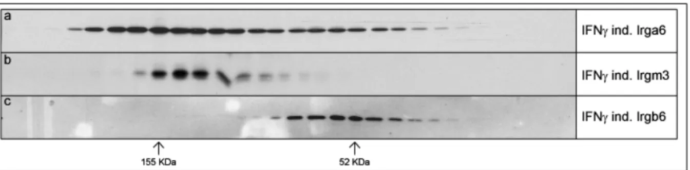
Analysis of cellular localisation of five of the IRG members, Irga6, Irgb6, Irgd, Irgm1 and Irgm3 (IIGP1, TGTP, IRG-47, LRG-47 and IGTP, in old nomenclature) in IFNγ-induced mouse fibroblasts and macrophages, revealed different levels of membrane association of these proteins (Martens et al., 2004). Irgm1 and Irgm3 were found almost exclusively bound to the membranes whereas, in contrast, Irgd protein was mainly soluble. Irga6 and Irgb6 partitioned roughly equally between the membrane bound and soluble fractions. Co-staining with different organelle markers localised Irgm1 and Irgm2 to the Golgi apparatus and Irgm3 to the endoplasmic reticulum (Martens, 2004; Martens et al., 2004). Irga6 and Irgb6 were found
predominantly co-localising in a reticular pattern with ER markers, contrasting findings reporting Irga6 association with Golgi markers (Zerrahn et al., 2002).
1.4.4.3. The involvement of IRG proteins in resistance to intracellular pathogens
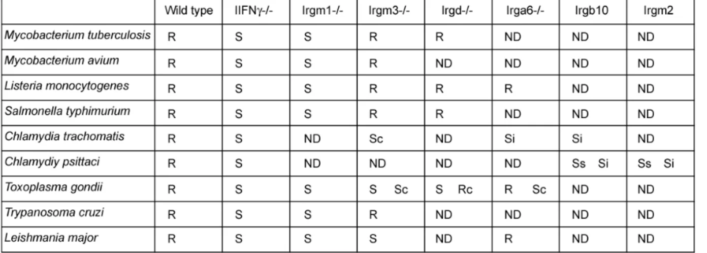
Consistent with their strong inducibility by IFNγ, many members of the IRG family have been implicated in resistance against intracellular pathogens in mice, both of bacterial and protozoan origin: Mycobacterium tuberculosis (MacMicking et al., 2003), Mycobacterium avium (Feng et al., 2004), Salmonella typhimurium (Taylor et al., 2004), Listeria monocytogenes (Collazo et al., 2001), Chlamydia trachomatis (Nelson et al., 2005; Bernstein-Hanley et al., 2006), Chlamydia psittaci (Miyairi et al., 2007), Tripanosoma cruzi (Santiago et al., 2005), Leishmania major (Feng et al., 2004) and Toxoplasma gondii (Taylor et al., 2000; Collazo et al., 2001; Halonen et al., 2001; Butcher et al., 2005; Martens et al., 2005; Ling et al., 2006). Involvement of different IRG proteins in resistance against these pathogens is shown in table 1.1.
Influence of IRG proteins in viral infections is limited to reports correlating overexpression of Irgb6 and relative reduced plaque formation by vesicular stomatitis virus but not herpes simplex virus in L cells (Carlow et al., 1998) and even weaker effect for Coxackie virus in Hela cells expressing Irgm2 (Zhang et al., 2003). Ebola virus and mouse cytomegalovirus have failed to reveal a susceptibility phenotype (Taylor et al., 2000).
Although several studies document susceptibility of IRG-deficient mice to various pathogens, little is known about the mechanism of their function. Irgm1- defficient mice infected with M. avium display severe lymphopenia at the site of bacterial replication (Feng et al., 2004) whereas pathology of T. gondii infected Irgm3-deficient mice was contributed to the overproduction of inflammatory cytokines (Taylor et al., 2000). Recently, infection with C. psittaci revealed that C57BL/6J mice were 105-fold more resistant than DBA/2J mice due to differential expression of both Irgb10 and Irgm2 proteins (Miyairi et al., 2007). Microarrays of infected peritoneal lavage showed over 10-fold upregulation of neutrophil-recruiting chemokines in susceptible mice and over 100-fold increase in macrophage differentiation genes in resistant mice. Massive neutrophil recruitment was seen in susceptible in comparison
to resistant mice indicating that the susceptibility pattern involves the stimulation of different inflammatory pathways.
Table 1.1. Susceptibilities of IRG-deficient mice and cells to intracellular pathogens (modified from (Martens and Howard, 2006)
R, wild type or knockout mouse resistant; S, knockout mouse sensitive; ND, not done; Rc, knockout cells resistant; Sc knockout cells sensitive; Ss, wild type mouse strain DBA/2J sensitive; Si, RNAi wild- type cells sensitive.
1.4.4.4. The involvement of IRG proteins in cell-autonomous resistance to intracellular pathogens
Loss of resistance of Irgm1- and Irgm3-deficient mice to infection with T.
gondii correlates with the loss of IFNγ-dependent resistance in infected cells in vitro (Halonen et al., 2001; Butcher et al., 2005), demonstrating the role of IRG proteins in cell-autonomous resistance. Effect of IRG proteins in challenging pathogen infections is not redundant (Collazo et al., 2001; Butcher et al., 2005; Bernstein-Hanley et al., 2006; Miyairi et al., 2007), indicating that they may regulate each other’s functions in infected cells. Five of the known IRG proteins, Irgb6, Irga6, Irgd, Irgm2 and Irgm3 were found concentrated at the parasitophorous vacuole membrane in IFNγ-induced astrocytes infected with T. gondii (Martens et al., 2005), already 15 min after infection. Both Irga6 and Irgm3 were found to be involved in T. gondii vacuole vesiculation and disruption in astrocytes and macrophages (Martens et al., 2005;
Ling et al., 2006), effectively striping parasite of its membranes. A series of disruption
events was proposed involving parasitophorous vacuole membrane (PVM) ruffling, PVM vesiculation, disruption, and parasite plasma membrane stripping, leading to lysosomal degradation of the parasite (Ling et al., 2006). Disruption of T. gondii containing vacuoles, measured by uracil incorporation, was slightly decreased in Irga6-deficient cells, although Irga6-deficient mice showed no significant susceptibility to T. gondii infection (Martens et al., 2005).
Irgm1 protein was not found on parasitophorous vacuole (Martens et al., 2005) but associated with phagosomes containing M. tuberculosis in IFNγ-induced macrophages (MacMicking et al., 2003). Recruitment of Irgm1 to the mycobacterial phagosome seems not to require a pathogen-derived signal since this relocalisation occurs as well during phagocytosis of latex beads in fibroblasts and macrophages (Martens et al., 2004). Absence of Irgm1 results in delayed and limited phagosome acidification in Irgm1-deficient cells (MacMicking et al., 2003).
A role of Irgm1 in induction of autophagy has been also proposed (Gutierrez et al., 2004). As autophagosome-like vacuoles were closely associated with disrupted T. gondii-containing vacuoles (Martens et al., 2005; Ling et al., 2006), susceptibility of Irgm1-deficient mouse to T. gondii infection could be explained by inability of cells to engulf disrupted vacuole into the autophagosome. Because autophagy also targets cytosolic pathogens (Rich et al., 2003; Deretic, 2005), its promotion by Irgm1 may also explain how Irgm1 helps mice to resist infections by L. monocytogenes (Collazo et al., 2001) and T. cruzi (Santiago et al., 2005).
1.4.4.5. Biochemical properties of IRG proteins
GTPase activities of three of the IRG proteins, Irga6, Irgb6 and Irgm3, have been reported. Partly purified GST-Irgb6 and GST-Irgm3 proteins were shown to hydrolyse GTP to GDP in vitro (Taylor et al., 1996; Carlow et al., 1998). Weak GTP hydrolysis was also demonstrated for FLAG-Irgm3 immunoprecipitated from NIH/3T3 fibroblasts (Taylor et al., 1997). It is suggested that cellular Irgm3 is predominantly GTP-bound (Taylor et al., 1997), although nucleotide-binding affinity were not measured. Both endogenous, IFNγ-induced Irgm3 and transfected FLAG-Irgm3 proteins were shown to be mostly in the GTP-bound form in cells (up to 96%), indicating that the IFNγ state of the cell does not influence the activity of this protein
(Taylor et al., 1997). However, recombinant Irga6 protein is the only IRG member whose biochemical and enzymatic properties were studied in detail.
Recombinant Irga6, purified from Escherichia coli, crystallized as a dimer in the nucleotide-free and GDP-bound state (Ghosh et al., 2004). Structure shows Irga6 folding into helical and G domain, consisting of 6-stranded β sheets surrounded by 6 α helices. Helical domain is unique, being built from both N- and C-terminal helices (figure 1.4.). Since recombinant Irga6wt contained a short non-canonical N-terminal extension derived from the GST fusion (Uthaiah et al., 2003) and, in addition, because prokaryotes do not expressed enzymes necessary for protein myristoylation (Heuckeroth et al., 1988), the structural position of the N-terminal myristoyl group could not be determined.
Figure 1.4. Structure of Irga6 (Ghosh et al., 2004)
Irga6 binds to GTP and GDP with dissociation constants in the micromolar range with at least 10 times higher affinity for GDP than for GTP (Uthaiah et al., 2003). Considering cellular nucleotide concentration, 330µM GTP and 120 µM GDP (Kleinecke and Soeling, 1979), Irga6 in cells should be predominantly GDP-bound. It hydrolyses GTP to GDP but not to GMP, and the GTPase activity is concentration- dependent with a maximum GTP turnover rate of 2 min-1. Magnesium ions were found to be essential for the GTPase reaction. Irga6 oligomerises in the presence of
GTP, and the oligomers resolve as GTP hydrolysis proceeds. Micromolar nucleotide affinities and oligomerisation-dependent hydrolytic activity are properties that Irga6 shares with other large GTPases, GBPs, Mx proteins and with the paradigm of the self-activating GTPases, the dynamins. As in all other large GTPases (Boehm et al., 1998; Prakash et al., 2000a; Danino and Hinshaw, 2001; Haller and Kochs, 2002;
Klamp et al., 2003), the catalytically important Gln61Ras in Irga6 is replaced by a hydrophobic amino acid, suggesting the requirement for a different catalytic residue (Mishra et al., 2005). Irga6 oligomer formation and GTP hydrolysis is necessary for AlFx to bind and trap the complex in AlFx-GDP transition state, indicating that the oligomer is the catalytically active form in which an additional catalytic residue can be provided in cis or in trans (Uthaiah et al., 2003).
Since recombinant Irga6wt, purified from E. coli was not myristoylated, it is not clear whether IFNγ-induced Irga6wt in cells share the same biochemical properties with the recombinant Irga6wt in vitro. It has been reported that the lipid modification affects enzymatic activity of Rac GTPase (Molnar et al., 2001) or the nucleotide binding affinities of small GTPase Arf1 (Randazzo et al., 1995). However, there is no information about the role of the myristoyl group in the enzymatic properties of Irga6.
1.5 Lipid modifications and membrane-binding properties of GTPases
Lipid modifications, both extra- and intracellularly oriented, are increasingly recognised as important mechanisms for targeting proteins within cells and for controlling their activity. Intracellularly oriented lipid modifications include reversible S-acylation (palmitoylation) as well as covalent myristoylation and prenylation (farnesylation and geranylgeranylation) (Magee and Seabra, 2005). These hydrophobic moieties are found on a wide variety of proteins and are thought to regulate signal transduction, movement of a signaling protein within the cell and its final destination and reversible protein-protein and protein-membrane associations (Resh, 2006).
1.5.1 S-acylation
S-acylation is a unique modification that is reversible in vivo and can therefore regulate protein localisation and function (Milligan et al., 1995). Nearly all S-acylated proteins contain palmitate, a saturated sixteen-carbon fatty acid, linked through thioester bond to one or more cysteine residues. Addition of palmitate is not limited to the N- or C-terminus of the protein and there is no consensus-binding motif.
Palmitoylated cysteine residues are often found near sites of N-myristoylation or prenylation where they increase membrane-binding energy, although mono- and di- palmitoylation alone can localise protein to the plasma membrane (McCabe and Berthiaume, 1999; Baker et al., 2003; Magee and Seabra, 2005) and the Golgi region (Perez de Castro et al., 2004; Magee and Seabra, 2005), respectively.
1.5.2. Prenylation
Protein prenylation is a posttranslational reaction that occurs in the cytosol. A 15-carbon (farnesyl) or 20-carbon (geranylgeranyl) isoprenoid are linked irreversibly through thioether bond to one or more cysteine residues at or near the C terminus of the protein. Many prenylated proteins contain a C-terminal ‘CaaX box’ (Cys-aliphatic- aliphatic-X) and the ‘X’ amino acid determines whether the cysteine within the CaaX box is farnesylated by farnesyltransferase (X is not leucine) or geranylgeranylated by geranylgeranyltransferase I (X is leucine) (Konstantinopoulos et al., 2007). Upon prenylation, proteins are translocated to the ER where the three C-terminal amino acids (aaX) are cleaved (Boyartchuk et al., 1997) and, subsequently, the C-terminal prenylated cysteine residue carboxymethylated (Gelb, 1997).
For small G proteins, the number of prenyl groups as well as their identity influence protein targeting. RhoB exists in farnesylated and geranylgeranylated forms; farnesylated RhoB is targeted to the plasma membrane, whereas geranylgeranylated RhoB localises to late endosomes (Gomes et al., 2003). Most Rab proteins are doubly geranylgeranylated at the two cysteine residues at the C- terminus (Zerial and McBride, 2001; Goody et al., 2005) and localise to endosomes, whereas monogeranylgeranylated Rabs mistarget to the ER and are nonfunctional (Calero et al., 2003; Gomes et al., 2003). Geranylgeranylated Rab proteins are in the
cytosol in complex with a GDI molecule, which sequester the prenyl groups to form a soluble complex. Rab-GDI interaction is strongly dependent on Rab prenylation and the presence of GDP (Sasaki et al., 1990; Sasaki et al., 1991). Upon association with appropriate membrane (Soldati et al., 1994), GDI is released from the Rab protein which, anchored to the membrane via its geranygeranyl groups, can interact with a membrane-localised GEF. Process of membrane association and GDI release often involves proteins known as GDI-displacement factors (GDFs), which are proposed to catalyse Rab-GDI dissociation at particular membrane surfaces (Pfeffer and Aivazian, 2004). GTP-bound Rab senses its target proteins and the interaction is terminated upon GAP-dependent GTP hydrolysis, allowing soluble Rab-GDI complex formation (Behnia and Munro, 2005). Similar mechanism was reported for Rho (Dransart et al., 2005) and Ras (Pechlivanis and Kuhlmann, 2006) GTPases.
GBP proteins are also characterized by presence of the C-terminal CaaX box, predicting protein modification either by farnesylation (hGBP1 and mGBP5) or geranygeranylisation (hGBP2, hGBP5, mGBP1 and mGBP2) (Vestal et al., 2000;
Vestal, 2005). However, in vivo prenylation was only shown for mGBP2 (Vestal et al., 2000) and hGBP1 (Modiano et al., 2005). Redistribution of soluble hGBP1 to the Golgi is GTP-dependent and requires both farnesylation and IFNγ-induced factor (Modiano et al., 2005).
1.5.3. Myristoylation
Approximately 0.5% of all eukaryotic proteins are modified by co-translational binding (Olson and Spizz, 1986; Wilcox et al., 1987) of myristate (rare 14-carbon fatty acid) to the N-terminal glycine residue (Kamps et al., 1985) on a variety of eukaryotic and viral proteins (Resh, 2006). After the initiating methionine is removed by methionine aminopeptidase, myristate is linked through an amide bond to the N- terminal glycine in the reaction catalysed by N-myristoyltransferase (Farazi et al., 2001). Myristoylation absolutely requires N-terminal glycine residue at position 1 and preferentially small, polar serine and, to a lesser extent, threonine residue at position 5 (Maurer-Stroh et al., 2002).
N-myristoylation alone does not provide sufficient hydrophobicity to stably anchor proteins to a lipid bilayer but rather promotes weak and reversible protein-
membrane and protein-protein interactions (Peitzsch and McLaughlin, 1993; Silvius and l'Heureux, 1994; Murray et al., 1997; Murray et al., 1998). Stable membrane association of N-myristoylated proteins requires a second lipid-binding motif. This secondary signal can be either palmitoyl group, attached by palmitoyl acyltransferases residing in the target membrane domain (Schroeder et al., 1996) or N-terminal stretch of basic amino acids associating electrostatically with negatively charged phospholipids (Murray et al., 1997; Murray et al., 1998).
Alternatively, regulated membrane association of myristoylated proteins is achieved by ligand-promoted conformational changes, so-called switches. In myristoyl-electrostatic switch, phosphorylation within the N-terminal polybasic motif introduces negative charge and reduces the electrostatic component of bilayer interaction, thus releasing the protein from the membrane (McLaughlin and Aderem, 1995). In the case of myristoyl-ligand switches, the myristoylated N-terminus is, in the absence of ligand, sequestered in a hydrophobic binding site located on the protein (Bhatnagar and Gordon, 1997). Binding of ligand induces a protein conformational change that exposes the myristoyl moiety, allowing the protein to associate with membranes (Zozulya and Stryer, 1992; Ames et al., 1995).
The small GTPase ADP-Ribosylation Factor-1 (ARF1) provides one example of a myristoyl-ligand switch, interacting with the membrane through two components:
the myristate, which gives a basal affinity for lipid regardless of the protein conformation (Franco et al., 1995), and a protein region that becomes available for membrane binding only when ARF switches to the active, ARF-GTP conformation. In the GDP-bound form, ARF1 is soluble, with hydrophobic residues of the N-terminal amphipathic peptide buried in the protein core. When ARF switches to the GTP state, these residues insert into membrane lipid layer (Antonny et al., 1997).
Eleven members of IRG protein family are shown to carry the amino-terminal myristoylation signal MGxxxS. However, lipid modification of only one of them, namely Irga6, was reported (Martens et al., 2004). Even though the N-terminus of Irga6 targets EGFP to membranes in a myristoyl-dependent manner, myristoylation is not absolutely necessary for membrane association of Irga6 itself since only a minor increase of cytosolic pool of nonmyristoylated Irga6, relative to the wild type protein, was found (Martens et al., 2004). Apart from the influence on the resting localisation of Irga6, nothing is known about the effect of the myristoyl group on Irga6 activity and its relocalisation to the PVM.
1.6. The aim of this study
Immunity-Related GTPases (IRG), represented by 25 coding units in the mouse genome, are implicated in resistance against a variety of bacterial and protozoal pathogens (table 1.1.). Several studies have documented the susceptible phenotype of IRG-deficient mice upon challenge with various pathogens, but the exact mechanism of their function in cell-autonomous immunity is still not known.
This study attempts to understand the role of IRG proteins in cell-autonomous immunity by analysis of one member of IRG family, Irga6. Identification of Irga6 interaction partners in IFNγ-induced cells could give more insight into the function of Irga6. Because IRG proteins show non-redundancy in resisting pathogen infection (Collazo et al., 2001; Butcher et al., 2005; Bernstein-Hanley et al., 2006; Miyairi et al., 2007) and, thus, probably regulate each other’s functions, association of Irga6 with other IRG proteins was also analysed.
Recombinant nonmyristoylated Irga6wt was shown to form GTP-dependent, catalytically active homooligomers in vitro (Uthaiah et al., 2003), characterized by increased GTPase activity. However, there was no information about Irga6 self- association in vivo. Therefore, the ability of Irga6 to form GTP-dependent homooligomers in vivo was studied in an attempt to analyse the nucleotide state of Irga6 in IFNγ-induced cells and possible changes of Irga6 activity during infection.
IRG proteins possess few obvious structural features, apart from the GTP- binding domain itself, which would indicate their function in vivo. One exception is Irga6, which has a myristoylation sequence (MGQLST) at the N-terminus.
Additionally, Triton X-114 partitioning demonstrated the hydrophobic character of native Irga6 (Martens et al., 2004) that was dependent on the integrity of the myristoylation motif, indicating, all together, that Irga6wt is probably myristoylated in vivo. In this study, it was attempted to analyse the role of the myristoyl group in Irga6 activity in vivo and in the ability of Irga6 to relocalise to the parasitophorous vacuole membrane of Toxoplasma gondii.
The structure and biochemical properties of purified, recombinant, nonmyristoylated Irga6 protein have been intensively studied in vitro and are well characterised (Uthaiah et al., 2003; Ghosh et al., 2004). However, there have been reports demonstrating that the lipid modification significantly affects the enzymatic properties of the GTPases in the presence of lipid vesicles and membranes. Addition
of membranes increased GTP hydrolysis of only prenylated Rac GTPase and had hardly any effect on the nonprenylated protein (Molnar et al., 2001). Myristoylation of the small GTPase Arf1 strongly influences its nucleotide binding affinities. In the absence of lipids, Arf1 has a higher affinity for GDP than for GTP, whereas, in the presence of phospholipids, Arf1 affinity for GTPγS is much higher than for GDP (Randazzo et al., 1995).
In addition to identifying the role of myristoyl group on Irga6 function in vivo, this study also aimed to purify the myristoylated Irga6 and analyse its GTPase activity as well as nucleotide binding affinities in the presence and absence of lipid vesicles.
The analysis of biochemical properties of myristoylated Irga6 would, hopefully, contribute to better understanding of the function of Irga6 in T. gondii infected cells and give more insight in its role in the vesiculation and disruption of the parasitophorous vacuole membrane and degradation of the parasite.
2. Material and Methods 2.1 Reagents and Cells
2.1.1 Chemicals, Reagents and Accessories
All chemicals were purchased from Aldrich (Steinheim), Amersham-Pharmacia (Freiburg), Applichem (Darmstadt), Baker (Deventer, Netherlands), Boehringer Mannheim (Mannheim), Fluka (Neu-Ulm), GERBU (Gaiberg), Merck (Darmstadt), Pharma-Waldhof (Düsseldorf), Qiagen (Hilden), Riedel de Haen (Seelze), Roth (Karlsruhe), Serva (Heidelberg), Sigma-Aldrich (Deisenhofen) or ICN biochemicals, Oxoid, (Hampshire UK). Developing and fixing solutions for Western Blot detection were from Amersham Pharmacia (Freiburg), Luminol from Sigma Aldrich (Deisenhofen), Coumaric acid from Fluka (Neu-Ulm). Deionised and sterile water (Seral TM) was used for all the buffers and solutions, Ultra pure water derived from Beta 75/delta UV/UF from USF Seral Reinstwassersysteme GmbH, (Baumbach) equipped with UV (185/254nm) and ultrafiltration (5000 kd cut off), or from Milli-Q- Synthesis (Millipore).
2.1.2 Equipment
Centrifuges used were: Biofuge 13, Heraeus; Sigma 204; Sigma 3K10; Labofuge 400R, Heraeus; Sorvall RC-5B, Du Pont instruments; Optima TLX Ultracentrifuge, Beckmann and Avanti J-20 XP, Beckman. BioRAD Gel dryer, Model-583; BioRad Power pack 300 or 3000; electrophoresis chambers from FMC Bioproducts (Rockland Maine US); Gel Electrophoresis Chamber, Cambridge electrophoresis;
Biorad Mini Protean II; PTC-100, MJ Research Inc.; ÄKTA P-920, OPC-900, Frac- 950, Amersham; Centrifuge tubes 15ml, TPP Switzerland; 50ml Falcon, BectonDickenson; Zeiss Axioplan II fluorescence microscope equipped with a Quantix cooledCCD camera.
2.1.3 Materials
Sterile filters FP 030/3 0,2 µm and ME 24 0,2 µm (Schleicher und Schüll, Dassel);
Nitrocellulose transfer membrane PROTRAN (Schleicher und Schüll, Dassel); 3MM Whatmann Paper (purchased via LaboMedic); 100 Sterican 0,50 x 16mm hypodermic needles (Braun AG, Melsungen); 0.2µm and 0.45µm sterile filters (Schleicher und Schuell, Dassel); X-OMAT LS and AR X-ray films, Kodak. All plastic ware for cell culture was from Sarstedt (Nümbrecht) or Greiner (Solingen).
2.1.4 Enzymes/ Proteins
Restriction enzymes (New England Biolabs); “Complete Mini” protease inhibitor cocktail (Roche, Mannheim); Pyrococcus furiosus (Pfu) DNA Polymerase (Promega, Mannheim); T4 DNA ligase (New England Biolabs); RNase A (Sigma); shrimp alkaline phosphatase (SAP) (USB, Amersham); PageRulerTM Prestained Protein Ladder (Fermentas); PageRulerTM Protein Ladder (Fermentas); SigmaMarkerTM Wide Range (Sigma); GeneRulerTM DNA Ladder Mix (Fermentas).
2.1.5 Kits
Plasmid Maxi and Midi kit (Qiagen, Hilden), Terminator-cycle Sequencing kit version 3 (ABI),
QuikChange TM Site directed mutagenesis kit (Stratagen), Rapid PCR product purification Kit (Roche, Mannheim),
2.1.6 Vectors
pGW1H (British Biotech), pGEX-4T2 (Amersham), pVL1393 (BD Biosciences),
pEGFP-N3 (BD Bioscience Clontech).
2.1.7 Cell lines
L929 (CCL-1) and gs3T3 (Invitrogen) mouse fibroblasts were cultured in IMDM or DMEM supplemented with 10% FCS (Biochrom AG, Berlin), 2 mM L-Glutamine, 1 mM sodium pyruvate, 100 U/ml penicillin and 100 µg/ml streptomycin, all from Gibco BRL. Hybridoma 10D7 and 10E7 cells were grown in IMDM, supplemented with 5%
FCS (Biochrom AG, Berlin), 2 mM L-Glutamine, 1 mM sodium pyruvate, 100 U/ml penicillin and 100 µg/ml streptomycin, all from Gibco BRL. Sf9 insect cells, cloned from IPLB-Sf21 derived from pupal ovarian tissue of the Fall armyworm, Spodoptera frugiperda, were grown in Insect-XpressTM Medium (Lonza) or in TNM-FH Insect Medium (Sigma).
2.1.8 Media
Luria Bertani (LB) medium
10 g bacto tryptone, 5 g yeast extract, 10 g NaCl, destilled water 1l LB plate medium
10 g bacto tryptone, 5 g yeast extract, 10 g NaCl, 15 g agar, destilled water 1l IMDM (Iscove’s Modified Dulbecco’s Medium) supplemented with 10% FCS
(Biochrom AG, Berlin), 2 mM L-glutamine, 1 mM sodium pyruvate, 1x non- essencial amino acids, 100 U/ml penicillin, 100 µg/ml streptomycin (all from Gibco BRL)
DMEM (Dulbecco’s Modified Eagle Medium) supplemented with 10% FCS (Biochrom AG, Berlin), 2 mM L-glutamine, 1 mM sodium pyruvate, 1x non-essencial amino acids, 100 U/ml penicillin, 100 µg/ml streptomycin (all from Gibco BRL) Insect-XpressTM Medium with L-glutamine (Lonza)
TNM-FH Insect Medium (Modified Grace’s Medium) with L-glutamine and sodium bicarbonate (Sigma)
2.1.9 Bacterial and protozoan strains
Escherichia coli DH5α: 80dlacZ ∆Μ15, recA1, endA1, gyrA96, thi-1, hsdR17 (rB-, mB+), supE44, relA1, deoR, ∆(lacZYA-argF)U169
Escherichia coli BL-21: E. coli B, F-, omp T, hsd S (rB-, mB-), gal, dcm Toxoplasma gondii Me49
2.1.10. Serological reagent
2.1.10.1. Primary antibodies and sera
Name Immunogen Species Concentration Dilution Origin
10D7 recombinant mouse Irga6wt
mouse monoclonal
1-3 µg/µl WB: 1:2000 IF: 1:500
IP: 10µl/ 50µl PAS
Jens Zerrahn, Berlin
10E7 recombinant mouse Irga6wt
mouse monoclonal
1-3 µg/µl WB: 1:2000 IF: 1:500
IP: 10µl/ 50µl PAS
Jens Zerrahn, Berlin
5D9 recombinant mouse
Irga6wt
mouse monoclonal
1-3 µg/µl IP: 10µl/ 50µl PAS Jens Zerrahn, Berlin
165 recombinant mouse
Irga6wt
rabbit polyclonal
1-3 µg/µl WB: 1:25000 IF: 1:8000 IP: 7µl/ 50µl PAS 2600 peptide
KLGRLERPHRD
rabbit polyclonal
IF: 1:4000
IP: 7µl/ 50µl PAS
EUROGENTEC
αIGTP clone 7
Mouse Irgm3 aa 283- 423
mouse monoclonal
0.25 µg/µl WB: 1:2000 BD Transduction laboratories A20
(sc11079)
N-teminal peptide of Irgb6
goat polyclonal
0.2 µg/µl WB: 1:500 Santa Cruz
2.1.10.2. Secondary antibodies and antisera
donkey-α-mouse Alexa 488, donkey-α-rabbit Alexa 546, donkey-α-mouse Alexa 546, donkey-α-rabbit Alexa 488 (all Molecular Probes), goat-α-mouse kappa light chain-
FITC (Southern Biotech), donkey-α-rabbit (Amersham), goat-α-mouse HRP (Amersham), donkey-α-goat HRP (Santa Cruz), DAPI (Roche).
2.2 Molecular Biology
All plasmids and constructs were amplified, cloned or propagated using protocols adapted from Sambrook, J., Fritsch, E.F., and Maniatis, T., Vol. 1, 2, 3 (1989), or from the cited references.
2.2.1 Agarose gel electrophoresis
DNA was analysed by agarose gel electrophoresis (1x TAE; 0.04 M Tris, 0.5 mM EDTA, pH adjusted to 7.5 with acetic acid) The DNA was stained with ethidium bromide (0.3 µg/ml), a fluorescent dye which intercalates between nucleotide bases, and the migration of the DNA molecules was visualized by using bromophenol blue.
2.2.2 Generation of Irga6 expression constructs
pGW1H-Irga6cTag1 construct was made by amplification of Irga6cTag1 sequence from pGEX-4T-2-Irga6cTag1 by using IIGP-SAL5 as forward and IIGPm3 as reverse primers. IIGPm3 primer introduces new SalI site after the stop codon (marked in red) and, additionally, mutates the internal SalI restriction site (residue in green). PCR product was digested with SalI and ligated into the SalI-digested pGW1H vector.
IIGP-SAL5 forward 5’-CCCCCCCCCGTCGACCACCATGGGTCAGCTGTTCTCTTCACCTAAG-3’
IIGPm3 reverse 5’-CCCCCCCCCGTCGACTCAGTCACGATGCGGCCGCTCGAGTCGGCCTAG-3
Following mutations were introduced in both pGW1H-Irga6wt and pGW1H- Irga6cTag1 constructs using listed primers:
pGW1H-Irga6-G2A forward 5’-GAGTCGACCACCATGGCTCAGCTGTTCTCTTCA-3’
pGW1H-Irga6-G2A reverse 5’- TGTAGAGAACAGCTGAGCCAGGGTGGTCGACTC-3’
∆7-12 forward 5’-GGGTCAGCTGTTCTCTAATAATGATTTGCCC-3’
∆7-12 reverse 5’- GGGCAAATCATTATTAGAGAACAGCTGACCC-3’
K82A forward 5’-GGGAGACGGGATCAGGGGCGTCCAGCTTCATCAATACCC-3’
K82A reverse 5’- GGGTATTGATGAAGCTGGACGCCCCTGATCCCGTCTCCC-3’
S83N forward 5’-GGAGACGGGATCAGGGAAGAACAGCTTCATCAATACCCTG-3’
S83N reverse 5’- CAGGGTATTGATGAAGCTGTTCTTCCCTGATCCCGTCTCC-3’
E106A forward 5’-GCTAAAACTGGGGTGGTGGCGGTAACCATGGAAAG-3’
E106A reverse 5’-CTTTCCATGGTTACCGCCACCACCCCAGTTTTAGC-3’
Primers listed below were used to introduce mutations only in pGW1H-Irga6wt construct.
T21I forward 5’-CCCTCCAGCTTTATTGGTTATTTTAAGAAATTTAATACGG-3’
T21I reverse 5’-CCGTATTAAATTTCTTAAAATAACCAATAAAGCTGGAGGG-3’
G22E forward 5’-CCCTCCAGCTTTACTGAGTATTTTAAGAAATTTAATACGG-3’
G22E reverse 5’-CCGTATTAAATTTCTTAAAATACTCAGTAAAGCTGGAGGG-3’
F24L forward 5’-GCCCTCCAGCTTTACTGGTTATTTGAAGAAATTTAATACGGG-3’
F24L reverse 5’-CCCGTATTAAATTTCTTCAAATAACCAGTAAAGCTGGAGGGC-3’
∆7-25 forward 5’-CCACCATGGGTCAGCTGTTCTCTAAATTTAATACGGG-3’
∆7-25 reverse 5’- CCCGTATTAAATTTAGAGAACAGCTGACCCATGGTGG-3’
∆2-12 forward 5’-CGAGATCTAGAGTCGACCACCATGAATAATGATTTGCCC-3’
∆2-12 reverse 5’- GGGCAAATCATTATTCATGGTGGTCGACTCTAGATCTCG-3’
Following primers were used to make deletions in pEGFP-N3-Irga6-1-33 construct, containing first 33 Irga6 amino acids. This way, three constructs were created containing first 20, 23 and 25 Irga6 amino acids, respectively.
∆21-33 forward 5’-CCTCCAGCTTTGTCGACGGTACCGCG-3’
∆21-33 reverse 5’-CGCGGTACCGTCGACAAAGCTGGAGG-3’
∆24-33 forward 5’-GCTTTACTGGTTATGTCGACGGTACCGCG-3’
∆24-33 reverse 5’- CGCGGTACCGTCGACATAACCAGTAAAGC-3’
∆26-33 forward 5’-GGTTATTTTAAGGTCGACGGTACCGCGGGC-3’
∆26-33 reverse 5’-GCCCGCGGTACCGTCGACCTTAAAATAACC-3’
To create pVL-Irga6wt construct, pGEX-Irga6wt was digested with SmaI and NotI restriction enzymes. Appropriate fragment containing Irga6wt was excised from the
agarose gel and ligated into the pVL1393 vector, which was already digested with SmaI and NotI. Thrombin cleavage site (underlined), between Irga6wt sequence and the stop codon was introduced by site directed mutagenesis using pVL-Irga6wt- thrombin forward and pVL-Irga6wt-thrombin reverse primers. Sequence encoding six histidine residues (in red) behind the thrombine cleavage site was introduced by pVL- Irga6wt-thrombin-His forward and pVL-Irga6wt-thrombin-His reverse primers. Site directed mutagenesis was used to create G2A and S83N mutation in pVL-Irga6wt- thrombin-his construct using pVL-Irga6wt-G2A, pVL-Irga6wt-G2A and S83N forward, S83N reverse primers, respectively.
pVL-Irga6wt-thrombin forward 5’-GAGATATGTTTAAGAAACCTGGTCCCCCGCGGCTCGTA GGTCGACTCGAGCGG-3’
pVL-Irga6wt-thrombin reverse 5’-CCGCTCGAGTCGACCTACGAGCCGCGGGGGACCAGGT TTCTTAAACATATCTC-3’
pVL-Irga6wt-thrombin-His forward 5’-GGTCCCCCGCGGCTCGCATCATCACCATCACCAT TAGGTCGACTCGAGCGG-3’
pVL-Irga6wt-thrombin-His reverse 5’-CCGCTCGAGTCGACCTAATGGTGATGGTGATGATG CGAGCCGCGGGGGACC-3’
pVL-Irga6wt-G2A forward 5’-GGGTCGACCACCATGGCTCAGCTGTTCTCTTCACC-3’
pVL-Irga6wt-G2A reverse 5’-GGTGAAGAGAACAGCTGAGCCATGGTGGTCGACCC-3’
2.2.3 Cloning of PCR amplification products
Amplified PCR products were purified using the rapid PCR purification Kit (Roche) and eluted with 100 µl 10mM Tris, pH 8.5. DNA yield was monitored by agarose gel electrophoresis and DNA fragments were digested with the appropriate restriction endonuclease (New England Biolabs) according to the suppliers’ protocol. Restriction enzymes were used at a 5-10 fold over-digestion. Following restriction, DNA fragments were again column purified using the rapid PCR purification Kit (Roche) and DNA yield was monitored by agarose gel electrophoresis.
2.2.4 Purification of DNA fragments from agarose gels
DNA fragments were loaded on agarose gels of the suitable percentage after incubation with appropriate restriction endonucleases. After proper separation of the fragments, DNA was visualized under a low energy UV source and cut out of the gel using a clean blade. DNA fragments were eluted from the gel with the rapid PCR purification Kit (Roche) according to the manufactures protocol. Purity and yield of the DNA was determined by agarose gel electrophoresis and UV spectroscopy.
2.2.5 Ligation
The appropriate cloning vector was cut with the respective restriction enzyme(s) (10 U/ 1 µg DNA) for 1 h under according to the restriction enzyme suppliers’ protocol.
After the first hour the same amount of restriction enzyme and 0.1 U of shrimp alkaline phosphatase were added to the reaction followed by 1.5 h incubation.
Following restriction, DNA fragments were column purified using the rapid PCR purification Kit (Roche) and DNA yield was monitored by agarose gel electrophoresis.
Vector and the appropriate cut insert were mixed at a ratio of 1:3 and ligated with T4- DNA ligase in a total volume of 10 µl at 16°C over-night according to the manufactures protocol. As control, the same reaction without insert was carried out which should not yield any colonies after transformation into competent DH5α.
2.2.6 Preparation of competent cells
A single colony from a particular E .coli strain was grown over-night in 2 ml LB medium with 0.02 M MgSO4/ 0.01 M KCl with vigorous shaking (~300 rpm). It was diluted 1:10 into fresh medium with the same constituents and grown for 90 min, at 37°C to an OD600 of 0.45. Cultures were incubated on ice for 10 min after which the cells were pelleted by centrifugation at 6000 rpm at 4°C for 5 min. Cells were resuspended in TFB I (30 ml/ 100 ml culture), incubated 5 min on ice, pelleted again by centrifugation at 6000 rpm at 4°C for 5 min and finally resuspended in TFB II (4 ml per 100 ml culture). 100 µl aliquots of the competent bacteria were frozen at –80°C.
Composition of the buffers:
TFB I (30 mM KOAc/ 50 mM MnCl2/ 100 mM RbCl2/ 10 mM CaCl2/ 15% w/v glycerin, pH 5.8)
TFBII (10 mM MOPS, pH 7.5/ 75 mM CaCl2/ 100 mM RbCl2/ 15% w/v glycerin) Both the solutions were sterilized and stored at 4°C.
2.2.7 Transformation of competent bacteria
100 µl of competent bacteria were thawed on ice and gently mixed 3-4 times. 5 µl of the ligation reaction was added to the cells followed by incubation for 20 min on ice Cells were then heat-shocked for 45 sec at 42°C followed by a further incubation on ice for 2 min. Antibiotic free LB medium was added to a total volume of 1 ml and cells were rolled at 37°C for 1 h. The culture was spun at 9000 rpm for 2 min and 800 µl of the supernatant was removed. The cell pellet was resuspended in the remaining 200 µl medium in the 1.5 ml reaction tube and plated on a LB agar plate supplemented with the appropriate antibiotics.
2.2.8 Plasmid isolation
For screening a large number of cultures for clones containing the desired insert, 4 ml LB cultures with the appropriate antibiotics were inoculated with single colonies picked from a ligation plate and grown over-night at 37°C, 250 rpm. All following steps were performed at room temperature. 1.5 ml of the cultures was transferred into a 1.5 ml reaction tube and pelleted by centrifugation at 23000 g for 5 min. The supernatant was discarded and pellet resuspended in 100 µl P1 (50 mM Tris, pH 8.0/
10 mM EDTA/ 100µg/ml RNase A). After addition of 100 µl P2 (200mM NaOH/ 1%
SDS) the reaction was gently mixed and incubated for 5 min. 140 µl of P3 (3M potassium acetate, pH 5.5) was added and the reaction was spun for 15 min at 23000 g. The supernatant (~340 µl) was transferred into a new tube and 700 µl of 100% ethanol was added. After mixing, the reaction was spun for 15 min at 23000 g and the supernatant was removed. The pellet was washed by addition of 700 µl of 70% ethanol and spun at 23000 g. After removal of the supernatant the pellet was