Analysis of the role of the p47 GTPase IIGP1 in Resistance against Intracellular Pathogens
Inaugural-Dissertation zur
Erlangung des Doktorgrades
der Mathematisch-Naturwissenschaftlichen Fakultät der Universität zu Köln
vorgelegt von Iana Angelova Parvanova
aus Bulgarien
Köln 2005
Berichterstatter: Prof. Dr. Jonathan C. Howard Prof. Dr.Jens Brüning
Tag der mündlichen Prüfung: 19.7.2005
To my family
1 INTRODUCTION 1
1.1 Infection and immunity- the never ending battle...1
1.2 Interferons, the central players in antimicrobial and antiviral immunity...3
1.3 Antimicrobial functions of IFNγ...5
1.4 IFN-induced cell-autonomous immunity ...6
1.5 IFN-inducible GTPases as cell-autonomous resistance factors...6
1.5.1 Mx proteins...7
1.5.2 p65-kDa GBPs...8
1.5.3 Very large inducible GTPases (VLIGs) ...8
1.5.4 p47 (IRG) GTPases ...9
1.5.4.1 IIGP1...11
1.6 Aims of this study...13
2 MATERIALS AND METHODS 14 2.1 Chemicals and reagents...14
2.1.1 Oligonucleotides...14
2.1.2 Enzymes ...14
2.1.3 Kits ...15
2.1.4 Serological reagents...15
2.1.4.1 Primary antibodies and antisera ...15
2.1.4.2 Secondary antibodies and antisera ...15
2.2 Media ...15
2.2.1 Luria Bertani (LB) medium...15
2.2.2 LB agar medium...15
2.2.3 EF medium ...16
2.2.4 ES medium ...16
2.2.5 Freezing medium...16
2.3 Cells and cell lines...16
2.3.1 Bacterial strains ...16
2.3.2 Bacterial pathogens ...16
2.3.3 Protozoan parasites...17
2.3.4 Mammalian cells and cell lines ...17
2.4 Methods ...17
2.4.1 Molecular biology ...17
2.4.1.1 Preparation of competent Cells ...17
2.4.1.2 Isolation of Plasmid DNA...17
2.4.1.3 Isolation of Genomic DNA from mouse tissues and cells ...17
2.4.1.4 Agarose gel electrophoresis purification of DNA fragments from agarose gels...18
2.4.1.5 Quantification of nucleic acids ...18
2.4.1.6 Polymerase Chain Reaction ...19
2.4.1.6.1 General PCR protocol 19 2.4.1.6.2 Mouse typing PCR for IIGP1 deletion 19 2.4.1.7 Cloning of PCR products ...19
2.4.1.8 Ligation...19
2.4.1.9 DNA sequencing...20
2.4.1.10 Southern Blot analysis ...20
2.4.1.11 Preparation of total RNA from mouse tissues and cells...20
2.4.1.12 Preparation of mRNA ...21
2.4.1.13 cDNA synthesis ...21
2.4.1.14 Real-Time PCR...21
2.4.1.14.1 Quantification of IIGP1 transcripts 21 2.4.1.14.2 Detection of A. phagocytophilum 22 2.4.1.14.3 Detection of C. trachomatis 23 2.4.2 Cell biology ...23
2.4.2.1 ES cell culture...23
2.4.2.1.1 Thawing of cells 23 2.4.2.1.2 Freezing of cells 23 2.4.2.1.3 Mitomycin C treatment of EF cells 24 2.4.2.1.4 Transfection of ES cells 24 2.4.2.1.5 G418 selection (positive selection) 24 2.4.2.1.6 Gancyclovir (GANC) selection (negative selection) 25 2.4.2.1.7 ES colony picking 25 2.4.2.1.8 Freezing of 96 well plates 25 2.4.2.1.9 Thawing and expansion of clones from 96 well plates 26 2.4.2.1.10 His-TAT-NLS-Cre transduction of ES cells 26 2.4.2.1.11 Preparation of ES cells for blastocyst injection 26 2.4.2.2 FACS analysis...27
2.4.2.3 In vitro passage of Toxoplasma gondii...27
2.4.2.4 Preparation and culture of murine primary astrocytes (mixed glial cell cultures) ...27
2.4.2.5 Toxoplasma gondii growth assay...28
2.4.2.6 Infection of primary astrocytes with T. gondii for immunofluorescence ...28
2.4.2.7 Indirect immunofluorescence...29
2.4.3 Mouse infection experiments...29
2.4.3.1 Preparation of Toxoplasma gondii cysts from mouse brain ...29
2.4.3.2 Infection of mice with Toxoplasma gondii...29
2.4.3.3 Infection of mice with Plasmodium berghei...30
2.4.3.4 Preparation of Leishmania major metacyclic promastigotes and infection of mice ..30
2.4.3.5 Infection of mice with Listeria monocytogenes...31
2.4.3.6 Measurement of L. monocytogenes load in spleen and liver...31
2.4.3.7 Infection of mice with A. phagocytophilum...31
2.4.3.8 Infection of mice with C. trachomatis...32
3 RESULTS 33 3.1 IIGP1 has seven homologues...33
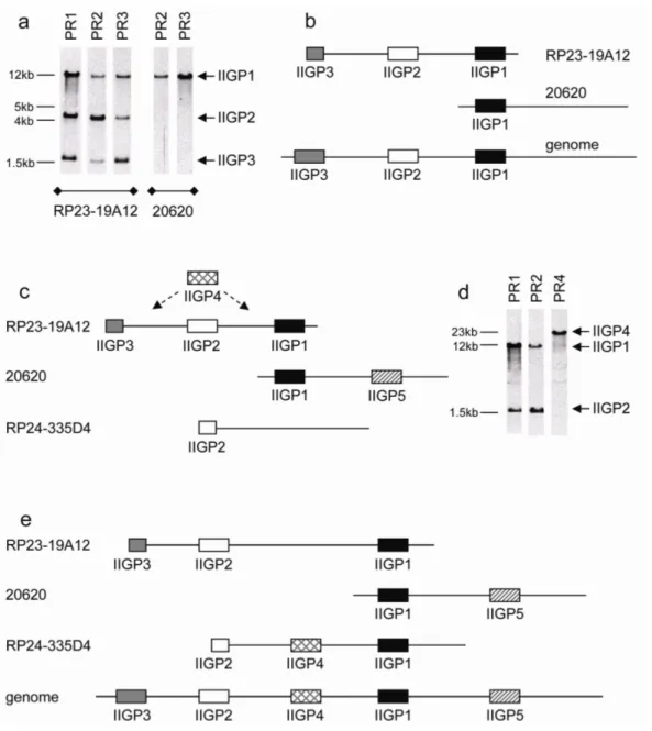
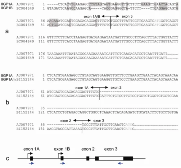
3.2 IIGP1 has two splice variants originating from two individual promoters ...35
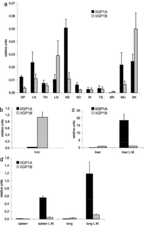
3.3 IIGP1A and IIGP1B have similar basal expression levels in all mouse organs except the liver ...36
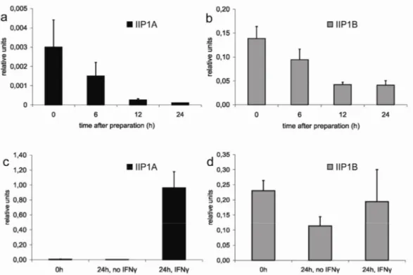
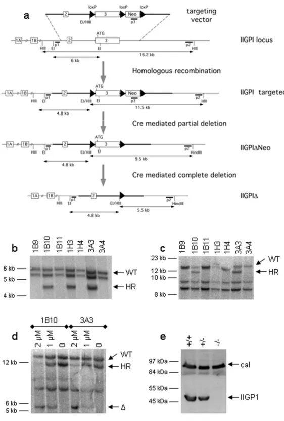
3.4 The response of the IIGP1A but not IIGP1B promoter to IFNγ induction is very strong 37 3.5 Generation of conditional IIGP1 allele and IIGP1-deficient mouse...40
3.6 Influence of IIGP1 deficiency on resistance against intracellular pathogens ...44
3.6.1 IIGP1 deficiency leads to a partial loss of IFNγ-induced inhibition of Toxoplasma gondii growth in primary astrocytes. ...44
3.6.2 The accumulation of other p47 GTPases at the membrane of T. gondii parasitophorous vacuoles does not depend on IIGP1...46
3.6.3 The effect of IIGP1 deficiency on susceptibility of mice to T. gondii is not yet clear ...47
3.6.4 IIGP1 deficient mice have higher incidence of development of cerebral malaria...49
3.6.5 Resistance of C57BL/6 mice against Leishmania major is not affected by the lack of IIGP1 ...51
3.6.6 IIGP1 deficient mice are not susceptible to infection with Listeria monocytogenes...52
3.6.7 IIGP1 deficient mice are resistant to infection with Anaplasma phagocytophilum...53
3.6.8 IIGP1 deficient mice are not susceptible to infection with Chlamydia Trachomatis...54
4 DISCUSSION 56 4.1 IIGP1 is required for resistance against some intracellular protozoan parasites ...56
4.1.1 IIGP1 and Toxoplasma...57
4.1.2 IIGP1 and Plasmodium...60
4.1.3 IIGP1 and Leishmania...62
4.2 IIGP1 seems not to be required for defense against intracellular bacteria ...63
5 REFERENCES 67
6 SUMMARY 83
7 ZUSAMMENFASSUNG 84 8 ACKNOWLEDGEMENTS 85 9 ERKLAERUNG 86 10 LEBENSLAUF 87
ABBREVATIONS
2'-5'-OAS 2'-5'-oligoadenylate synthetase
ADAR adenosine deaminases that act on double-stranded RNA
BAC bacterial artificial chromosome
bp base pair
BSA bovine serum albumine
CDS coding sequence
CFU colony-froming units
CM cerebral malaria
cpm counts per minute
DMEM Dulbecco Modified Eagles Medium
DMSO dimethylsulfoxid
EF cells embryonic feeder cells
ES cells embryonic stem cells
ER endoplasmatic reticulum
FCS foetal calf serum
GAP GTPase activating protein
GBP guanylate binding protein
GDP guanosine diphosphate
GTP guanosine triphosphate
mHPRT mouse hypoxanthine guanine phosphoribosyl transferase
IDO indoleamine 2,3-dioxygenase
IF immunofluorescence
IFN Interferon
IFNGR IFN-γ receptor
IFNAR IFN-α receptor
iNOS inducible nitric oxide synthetase
i.p. intraperitoneal
ISG20 interferon stimulated gene 20
JAK janus kinase
kb kilobase
kDa kilodalton
LPS lipopolysaccharide
M molar
MEF mouse embryonic fibroblats
OD optical density
PBS phosphate buffered saline
PCR polymerase chain reaction
PFA paraformaldehyde
Phox phagosome oxidase
p.i. post infection
PKR protein kinase R
PML promyelocytic leukaemia
PV parasitophorous vacuole
PVM parasitophorous vacuole membrane
RNAse ribonuclease
rpm rounds per minute
RT room temperature
STAT signal transducer and activator of transcription
TNF-α tumor necrosis factor α
TRIM5α tripartite motif 5α
U unit
ZAP Zinc-finger Antiviral Protein
1 INTRODUCTION
1.1 Infection and immunity- the never ending battle
Millions of years of co-evolution and reciprocal adaptation have created a complicated system of interactions between pathogens and their hosts [1, 2]. The aim of all pathogens is to invade the host and successfully establish an infection, which allows them to exploit resources, propagate and eventually spread to other hosts. In order to achieve these goals pathogens have developed numerous evasion mechanisms interfering with host resistance processes [3-8]. To answer the pathogen challenge living organisms have developed a sophisticated multilayer immune system. The function of this system is to recognize invaders, interfere with essential steps in their propagation and destroy them. All multicellular organisms possess a complex of evolutionary conserved immune mechanisms, known as the innate immune system. In vertebrates, there is a second and more sophisticated layer of defense mechanisms, the adaptive immune system. All elements of the immune system, innate and adaptive, are subjected to complex regulation in order to guarantee elimination of invading agents with minimal host damage.
The innate immune system provides the first line of defense against invasion. This system senses the invaders through a variety of germline-encoded pattern recognition receptors (PRRs) recognizing conserved products of microbial metabolism designated as pathogen associated molecular patterns (PAMPs) [9]. The lists of PRRs includes cell surface molecules such as Toll-like receptors (TLRs) [10] and scavenger receptors [11], intracellular receptors like NODs [12], PKR [13], 2’-5’-oligoadenylate synthase (OAS) [14] and some molecules secreted in the bloodstream and tissue fluids, for example mannose-binding protein (MBP) [15] and C-reactive protein (CRP) [16]. Essential components of the innate immune system are numerous cells that bear PRRs; these include macrophages, dendritic cells (DCs), mast cells, neutrophils, eosinophils, natural killer (NK) cells. They can rapidly become activated during an inflammatory response and differentiate to short-lived effector cells whose major role is to fight the infection. Important innate elements of host defense are also the different antimicrobial peptides [17, 18] and the complement system [19]. The mechanisms by which the innate immune system fights infections include
opsonization and phagocytosis, activation of complement and coagulation cascades, induction of apoptosis, activation of proinflamatory signaling pathways.
In vertebrates, the immune system is more complicated. It includes a complex of mechanisms known as the adaptive immune system. It becomes activated with the help of the innate immune system, which induces the production of co-stimulatory molecules, secretion of chemokines and cytokines and triggers DC maturation, thus directing the cells of the adaptive immune system to the place of inflammation. The adaptive immune system provides some big advantages to the host, especially in immune recognition. The cells of the adaptive immune system, T and B-lymphocytes, express surface receptors known as T-cell receptor (TCR) and B-cell receptor (BCR), respectively. The genes encoding these receptors are assembled by recombination of gene segments during lymphocyte development. This assembling process generates a huge variability of receptors, which potentially could recognize every unknown antigen the organism can encounter. The pathogens, which manage to go through the barriers of the innate immune system, meet the pool of lymphocytes and select among them the cells bearing receptors with the right specificity. These cells clonally expand and produce large numbers of effector cells, which fight the invaders. Because of the processes of selection and clonal expansion, the adaptive immune response is very specific but also delayed in time when compared to innate immunity. In the process of clonal expansion the adaptive immune system produces also long-lived cells, thus providing the host with immunological memory and allowing it to mount a stronger and more specific response in case of re-encounter with the same pathogen. The cells of the adaptive immune system are specialized with respect to their anti-pathogenic effector functions. B cells differentiate into plasma cells, which produce antibodies targeting extracellular pathogens. T cells from the CD8+ subset directly lyse infected cells or neutralize pathogens in non-cytolytic manner by secreting cytokines, mainly IFNγ [20]. The CD4+ T cells do not have direct antimicrobial functions but they orchestrate the complicated actions of the immune system by secreting diverse cytokines. Cytokines are small regulatory proteins secreted by various cells of the body in response to activating stimuli. They control important processes such as cell proliferation and chemotaxis, thus contributing to both innate and adaptive immunity.
Some cytokines have direct antimicrobial and antiviral functions. In this respect, very important molecules are the IFNs.
1.2 Interferons, the central players in antimicrobial and antiviral immunity
Interferons (IFNs) are members of a multigene family of small inducible cytokines, originally described as substances protecting cells from viral infection [21]. The family subdivides in three groups: IFN type I, II and III. The group of type I IFNs includes multiple IFNα proteins [22-23], as well as IFNβ [24], IFNκ [25], IFNε [26]
and IFNτ [27], each with a single member. All proteins from this list are found in both mice and humans. Mouse limitin [28], pig IFNδ [29] and human IFNω [30] also belong to the group of type I IFNs. The IFNα species exhibit antiviral, antiproliferative and immunomodulatory activities. Antiviral activity has also been demonstrated for IFNβ, IFNω and IFNκ. The group of type III IFNs consists of three IFNλ proteins [31-32] which are also induced by virus infection and have antiviral activity.
The type II IFN group has only one member- IFNγ. IFNγ is produced by cells of the immune system (NK cells, CD4 cells, CD8 CTLs, macrophages) [33] in response to diverse activating stimuli. The synthesis of IFNγ is regulated mainly by IL-12 and IL- 18, which synergistically induce its production. IFNα/β also can promote IFNγ expression.
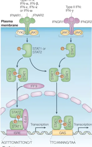
IFNγ mediates its functions through the Jak-Stat signaling pathway [34] (Fig. 1). The receptor for IFNγ (IFNGR) is expressed on the cell surface of all nucleated cells and consists of two heterologous subunits, IFNGR-1 and IFNGR-2. Upon binding of the ligand, the IFNGR dimerizes and induces a cascade of intracellular phosphorylation and activation events involving members of the Janus family tyrosine kinases (Jak-1 and Jak-2) and subsequently Stat-1, a transcription factor from the STAT (signal transducer and activator of transcription) family. Phosphorylated Stat-1 forms a homodimer, known as gamma activation factor (GAF); the dimer translocates to the nucleus and binds to the gamma activation site (GAS) response element present in IFNγ-responsive promoters, thus leading to activation of transcription.
IFNα and β also signal through a heterodimeric receptor and a pathway similar of that of IFNγ (Fig.1). Receptor dimerization is, however, not required and the set of Jak and Stat components involved is different. This pathway employs the kinases Jak-1 and Tyk-2 and the transcription factors Stat-1 and Stat-2. Upon activation, the latter two form a trimer with another transcription factor, IRF-9 (IFN regulatory factor 9);
the trimer is known as ISGF3. This trimer translocates to the nucleus, binds to the IFN-stimulated response element (ISRE) promoter sequence and activates transcription of IFNα/β-inducible genes.
The cellular response to IFNs and especially to IFNγ in particular, is extremely complex. Current data shows that IFNγ induces the expression of more then 800 genes. The products of these genes mediate the multiple effects of IFNγ on all aspects of immunity [35-38]. The complexity of the IFNγ response, however, makes the analysis of the functions of the individual genes involved in it very difficult.
Therefore, the mechanisms by which IFNγ mediates its functions are still largely unknown.
Figure 1. Interferon receptors and activation of the JAK–STAT pathways by type I and type II interferons. ( from [219]) Type I interferons (IFNs) bind to a common receptor (type I IFN receptor) The type I IFN receptor is composed of two subunits, IFNAR1 and IFNAR2, which are associated with the tyrosine kinases TYK2 and JAK1, respectively. IFN- , binds to a distinct cell-surface receptor (type II IFN receptor). This receptor is also composed of two subunits, IFNGR1 and IFNGR2, which are associated with JAK1 and JAK2, respectively. Activation of the JAKs that are associated with the type I IFN receptor results in tyrosine phosphorylation of STAT2 and STAT1; this leads to the formation of STAT1–STAT2–IRF9 complexes, which are known as ISGF3 complexes. These complexes translocate to the nucleus and bind ISREs in DNA to initiate gene transcription. Both type I and type II IFNs also induce the formation of STAT1–STAT1 homodimers (GAFs) that translocate to the nucleus and bind GAS elements that are present in the promoter of certain ISGs, thereby initiating the transcription of these genes. The consensus GAS element and ISRE sequences are shown.
1.3 Antimicrobial functions of IFNγ
Pathogenesis studies using mouse mutants with disrupted genes encoding IFNγ, IFNGR-1, IFNGR-2 and Stat-1 provide strong evidence for the importance of IFNγ in host response to microbial and viral pathogens. If kept in pathogen-free environment these null mutants show no developmental or physiological abnormalities but their ability to mount an immune response against infections is largely compromised.
These include infections with a variety of intracellular bacteria, protozoa and viruses some of which are listed in Table 1.
Table1
Pathogens References Intracellular bacteria
Mycobacterium spp. [41, 42]
Chlamydia spp. [43, 44]
Salmonella typhimurium [45, 46]
Listeria monocytogenes [47-50]
Shigella flexneri [51]
Yersinia enterocolitica [52]
Legionella pneumophilla [53]
Bordetella pertussis [54]
Intracellular protozoa
Toxoplasma gondii [55]
Plasmodium spp. [56]
Trypanosoma cruzi [57]
Leishmania major [58, 59]
Viruses
herpes simples virus (HSV) [60]
lymphocytic choriomeningitis virus (LCMV) [61]
Vaccinia virus [47]
murine cytomegalovirus (MCMV) [62]
vesicular stomatitis virus (VSV) [49]
The classical view about IFNγ is that this cytokine is mainly involved in resistance against bacteria while the antiviral defense is a function of the type I IFNs. IFNγ
indeed plays a critical role in innate host response against microbes but it also contributes to the protection against viruses, especially in long-term control of viral infections. In some diseases, IFNγ plays important role in both protection and pathogenesis. For example, in a Helicobacter pylori infection model, increased production of IFNγ in absence of IL-4 promotes mucosal inflammation [39]. IFNγ is also proved to be responsible for persistence of Chlamydia spp. infection [40].
In many cases, the antimicrobial and antiviral effects of IFNγ are due to its general immunostimulatory functions. However, there is growing evidence supporting the idea that the potent direct negative effect of many IFNγ-induced proteins on growth of intracellular pathogens is essential for host defense. Extensive data shows that IFNγ directly induces intracellular resistance programs and these cell-autonomous effects play a critical role in resistance against intracellular pathogens from all classes.
1.4 IFN-induced cell-autonomous immunity
By definition, cell-autonomous resistance is mediated by any cell without assistance from specialized cells of the immune system. Such assistance might be needed initially for the induction of a cell-autonomous resistance factor but once induced and synthesized in the cell this factor confers resistance by itself without further specialized help. In recent years, many IFN-induced molecules have been implicated in cell-autonomous resistance mechanisms. The list of such molecules includes well- known and extensively studied antiviral factors as PKR [63, 64] and 2’-5’
OAS/RNaseL [65, 66], as well as molecules with activity against a large spectrum of pathogens from all classes, for example iNOS [67, 68, 69], IDO [70, 71, 72] and the phox complex [73]. The list of cell-autonomous resistance factors is still growing, relative newcomers in it being ADAR1 [74], ISG20 [75], ZAP [76] and TRIM5α [77]. It is becoming clear that IFN-induced GTPases also play an important role in cell-autonomous resistance against intracellular pathogens.
1.5 IFN-inducible GTPases as cell-autonomous resistance factors Among the genes highly induced by IFNs are the members of four families of GTPases. Type I IFNs induce the Mx proteins. Type II IFN induces three other families of GTP binding proteins: the p65 guanylate-binding proteins (GBPs), the
very large inducible GTPases (VLIGs) and the p47 GTPases. Members of all these families except the VLIGs have already been implicated in cell-autonomous resistance mechanisms.
1.5.1 Mx proteins
The first mouse Mx gene was identified as a locus in A2G mice conferring resistance to influenza virus [78]. The gene was subsequently mapped and cloned [79, 80] and is currently known as Mx1. A second mouse family member (Mx2) [81] and two human proteins (MxA and MxB) were identified later [82, 83]. Mx GTPses are strongly induced by IFNα and IFNβ in cultured cells. They are also shown to be abundant in most tissues of mice infected with viruses or treated with IFNα or IFNβ. Other cytokines, including IFNγ are poor inducers of Mx GTPases.
Mx proteins, in particular MxA and Mx1, attract a great deal of attention due to their potent antiviral activity demonstrated in induced or stably transfected cell culture systems. The Mx proteins protect cells against a variety of negative-strand RNA viruses form the families Orthomyxoviridae, Bunyaviridae and Hepadnaviridae. This group of viruses includes some human pathogens causing severe diseases, for example influenza [84], hepatitis B (HBV) [85], hemorrhagic fever with high mortality rate (Hantaan virus and Crimean-Congo hemorrhagic fever virus) [86, 87], fever epidemics (Rift Valley fever virus) [87] or encephalitis (La Crosse virus) [87].
The antiviral activity of the Mx proteins is further demonstrated by extensive in vivo studies. It is long known that most inbred mouse strains naturally carry nonfunctional Mx genes [88]. This explains their strong susceptibility to infections with orthomyxoviruses [89, 90]. The higher resistance of MxA-transgenic mice to certain viral infections [91] also confirms the function of human MxA as a potent antiviral effector in vivo. Furthermore, MxA protects these transgenic mice from viruses even in the absence of functional IFNα/β system [92], a fact, undoubtedly proving that the protein does not need help from other IFN-induced proteins to perform its antiviral activity. MxA is a powerful antiviral agent on its own, once induced and synthesized it works without external help. This makes the protein a paradigm for a GTPase functioning as a cell-autonomous resistance factor.
The mechanism of action of Mx proteins is still unclear. The direct interaction of MxA with viral particles has been demonstrated and proposed to be important for its
function [93]. The protein was shown to block nuclear import of viral nucleocapsids [94] probably by sequestering them into perinuclear complexes [95] in a process involving smooth ER membranes [96]. Like certain other large GTPases, Mx proteins also form highly organized oligomers in nucleotide-dependent manner [97, 98] and exhibit high GTP hydrolysis rate [97, 99]; however, a report on a MxA mutant unable to oligomerize and hydrolyze GTP but still exhibiting antiviral activity, makes the functional significance of these processes questionable [100]. MxA was also shown to self-assemble into rings, which tubulate lipids in vitro, but its still unknown if membrane deformation is important for its function [101]. Mouse Mx1 is a nuclear protein accumulating in distinct nuclear dots that frequently associate with promyelocytic leukemia protein (PML) nuclear bodies. However, recently the protein was shown to be functionally independent from them [102].
1.5.2 p65-kDa GBPs
GBPs represent a family of GTPases conserved in vertebrates [103]. They are highly induced by IFNγ and less by IFNα/β [103]. The function of the members of this family is still not completely clear. When stably expressed in HeLa cells human GBP1 (hGBP1) shows activity against vesicular stomatitis virus (VSV) and encephalomyocarditis virus (ECMV) [104]. A similar but smaller effect against these two viruses was also recently reported for murine GBP2 (mGBP) [105]. GBPs were also shown to be involved in regulation of cell proliferation: mGBP2 alters the growth characteristics of fibroblasts [106] and hGBP1 controls proliferation and angiogenic capability of endothelial cells [107, 108] only in the presence of a functional GTP- binding domain. Some family members are isoprenylated at the C-terminus and this appears to be necessary for targeting to intracellular vesicular structures [109]; the functional importance of this modification for the GBP proteins is, however, still unclear.
1.5.3 Very large inducible GTPases (VLIGs)
The VLIGs are the most recently discovered family of IFN-inducible GTPases [110].
In mice, this family consists of at least six genes, in humans there is only one homologue. The prototype VLIG, VLIG-1, is a cytosolic and nuclear protein which is induced by both type I and type II IFNs and has a canonical GTP-binding domain.
The molecular mass of VLIG-1 is 280 kDa, which makes it the largest known GTPase in any species. No resistance function for this protein has been shown so far but VLIG-1 displays highest homology to the GTPases mediating cell-autonomous resistance. This, in addition to the IFN inducibility, suggests a possible role of the VLIGs in intracellular defense mechanisms.
1.5.4 p47 (IRG) GTPases
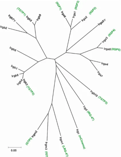
Extensive data generated in the last few years clearly show that the p47 GTPases are among the most efficient cell-autonomous resistance mediators found in the mouse so far [111]. The family consists of 23 members in C57BL/6 mice [112], six of which have been previously described, namely TGTP/Mg21 [113], IRG-47 [114], IIGP1 [103], GTPI [103], IGTP [115] and LRG-47 [116]. IIGP1, TGTP and IRG-47 contain the three classical GTP binding motifs [117]: GX4GKS/T (G1; phosphate binding P loop), DXXG (G3) and (N/T)(K/Q)XD (G4; responsible for the base specificity). In the three other published members the universally conserved GKS sequence in the P loop is substituted by GMS, which is so far a unique feature among all known GTPases. The substitution correlates with other sequence similarities between these three proteins and defines a distinct GMS subgroup in the family [103]. p47 GTPase families with different degree of complexity are found in jawed fish and mammals [112]. Only two genes are present in humans: a homologue of mCINEMA, called hCINEMA, and a gene fragment homologous to the members of the mouse GMS subgroup [112]. Recently a new general nomenclature for the p47 GTPase family was introduced [112]. It is based on phylogenetic principles and the names of the genes originate from the stem name IRG (Immunity-Related GTPases). The family tree shown in Fig. 2 presents the new, as well as the old names of the members of the mouse p47 GTPase (IRG) family. The old names will to be used in this thesis.
The p47 GTPases are classified as immediate-early genes because their expression does not require de novo protein synthesis of transcription factors [118, 116, 111]. A hallmark feature of the members of the p47 family is their strong and rapid induction in response to IFNγ stimulation from almost undetectable basal levels in all cell lines and primary cells tested [115, 114, 103, 116, 112]. The only exception in this respect is CINEMA, which is not IFN inducible [112]. IFNα/β and LPS also induce the expression of the p47 GTPases but less than IFNγ [116, 119]. Other bacterial cell wall
components also stimulate synthesis of some family members, probably via TLR signaling and indirect secretion of IFNβ [120]. The p47 GTPases seem not to be induced by other cytokines, including some interleukins (IL-1α and β, IL-2, IL-4, IL- 6, IL-10), tumor necrosis factor α (TNFα) and granulocyte-macrophage colony stimulating factor (GM-CSF) [116, 118, 119]. The p47 GTPases are also shown to be highly induced in vivo after infection with bacteria and protozoa [103, 121, 122].
Interestingly, at least the six published family members have low but detectable basal expression levels in almost all mouse organs tested and apparently this basal production does not depend on IFN, as it is still present in mice lacking receptors for IFNα/β or/and IFNγ (Jia Zeng, personal communication). The in vivo expression pattern of CINEMA is also an exception from the rule: the protein is expressed only in
Figure 2: Unrooted tree of nucleotide sequences of the G-domains of the 23 members of the mouse p47 GTPase (Irg) family (p-distance based on a Neighbour-Joining method). The names generated according to the new nomenclature [112] are depicted in black, the old names, in green (courtesy to Cemalettin Bekpen)
testis in both mouse and man ([112] and Christoph Rohde, personal communication).
Mice with targeted deletions of three individual members of the p47 GTPase family, namely IGTP, IRG-47 and LRG-47, display complete loss of resistance to several intracellular pathogens of bacterial and protozoan origin despite having an intact and functional adaptive immune system and IFNγ production [111, 121, 122]. The data on susceptibility of these deficient mice is summarized in Table 2.
Table 2. This table (from Taylor et al. [111]) summarizes the phenotypes of the three available p47 GTPase deficient mice in comparison to the IFNγ deficient mouse (S, susceptible, R, resistant, N.T., not tested)
The cellular mechanisms underlying the essential resistance functions of the p47 GTPases are not yet understood. Currently available data is concentrated mainly on the interactions between LRG-47 and phagosomes. The protein was reported to co- purify with phagosomes from Mycobacterium-infected macrophages and promote the acidification, and thus maturation of these phagosomes [123]. In resting cells, LRG- 47 was shown to localize to cis-Golgi and ER [124]. Upon phagocytosis LRG-47 translocates to the plasma membrane at the forming phagocytic cups and in fibroblasts also at phagocytosis-induced membrane ruffles; the protein then stays associated with the maturing phagosome and reaches the lysosomal compartment [124]. Other family members have also been shown to associate with intracellular membrane compartments: IIGP1 and IGTP both localize to the ER [124, 125], GTPI is a Golgi protein [126] and TGTP1 partitions between intracellular membranes and cytosol [126]. The intracellular behavior of the p47 GTPases suggests that these proteins work by interfering with the lifestyle of pathogens whose survival and propagation in host cells is critically dependent on exploitation of membrane compartments.
1.5.4.1 IIGP1
From all published members of the p47 GTPase family IIGP1 is the best characterized with respect to biochemical properties and enzymatic activity in vitro, features, which relate the protein to the large GTPases including dynamins and p65 GBPs [127-129].
Purified recombinant IIGP1 binds GDP and GTP in the micromolar range, hydrolizes
GTP to GDP in a cooperative manner and forms enzymatically active oligomers in presence of GTP in vitro [130]. In contrast to the large GTPases, however, IIGP1 has higher affinity for GDP than for GTP; its intrinsic enzymatic activity is also relatively low, which suggests that the protein might need additional help for GTPase activation.
Such GAP (GTPase-activating protein) activity is an intrinsic feature of the related dynamins and p65 GBPs [127, 131, 132]. It is possible that the IIGP1 molecules are activated in trans during the oligomerization process.
The crystal structure of IIGP1 shows a typical Ras-like G domain between an N- terminal three-helix bundle and a complex system of C-terminal helices and loops [133]. The protein crystallizes as a dimer, which is required for cooperative GTP hydrolysis and GTP-dependent oligomerization as shown by analysis of dimer interface mutants. Sequence comparison and secondary structure prediction suggests that the structure of IIGP1 can be a valid model for the p47 GTPase family.
IIGP1 associates to the endoplasmic reticulum in fibroblast, macrophage and hepatocyte cell lines [124]; it was also previously reported to be a predominantly Golgi protein in bone-marrow derived macrophages [119]. Notably, IIGP1 translocates to the parasitophorous vacuole (PV) in primary astrocytes infected with Toxoplasma gondii and this process correlates with subsequent disruption of the PV [134]. IIGP1 behaves as a classical peripheral membrane protein with relatively weak interactions to the membrane [124]. The protein is partly membrane associated but it also has a substantial cytosolic pool. IIGP1 is targeted to membranes by N-terminal myristoylation. There is, however, also a residual membrane interaction signal because the non-myristoylated IIGP1 mutant is still partly membrane-bound. IIGP1 probably could interact directly with lipids in cellular membranes as suggested by its ability to bind to synthetic phosphatidylserine lipid vesicles in vitro [124].
The only interaction partner of IIGP1 reported until now is the microtubule binding protein Hook3 (mHk3) [135]. The interaction was identified by yeast-two-hybrid screen using the complete IIGP1 protein as a bait. It was also confirmed by co- immunoprecipitation of the two proteins in lysates of IFNγ-activated bone marrow- derived macrophages. The hook proteins were previously proposed to participate in proper assembly and/or positioning of membranous compartments and contribute to their ordered dynamic formation, maturation and trafficking [136]. Therefore, the interaction between IIGP1 and mHk3 was reported as evidence for participation of IIGP1 in intracellular trafficking [135].
1.6 Aims of this study
The introductory remarks in the previous chapter summarized our current knowledge about IIGP1 with respect to biochemical characteristics and intracellular behavior.
Our aim was to investigate the proposed defense function of the protein by generating an IIGP1 deficient mouse strain and analyzing the susceptibility of these animals to infection with different pathogens. We also planned to characterize in detail the expression profile of IIGP1 and the genomic organization of its homologous genes.
2 MATERIALS AND METHODS
2.1 Chemicals and reagents
All chemicals were purchased from Aldrich (Steinheim), Amersham-Pharmacia (Freiburg), Applichem (Darmstadt), Baker (Deventer, Netherlands), Boehringer Mannheim (Mannheim), Fluka (Neu-Ulm), GERBU (Gaiberg), Merck (Darmstadt), Pharma-Waldhof (Düsseldorf), Qiagen (Hilden), Riedel de Haen (Seelze), Roth (Karlsruhe), Serva (Heidelberg), Sigma-Aldrich (Deisenhofen) or ICN biochemicals, Oxoid, (Hampshire UK).
2.1.1 Oligonucleotides
Oligonucleotidess were purchased from Invitrogen (Carisbad, USA) and are listed in Table 3
Table 3
Name Sequence 5’- 3’ Usage
RT1.1FA TGCTTCCTGAAGCTGAACTA Real-Time PCR (IIGP1A)
RT1.2FA ACCGAGGGCTATTCCTCTCA Real-Time PCR (IIGP1B)
RT1R CAGAGAAGGGATGATATTCAC Real-Time PCR (IIGP1A and B)
mHPRT FA ATTAGCGATGATGAACCAGG Real-Time PCR (HPRT)
mHPRT R2A TGGCCTATAGGCTCATAGTG Real-Time PCR (HPRT)
5HAF CCGCTCGAGGTACTGTTGAAAGCAATGATT 5HAR1 CCGCTCGAGGAATTCTATACAAAACTTTCC
CAGTAG
Targeting vector (5’ homology arm)
3HAF CAGGATCCCGCAGAAGGTTTG
3HAR CGCGGATCCATAATGTTTTCATCTCTAATC
Targeting vector (3’ homology arm)
5DEL TTGTTATTCAGGGAAGCTAAG 3DEL TGTCTGGTGATTCTCATTAGC 5’EX2 CTCAGGTTATCTAACATTCTG
IIGP1 mouse typing
G6PDH 1f ACAGGGACAGAGGGAGAA
G6PDH 1r AACGCAAAGCTGAAGTGA
Real-Time PCR
A. phagocytophilum detection CL Forward GGA GGC TGC AGT CGA GAA TCT
CL Reverse TTA CAA CCC TAG AGC CTT CAT CAC
Real-Time PCR C. trachomatis detection
2.1.2 Enzymes
Restriction Enzymes were purchased from New England Biolabs (Bad Schwalbach);
T4 DNA ligase (New England Biolabs); RNase A (Sigma); shrimp alkaline
phosphatase (SAP) (USB, Amersham); Thermus aquaticus (Taq) polymerase was prepared by Rita Lange; Pyrococcus furiosus (Pfu) DNA Polymerase (Promega, Mannheim)
2.1.3 Kits
Plasmid Maxi and Midi kit (Qiagen, Hilden), Terminator-cycle Sequencing kit version 3 (ABI), Rapid PCR product purification Kit (Boehringer, Ingelheim), pGEM- T Easy Vector System I (Promega, Madison, USA), Ladderman labeling kit (TaKaRa) RNeasy Mini Kit (Qiagen, Hilden), Oligotex mRNA Mine Kit (Qiagen, Hilden), SuperScript First-Strand Synthesis System (Invirtogen, Carisbad, USA), QuantiTect SYBR Green PCR KIT (Qiagen, Hilden)
2.1.4 Serological reagents.
2.1.4.1 Primary antibodies and antisera (Table 4)
name target species dilution origin
10D7 IIGP1 mouse monoclonal IB: 1:1000
IF: 1:200 Jens Zerrahn, Berlin
A20 TGTP1 goat polyclonal IF: 1:100 Santa Cruz Biotechnology, Santa Cruz, CA
I68120 IGTP mouse monoclonal IF: 1:250 BD Transduction Laboratories, Lexington, KY
5-241-178 GRA7 mouse monoclonal IF: 1:30000 R. Ziemann, Abbott Laboratories, Abbott Park, IL
2.1.4.2 Secondary antibodies and antisera
goat anti-mouse Alexa 546/488, goat anti-rabbit Alexa 546/488, donkey anti-goat Alexa 546/488, donkey anti-mouse Alexa 488, goat anti-mouse Alexa 680 (all Molecular Probes), goat anti-mouse HRP (Amersham)
2.2 Media
2.2.1 Luria Bertani (LB) medium
10g bactotryptone, 5g yeast extract, 10g NaCl, 1l dH2O
2.2.2 LB agar medium
10g bactotryptone, 5g yeast extract, 10g NaCl, 15g agar, 1l dH2O
2.2.3 EF medium
DMEM (Dulbecco’s Modified Eagle Medium) with Glutamax (no sodium piruvate, 4500 mg glucose, with pyridoxine) supplemented with 10% EF FCS, 1 mM sodium piruvate, 100 µg/ml penicillin/streptomycin (optional)
2.2.4 ES medium
DMEM (Dulbecco’s Modified Eagle Medium) supplemented with 15% ES FCS (tested for germline transmission), 1 mM sodium piruvate, 2 mM L-glutamine, 1x non-essential amino acids, 1 mM β-mercaptoethanol, 100 µg/ml penicillin/streptomycin (optional, not recommended), LIF (leukemia inhibitory factor;
a supernatant from LIF-transfected CHO cells line 8/24 720 LIFD(.1) from Genetics Institute, Cambridge, Massachusetts; the amount used depends on the concentration of the batch)
Mammalian tissue culture media and supplements were bought from Gibco BRL, (Eggelstein), ES and EF FCS from Gibco BRL, (Eggelstein), LIF was provided by the Center for Mouse Genetics, Institute for Genetics, Cologne
2.2.5 Freezing medium
10% DMSO, 90% FCS, sterile filtered, kept in aliquots at -20◦C 2x Freezing media (for 96 well plates)
20% DMSO, 80% FCS, sterile filtered, kept in aliquots at -20◦C
2.3 Cells and cell lines 2.3.1 Bacterial strains
Escherichia coli DH5α: 80dlacZ∆M15, recA1, gyrA96, thi-1, hsdR17 (rB-, mB+), supE44, relA1, deoR, ∆(lacZYA-argF)U196
2.3.2 Bacterial pathogens
Listeria monocytogenes strain EDG, serotype 1/2a Chlamydia trachomatis L2
Anaplasma phagocytophilum strain MRK
2.3.3 Protozoan parasites
Toxoplasma gondii strain ME49, DX Plasmodium berghei strain ANKA
Leishmania major clone V1 (MHOM/IL/80/Friedlin)
2.3.4 Mammalian cells and cell lines
Embryonic feeder (EF) cells: primary, prepared from day 13-14 embryos from 129 mouse strain harboring pSV2 neo [137]
Embryonic stem (ES) cells: Bruce4 cell line derived from C57BL/6 mouse strain [138]
2.4 Methods
2.4.1 Molecular biology
All common methods molecular biology methods were performed according to standard protocols [139] or cited references.
2.4.1.1 Preparation of competent Cells
Competent Escherichia coli DH5α or BL21 cells were prepared according to the protocol of Inoue et al. [140] and used in heat shock transformations of plasmid DNA.
2.4.1.2 Isolation of Plasmid DNA
Plasmid DNA was isolated from transformed bacteria with an alkaline lysis method [141] following the standard protocol [139]. Plasmid DNA of a higher amount and purity was prepared with Oiagen Plasmid Midi kit (Qiagen, Hilden) following the supplier's instructions. BAC DNA was isolated using Oiagen Plasmid Midi kit (Qiagen, Hilden) following the protocol for preparation of very low copy number plasmids provided by the company.
2.4.1.3 Isolation of Genomic DNA from mouse tissues and cells
Mouse tissue was incubated o/n at 56°C in lysis buffer (l0 mM NaC1, 10 mM Tris- HCl pH 7.5, l0 mM EDTA, 0.2% SDS, 0.4 mg/mI Proteinase K [freshly added each time]). Debris were pelleted by centrifugation and supernatant was mixed with an equal volume of isopropanol to precipitate DNA. DNA was washed with 70% EtOH, dried and resuspended in ddH2O or TE buffer.
Preparation and restriction digest of ES cell DNA in 96 well plates was performed as follows. 50 µl of lysis buffer (l0 mM NaC1, 10 mM Tris-HCl pH 7.5, l0 mM EDTA, 0.5% Sarcosyl, 0.4 mg/mI Proteinase K [freshly added each time]) was added to each well of the 96 well plate. The plate was wrapped in parafilm, transferred to pre warmed at 56◦C humidified chamber and incubated at 56◦C overnight. At the next day, the plate was allowed to cool down at room temperature for 1 hour. 100 µl of 100%
EtOH were added to each well and the plate was let to sit at room temperature for 1 hour during which the DNA strands became visible at lower magnification. The plate was inverted and carefully drained on paper towels allowing DNA to remain attached to the plastic walls of the wells. The plate was washed 3 times with 100µl 70% EtOH and dried at room temperature. 35 µl restriction mix (1x restriction buffer, 1 mM spermidine, 1 mM DTT, 100 µg/ml BSA, 20-30 units of restriction enzyme per reaction) were added to each well and the plate was incubated overnight at the appropriate temperature in a humidified chamber. At the next day, the digested DNA samples were fractionated on agarose gel.
2.4.1.4 Agarose gel electrophoresis purification of DNA fragments from agarose gels
Size of DNA fragments was analyzed by agarose gel electrophoresis. DNA was run on 0.7% - 2% gels in 1 x TAE [139] and stained with 0.3 µg/ml ethidium bromide;
migration of the DNA molecules was visualized by using Bromophenol blue or Orange G. DNA fragments were eluted from the gel with the rapid PCR purification Kit (Boehringer) according to the manufacture’s protocol. Purity and yield of the DNA was determined by agarose gel electrophoresis and measurement of OD260.
2.4.1.5 Quantification of nucleic acids
The concentration of DNA and total RNA was determined by measuring the absorption of the sample at 260 nm in a spectrophotometer (Pharmacia). An OD260 of 1 corresponds approximately to 50 µg/ml for double stranded DNA or 40 µg/ml RNA.
2.4.1.6 Polymerase Chain Reaction 2.4.1.6.1 General PCR protocol
Polymerase chain reaction (PCR) [142] was used for the cloning of the short and long arm of homology of the targeting vector, screening for deleted clones after in vitro neo deletion and typing of transgenic mouse strains. All PCR reactions except the amplifications of the homology arms were made with Thermus aquaticus (Taq) DNA polymerase prepared by Rita Lange; for the homology arms 1:20 mix of KlenTherm Taq (BIOFIDAL, Vaulcs-En-Velin, France) and Pyrococcus furiosus (Pfu) DNA Polymerase (Promega, Mannheim) was used. The general reaction mix contained 1 µl DNA, 1x Hepes PCR buffer, 10 pM of each primer, 200 pM dNTP-mix, various amount of 50 mM MgCl2, 2,5U Taq-Polymerase, and water up to 50 µl. Primers were bought from Invitrogen and are listed in Table 3
2.4.1.6.2 Mouse typing PCR for IIGP1 deletion
The reaction mix contained 1 µl genomic tail DNA, 1x Hepes PCR buffer, 10 pM of each primer (5DEL, 3DEL, 5’EX2) (Table 3), 200 pM dNTP-mix, 3 mM MgCl2, 2,5U Taq-Polymerase, and water up to 50 µl. The PCR program was 94°C for 2 min, followed by 35 cycles of 94°C for 30 sec, 58° for 30 sec and 72°C for 45 sec. The sizes of the amplified bands were 260, 330 and 500 bp for wild type, floxed and deleted IIGP1 allele, respectively.
2.4.1.7 Cloning of PCR products
Amplified PCR products were purified using the rapid PCR purification Kit (Boehringer). DNA yield was monitored by agarose gel electrophoresis. Purified fragments were cloned in pGEMTeasy vector according to supplier’s protocol.
2.4.1.8 Ligation
Vector was cut with the respective restriction enzyme(s) (10U/1µg DNA) usually for 1h under temperature and buffer conditions optimal for the enzyme(s) used. After the first hour the same amount of restriction enzyme(s) and 0.1U of shrimp alkaline phosphatase were added to the reaction followed by 1.5h incubation under the same conditions. Following restriction DNA fragments were ran on agarose gel and purified using the rapid PCR purification Kit (Boehringer). DNA yield after purification was monitored by agarose gel electrophoresis. Vector and insert were mixed at a ratio of
1:3 and ligated with T4-DNA ligase in total volume of 10µl at 16◦C over night according to the manufacture’s protocol. Two control reactions were set in parallel: a ligation of vector without insert (control for dephosphorylation of the vector) and a ligation of insert only (control for purity of the insert). At the next day 5 µl of each ligation reaction were transformed in competent bacteria.
2.4.1.9 DNA sequencing
Plasmid DNA and PCR products were sequenced using the ABI PrismR BigDyeTM Terminator Cycle Sequencing Ready Reaction Kit (PE Applied Biosystems) and the automatic sequencers ABI373 and ABI377 with the help of Rita Lange. The method is based on the dideoxy-chain termination reaction with fluorescently labeled dNTPs [143].
2.4.1.10 Southern Blot analysis
Gels were treated subsequently with 0.25 M HCl and 0.4 M NaOH for 20 min and placed onto a prewet Hybond N+ thansfer membrane (Amersham). The membrane was placed on stacks of paper towels covered with 3 prewet Whatman paper sheets.
The gels were overlaid with prewet Watman paper sheets and connected via a Wantman paper bridge to a reservoir with transfer solution (0.4 M NaOH, 0.6 M NaCl). Transfer for performed for at least four hrs, then the blot was dissembled. The membrane was incubated with hybridization solution (1 M NaCl, 50 mM Tris pH 7.5, 10% Dextransulfat, 1% SDS, 250 µg/ml sonicated salmon sperm DNA) over night or at least 2 hrs at 65ºC in a hybridization oven (Techne Hybridizer HB-1D, Techne, Cambridge, UK). Probes were labeled with Ladermann labeling kit (Takara, Japan) according to manufacturers instructions, cleaned from residual radioactive nucleotide over ProbeQuant G-50 Micro Columns (Amersham Biosciences, UK) added to the hybridization solution and incubated overnight at 65ºC in hybridization oven. At the next day blots were subsequently washed for 10 min at 65ºC with 2xSSC, 1xSSC, 0.5xSSC 1%SDS for 10 min at 65ºC. Radioactive signals were measured with Fujix BAS 1000 phosphoimager and analyzed with Aida Image Analyser v.3.43 software.
2.4.1.11 Preparation of total RNA from mouse tissues and cells
Mice were killed with CO2; tissues were prepared and immediately placed in the appropriate amount of RNAlater RNA Stabilization Reagent (Qiagen). According to
manufacturer’s instructions, tissues were kept in the reagent overnight at 4◦C and later processed or kept for archival storage at -20◦C.
Total RNA from tissues was prepared with RNeasy Mini Kit (Qiagen); the only exceptions were testis and brain tissue, which were processed with RNeasy Lipid Tissue Kit (Qiagen). In all cases, the procedures recommended by the manufacturer were followed including an additional on-column DNaseI digestion with RNase-Free DNase Set (Qiagen). Prior to RNA preparation tissues were mechanically disrupted in the appropriate volume of lysis buffer from the kit and homogenized through QIAshredder Homogenizers (Qiagen).
Total RNA from cells was also prepared with RNeasy Mini Kit (Qiagen) following manufacturer’s instructions.
The integrity and size of purified total RNA was evaluated by agarose gel electrophoresis and ethidium bromide staining. RNA was stored at -80◦C in water.
2.4.1.12 Preparation of mRNA
PolyA mRNA was prepared from total RNA with Oligotex mRNA kit (Qiagen) according to the procedure recommended by the supplier. mRNA was stored at -80◦C.
2.4.1.13 cDNA synthesis
cDNA was synthesized from mRNA using SuperScript First-Strand Synthesis System (Invirtogen) according to manufacturers instructions. 10 to 100 ng mRNA was used as template; synthesis was primed by Oligo-dT. cDNA diluted in water 1:1 to reduce the concentration of MgCl2, which might interfere with further amplification and stored at -20◦C.
2.4.1.14 Real-Time PCR
2.4.1.14.1 Quantification of IIGP1 transcripts
The amount of IIGP1A and IIGP1B transcripts was detected by a quantitative PCR assay using the LightCycler System (Roche). cDNA was used as a template.
Fragments from IIGP1A (969 bp) and IIGP1B (972 bp) transcripts were amplified using primer pairs RT1.1FA/RT1R and RT1.2FA/RT1R, respectively. The amount of measured transcripts was normalized to the amount of the mouse HPRT transcript in the probes. The 827 bp fragment from this transctipt was amplified in a separate
reaction using the primers mHPRT FA and mHPRT R2A. The sequences of all primers are listed in Table 3.
The PCR reaction mixtures (20 µl) contained 1x QuantiTect SYBR Green PCR Master Mix (Qigen, Hilden), 10 pM of each primer, 2 µ l of template cDNA and 1U Taq polymerase (prepared by R.Lange). The LightCycler PCR program consisted of 95°C for 3 min followed by 35 cycles of 95°C for 30 s, 60°C for 30 s, and 72°C for 1 min and 78°C for 1 min; additional melting step was added at the end of each run (95°C for 15 min). Melting curve analysis was performed after each run. The quantitation of all transcripts was achieved by external standards, five serial tenfold dilutions of IIGP1A containing pGEMTeasy plasmid; the dilutions were ranging from 106 to 102 plasmid copies/dilution.
2.4.1.14.2 Detection of A. phagocytophilum
This analysis was preformed in the laboratory of Prof. Christian Bogdan, Institute of Medical Microbiology and Hygiene, University of Freiburg
A. phagocytophilum was detected by a quantitative PCR assay using the LightCycler System (Roche). The 444-bp fragment of the A. phagocytophilum ankA gene was amplified using the primers LA1 and LA6 [144]. For the sequence-specific detection of the amplicon the hybridization probes Ephago-HP-3 and Ephago-HP-4 were used.
The amount of bacterial DNA was normalized to the amount of mouse genomic DNA in the probes. The 429-bp fragment of the mouse glucose-6-phosphate dehydrogenase gene (G6PDH) was amplified in a separate reaction using the primers G6PDH 1f and G6PDH 1r (Table 3). The amplicon was detected by the hybridization probes G6PDH-HP-3 and G6PDH-HP-4. The hybridization probes were synthesized by TIB MOLBIOL (Berlin) and had the following sequence:
Ephago-HP-3 TAAAGCATGTAAAATACTACTAAAGTCT-fluorescein Ephago-HP-4 LC Red 640-CGTCAGTATCAGTCGTGAATGTAGA-Ph G6PDH-HP-3 TCATTACGCTTGCACTGTTGGTGGA-fluorescein G6PDH-HP-4 LC Red 640-TCACCTGCCACGTCTCGGAACTGC-Ph
The PCR reaction mixtures (20 µl) contained 1×LightCyclerFastStart DNA Master Hybridization Probes (Roche), 3 mM MgCl2, 0.5 µM of each primer, 0.2 µM of the respective hybridization probes, and 2 µ l of template DNA. The LightCycler PCR
program consisted of 95°C for 10 min followed by 50 cycles of 95°C for 10 s, 55°C for 20 s, and 72°C for 30 s. The quantitation of bacterial DNA and mouse genomic DNA was achieved by external DNA standard preparations, which consisted of five serial tenfold dilutions (ranging from 106 to 102 plasmid copies/dilution).
2.4.1.14.3 Detection of C. trachomatis
C. trachomatis was detected by a quantitative PCR assay by amplifying a fragment of the C. trachomatis 16s gene using primers CL Forward and CL Reverse (Table 3). For the sequence-specific detection of the amplicon, the following hybridization probe was used:
5'-[6-FAM]-TCG TCA GAC TTC CGT CCA TTG CGA-[TAMRA]-3'
The amount of bacterial DNA was normalized to the amount of mouse genomic DNA in the samples by amplifying a fragment of the mouse GAPDH gene in a separate reaction using primers and probe from Applied Biosystems.
2.4.2 Cell biology
2.4.2.1 ES cell culture
ES cell culture was performed according to the protocols published in Laboratory Protocols for Conditional Gene Targeting [145] with modifications used in the laboratory of Ari Waisman.
2.4.2.1.1 Thawing of cells
Cells were thawed fast at 37◦C in a water bath, immediately transferred in 10 ml media to dilute DMSO and pelleted by centrifugation for 5 min 1200 rpm, 4◦C. The pellet was resuspended in appropriate amount of media and plated.
2.4.2.1.2 Freezing of cells
Plates were washed 2 times with PBS and trypsinized for 3-5 min at 37◦C. The reaction was stopped by adding equal volume of media. Cells were pelleted for 5 min 1200 rpm, 4◦C. The pellet was resuspended in 1 ml freezing media, immediately transferred on ice and slowly frozen at -80◦C. At the next day, the frozen tubes were transferred in liquid nitrogen for storage.
2.4.2.1.3 Mitomycin C treatment of EF cells
ES cells were grown on a layer of EFs, mitotically inactivated by treatment with mitomycin C (MMC). Mitomycin C was diluted in EF media to final concentration 10 µg/ml and aliquoted. Aliquotes were kept at -20◦C. EFs were grown to confluence on 15 cm tissues culture plates and treated with 10 ml MMC media for 3 hours. After the treatment, cells were washed 2 times with PBS, trypsinized, counted and plated on gelatinized 9 cm tissue culture plates.
2.4.2.1.4 Transfection of ES cells
2 individual transfections were made. 30 µg of linearized pEF-IIGP1 were used for each transfection. ES cells were fed with fresh media 4 hours before transfection.
DNA was dried under sterile hood and dissolved in 0.8 ml RPMI without phenol rot.
ES cells were trypsinized and counted. 103 cells were plated for ES cell viability control and 2 aliquots of 107 cells were kept for transfection. These cells were centrifuged for 10 min at 1200 rpm, 4◦C. The cell pellet was dissolved in 0.4 ml RPMI without phenol rot and 0.4 ml of dissolved DNA was added. The mix was transferred to an electroporation cuvette, allowed to sit for 10 min at room temperature and electroporated at 240V/500µF. 103 cells from each transfection were plated on individual EF covered 6 cm tissue culture plates to control the survival rate of the ES cells after the transfection; additional 105 cells were pated for stringency of selection control. The rest of the transfected cells were diluted in media and plated on 10 cm tissue culture plates with MMC treated EF (5 plates per transfection). The cells were grown for 2 days with normal ES media. At day 2 the G418 process started.
The cell viability controls were analyzed by counting and comparing the number of colonies formed from the transfected and non-transfected ES cells. For both transfections the cell survival was around 10 %.
2.4.2.1.5 G418 selection (positive selection)
For Bruce4 ES cells, G418 is generally used in final active concentration 200 µg/ml.
The activity differs between the batches of G418 and has to be experimentally assessed for each new batch. The stock used for the experiments in this thesis was 70% active, i.e. 1.4 ml G418 were used per bottle (600 ml) of ES media. G418 was kept in aliquots at -80◦C. The selection process started at day 2 after transfection.
2.4.2.1.6 Gancyclovir (GANC) selection (negative selection)
GANC was used in working concentration 2x10⁻⁶ M and prepared fresh every day from a stock kept at -80◦C. To prepare the stock 4.3 mg GANC sodium salt were dissolved in 80 µl ddH2O to obtain a concentration 2x10⁻¹ M. 10 µl of the stock were further diluted in 1 ml of ES media, sterilized by filtering through a 0.22 µm filter, kept at -20◦C and used for preparation of the final working dilution of GANC. The selection process took 3 days from day 5 to day 8 after transfection with the targeting vector.
2.4.2.1.7 ES colony picking
At day 9-10 after transfection with the targeting vector plates with ES cells were washed three times with PBS and left in PBS after the last washing. Colonies were picked in a sterile hood. Individual colonies were taken with a P20 pipette and transferred to individual wells of a 96 well round bottom plate previously filled with 50µl trypsin/EDTA. After 20 min of picking, the plates incubated at 37◦C for 3-4 min.
Subsequently trypsinization process was stopped by adding 150µl of ES cell media and picking was continued. After one hour of picking the trypsinized ES cells were distributed into three gelatinized and EF covered flat bottom 96 well tissue culture plates and the wells were filled with media up to 200µl. At least 300 colonies were picked from each transfection.
The cells were grown for 2 to 3 days; two plates were frozen on two subsequent days and kept at -80◦C. The third plate was washed 2 times with PBS and trypsinized. The cells were equally distributed to three gelatinized flat bottom 96 well tissue culture plates and grown to complete confluence. The wells were subsequently washed 2 times with 100µl PBS and the plates were frozen at -20◦C.
2.4.2.1.8 Freezing of 96 well plates
The wells were washed 2 times with 100 µl PBS. The colonies were incubated for 3- 5 min with 50 µl trypsin/EDTA. The reaction was stopped by adding 50 µl ice cold 2x freezing media. The plates were immediately placed on ice, wrapped in parafilm and frozen slowly (in Styrofoam box) at -80◦C.
2.4.2.1.9 Thawing and expansion of clones from 96 well plates
The 96 well plates were thawed by incubation at 37◦C in a water bath or on a heating block. Immediately after thawing the cells were transferred to previously prepared tubes with 5 ml ES media and centrifuged for 5 min at 1200 rpm, 4◦C. Each cell pellet was dissolved in 1 ml ES media and plated in an individual well of gelatinized and EF covered 24 well tissue culture plate. Clones were expanded by subsequent transfer to gelatinized and EF covered 6 well tissue culture plates, 6- and 9 cm tissue culture dishes with growing for 2 to 3 days between the transferring steps. Three aliquots were frozen from each individual ES clone.
2.4.2.1.10 His-TAT-NLS-Cre transduction of ES cells
2x10⁵ ES cells were plated in individual wells of gelatinized 6 well tissue culture plate. Cells were allowed to attach for 4-5 hours and media was aspirated. His-TAT- NLS-Cre protein was diluted to 1 mM or 2 mM final concentration in ES media without FCS and sterile filtered. 600 µl were added per well and the plates were incubated for 20 hours at 37◦C. After the incubation His-TAT-NLS-Cre containing media was changed with normal ES cell media and cells were grown to confluence.
His-TAT-NLS-Cre protein was kindly provided by Thomas Wunderlich.
2.4.2.1.11 Preparation of ES cells for blastocyst injection
ES cells were thawed and plated on gelatinized and EF covered 9 cm tissue culture dishes 2 days prior injection. At the day of injection, plates were washed 2 times with PBS and trypsinized for 4-5 min at 37◦C. The reaction was stopped by adding equal volume of ES media. Cells were pelleted for 5 min at 1200 rpm, 4◦C, and resuspended in 10 ml ES media. The cell suspension was plated on gelatinized 9 cm tissue culture plate and incubated for 30 min at 37◦C to deplete from EF cells. After incubation, the supernatant containing the ES cells was centrifuged for 5 min at 1200 rpm, 4◦C and the cell pellet was resuspended in 1 ml injection media. The plate was washed with 10 ml ES media to harvest potentially weakly attached ES cells. This media was also centrifuged for 5 min at 1200 rpm, 4◦C and the cell pellet was resuspended in 1 ml injection media. ES cells from both pellets were compared under microscope and usually both were used for blastocyst injection.
![Table 2. This table (from Taylor et al. [111]) summarizes the phenotypes of the three available p47 GTPase deficient mice in comparison to the IFNγ deficient mouse (S, susceptible, R, resistant, N.T., not tested)](https://thumb-eu.123doks.com/thumbv2/1library_info/3625625.1501995/19.892.134.760.303.421/summarizes-phenotypes-available-deficient-comparison-deficient-susceptible-resistant.webp)