Structural and functional analysis of the GTPase Activating Protein of the small guanine nucleotide
binding protein Rap1
Inaugural-Dissertation zur
Erlangung des Doktorgrades
der Mathematisch-Naturwissenschaftlichen Fakultät der Universität zu Köln
Vorgelegt von Dipl. Biol.
Oliver Daumke aus Freiburg
Köln 2004
Die vorliegende Arbeit wurde im Zeitraum Februar 2001 bis März 2004 in der Abteilung I - Strukturelle Biologie - am Max-Planck-Institut für molekulare Physiologie in Dortmund unter der Anleitung von Prof. Dr. Alfred Wittinghofer angefertigt.
1. Berichterstatter: Prof. Dr. Helmut W. Klein 2. Berichterstatter: Prof. Dr. Jonathan C. Howard 3. Berichterstatter: Prof. Dr. Alfred Wittinghofer
Mündliche Prüfung: 18. Mai 2004
Index
1 Abstract ...1
2 Introduction ...2
2.1 Guanine nucleotide binding proteins ...2
2.1.1 The molecular switch... 2
2.1.2 Overview of the GTPase superclass ... 3
2.1.3 Biochemical and structural features of small GNBPs... 5
2.1.4 The hydrolysis reaction... 8
2.1.5 The Ras signal transduction pathway ... 9
2.1.6 The Rap GNBPs... 10
2.2 GTPase Activating Proteins...12
2.2.1 RasGAP and the arginine finger... 13
2.2.2 RhoGAPs... 14
2.2.3 Gα proteins and regulators of G protein signalling... 15
2.2.4 Other GAPs with an arginine finger... 16
2.2.5 ArfGAP... 18
2.2.6 RanGAP – catalysis without an arginine finger ... 19
2.2.7 The signal recognition particle and its receptor... 20
2.3 GTPase activating proteins of the Rap1GAP family ...22
2.3.1 Rap1GAP ... 22
2.3.2 Signal-induced proliferation-associated protein 1 (Spa-1) ... 23
2.3.3 E6TP-1... 24
2.3.4 Tuberin... 24
2.3.5 Biochemistry of the Rap1-Rap1GAP system ... 25
2.4 Objectives of this work...26
3 Materials and Methods ...27
3.1 Materials...27
3.1.1 Chemicals... 27
3.1.2 Enzymes... 27
3.1.3 Kits... 27
3.1.4 Microorganisms ... 27
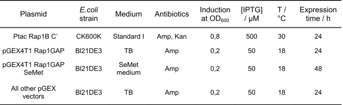
3.1.5 Media and antibiotics... 28
3.1.6 Buffers ... 28
3.2 Molecular biology methods...29
3.2.1 Agarose gels... 29
3.2.2 Isolation of plasmid DNA ... 29
3.2.3 Polymerase chain reaction (PCR) ... 29
3.2.5 Ligation ... 29
3.2.6 Competent cells... 29
3.2.7 Transformation ... 30
3.2.8 Bacteria storage ... 30
3.2.9 Site specific mutagenesis... 30
3.2.10 DNA sequencing... 30
3.2.11 Constructs ... 31
3.2.12 Point mutants... 31
3.3 Biochemical methods ...32
3.3.1 Sequence alignment... 32
3.3.2 SDS-PAGE ... 32
3.3.3 Determination of protein concentration ... 32
3.3.4 Matrix assisted laser desorption ionisation (MALDI) ... 32
3.3.5 Test expression ... 32
3.3.6 Protein overexpression and preparation of soluble bacteria extract ... 33
3.3.7 Rap1GAP purification ... 33
3.3.8 Purification of other GST fusion proteins... 34
3.3.9 Assignment of degradation bands... 34
3.3.10 Partial digest... 35
3.3.11 Purification of Rap1B C’ ... 35
3.3.12 Rap1-Aedans preparation ... 36
3.3.13 Nucleotide exchange ... 36
3.3.14 Nucleotide detection using reversed-phase HPLC ... 36
3.3.15 Radioactive charcoal assay... 37
3.3.16 Fast kinetics using stopped-flow measurement ... 38
3.3.17 CD spectrometry... 39
3.4 Crystallographic methods ...40
3.4.1 Crystallisation ... 40
3.4.2 Data collection and processing ... 41
3.4.3 Phase determination... 42
3.4.4 Refinement ... 44
3.4.5 Figure preparation ... 46
4 Results...47
4.1 Initial characterisation of Rap1GAP...47
4.1.1 Expression... 47
4.1.2 Biochemical characterisation of Rap1GAP ... 47
4.1.3 Arginine mutants... 49
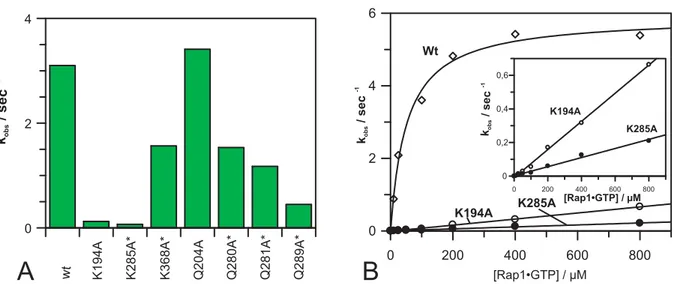
4.1.4 Lysine and glutamine mutants... 50
4.2 The Rap1GAP structure ...53
4.2.1 Purification... 53
4.2.2 Degradation and partial digest ... 53
4.2.3 High throughput cloning... 54
4.2.4 Crystallisation ... 55
4.2.5 Structure determination ... 57
4.2.6 Topology... 60
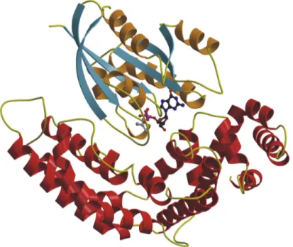
4.2.7 Description of the dimer... 63
4.2.8 The catalytic centre ... 65
4.3 The hydrolysis mechanism of Rap1GAP ...69
4.3.1 Dimerisation and Rap1GAP function... 69
4.3.2 Involvement of both domains in the GAP mechanism ... 70
4.3.3 Role of helix stabilisation for Rap1GAP function... 72
4.3.4 The catalytic residue... 73
4.3.5 Binding to a transition state analogue of GTP hydrolysis... 76
4.3.6 Other important residues in Rap1GAP... 77
4.3.7 Role of the carboxamide for GAP activity... 78
4.3.8 Implications for Tuberous sclerosis ... 80
5 Discussion...83
5.1 Dimerisation and role of two domains in Rap1GAP...83
5.2 Interacting partners of Rap1GAP...85
5.3 The novel GAP mechanism...88
5.4 Proposed model of the Rap1GAP-Rap1 complex ...90
5.5 Implications for Tuberous Sclerosis and cancer ...93
5.6 Evolution of the Rap1GAP system ...95
6 References ...97
7 Appendix ...111
7.1 Abbreviations...111
7.2 Deduction of equations...113
7.2.1 Determination of kon and koff via kobs... 113
7.2.2 Fluorescence titration ... 115
8 Zusammenfassung ...117
Danksagung...119
Teilpublikationen dieser Arbeit...121
Erklärung ...122
Lebenslauf ...123
1 Abstract
Rap1GAP is the founding member of a family of GTPase activating proteins (GAPs) for the small guanine nucleotide binding protein (GNBP) Rap1, which show no sequence homology to GAPs of other small GNBPs. Rap1 does not have a catalytic glutamine residue which is essential for the intrinsic and GAP mediated GTP hydrolysis of all other small GNBPs. In this thesis, the structure and the mechanism of GAP catalysed GTP hydrolysis are examined.
Most GAPs provide a catalytic arginine residue to the GNBP to complement the incomplete catalytic machinery. However, site-directed mutagenesis revealed that Rap1GAP does not employ a catalytic arginine. To understand the novel reaction mechanism, the structure of Rap1GAP was determined by X-ray crystallography to a maximal resolution of 2,9 Å. Initial phases were obtained by selenomethionine substituted crystals and a SIRAS phasing protocol. The structure was built and refined to an Rcryst= 23,4% and an Rfree of 27,6%.
Two Rap1GAP dimers were observed in the asymmetric unit, consistent with gel filtration experiments in which also dimerisation was observed. A Rap1GAP monomer consists of two domains. Both domains show a mixed α-β fold and were named dimerisation and catalytic domain, respectively. Surprisingly, the catalytic domain has structural similarity to the G domain of GNBPs itself suggesting a common evolutionary origin. No structural similarity to any other GAP was observed.
By site-directed mutagenesis, it was shown that dimerisation is not required for GAP function. However, both domains are necessary for full catalytic activity. Mutations around a highly invariant helix, the putative interaction helix, dramatically reduced GAP activity. Using a single-turnover fluorescence reporter assay it could be conclusively proven that Rap1GAP employs a catalytic asparagine from the interaction helix to stimulate GTP hydrolysis in Rap1. In the absence of this asparagine side-chain Rap1GAP was completely inactive but could still bind to Rap1•GTP. In contrast to the wild-type, the Rap1GAPN290A mutant can not associate with a transition state mimic of Rap1 GTP hydrolysis. Thus, Rap1GAP is the first example of a GAP which provides a catalytic asparagine for catalysis. Based on the analysis of various mutants, a model for the interaction of Rap1GAP with Rap1 is proposed.
The results of this thesis have implications for the disease Tuberous sclerosis. Loss of function mutations in the Rap1GAP homologue Tuberin are associated with this disease and can be rationalised in the view of this work.
2 Introduction
2.1 Guanine nucleotide binding proteins
2.1.1 The molecular switch
Guanine nucleotide binding proteins (GNBPs) are involved in a wide range of cellular processes including protein synthesis, sensual perception, vesicular transport and signal transduction cascades leading to cell proliferation and cell differentiation. Most GNBPs regulate these processes rather than provide energy for mechano-chemical work or chemical synthesis. Typically, GNBPs exist in an active GTP bound conformation and in an inactive GDP bound state (Bourne et al., 1990), therefore acting as a molecular switch (Figure 1). The GTP bound state has high affinity for interacting proteins which are referred to as effector molecules whereas the GDP bound state has low affinity and poorly interacts. In turn, the effectors are relocalised to their specific subcellular sites by this interaction or their activity is modulated.
To reach the active state, GDP has to be released and a new GTP molecule has to be bound. Since this reaction is intrinsically too slow to regulate cellular processes it can be accelerated by the action of guanine nucleotide exchange factors (GEFs).
GEFs catalyse nucleotide dissociation and allow GTP which is more abundant in the cell to bind. Also the intrinsic GTPase reaction of GNBP is slow. For efficient inactivation, the intrinsic reaction can be drastically stimulated by the action of GTPase activating proteins (GAPs).
GNBP GDP GTP
GNBP
GTP GDP
H O2 Pi
Effector proteins
off on
GAP GEF
Figure 1. The molecular switch of guanine nucleotide binding proteins. For explanation see text.
2.1.2 Overview of the GTPase superclass
Nucleotide binding proteins appear in a number of different folds (Vetter and Wittinghofer, 1999) including the di-nucleotide binding (Rossmann) fold, the protein and histidine kinase fold and the mono-nucleotide binding fold (P-loop containing nucleotide triphosphate (NTP) hydrolases). P-loop containing NTP hydrolases share the most abundant protein fold in most organisms and comprise 10 to 18% of all gene products (Koonin et al., 2000). This family can be divided in at least seven further lineages one of which is the GTPase superclass (Leipe et al., 2002).
The GTPase superclass was recently classified anew based on sequence and structure alignments of all available bacterial and eukaryotic GNBPs (Leipe et al., 2002). Two major branches could be identified which were named the TRAFAC (translation factor-related) and the SIMBI class (signal recognition particle, MinD and BioD). The best known members of the SIMBI class are the signal recognition particle GTPase (Ffh in bacteria) and the α-subunit of the signal recognition particle receptor (FtsY in bacteria) (Freymann et al., 1997). They exist in bacteria and eukaryotes and are involved in co-translational cellular targeting of nascent secretory and membrane proteins. The TRAFAC class contains most notably the translation factor superfamily, the Myosin-kinesin and the Ras-like superfamily.
Initiation factors IF2 (eIF5B in eukaryotes), eIF2γ (in some bacteria SelB) and elongation factors EF-Tu and EF-G are the four members of the translation factor superfamily that appear ubiquitously in bacteria and eukaryotes (Leipe et al., 2002).
eIF2γ forms a ternary complex with GTP and Met-tRNAiMet and mediates with other initiation factors the binding of tRNAiMet to the ribosome (reviewed in Sonenberg and Dever, 2003). GTP hydrolysis in eIF2γ is triggered in a reaction which requires among others initiation factor IF2/eIF5B. EF-Tu is a three-domain protein that forms a ternary complex with aminoacyl-tRNA and controls incorporation of the correct amino acids into the peptide chain (reviewed in Ogle et al., 2003). The five-domain protein EF-G catalyses translocation of tRNAs on the ribosome. The origin of other factors involved in protein biosynthesis, e.g. the release factors, differs in the three kingdoms of life (Leipe et al., 2002).
The eukaryotic cellular motor ATPases kinesin and myosin were grouped in the myosin-kinesin superfamily of TRAFAC GTPases. It has been argued that these proteins have evolved from an ancestral GTPase at the onset of eukaryotic evolution and have lost later their specificity towards GTP (Leipe et al., 2002). Members of the
dynamin family which are involved in the budding of clathrin-coated vesicles (Urrutia et al., 1997), and the GB1 family including GBPs (Prakash et al., 2000), are also grouped in this superfamily.
Heterotrimeric G-proteins appear exclusively in eukaryotes and more than 20 members are known (Sprang, 1997). They are activated by cell-surface receptors of the seven-transmembrane-helix class and regulate intracellular effectors such as adenylyl-cyclase or phospholipase Cβ.
Members of the Ras superfamily exist predominantly in eukaryotes. The exact classification of this superfamily varies (Bourne et al., 1990; Garcia-Ranea and Valencia, 1998; Leipe et al., 2002). The group of 20-25 kD GNBPs with Ras as most prominent member was called the small GNBPs. They were first grouped as a separate superfamily composed of the Ras, Rab, Rho, Ran and Arf/Sar1 family (Garcia-Ranea and Valencia, 1998), albeit it was recently suggested that the family of hetero-trimeric GNBPs should also be included in this superfamily (Leipe et al., 2002). More than 100 small GNBPs have been identified in eukaryotes (reviewed in Takai et al., 2001). Ras family members mainly regulate gene expression, whereas GNBPs of the Rho/Rac family control gene expression and cytoskeletal reorganisation. Members of the Ran family regulate nucleo-cytoplasmic transport during G1, S, G2 and among others microtubule organisation during M phase. Rab and Arf GNBPs control intracellular vesicle trafficking. A dendrogram showing the evolutionary relation between the small GNBPs is depicted in Figure 2.
2.1.3 Biochemical and structural features of small GNBPs
Ras consists of 189 amino acids and is the best characterised small GNBP (Wittinghofer and Waldmann, 2000). It is considered a paradigm for the complete family and its biochemical features are often representative for the other small GNBPs as well.
Ras is located in the plasma membrane by means of a farnesyl and a palmitoyl anchor which is required for its function in vivo. As most other small GNBPs, it has an extremely high affinity for both GDP and GTP with a KD in the picomolar range (Wittinghofer and Waldmann, 2000). No other standard nucleotide binds to Ras with a comparable affinity showing the high specificity for guanine nucleotides. The β-phosphate is required for high affinity binding since guanosin monophosphate (GMP) has a 106-fold reduced affinity in comparison to GDP/GTP. The binding affinity strongly depends on the presence of magnesium ions. When magnesium ionsare absent, the dissociation rate constant (koff) increases by many hundredfold with a concomitant increase in the equilibrium dissociation constant (KD) (John et al., 1988).
Available crystal structures demonstrate that Ras shares a common structural core - the G domain fold - with the complete class of TRAFAC GTPases (Leipe et al., 2002;
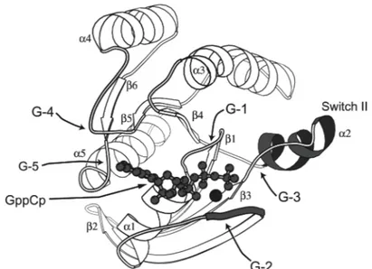
Sprang, 1997). The G domain fold comprises approximately 200 residues and consists of a six stranded mixed β-sheet surrounded by five α-helices (Figure 3). The most highly conserved elements in this domain are the five polypeptide loops that form the guanine nucleotide-binding site called G1 through G5 (Figure 3, Bourne et al., 1991).
Figure 3. Structure of the Ras protein in the GppCp•Mg bound form with the conserved elements and G-box regions labelled (from Sprang, 1997).
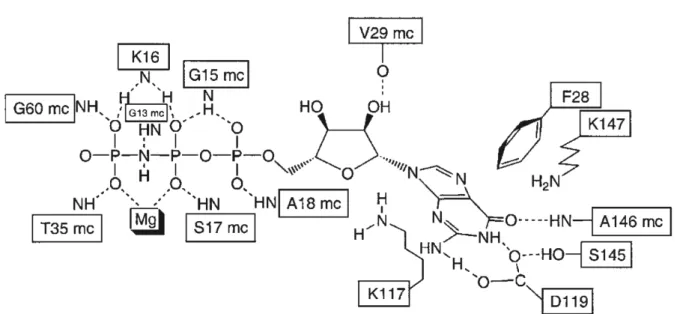
The G1 motif, better known as P-loop, connects strand β1 to helix α1. It contains the consensus motif GxxxxGK(S/T) (in Ras amino acids 10-17) and forms a ring-like structure which wraps tightly around the β-phosphate of GTP and GDP. The C-terminal serine of this motif (serine 17 in Ras) is complexed to the magnesium ion which is essential for high affinity binding of the nucleotide.
The connection between helix α1 and strand β2 contains a conserved threonine residue of the G2 motif involved in Mg2+ coordination and in direct binding to the γ-phosphate (threonine T35 in Ras). Also the G3 motif (DxxGQ/H/T) is involved in phosphate binding. The conserved aspartate (aspartate D57 in Ras) binds the magnesium ion via a water molecule and glycine G60 binds via a main chain contact directly to the γ-phosphate. Motif G4 (consensus NKxD with asparagine N116 in Ras) is located between strand β5 and helix α4 and is partly responsible for the recognition of the guanosin base. The (T/G)(S/C)A(K/T/L) (G5) motif (serine S145 in Ras) is located between strand β6 and helix α5. It does not participate directly in the guanine base recognition besides a main chain interaction of alanine A146. However, it stabilises residues of the G4 motif and hydrophobic residues involved in binding of the guanine base (e.g. phenylalanine F28 in Ras). The network of conserved interactions between Ras and the GTP analogue GppNHp is shown in Figure 4.
Figure 4. Interactions of the Ras and the GTP analogue GppNHp with selected, conserved residues (from Wittinghofer and Waldmann, 2000)
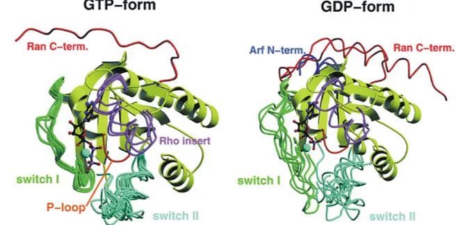
Two elements in GNBPs have been shown to undergo major conformational changes upon GTP hydrolysis, the switch I and switch II region (reviewed in Vetter and Wittinghofer, 2001). Conformational changes in switch I (amino acids 32-40 in Ras) are mediated mainly by the conserved threonine T35 of motif G2, conformational changes in switch II (amino acids 61-67 in Ras) by the conserved glycine G60 of motif G3. Due to the interaction of these residues with the γ-phosphate, the switches become stabilised in the GTP bound form. In contrast, the switches are rather mobile in the GDP bound form (Figure 5).
Figure 5. Superposition of selected Ras-related proteins in the GDP and the GTP bound form.
Whereas in the GTP bound form, all switches are stabilised in a similar conformation, they are much more flexible in the GDP bound form and show divergent conformations. Extra elements in the structures of Rho, Arf and Ran are highlighted (from Vetter and Wittinghofer, 2001).
The switches are the main determinants for binding of effector molecules. In the case of Ras, switch I was shown to bind to an 80 residues Ras Binding Domain (RBD) of effectors (Nassar et al., 1995; Huang et al., 1998; Vetter et al., 1999; Pacold et al., 2000). Although there is no sequence homology among the RBDs of different effectors, all examined RBDs show a ubiquitin-like fold. The RBD and Ras•GTP interact via an interprotein β-sheet. Besides some main chain interactions, most contacts are mediated by hydrophilic side chains. RBDs from different proteins, however, use different residues for interaction (Joneson et al., 1996).
2.1.4 The hydrolysis reaction
The intrinsic GTPase reaction of small GNBPs is slow, for Ras in the range of 0,02 min-1, and is strictly dependent on the presence of divalent cations (Wittinghofer and Waldmann, 2000). A water molecule close to the γ-phosphate and in hydrogen bond distance to glutamine Q61 is considered to be the attacking nucleophile, and mutations of this glutamine to most other amino acids dramatically reduce the intrinsic GTP hydrolysis (Der et al., 1986; Krengel et al., 1990; Prive et al., 1992).
Based on NMR studies with various Ras mutants and by determining intrinsic GTP hydrolysis reaction rates at various pH, a substrate assisted catalysis was proposed in which the γ-phosphate itself activates the attacking water molecule by abstracting a hydrogen atom (Schweins et al., 1995; Schweins et al., 1997). Both the less charged γ-phosphate and the more reactive nucleophile (the hydroxide ion) were suggested to promote catalysis. The role of glutamine Q61 is thought to be the positioning of the attacking water molecule in vicinity of the γ-phosphate.
A continuum of possible transition states of the GTP hydrolysis reaction ranging from purely dissociative to purely associative can be described (Figure 6) (Maegley et al., 1996). The dissociative transition state is dominated by bond cleavage, so that the bond to the leaving group is fully broken and the bond to the incoming nucleophile is barely formed. To maintain charge, this leads to a loss of negative charge on the terminal phosphoryl group during the transition (Figure 6) and to an increase in charge at the β-phosphate, especially at the β-γ bridging oxygen. On the other hand in the associative transition state, there is a large amount of bond formation to the incoming nucleophile but only a small amount of bond cleavage to the leaving group.
To maintain charge, this will result in an increase in negative charge on the terminal phosphoryl group (Admiraal and Herschlag, 1995).
In solution, GTP hydrolysis seems to proceed via a dissociative transition state (Admiraal and Herschlag, 1995). It is however controversially discussed whether in the protein environment the transition state has a dissociative or associative character (Maegley et al., 1996; Cepus et al., 1998; Allin and Gerwert, 2001;
Schweins et al., 1995; Scheffzek et al., 1997; Glennon et al., 2000; Seewald et al., 2002).
2.1.5 The Ras signal transduction pathway
The Ras gene was originally identified as the active principle of sarcoma viruses and was called an oncogene (reviewed in Takai et al., 2001). Later, it was realised that cellular counterparts exist in mammals.
In humans, three isoforms of Ras exist, H-Ras, N-Ras and K-Ras with a molecular weight of 21 kD which differ mainly at the C-terminus. From studies in Drosophila and C. elegans Ras was recognised as a central component in signal transduction pathways linking activation of cell surface receptors to gene expression in the nucleus (reviewed in Pawson and Saxton, 1999; Wittinghofer and Waldmann, 2000).
Growth factors such as EGF or PDGF bind to the extracellular domain of specific receptor tyrosine kinases. This leads to receptor dimerisation and subsequent trans- phosphorylation of tyrosine residues in the cytoplasmic part of the receptor.
Phosphorylated tyrosines in a sequence specific context are recognised by proteins possessing specialised phospho-tyrosine binding domains, e.g. SH2 (src homology 2) domains. In this way, the adapter protein Grb2 binds via its SH2 domain to phosphorylated growth factor receptor. SOS, an exchange factor for Ras and associated with Grb2, is thereby recruited to the membrane where it promotes GDP- GTP exchange on Ras. Ras in the GTP bound form activates several effectors of which the best characterised is the protein kinase Raf. Raf phosphorylates the protein kinase MEK which activates the protein kinase ERK. Finally, ERK phosphorylates a class of transcription factors, the ternary complex factors, which associate with a second class of transcription factors, the serum response factors.
The resulting protein complex initiates transcription by binding to a conserved promoter element, the serum response element which is present in a variety of genes, e.g. many transcription factors. The Ras signal is terminated by the action of GAPs targeted to the intracellular part of the receptor and by dissociation of the SOS-Grb complex upon phosphorylation by ERK.
Ras acts also in different pathways. It is activated by G-protein coupled receptors and cytoplasmic tyrosine kinases (Weiss and Littman, 1994), and it also triggers different pathways, e.g. by activating PI-3-Kinase or the nucleotide exchange factor for the small GNBP Ral, RalGEF.
Point mutations of Ras at either position 12, 13 or 61 render the protein unable to hydrolyse GTP, even in the presence of GAP (Trahey and McCormick, 1987;
Downward, 1998) thus leading to constitutively active Ras. These mutations were found in human tumours (e.g. Almoguera et al., 1988) indicating that a constitutive active Ras pathway leads to malignant transformation. It is now estimated that 30%
of all human tumours carry activated forms of the Ras gene. Thus, the Ras pathway is an important target for cancer therapy (Wittinghofer and Waldmann, 2000).
2.1.6 The Rap GNBPs
Rap1 was identified by low stringency hybridisation of various cDNA libraries with Ras cDNA (Pizon et al., 1988) and, at the same time as K-rev1 in a screen for cDNAs which revert the phenotype of K-Ras transformed fibroblast (Kitayama et al., 1989).
With more than 50% sequence identity, Rap1 is the closest relative of Ras, especially Switch I is highly conserved. Unlike Ras, it is modified by a geranyl-geranyl anchor rather than a farnesyl anchor.
Rap1 homologues are found in all vertebrates, in Drosophila and in yeast (reviewed in Bos et al., 2001). The yeast homologue Bud1 is involved in selecting the new budding site by recruiting and activating effectors to the selected region in the cell (Park et al., 2002). In Drosophila, maternal and zygotic Rap1 expression is essential for development of the embryo, imaginal disc development and oogenesis (Asha et al., 1999). Drosophila Rap1 was shown to be enriched at adherens junctions, particularly between newly divided sister cells, and to regulate the position of these junctions thereby regulating cell adhesion (Knox and Brown, 2002).
Four isoforms of Rap exist in humans, namely Rap1a, Rap1b, Rap2a and Rap2b.
Most work has been done on Rap1a and Rap1b, which share more than 90%
sequence identity, and in most experimental approaches, no discrimination between Rap1a and Rap1b (from here Rap1) has been made (Bos et al., 2001).
Rap1 in human was shown to be ubiquitously expressed. In fibroblast, it is located in the mid-Golgi compartment and early and late endosomes (Beranger et al., 1991;
granules to the plasma membrane upon GTP loading caused by a variety of different stimuli (Franke et al., 1997). In malignant oral keratinocytes, it was recently demonstrated to translocate from a perinuclear distribution to the nucleus upon GTP loading. Rap1 is thus, besides Ran, the only other small GNBP which is present in the nucleus (Mitra et al., 2003). By fluorescent resonance energy transfer studies it was established that activation of Rap1 in COS cells takes place at internal perinuclear membranes rather than at the plasma membrane (Mochizuki et al., 2001).
Based on the striking similarity of the Ras and Rap1 switch I region, Rap1 interaction with Ras effectors was analysed (e.g. Nassar et al., 1996). All Ras effectors examined are able to interact with the GTP bound form of Rap1 as well. However, this interaction does not activate all the effectors, as shown for the Raf-Kinase isoform, Raf-1 (Shirouzu et al., 1998). In line with the suppression of K-Ras action in fibroblasts and the recent finding that Rap1•GTP can inhibit cell proliferation in keratinocytes (Mitra et al., 2003), this suggested initially that Rap1 has an antagonistic role in Ras signalling by sequestering mutual effectors in an inactive state.
However, many lines of evidence suggest now that Rap1 is also able to activate signal transduction pathways independently of Ras. When micro-injected into Swiss3T3 fibroblasts, Rap1 is able to induce DNA synthesis and morphological changes (Altschuler and Ribeiro-Neto, 1998). In fibroblasts, Rap1 activation fails to interfere with Ras-dependent ERK activation (Zwartkruis et al., 1998). In PC12 cells, the Raf-isoform B-Raf is activated by Rap1•GTP and activates ERK, independently of Ras action (Vossler et al., 1997; Kao et al., 2001). B-Raf and likewise Ral-GEF activity are also stimulated in vitro by Rap1•GTP (Ohtsuka et al., 1996).
The best characterised role of Rap1, however, is the activation of integrin mediated cell-adhesion referred to as inside-out signalling (Katagiri et al., 2000; Reedquist et al., 2000; Sebzda et al., 2002; de Bruyn et al., 2002). A new Rap1 effector molecule, RAPL, was identified which mediates this effect by linking Rap1 to the integrin LFA-1 (Katagiri et al., 2002). This relocalisation is accompanied by an increase in integrin- mediated cell adhesion.
Furthermore, Rap1 was shown to be involved in the process of learning and memory (Morozov et al., 2003), in the development of leukaemia (Ishida et al., 2003), in angiogenesis and cerebrovascular diseases (Sahoo et al., 1999).
Activation of Rap1 can be observed upon a wide variety of stimuli, e.g. activation of tyrosine kinases, hetero-trimeric G-protein-coupled receptors and cell-adhesion molecules (McLeod et al., 1998; Posern et al., 1998; M'Rabet et al., 1998; York et al., 1998; Zwartkruis et al., 1998). Common second messengers such as cyclic AMP, Ca2+ and diacylglycerol (DAG) are involved in transducing extracellular signals to Rap1 (Bos et al., 2001). This activation is mediated by a variety of Rap1 specific GEFs. C3G was the first RapGEF to be identified (Gotoh et al., 1995). Analogous to the Ras exchange factor SOS, it is recruited to the membrane by an SH2 containing adapter protein Crk and allows consequently GDP-GTP exchange of membrane bound Rap1 (Okada et al., 1998). Another class of GEFs are the recently identified Epacs which are directly activated by cyclic AMP (de Rooij et al., 1998; Kawasaki et al., 1998a). CalDAG-GEFs can be activated by Ca2+ influx or by diacylglycerol (Kawasaki et al., 1998b) . A fourth group of RapGEFs are the PDZ-GEFs which may be responsible for signal amplification (de Rooij et al., 1999). It was suggested that PDZ-GEFs are recruited by Rap1•GTP via their RBDs to the membrane and catalyse further GDP-GTP exchange in Rap1 (Gao et al., 2001b). The Rap1 signal is terminated by GTP hydrolysis in Rap1 which can be stimulated by the action of GTPase activating proteins (see below).
2.2 GTPase Activating Proteins
The intrinsic GTPase reaction of most GNBPs is too slow to regulate signal transduction processes in a meaningful time frame (reviewed in Vetter and Wittinghofer, 2001). Thus, GTP hydrolysis can be accelerated by GTPase Activating Proteins (GAPs) which typically down-regulate GNBPs in the range of a few seconds (Scheffzek et al., 1998). The slow intrinsic GTPase activity of EF-Tu is for example dramatically stimulated by the mRNA charged 70S ribosome (reviewed in Ogle et al., 2003). GTP hydrolysis in Gα proteins can be stimulated by a family of proteins called regulators of G protein signalling (RGS) (Tesmer et al., 1997). Furthermore, GAPs specific for the Ras, Rho, Rab, Ran and Arf family members were discovered. GAPs for one family of small GNBPs show generally, albeit not always, sequence homology to each other. However, GAPs of different families share no sequence homology.
Many GAPs have a modular architecture to fulfil various other functions in the cell.
2.2.1 RasGAP and the arginine finger
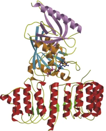
RasGAP was discovered when it was found that Ras•GTP microinjected in cells was rapidly converted into Ras•GDP (Trahey and McCormick, 1987). The protein responsible for this activity was identified and is now known as p120GAP. A fragment comprising 334 amino acids (GAP-334) was shown to be sufficient for catalysis. The structure of GAP-334 showed a helical, elongated protein with a central domain of 218 amino acids that is conserved among all RasGAPs (Scheffzek et al., 1996).
Later, the structure of a complex of RasGAP and Ras•GDP was solved (Scheffzek et al., 1997) in the presence of aluminium fluoride which along with GDP/ADP is a mimic of the transition state of many phosphoryl transferring enzymes (Figure 7, Chabre, 1990).
GAP-334 interacts predominantly with the switch regions and the P-loop of Ras. An exposed loop of RasGAP containing the highly conserved arginine R789 complements the catalytic site of Ras, and the guanidium group of arginine R789 interacts with the β-phosphate and with AlF3 (Figure 8). Additionally, the main-chain carbonyl oxygen of arginine R789 makes a hydrogen bond to the side-chain amide group of the catalytic glutamine Q61 in Ras thereby stabilising its position. Since arginine R789 is located in a flexible loop and points into the active site, it has been called the ‘arginine finger’ (Scheffzek et al., 1998).
Figure 7. Structure of RasGAP in complex with Ras•GDP•AlF3 (PDB accession code 1WQ1).
RasGAP is shown in red, Ras in orange and blue.
The principles of GTPase stimulation of GAP are thought to be the stabilisation of glutamine Q61 leading to correct positioning of the attacking water molecule and the provision of positive charge by the arginine residue involved in stabilising the transition state.
This structure also helped to explain why mutations in glycine G12 of Ras interfere with the activation by GAPs. Glycine G12 is within van-der-Waals distance of the catalytic glutamine of Ras and the catalytic arginine of RasGAP and any mutation interferes sterically with the arrangement of these residues in the transition state.
Figure 8. The active site of the RasGAP•Ras•GDP•AlF3 complex showing important elements of catalysis (from Scheffzek et al., 1997).
2.2.2 RhoGAPs
Numerous GAPs specific for Rho/Rac/Cdc42 have been described and several structures have been solved (Barrett et al., 1997; Rittinger et al., 1997a; Rittinger et al., 1997b; Nassar et al., 1998). Similar to GAPs for Ras, these proteins are purely helical (Figure 9). Based on their similar three-dimensional architecture, it was proposed that RhoGAPs and RasGAPs have a common evolutionary origin although no obvious sequence similarity is observed (Rittinger et al., 1998).
The structure of the RhoGAP•Rho•GDP•AlF3 complex revealed that the mechanism of GTPase stimulation involves - as in the case of RasGAP – the introduction of a catalytic arginine from the GAP and stabilisation of the catalytic glutamine in Rho (Rittinger et al., 1997b).
Figure 9. Structure of RhoGAP in complex with Rho•GDP•AlF−4 (PDB accession code 1AM4).
RhoGAP is in coloured in red, Rho in orange and blue.
2.2.3 Gα proteins and regulators of G protein signalling
Gα proteins transduce signals from seven-helix-transmembrane receptors to downstream targets. The crystal structure of Gαtα in the GTPγS bound state showed that these proteins have a Ras-like G domain fold with a helical insertion shortly behind the switch I region (Noel et al., 1993). The structure of a Gα protein bound to GDP and aluminium fluoride revealed that the mechanism of GTP hydrolysis involves stabilisation of the attacking water molecule by a catalytic glutamine (homologous to glutamine Q61 in Ras) and by a threonine from the helical domain (Coleman et al., 1994). Strikingly, the GTPase reaction was highly dependent on an arginine residue supplied by the helical domain which makes a hydrogen bond to aluminium fluoride mimicking the transition state of GTP hydrolysis. This arginine residue provided by Gα in cis (from Gα itself) is the functional equivalent of the arginine finger of RasGAP and RhoGAP which is provided in trans (from a second molecule).
Regulator of G protein signalling (RGS) proteins accelerate the intrinsic reaction of Gα proteins but do not share sequence homology to any other GAPs (Druey et al., 1996). They bind with modest affinity to the GTP bound forms of Gα and with high affinity to the GDP•AlF−4 bound form and stimulate GTP hydrolysis by at least 50-fold.
The structure of RGS4 bound to Giα1•GDP•AlF −4 showed that RGS proteins are helical, bind to the switch regions of Gα and stabilise them (Figure 10; Tesmer et al., 1997). However, they do not contribute catalytic residues to the active site of Gα except one asparagine residue which stabilises the catalytic glutamine of Gα. It was suggested that this asparagine could assist in the positioning of the hydrolytic water molecule.
Figure 10. Complex structure of RGS4 bound to Giα1•GDP•AlF−4 (PDB code 1AGR). Giα1 is shown in orange and blue, its helical insert domain in purple and RGS4 in red.
2.2.4 Other GAPs with an arginine finger
RabGAPs comprise a family of GAPs for the small GNBP Rab (Ypt in yeast) involved in vesicle trafficking. The structure of the yeast GAP Gyp1 revealed a fully α-helical
proteins (Rak et al., 2000). By mutational analysis, an arginine was identified which is critical for the catalytic activity (Albert et al., 1999). However, the exact mode of interaction between RabGAP and Rab is currently unknown, since no structure of a RabGAP-Rab complex has been described yet.
Some bacterial toxins such as ExoS from Pseudomonas aeruginosa, SptP from S. typhimurium and YopE from Yersinia sp. are introduced in eukaryotic cells and act as Rho-specific GAPs to reorganise the cell’s cytoskeleton (Goehring et al., 1999;
Fu and Galan, 1999; Pawel-Rammingen et al., 2000). The structure of the GAP domain of ExoS (130 residues, from here on ExoS) in complex with Rac•GDP•AlF−4 was solved (Figure 11B; Wurtele et al., 2001). It revealed that ExoS has a helical fold with a small two-stranded β-sheet. No structural homology was observed to any other known GAP structure. However, the mechanism of GTPase stimulation is similar to other GAPs since ExoS also provides an arginine finger into the active site of Rac.
Similarly, the structure of SptP in complex with Rac1•GDP•AlF3 revealed that SptP provides a catalytic arginine to Rac1 (Stebbins and Galan, 2000). It was suggested that bacterial GAPs and RhoGAPs have most likely evolved independently.
Figure 11. A) Ypt GAP (PDB code 1FKM) is a purely helical protein with no structural homology to other GAPs. B) The GAP domain of ExoS (in red and green) in complex with Rac•GDP•AlF−4 (orange and blue) (PDB code 1HE1).
2.2.5 ArfGAP
Arf is a small GNBP which is involved in vesicle trafficking in eukaryotic cells (for review see Spang, 2002). Arf in the GDP bound state translocates from the cytosol to the membrane and is activated by Arf specific exchange factors (Renault et al., 2003). Arf in its GTP bound form associates with the membrane and recruits a seven- subunit complex called coatomer. This process is followed by budding of a vesicle containing Arf and the coatomer complex. When the membrane curvature of the vesicle increases, the activity of ArfGAP also present in this complex is stimulated dramatically, leading to GTP hydrolysis and Arf•GDP dissociation (Bigay et al., 2003).
The catalytic domain of the ArfGAPs family comprises 140 residues including a zinc finger motif (Cukierman et al., 1995). The structure of the ArfGAP domain including four C-terminal ankyrin repeats (Mandiyan et al., 1999) and of the ArfGAP domain in complex with Arf•GDP (Goldberg, 1999) was solved. It revealed that the zinc finger of ArfGAP – consisting of four strands and one helix – is embedded in an irregular array of six α-helices and one β-strand (Figure 12). Clearly, no structural similarity to any GAPs of another GNBP family was observed.
Figure 12. Crystal structure of ArfGAP (in red and green) in complex with Arf•GDP coloured in orange and blue (pdb coordinates provided by J.Goldberg). ArfGAP binds to Arf at a position far away from the nucleotide binding site.
Mutagenesis studies in ArfGAP revealed an arginine residue whose mutation to alanine dramatically reduced the GAP activity (Mandiyan et al., 1999). However, the structure of the complex showed that ArfGAP binds to a region of switch II of Arf
proposed that the GTPase reaction is not stimulated by a catalytic residue but rather by rearranging switch II of Arf leading to a better positioning of the catalytic glutamine in switch II. In further experiments it was shown, that upon addition of coatomer to Arf•GTP and ArfGAP, the GTPase reaction was a further 1000-fold stimulated. This suggests that the coatomer itself is involved in the GAP reaction and that an arginine finger is provided by the coatomer (Goldberg, 1999).
Sar1 is an Arf related protein which is involved in the formation of so-called COPII coated vesicles on the endoplasmic reticulum (Pasqualato et al., 2002). COPII consists of Sar1 and two large heterodimeric complexes Sec23/24 and Sec13/31 (Barlowe et al., 1994).
Sar1 has a histidine residue at the equivalent position of the catalytic glutamine in most other small GNBPs, and the intrinsic GTP hydrolysis of Sar1 is very slow.
However, GTP hydrolysis can be stimulated by the Sec23 subunit which does not show sequence similarity to other GAPs (Yoshihisa et al., 1993). The crystal structure of Sec23 in complex with Sar1•GppNHp revealed that Sec23 provides a catalytic arginine into the active site of Sar1, and that the catalytic histidine of Sar1 positions the attacking water molecule (Bi et al., 2002). This reaction employing a catalytic histidine for positioning of the attacking water is unique for small GNBPs but might be similar for EF-Tu (Cool and Parmeggiani, 1991; Berchtold et al., 1993; Vogeley et al., 2001; Mohr et al., 2002).
2.2.6 RanGAP – catalysis without an arginine finger
RanGAPs stimulate the GTPase reaction of the small GNBP Ran (Seewald et al., 2002) which is involved in nuclear transport. A third protein, RanBP, binds to Ran and increases its affinity for RanGAP (Seewald et al., 2003). RanGAPs consist of a modular architecture with a 330-350 residue leucine-rich repeat (LRR) domain followed by an acidic region of approximately 40 residues (Hillig et al., 1999). The LRRs appear in proteins with various function, e.g. in the ribonuclease A inhibitor or Drosophila Toll-like receptor (Kobe and Deisenhofer, 1995). A single LRR forms a β-α hairpin consisting of a β-strand, a loop and an α-helix roughly parallel to the β-strand. The structure of yeast RanGAP showed that eleven LRR repeats form a crescent shaped molecule (Hillig et al., 1999). RanGAP does not have any structural similarity to the purely helical GAPs of Ras and Rho.
The complex between RanGAP, RanBP and Ran in the presence of a GTP analogue and in the presence of GDP and aluminium fluoride was solved (Figure 13; Seewald et al., 2002). Surprisingly, the only arginine in the vicinity of the active site was bent away from the γ-phosphate or the aluminium fluoride, and it was previously shown that this arginine is not important for catalysis (Hillig et al., 1999). This led to the proposal that RanGAP mediates GTP hydrolysis without an arginine finger. It was suggested that the basic machinery of fast GTP hydrolysis is provided exclusively by Ran and that binding of RanGAP leads to correct positioning of the catalytic glutamine in Ran.
Figure 13. Structure of RanGAP in complex with Ran•GDP•AlF3 and RanBP1 (PDB code 1K5D).
RanGAP is shown in green and red, Ran in yellow and blue and RanBP1 in purple.
2.2.7 The signal recognition particle and its receptor
The signal recognition particle (SRP) and its receptor (SR) target newly synthesised proteins destined for secretion or membrane integration to the endoplasmic reticulum (Keenan et al., 2001). Both SRP and SR are conserved across all kingdoms of life. In prokaryotes, SRP consists of a single 48 kD GTPase called Ffh and a 110 nucleotide 4,5S RNA, whereas SR consists of the GTPase called FtsY. When both proteins are
loaded with GTP, they can bind to each other leading to a reciprocal stimulation of their GTPase activities, GTP hydrolysis and concomitant dissociation of the complex.
Very recently, the mechanism of this reciprocal stimulation was elucidated by solving the structure of the complex between the two GTPases in the presence of a non-hydrolysable GTP analogue (Egea et al., 2004; Focia et al., 2004). The two GTPases form a quasi symmetric heterodimer, and the two nucleotides are aligned in a nearly symmetrical composite active site (Figure 14). In each chain, a catalytic aspartate was identified which is thought to activate the attacking nucleophilic water molecule in cis. Additionally, an arginine and a glutamine residue are provided from both molecules into the active site and interact in cis and possibly in trans with the β- and γ-phosphate groups. Strikingly, the 3’ OH group of each GTP molecule contacts the γ-phosphate group of the opposing GTP molecule and reduces the negative charge on the γ-phosphate. This hydroxyl group was shown to be essential for association, reciprocal activation and catalysis (Egea et al., 2004).
Figure 14. Ribbon type presentation of the SRP and SR heterodimer both in the GppNHp bound form (PDB code 1OKK). The nucleotides are buried in a common active site. SRP is shown in green and red, SR in orange and blue.
2.3 GTPase activating proteins of the Rap1GAP family
2.3.1 Rap1GAP
Rap1GAP (formerly GAP3) was isolated as a soluble and a membrane-associated isoform from cytosolic brain extract (Kikuchi et al., 1989; Polakis et al., 1991) which specifically stimulate GTP hydrolysis in Rap1, and to a smaller extent in Rap2 (Janoueix-Lerosey et al., 1992). The protein responsible for this activity was sequenced and the corresponding gene was cloned from a human brain cDNA (Rubinfeld et al., 1991). It turned out to encode a polypeptide of 663 amino acids with a molecular weight of 73 kD which in human is expressed most abundantly in brain, foetal tissue, undifferentiated cells and certain tumour cell lines. By deletion analysis it was shown that a protein fragment consisting of amino acids 75-416 is necessary and sufficient to retain full GAP activity (Rubinfeld et al., 1992). This fragment did not show any sequence similarity to GAPs of other small GNBPs. However, homologous sequences have been identified in the human genes Spa1 (Hattori et al., 1995), E6TP1/SpaR/SpaL (Gao et al., 1999; Roy et al., 1999) and Tsc2 (Figure 15;
Tuberous Sclerosis Consortium,1993). Homologues of these genes are found in all higher organisms.
Rap1GAP was shown to be regulated by protein degradation and relocalisation from cytosol to the membrane. It is phosphorylated in vivo at four distinct sites which are located C-terminally of the catalytic fragment (Polakis et al., 1992; Rubinfeld et al., 1992). Recently, it was demonstrated that in thyroid cells phosphorylation of Rap1GAP by GSK3β is associated with proteasome-mediated degradation (Tsygankova et al., 2004).
A splice isoform of Rap1GAP, RapGAPII, is expressed in heart, liver, kidney and cerebrum and contains an N-terminal GoLoco motif which specifically binds to the α-subunits of the Gi family of heterotrimeric G-proteins (Mochizuki et al., 1999;
Kimple et al., 2002). Upon stimulation of Gi, RapGAPII translocates from the cytosol to the membrane, followed by a decrease of Rap1•GTP and an activation of the ERK pathway, thus linking Gi and Rap1 signalling pathways. Rap1GAP was also shown to bind to the GTP bound form of Gαz (Meng et al., 1999) accompanied by a decrease of cellular Rap1•GTP (Meng and Casey, 2002) and to the GDP bound form of Gα0
accompanied by an increase of cellular Rap1•GTP (Jordan et al., 1999).
Another possible interaction partner of Rap1GAP is the cytoskeleton-anchoring protein AF-6 (Su et al., 2003)which was earlier described as a putative Rap1 effector (Linnemann et al., 1999; Boettner et al., 2000). It was proposed that the interaction is mediated by the PDZ domain of AF-6 which should bind to an internal VVF motif of Rap1GAP located in the catalytic domain (Su et al., 2003).
1807 1042
663
Spa1 Rap1GAP
Tuberin
GAP
GAP
GAP PDZ
181 397
GOLOCO 1 17
Leucine Zipper
CC CC TAD TAD CAM
CC
75
-
415
SPAR Act1 GAP PDZ Act2 GKBD CC
E6TP1
1804
Figure 15. Schematical view of the Rap1GAP family showing the domain structure of the involved proteins. Abbreviations used here: CC: coiled coil, Act: actin regulatory domain, GKBD: guanylate kinase-like binding domain, TAD: Transcription activation domain CAM: Calmoduline binding domain. The GoLoco domain is present in RapGAPII but not Rap1GAP. This figure was kindly provided by P. Chakrabarti.
2.3.2 Signal-induced proliferation-associated protein 1 (Spa-1)
Spa-1 encoding a 130 kD protein was identified as a gene which was little expressed in a quiescent murine lymphoid cell line but was induced upon interleukin-2 stimulation (Hattori et al., 1995). The corresponding protein contains the Rap1GAP domain, a PDZ domain involved in protein-protein interaction and a C-terminally located coiled-coil region (Figure 15). In human, Spa-1 is most abundantly expressed in lymphoid tissues such as thymus, spleen but not in tissues expressing Rap1GAP such as brain, kidney and pancreas (Kurachi et al., 1997). Baculo-virus expressed Spa-1 showed specific GAP activity towards Rap1 and Rap2 (Kurachi et al., 1997).
As for Rap1GAP, AF-6 was described as possible interaction partner (Su et al., 2003). Recently, Spa-1 was shown to be a tumour-suppressor gene since Spa-1 knockout mice developed a spectrum of myeloid disorders that resembled human chronic myelogenous leukaemia (Ishida et al., 2003). Furthermore, it was demonstrated that the increased Rap1•GTP levels caused by Spa-1 deletion were responsible for increased cell proliferation of hematopoietic progenitors.
2.3.3 E6TP-1
E6TP1 (E6 targeted protein 1) was identified as a 200 kD protein which is targeted to proteasome-mediated degradation by the papilloma virus protein E6 (Gao et al., 1999). Degradation of E6TP1 was correlated with the ability of E6 to immortalise mammary epithelial cells (Gao et al., 2001a), thus establishing the role of E6TP1 as a tumour suppressor gene. The 1804 residue protein is widely expressed in tissues and in-vitro cultured cell lines. Like Spa-1, it contains a Rap1GAP homology domain (residues 489–819), a PDZ domain and a coiled-coil region (Figure 15). Additionally, it has two actin binding domains (Pak et al., 2001). GAP activity towards Rap1 and Rap2 was demonstrated (Singh et al., 2003).
SPAR/SPAL is the rat homologue of E6TP1 and was found to form a complex with PSD-95 and NMDA receptors in neurons. This complex has a crucial role for establishing dendritic spines (Pak et al., 2001) which are postsynaptic protrusions where glutamate-mediated neuronal transmission takes place (Meyer and Brose, 2003). Phosphorylation of SPAR1 by serum-inducible kinase leads to its degradation accompanied by loss of the dendritic spine (Pak and Sheng, 2003). Thus, SPAR is a critical component in the formation of neuronal activity albeit the role of Rap1 regulation is unclear to date.
2.3.4 Tuberin
Tuberous sclerosis (Tsc) is an autosomal dominant inherited disease with a prevalence of 1 in 6000 births characterised by benign tumours called hamartomas in a variety of tissues, as well as rare malignancies (reviewed in Manning and Cantley, 2003; Li et al., 2004). Other symptoms include mental retardation and epilepsy. This disease is caused by loss of function mutations in either of the two tumour suppressor genes Tsc1 and Tsc2 encoding Hamartin (130 kD) and Tuberin (200 kD), respectively (Tuberous Sclerosis Consortium, 1993; van Slegtenhorst et al., 1998). In vivo, Tuberin and Hamartin form a heterodimer which is thought to stabilise the two proteins. This complex functions in the insulin/mTOR pathway to inhibit cell growth and proliferation (Potter et al., 2002; Tee et al., 2002).
Tuberin was shown to have sequence homology to a C-terminal fragment of the Rap1GAP domain, and in-vitro GAP activity towards the small GNBPs Rap1 and Rab5 was reported (Wienecke et al., 1995; Xiao et al., 1997). However, it is evident
2003; Garami et al., 2003; Castro et al., 2003; Zhang et al., 2003). It is currently unclear how RheB in the GTP bound form can activate the downstream effector kinase mTOR.
2.3.5 Biochemistry of the Rap1-Rap1GAP system
A catalytic glutamine (glutamine Q61 in Ras) is crucial for the intrinsic and GAP stimulated GTP hydrolysis of Gα, Ras, Rho, Rab and Ran GNBPs. In contrast, Rap1 does not have a catalytic glutamine but a threonine at the equivalent position. As a result, the intrinsic GTP hydrolysis rate of Rap1 is 10-fold slower than for Ras but can be reconstituted by exchanging threonine T61 for a glutamine (Frech et al., 1990).
Furthermore, threonine T61 is not required for the intrinsic and GAP stimulated GTP hydrolysis in Rap1 (Maruta et al., 1991). It is therefore anticipated that the mechanism of hydrolysis is different between Rap1 and the other GNBPs.
Glycine G12 mutations in Ras and other GNBPs render the protein inactive to GAPs since any side-chain at position 12 would interfere with the positioning of the arginine finger and the catalytic glutamine (see above). However, the G12V mutant of Rap1 can still be down-regulated by Rap1GAP, albeit with an 8-fold reduced rate (Brinkmann et al., 2002). This again indicates that Rap1GAP stimulates GTP hydrolysis in a completely different way than all other GAPs.
A fluorescence assay was developed to monitor a single turnover reaction cycle of the Rap1GAP catalysed reaction in Rap1 (Kraemer et al., 2002). It was proposed that the rate limiting step of the reaction was not GTP hydrolysis but the release of inorganic phosphate, similarly as already proposed for RasGAP-Ras system (Allin et al., 2001).
2.4 Objectives of this work
All available data indicated that the Rap1GAP stimulated reaction is fundamentally different from all other GAP systems described hitherto (see above). Therefore, this new mechanism was explored by biochemical and structural analysis.
In a first step, the reaction should be characterised and residues important for catalysis should be identified. Consequently, the structure of one member of the Rap1GAP family should be solved. It was expected that Rap1GAP has a completely novel GAP fold since no sequence similarities to any other GAP could be detected.
Based on the structure and the available biochemical data, the catalytic centre should be identified and further characterised by mutational and biochemical analysis.
This work also aimed to understand the molecular basis for diseases such as Tuberous sclerosis since single point mutations in the Rap1GAP domain of Tuberin were described in Tuberous sclerosis patients. However, the role of these mutations in disease was unclear and could be unravelled by understanding the involved reaction mechanism.
3 Materials and Methods
3.1 Materials
3.1.1 Chemicals
Chemicals from the following companies were used: Amersham-Pharmacia (Freiburg), Baker (Deventer, Niederlande), Fluka (Neu-Ulm), GERBU (Gaiberg), Merck (Darmstadt), Pharma-Waldhof (Düsseldorf), Qiagen (Hilden), Riedel-de-Haen (Seelze), Roche (Mannheim), Roth (Karlsruhe), Serva (Heidelberg) und Sigma- Aldrich (Deisenhofen).
3.1.2 Enzymes
DNAase-I Roche (Mannheim)
Pfu DNA polymerase New England Biolabs (Schwalbach) Restriction enzymes New England Biolabs (Schwalbach) T4 DNA ligase New England Biolabs (Schwalbach) Trypsin, α-chymotrypsin Sigma (Deisenhofen)
Thrombin Serva (Heidelberg)
3.1.3 Kits
QIAprep Spin Miniprep Kit Qiagen (Hilden) QIAquick Gel Extraction Kit Qiagen (Hilden)
BigDye Terminator Sequencing Kit Applied Biosystems (Langen) λ-DNA standard Invitrogen (Karlsruhe) Wide Range, SDS7 protein marker Sigma (Deisenhofen)
3.1.4 Microorganisms
E. coli CK600K supE, hsdM+, hsdR-, kanR (Hoffmann-Berling, Heidelberg) E. coli TG1 K12, supE, hsd∆5, thi, ∆(lac-proAB), F’[traD36, proAB+, lacIq,
lacZ∆M15] (Promega)

![Figure 17. Michaelis-Menten kinetic of the Rap1GAP-stimulated reaction. 100 nM Rap1GAP or GST-Rap1GAP were used as enzyme and increasing concentrations of Rap1•[γ- 32 P]GTP as substrate, in standard buffer at 25 °C](https://thumb-eu.123doks.com/thumbv2/1library_info/3660928.1503748/53.892.256.680.95.430/figure-michaelis-stimulated-reaction-increasing-concentrations-substrate-standard.webp)