Descending Octopaminergic Neurons in the Stick Insect:
Their Inputs and their Output Effects on the Locomotor System
Inaugural-Dissertation zur
Erlangung des Doktorgrades der Mathematisch- Naturwissenschaftlichen Fakultät
der Universität zu Köln
vorgelegtvon
Thomas Stolz
aus Aichach (Bayern)
Köln 2018
Berichterstatter:
PD Dr. Joachim Schmidt Prof. Dr. Peter Kloppenburg
Tag der mündlichen Prüfung: 12.06.2018
i
Abstract
The neural networks controlling locomotion (walking) must exhibit a high degree of flexibility for task-specific adaptation of behavior to environmental influences and changes in internal state. Neuromodulatory influences are very important for this flexibility, as they can regulate the activity of all neurons in the walking system and the strengths of their synaptic connections. To fully understand the neural control of walking, it is crucial to identify the neurons that release neuromodulators and to determine their activity patterns during behavior and analyze the properties of their output effects.
Octopamine, one such neuromodulator, is considered the invertebrate homolog to the vertebrate noradrenaline. It is a significant modulator in insect locomotor systems, both acting in the peripheral and central nervous systems. Octopamine modulates muscle metabolism, neuromuscular transmission, sensory sensitivity, excitability of motor neurons, and activity in the central pattern generating networks that control locomotion. The neural source of octopamine acting in the central nervous system of insect thoracic segments has not yet been identified. Thus, it is unknown to what extent effects of application of octopamine to thoracic ganglia in previous studies reflect the physiological role of octopamine.
In the current thesis, I hypothesized that dorsal unpaired median neurons with bilaterally descending axons (desDUM neurons) are a source of octopaminergic modulation of activity in thoracic neural networks for the control of walking in the stick insect Carausius morosus. I revealed the morphology of desDUM neurons in the gnathal ganglion by intracellular staining. Employing the newly developed method of direct MALDI-TOF mass spectrometry, I could show that stick insect desDUM neurons are octopaminergic.
Using semi-intact preparations and intracellular recordings of desDUM neurons, I found that they are phasically activated during six-legged walking and single-leg stepping. The phasic excitatory input to desDUM neurons during walking does not arise from coupling to activity of mesothoracic central pattern generating networks, but most likely from activation of mechanosensory organs of all six legs. Passive leg movement and stimulation of mesothoracic
ii
campaniform sensilla excited desDUM neurons. Furthermore, stimulation of the mesothoracic femoral chordotonal organ (fCO) had a weak excitatory influence on their activity.
Further, I investigated the output effects of desDUM neurons on reflex-evoked, spontaneous, and centrally generated activity of mesothoracic motor neurons with activation of single desDUM neurons. I could show that distinct desDUM neurons mediate differential effects.
Some neurons induce a decrease and others an increase, in the magnitude of reflex-induced motor neuron activity. The neurons which mediated an excitatory influence additionally increased the frequency of reversal of an fCO-induced postural reflex. Some desDUM neurons mediated an increase in the frequency of centrally generated rhythmic motor neuron activity. Collectively, the results of the current thesis provide a comprehensive characterization of desDUM neurons and their complex roles in the stick insect locomotor system.
The identity of direct neural target structures for the modulatory action of desDUM neurons, as well as the net output effects of the entire population of desDUM neurons during walking remain open questions. In future experiments, genetic access to desDUM neurons could aid in the activation, silencing, or visualization of their activity, which would collectively contribute to comprehensive answers to the open questions.
iii
Zusammenfassung
Das neuronale Netzwerk zur Steuerung der Fortbewegung (Laufen) muss ein hohes Maß an Flexibilität für die aufgabenspezifische Anpassung des Verhaltens an Umwelteinflüsse und innere Zustandsänderungen aufweisen. Neuromodulatorische Einflüsse sind sehr bedeutend für die Flexibilität, da sie die Aktivität aller Neuronen des Laufsystems und die Stärke ihrer synaptischen Verbindungen modulieren können. Um die neurale Kontrolle des Laufens vollständig zu verstehen, ist es wichtig, die Neuronen zu identifizieren, die Neuromodulatoren freisetzen, ihre Aktivitätsmuster während des Verhaltens zu bestimmen und die Eigenschaften ihrer Ausgangseffekte zu analysieren.
Der Neuromodulator Octopamin, gilt als Invertebraten-Homolog zum Noradrenalin der Wirbeltiere. Octopamin ist ein signifikanter Modulator im Laufsystem der Insekten, wo es sowohl im peripheren als auch zentralen Nervensystem wirkt. Octopamin, moduliert den Muskelstoffwechsel, die neuromuskuläre Übertragung, die sensorische Sensitivität, die Erregbarkeit von Motorneuronen und die Aktivität in zentralen Muster erzeugenden Netzwerken, die die Fortbewegung steuern. Die neurale Quelle von Octopamin, das im Zentralnervensystem der thorakalen Segmente von Insekten wirkt, ist noch nicht identifiziert worden. Es ist daher nicht bekannt, inwieweit Effekte infolge der Applikation von Octopamin auf thorakale Ganglien, in früheren Studien, die physiologische Rolle von Octopamin widerspiegeln.
In der vorliegenden Arbeit stellte ich die Hypothese auf, dass dorsale ungepaarte mediane Neuronen mit bilateral absteigenden Axonen (desDUM-Neuronen) eine Quelle der oktopaminergen Modulation der Aktivität in thorakalen neuronalen Netzwerken zur Steuerung des Laufens bei der Stabheuschrecke Carausius morosus sind. Ich habe die Morphologie der desDUM-Neuronen im Unterschlundganglion durch intrazelluläre Färbung aufgedeckt. Mit der neu entwickelten Methode der direkten MALDI-TOF Massenspektronomie konnte ich zeigen, dass desDUM Neuronen in der Stabheuschrecke oktopaminerg sind.
Mittels semiintakter Präparate und der intrazellulären Aufnahme des Membranpotentials von desDUM-Neuronen fand ich heraus, dass die Neuronen während des sechsbeinigen Laufens
iv
und des einbeinigen Schreitens phasisch aktiviert werden. Der phasisch exzitatorische Eingang in desDUM-Neuronen während des Laufens ergibt sich nicht aus einer Kopplung an Aktivität von mesothorakalen zentralen Muster erzeugenden Netzwerken, sondern höchstwahrscheinlich aus einer Aktivierung von meschanosensorischen Organen aller sechs Beine. Passive Beinbewegung und Stimulation der mesothorakalen campaniformen Sensillen waren erregend. Weiterhin hatte die Stimulation des mesothorakalen femoralen Chordotonalorgans (fCO) einen schwachen exzitatorischen Einfluss auf die Aktivität von desDUM Neuronen.
Ich untersuchte die Effekte von desDUM Neuronen auf reflexinduzierte, spontane und zentral generierte Aktivität von mesothorakalen Motoneuronen anhand der Aktivierung von einzelnen desDUM Neuronen. Ich konnte zeigen, dass verschiedene desDUM-Neuronen unterschiedliche Wirkungen vermitteln. Einige Neuronen induzieren eine Abnahme und andere eine Zunahme der Stärke der reflexinduzierten Aktivität von Motoneuronen. Die Neuronen, die einen exzitatorischen Einfluss vermittelten, erhöhten zusätzlich die Häufigkeit der Umkehrung eines fCO-induzierten Haltungsreflexes. Einige desDUM-Neuronen vermittelten eine Zunahme der Frequenz von zentral erzeugter rhythmischer Motoneuronenaktivität. Zusammenfassend liefern die Ergebnisse der vorliegenden Arbeit eine umfassende Charakterisierung der desDUM-Neuronen und ihrer komplexen Rolle im Laufsystem der Stabheuschrecke.
Die Identität direkter neuronaler Zielstrukturen für die modulatorische Wirkung von desDUM-Neuronen sowie der Nettoeffekt der gesamten Population von desDUM-Neuronen beim Laufen bleiben offen. In zukünftigen Experimenten könnte der genetische Zugang zu desDUM-Neuronen bei der Aktivierung, Hemmung und Aufzeichnung ihrer Aktivität helfen.
Die könnte dazu beitragen die offenen Fragen zu beantworten.
v
Contents
Abstract ... i
Zusammenfassung ... iii
Contents ... v
Abbreviations ... vii
1 Introduction ... 1
2 Materials and Methods ... 17
2.1 Basic semi-intact stick insect preparation ... 17
2.2 Intracellular recordings ... 17
2.3 Intracellular neurobiotin staining of desDUM neurons ... 18
2.4 Extracellular recording from leg-nerves ... 18
2.5 Analysis of octopamine/tyramine content with MALDI-TOF MS ... 19
2.6 Single-legged stepping and six-legged walking preparations and passive leg movement ... 20
2.7 Pilocarpine-evoked rhythmic motor neuron activity ... 21
2.8 Stimulation of the femoral chordotonal organ (fCO) ... 22
2.9 Stimulation of campaniform sensilla (CS) ... 25
2.10 General information on data analysis and statistics ... 25
3 Results ... 27
3.1 Identification of stick insect desDUM neurons ... 27
3.1.1 Morphology of desDUM neurons ... 27
3.1.2 Octopamine content of desDUM neurons ... 29
3.2 Input physiology ... 32
3.2.1 Activity of desDUM neurons during walking ... 32
3.2.2 Influence of CPGs on activity of desDUM neurons ... 35
3.2.3 Effect of passive leg movement on activity of desDUM neurons ... 36
3.2.4 Effect of femoral chordotonal organ stimulation on activity of desDUM neurons ... 37
3.2.5 Effect of campaniform sensilla stimulation on activity of desDUM neurons .... 40
3.3 Output effects of desDUM neurons ... 42
3.3.1 Effects of desDUM neurons on activity of ExtTi MNs... 42
vi
3.3.1.1 Modulation of fCO-induced reflex responses in ExtTi MNs ... 42
3.3.1.2 Effects of desDUM neurons on spontaneous ExtTi MN activity ... 66
3.3.2 Effect of desDUM neurons on activity of ProCx and RetCx MNs ... 70
3.3.2.1 Effect on CS stimulation-induced MN activity ... 71
3.3.2.2 Effects on spontaneous RetCx and ProCx MN activity ... 73
3.3.2.3 Effects on centrally generated RetCx and ProCx MN activity ... 75
4 Discussion ... 78
4.1 Identification of stick insect desDUM neurons ... 78
4.1.1 Morphology of desDUM neurons ... 78
4.1.2 Electrophysiological identification of desDUM neurons ... 81
4.1.3 Octopamine content... 83
4.2 Input physiology of desDUM neurons ... 83
4.2.1 Activity of desDUM neurons during walking ... 83
4.2.2 Activity of desDUM neurons during centrally generated rhythmic leg MN activity ... 84
4.2.3 The role of sensory organs in recruiting desDUM neurons... 86
4.2.4 Possible mechanisms of signal integration ... 89
4.3 Output effects of desDUM neurons ... 91
4.3.1 Involvement of OA in mediating the effects of desDUM neuron ... 92
4.3.2 Individual neurons of the desDUM neuron population have differential effects on MN activity ... 93
4.3.3 Functional implications for desDUM neuron output ... 97
4.3.4 Assumptions on the net output of the desDUM neuron population ... 100
4.4 Summary and Conclusions ... 101
4.5 Outlook... 102
List of Figures ... 104
References ... 105
Danksagung... 122
Erklärung ... 123
vii
Abbreviations
AEP anterior extreme position
AP action potential
CA 4-hydroxy-3-methoxycinnamaldehyde
cAMP 3’,5’-cyclic adenosine monophosphate
CNS central nervous system
CPG central pattern generator
CS campaniform sensilla
DAG diacylglycerol
desDUM descending unpaired median
DUM dorsal unpaired median
EPSP excitatory postsynaptic potential
ExtTi extensor tibiae
fCO femoral chordotonal organ
FT femur-tibia
GABA gamma-aminobutyric acid
GNG gnathal ganglion
IP3 inositol 1,4,5-triphosphate
MALDI-TOF matrix assisted laser desorption/ionization-time of flight
MN motor neuron
MS mass spectrometry
OA octopamine
OAR octopamine receptor
PEP posterior extreme position
PKA protein kinase A
viii
PNS peripheral nervous system
ProCx protractor coxae
RetCx retractor coxae
RMP resting membrane potential
TA tyramine
TC thorax-coxa
UM unpaired median
VUM ventral unpaired median
1
1 Introduction
Animal locomotion is often necessary for survival. With the ability to move, animals can access food sources and mating partners and escape predators and negative environmental influences. Effective locomotion requires both robustness to perturbations and flexibility of the locomotor system. This is particularly important for terrestrial legged locomotion, in which walking movements must be adapted to the irregular and unpredictable nature of the ground. Furthermore, temporal coordination of movement between different legs and different joints of single legs has to be adjustable to meet the demands of different gaits and changes in walking direction and speed as well as varying environmental influences in real time. Therefore, the underlying neural networks must exhibit a high degree of flexibility for motor control to be adaptive. In general, legged locomotion is based on contractions of a large number of skeletal muscles that are controlled by motor neurons (MNs). The activity of MNs is under the control of interneurons that are part of central pattern generating networks (central pattern generators; CPGs) located in the spinal cord in vertebrates or thoracic ganglia in arthropods (Bässler and Büschges, 1998; Brown, 1911; Büschges et al., 1995; Chrachri and Clarac, 1987; Kiehn, 2006; Marder and Bucher, 2001; Marder and Rehm, 2005). In legged animals, additional interneurons coordinate the activity of muscle groups between joints of one leg or coordinate muscle contractions among different legs (Bidaye et al., 2018). Feedback from leg sensory organs to the CPGs, coordinating interneurons, or directly to MNs plays an essential role in allowing for plasticity (Bässler and Büschges, 1998; Bidaye et al., 2018; Burrows, 1996;
Büschges et al., 2011; Tuthill and Wilson, 2016; Windhorst, 2007). In addition, descending commands from the brain control coordination and the selection of task- specific behaviors (Bidaye et al., 2014; Bidaye et al., 2018; Gal and Libersat, 2006; Hsu et al., 2017; Martin et al., 2015; Mu and Ritzmann, 2008a; 2008b; Szczecinski et al., 2015). On top of that, the intrinsic properties of all these neuronal components of locomotor networks and the strengths of their synaptic connections can be shaped by neuromodulatory input (Katz, 1995; Katz, 1999; Katz and Frost, 1996; Marder, 2012;
Miles and Sillar, 2011). Neuromodulators can substantially extend the working range of a
Introduction
2
given neuronal network for locomotion. Thus, on one hand, deciphering the topology of locomotor networks (neurons, synapses) alone is not sufficient for the understanding of the mechanisms of locomotor control. On the other hand, knowledge of the constituents of motor circuits and their connectivity is the basis for studying neuromodulatory action.
The current thesis was focused on neuromodulatory influences on the activity of thoracic neural networks for the control of walking in the stick insect. At first I want to introduce general aspects of network properties for the generation of locomotion in greater detail.
General mechanisms for flexibility of locomotor control networks
Nervous systems have been clearly shown to possess the ability for intrinsic generation of rhythmic locomotor-like activity in the absence of phasic or rhythmic input (e.g., locust flight: (Wilson, 1961); leech crawling: (Kristan and Calabrese, 1976); stick insect walking: (Bässler and Wegner, 1983); cat hindlimb movements: (Grillner and Zangger, 1979); turtle rostral scratch: (Stein, 2008)). Using the application of muscarinic acetylcholine receptor agonists such as pilocarpine to the isolated ventral nerve cord, it was revealed that the underlying CPG networks for the generation of rhythmic leg MN activity in insects are situated in thoracic ganglia (e.g., locust: (Ryckebusch and Laurent, 1993) stick insect: (Büschges et al., 1995); hawkmoth: (Johnston and Levine, 2002);
cockroach: (Fuchs et al., 2011)). Homologous locomotor CPGs in vertebrates are located in the spinal cord (Grillner, 2003; Kiehn, 2006). In the lamprey, application of N-methyl- D-aspartate, a synthetic amino acid derivative that mimics glutamate, to an isolated spinal cord preparation induces a swimming-like motor rhythm (Grillner, 2003). For some physiological processes, in some systems, the neural components of the CPGs generating and controlling rhythmicity have been identified (e.g., pyloric rhythm in crustaceans:
(Miller and Selverston, 1982); leech heartbeat: (Arbas and Calabrese, 1987); crayfish swimmeret movement: (Mulloney and Smarandache-Wellmann, 2012)). In contrast, the identities of the neural constituents of CPGs controlling terrestrial legged locomotion remain rather elusive (Büschges, 2005, Bidaye et al., 2018). Nevertheless, some aspects of the results from experiments on isolated nerve cord preparations (Büschges et al., 2004), as well as computational modelling approaches, suggest that the CPGs for walking resemble half-center oscillators that control alternating rhythmic activation via mutual inhibition (Daun-Gruhn and Büschges, 2011; Ekeberg et al., 2004). Furthermore the
Introduction
3
pattern generators for leg movements appear to comprise multiple CPG networks combined with a modular organization. Some of the evidence for this assumption comes from experiments on pharmacologically induced rhythmic MN activity in the stick insect (Büschges et al., 1995) and the rostral scratch behavior in turtles (Stein, 2008; Stein et al., 2016). Specifically, investigations of rhythmic properties of pilocarpine-induced alternating MN activity in isolated ventral nerve cord preparations in the stick insect suggest that each of the three proximal leg joints is controlled by its own CPG (Büschges, 1995). In the underlying experiments, activity alternated between antagonistic MN pools that control movement at one leg joint, but coupling of the burst frequency to MN pools controlling movement at adjacent joints in the same leg was extremely rarely observed (Büschges, 1995; Büschges, 2005).
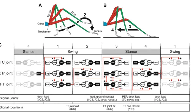
In some animals, this centrally generated rhythmic activity closely resembles activity during locomotion (e.g., crayfish fictive swimmeret movement: (Mulloney and Smarandache-Wellmann, 2012); lamprey fictive swimming: (Grillner, 2003); cockroach fictive walking: (Fuchs et al., 2011); hawkmoth fictive walking: (Johnston and Levine, 2002)). However, when flexibility of the locomotor system is demanded, it becomes obvious that the centrally generated output must be shaped by additional coordinating and adapting mechanisms. This holds especially true for the generation of six-legged walking in insects (Bässler and Büschges, 1998; Bidaye et al., 2018). From the perspective of a single leg, walking consists of two phases, stance and swing (Fig. 1.1A and B). During stance phase, the tarsus (foot, distal end of the leg) is placed on the ground, and force is exerted to propel the body in the walking direction. During swing phase, the leg is moved through the air to the initial position of the subsequent stance phase (Graham, 1972). Both phases, and the transitions between them, require appropriate temporal sequential activation of MNs that control contractions of muscles resulting in distinct movements around leg joints (Fig. 1.1C; (Rosenbaum et al., 2010). Additionally, the movements of adjacent legs need to be coordinated well for effective locomotion. When insects change gaits, walking speed, and direction, or when they make turns, the temporal sequential activation of MNs has to be adjusted in a task-specific manner (Borgmann and Büschges, 2015; Grabowska et al., 2012; Gruhn et al., 2016; Rosenbaum et al., 2010; Rosenbaum et al., 2015; Wosnitza et al., 2013). The question remains incompletely answered, though, of what neural mechanisms contribute to coordination and adaptation of CPG-derived output
Introduction
4
for walking. In stick insects, for instance, temporal coordination of the movement of single legs is only weakly organized by central drive. In pilocarpine-activated isolated ventral nerve cord preparations, so-called spontaneous recurrent patterns that resemble the motor output during step phase transitions occur only rarely (Büschges et al., 1995).
Furthermore, central influences coordinating CPGs supplying adjacent legs are only weak in isolated pharmacologically activated stick insect preparations and do not account for the generation of any of the motor patterns observed during walking (Mantziaris et al., 2017). In contrast, there appear to be rather strong direct connections between CPGs in animals where pharmacologically induced motor output closely resembles the motor output during walking (e.g., cockroach: (Fuchs et al., 2011); hawkmoth: (Johnston and Levine, 2002)).
Studies in insects highlight the importance of the integration of sensory signals with central motor commands for shaping the timing and magnitude of motor output (review:
(Bässler and Büschges, 1998; Bidaye et al., 2018; Büschges, 2005; Tuthill and Wilson, 2016). Insect legs comprise a large number of extremely diverse and specialized sensory organs that update the motor system about intrinsic and external mechanical changes (Tuthill and Wilson, 2016). Much of the processing of information from leg sensory organs is already carried out in the PNS, or at least directly between afferents of peripheral sensory organs. There is, for example, presynaptic inhibition in sensory afferents of a stick insect proprioceptive sense organ (Sauer et al., 1997). In general, sensory afferents only rarely exhibit direct synaptic connections to MNs (Burrows, 1987;
Parker and Newland, 1995; Pearson et al., 1976). Moreover, in insects, computation of distinct mechanoreceptive signals in the first layers of the central nervous system (CNS) is performed in parallel and locally in nonspiking interneurons of the thoracic ganglia (Bässler and Büschges, 1998; Burrows, 1987; Burrows, 1996; Burrows and Pflüger, 1986;
Sauer et al., 1996; Tuthill and Wilson, 2016; Watkins et al., 1985). This allows for sensory information to be processed at the necessary speed and markedly reduces the number of high-level CNS neurons required. Furthermore, intercalated nonspiking interneurons that receive excitatory sensory input are themselves either excitatory or inhibitory. Thus, the actual motor output that controls leg movement is markedly shaped by weighting of these low-level antagonistic pathways of sensory information processing (Bässler and Büschges, 1998; Tuthill and Wilson, 2016). Modification of these local
Introduction
5
control modules not only affects the magnitude, but also the sign of motor output in a state-dependent manner. In inactive stick insects, for example, tibial flexion signals, mediated by the femoral chordotonal organ (fCO) lead to an increase in extensor MN activity counteracting the imposed movement. This is and thought to help the animals maintain their posture while standing. This postural reflex is called the resistance reflex.
In active animals, this reflex is reversed. Flexion signals now induce an inhibition in extensor MN activity, whereas extension signals induce further extension, which likely facilitates the continuation of movement (Bässler, 1976; 1993; Bässler and Büschges, 1998; Hellekes et al., 2012).
Sensory signals have been shown to be relevant for the patterning of MN activity across joints of a single leg (review: (Bidaye et al., 2018; Tuthill and Wilson, 2016)). In the stick insect, for instance, position and load information was shown to facilitate the generation of motor patterns for the contraction of muscles that underlie step phase transitions (Akay et al., 2004; Zill et al., 2017). Load signals at the coxa-trochanter joint have an influence on the magnitude of motor activity at the femur-tibia (FT) joint (Akay et al., 2001), and the processing of fCO mediated sensory signals of position of the femur in relation to tibia, and movement around the FT joint (Akay and Büschges, 2006). Furthermore, these inter-joint reflexes can couple the activity of distributed CPGs (Akay et al., 2001; Akay et al., 2004; Bässler, 1986; Bucher et al., 2003; Büschges et al., 2008; Hess and Büschges, 1999). In addition, there is a role for sensory signals in the intersegmental modulation of motor activity. Walking legs have been shown to increase and pattern the motor output to neighboring legs (Borgmann et al., 2007; Ludwar et al., 2005). These modulations have been shown to be, in part, based on sensory signals that result from front leg stepping and can entrain the activity of mesothoracic thorax-coxa joint CPGs (Borgmann et al., 2009).
When a mesothoracic leg was left attached to the prepared animal, activity of metathoracic CPGs was also influenced (Borgmann et al., 2009). The authors concluded that intrasegmental sensory information appears to override intersegmental signaling (Borgmann et al., 2009; Borgmann et al., 2011).
Introduction
6
Figure 1.1 Control of single-leg stepping movements in stick insect. (A) Leg movement during swing phase. (B) Leg movement during stance phase. (C) Stepping consists of the cyclic repetition of swing and stance phases. Swing and stance phase can each be subdivided into two distinct phases. There is one CPG that drives MN activity which, in turn, activates muscles for each of the thorax-coxa (TC), coxa-trochanter (CTr), and femur-tibia (FT) joints. Sensory signals (load and position) have a pivotal role in coupling the activity of the distributed CPGs to enable transitions between the different phases of a step cycle. This figure was taken from Bidaye et al. (2018). For further detailed information see Büschges, 2005.
Based on these findings, we can assume that the basic neural circuitry for control of coordinated walking movements in insects is situated in the ventral nerve cord. At the thoracic level, motor control for coordinated legged locomotion is not achieved by a rigid hierarchical command structure but is due to a dynamic interplay between CPG-derived motor activity and sensory signals. However, descending signals from higher centers like the brain or gnathal ganglion (GNG) are crucial for the initiation, cessation, and modulation of effective thoracic motor activity (Bidaye et al., 2018). Furthermore, descending neurons are responsible for the task-specific selection of downstream motor programs (e.g., (Bidaye et al., 2014; Philipsborn et al., 2011; Reyn et al., 2014)). In insects, neurons in the GNG appear to facilitate the maintenance of ongoing behavior (Gal and Libersat, 2006; Kien and Altman, 1992; Ridgel and Ritzmann, 2005; Roeder, 1937). In stick insects, tonic depolarization of MNs contributes to their activity pattern during walking (Gabriel and Büschges, 2007; Ludwar et al., 2005; Westmark et al., 2009). It has been shown that this tonic depolarization does not derive from signals of leg sensory organs but appears to be the result of descending drive (Ludwar et al., 2005;
Introduction
7
Westmark et al., 2009). With respect to the influence of the GNG on walking, this might be where the neural source for the tonic depolarization in MN membrane potential is situated.
Inputs from higher brain centers can influence sensorimotor loops in thoracic ganglia.
Martin et al. (2015) showed that stimulation of central complex neurons induces the reversal of a postural reflex in the cockroach (Martin et al., 2015). In general, the task- dependent modulation of reflex responses, for example, during curve walking appears to depend on descending signals (Gruhn et al., 2016; Mu and Ritzmann, 2008a; 2008b). In addition, descending commands are crucial for the initiation and selection of motor activity in thoracic ganglia (e.g., fruit fly backward walking: (Bidaye et al., 2014; Sen et al., 2017); initiation of fruit fly voluntary walking: (Yellman et al., 1997); fruit fly courtship behavior: (Philipsborn et al., 2011); fruit fly escape behavior (Reyn et al., 2014;
Reyn et al., 2017)). Overall, in animals, the selection of different motor programs does not appear to be accomplished by changes in high-level behavior, but rather by modification of low-level thoracic motor control networks by top-down commands (Bidaye et al., 2018). Basically, all of the neurons in thoracic motor control networks and the synaptic connections between them are subject to neuromodulation (Katz, 1999;
Marder, 2012; Miles and Sillar, 2011). Thus, neuromodulatory neurons are an extremely important source of flexibility of the underlying neuronal networks for locomotion and the motor output they generate.
Introduction
8
Neuromodulation largely extends the working range of motor control networks A wide variety of chemical substances have been shown to exert effects on motor activity in both vertebrates and invertebrates. The major neuromodulators can be grouped into three categories. There are biogenic amines like serotonin, dopamine, and noradrenaline (octopamine in invertebrates); amino acids like gamma-aminobutyric acid (GABA) and glutamate; and peptidergic neuromodulators like substance P and proctolin (Katz, 1999;
Miles and Sillar, 2011). With respect to the compartments these substances are released to, and the receptors they activate many of them can act as neuromodulators, neurohormones, and even neurotransmitters. Typically, neuromodulators exert their effects by acting as agonists of metabotropic receptors (Katz, 1999). With respect to the intracellular second-messenger cascades that modulators activate, the effects usually have a slow time course in contrast to the effects of synaptic neurotransmission via ionotropic receptors (Hultborn and Kiehn, 1992; Katz, 1995; Levitan, 1988; Lopez and Brown, 1992). Neuromodulators do not fixedly transmit excitation or inhibition to their target structures, but rather alter cellular or synaptic properties, which influences the impact of subsequent neurotransmission (Katz, 1995). Despite the aforementioned properties of neuromodulatory action, finding a rigid comprehensive definition for the term
“neuromodulation” is difficult. This becomes apparent when a statement of Katz (1999) is considered, who claimed that “any communication between neurons, caused by release of a chemical, that is either not fast, or point-to-point, or not simply excitation or inhibition will be classified neuromodulatory.”
Neuromodulators are released by neurons, and this can be distinguished between intrinsic and extrinsic at the level of integration of neuromodulatory neurons in neural control circuits (Katz, 1995). Intrinsic neuromodulation implies that the neuromodulatory neuron is an integral part of the neural circuit it modulates. One form of intrinsic neuromodulation is the simultaneous co-transmission of neuromodulatory substances and conventional fast transmitters at a synapse (Katz, 1999; Miles and Sillar, 2011). As an example for intrinsic neuromodulation, serotonergic neurons have been shown to be constituents of a CPG network that controls escape swimming in a mollusk, and these neurons modulate activity of other constituents of the same control network (McClellan et al., 1994). With regard to intrinsic neuromodulation, the release of a neuromodulator is
Introduction
9
dependent on the activity of the neural circuit they are part of. This is in contrast to extrinsic neuromodulation, where the respective neuromodulatory neuron is located outside the neural circuit it affects. Thus, the release of neuromodulator is largely independent of the activity of the affected neural circuit. The best studied neural circuit influenced by extrinsic neuromodulation is the pyloric CPG of the spiny lobster stomatogastric ganglion (STG) (Marder, 2012). In this system, extrinsic modulatory input originates in central ganglia or sensory organs (Katz and Harris-Warrick, 1990a; 1990b).
In general, extrinsic modulatory neurons are not part of the circuits they modulate but are often activated in parallel to the neural circuits they affect (Katz, 1995). Octopaminergic efferent dorsal unpaired median (DUM) neurons in locust thoracic ganglia, for example, affect neuromuscular transmission to leg muscles and are activated in parallel to MNs responsible for walking (Baudoux et al., 1998; Duch et al., 1999). Extrinsic neuromodulation often comprises modulation of activity of more than one neural circuit (Katz, 1999). With respect to the sign and magnitude of neuromodulatory effects on a circuit level, not only the identity of the neuroactive substance but the timing and amount of its release, the type of receptor it activates, and the output properties of its target neurons are crucial (Katz, 1999; Marder, 2012; Miles and Sillar, 2011). The pyloric CPG of the spiny lobster, for instance, is differentially modulated by three neuromodulatory neurons that all contain proctolin (Nusbaum et al., 2001). Serotonin alike has been shown to exert different effects on pyloric CPG neurons as a result of activation of distinct serotonin receptors (Zhang and Harris-Warrick, 1994). Miles and Sillar (2011) note that all individual constituents of neural control networks for locomotion perpetually face a varying neuromodulatory environment, which is the pivotal aspect of flexibility.
Octopamine is a prominent neuromodulator in insect locomotor control
In the current thesis, I focused on the neuromodulatory substance octopamine (OA) and its effects on the generation of legged locomotion. This biogenic amine is considered to be the invertebrate homologue to the vertebrate noradrenaline, both with regard to its structure and physiological role (Roeder, 2005). Acting as a neurohormone, OA released to the haemolymph affects cell metabolism by mobilizing lipids and increasing glycolytic activity in muscles (Goosey and Candy, 1980; Mentel et al., 2003; Pflüger and Duch, 2011). Acting as a neuromodulator, OA has a prominent role in influencing multiple
Introduction
10
physiological events both in the PNS and the CNS (Bräunig and Pflüger, 2001; Roeder, 2005; Verlinden et al., 2010). With respect to locomotion, the peripheral effects of OA are modulation of neuromuscular transmission and changes in the contraction kinetics of muscles (e.g., (Evans and O'Shea, 1977; Evans and Siegler, 1982)). Furthermore, OA affects the activity of sensory organs (Farooqui, 2007). In stick insects, for example, OA selectively increases the excitability of position-sensitive fCO neurons, but does not affect velocity-sensitive sensory neurons (Ramirez et al., 1993). Similarly, OA sensitizes sensory neurons of the locust forewing stretch receptor (Ramirez and Orchard, 1990). In addition to its effects in the PNS, OA has been shown to affect a wide array of complex behaviors. This has been demonstrated, for example, for fruit fly sleep/wake behavior (Crocker et al., 2010), aggression in crickets (Rillich et al., 2011; Rillich and Stevenson, 2011), division of labor in honeybee and ant colonies (Schulz and Robinson, 2001; Seid and Traniello, 2005), and learning and memory in honeybees (Hammer, 1993; Mercer et al., 1983; Schwaerzel et al., 2003).
With respect to locomotor activity generated in thoracic ganglia, OA has been shown to affect the activity of MNs, coordinating neurons, and CPG neurons (review: (Bräunig and Pflüger, 2001; Marder, 2012; Roeder, 2005; Verlinden et al., 2010)). OA was, for example, shown to induce bursting and plateau potentials in interneurons of the locust flight pattern generator (Ramirez and Pearson, 1991a; 1991b). Additionally, it mediates sensitization and dishabituation in a locust postural reflex (Sombati and Hoyle, 1984a), and it can evoke a fictive flight motor pattern in locusts (Claassen and Kammer, 1986;
Rillich et al., 2013). Moreover, OA decreases the inhibitory postsynaptic potentials evoked by stretch receptor activity in locust flight MNs (Leitch et al., 2003). In inactive stick insects, OA selectively alters the response properties of a leg proprioceptive feedback system toward those that characterize the active state of animals (Büschges et al., 1993). Furthermore, OA modulates the tonic depolarization ubiquitous in mesothoracic leg MNs during walking in stick insects (Westmark et al., 2009). In summary, OA modulates muscle metabolism, neuromuscular transmission, sensory sensitivity, MN excitability, and activity in CPG circuits that control locomotion.
The neuromodulatory effects of OA are mediated by octopamine receptors (OARs) that are expressed in pharmacologically distinct forms and localized to the membrane of target
Introduction
11
neurons. OARs are coupled to G proteins and activate various second-messenger cascades that can include 3’,5’-cyclic adenosine monophosphate (cAMP), inositol 1,4,5- triphosphate (IP3), and diacylglycerol (DAG). These second messengers not only modulate levels of intracellular Ca2+ but also regulate phosphorylation of various signaling proteins involved in differential fine-tuning of neuronal output. The second messenger cAMP activates protein kinase A, IP3 mobilizes calcium from intracellular stores, and DAG activates protein kinase C. Through the various effects of their activation, OARs can markedly influence the electrical properties of target neurons. On a cellular level, OAR activation can modulate persistent inward currents, which can depolarize the neuron to facilitate AP generation or induce plateau potentials. OA can broaden action potentials (APs) and modulate calcium-dependent potassium currents that shape AP afterhyperpolarizations (Blenau and Baumann, 2001; Evans, 1993; Evans and Robb, 1993; Farooqui, 2007; Grohmann et al., 2003; Roeder et al., 1995; Roeder, 2005).
In drosophila sleep/wake behavior, for example, OA signals via cAMP and protein kinase A (PKA) to selectively decrease the potassium current from calcium-dependent Slowpoke channels in target neurons, which leads to increased excitability (Crocker et al., 2010).
Classification and distribution of octopaminergic neurons
OA and octopaminergicneural structures (dendritic processes) are remarkably abundant in the insect CNS and PNS (Axelrod and Saavedra, 1977; Evans, 1978). In contrast, the number of octopaminergic neurons in the nervous system is surprisingly small. Roeder (2005) suggests that there are only about 100 such neurons in large insects like locusts (Roeder, 2005). Busch et al. (2009) note that the number of octopaminergic neurons in the relatively small fruit fly is also about 100. The major cellular source for OA acting in the PNS of thoracic and abdominal segments is the unpaired median (UM) neurons. These neurons appear as individuals (unpaired) and have symmetrical bilaterally projecting axons with extensive branches. Their large somata are located either at the dorsal (DUM) or ventral (VUM) midline of the respective ganglia (Bräunig and Pflüger, 2001). Most of the thoracic UM neurons were only shown to possess peripheral release sites for OA (Kononenko and Pflüger, 2007; Stocker et al., 2018; Watson, 1984). They directly affect contraction properties of muscles, neuromuscular transmission, muscle metabolism, or activity of sense organs in the periphery (Bräunig and Pflüger, 2001; Roeder, 2005).
Introduction
12
Efferent DUM neurons are activated in parallel to locomotion and are responsive to mechanosensory signals in a multimodal fashion (Duch et al., 1999; Field et al., 2008;
Gras et al., 1990; Mentel et al., 2008). In addition to UM neurons, there are relatively few paired octopaminergic neurons with small somata in the thoracic and abdominal ganglia (Stevenson and Spörhase-Eichmann, 1995); their physiological functions are unknown.
Similarly, the neural source of OA acting as a neuromodulator in the CNS of thoracic segments has not yet been identified.
Further octopaminergic UM neurons are situated in the GNG, but not in the brain. The majority of GNG UM neurons project intersegmentally (Bräunig, 1991; Bräunig and Burrows, 2004; Bräunig and Pflüger, 2001; Busch et al., 2009). In the posterior locust GNG, for instance, there are six DUM neurons that send bilaterally descending axons to the thoracic ganglia (abbreviated desDUM neurons; Fig.1.2 (Bräunig and Burrows, 2004)). Bräunig and Burrows (2004) showed that these neurons arborize in regions of neuropil where information arising from leg sensory organs is processed. The population of six locust desDUM neurons comprises three morphologically heterogeneous subpopulations (Bräunig and Burrows, 2004). The presence of homologues neurons was demonstrated in several other insects like the hawk moth (Cholewa and Pfluger, 2009;
Dacks et al., 2005); honeybee (Schröter et al., 2007; Kreissl et al., 1994; Sinakevitch et al., 2005); fruit fly (Busch et al., 2009; Selcho et al., 2012); and cockroach (Sinakevitch et al., 2005; Stevenson and Spörhase-Eichmann, 1995). With regard to the number of desUM neurons and their basic morphological properties, there are marked similarities between different insect species. Despite an investigation of input to locust desDUM neurons from electrical stimulation of leg nerves (Bräunig et al., 2004), as well as coupling of larval hawk moth desVUM neurons to a centrally generated motor rhythm (Cholewa and Pfluger, 2009), the physiology of desUM neurons has not been addressed to date. This holds especially for the output physiology of the neurons.
Introduction
13
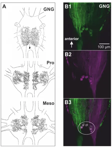
Retrograde labeling of the posterior connectives in stick insects revealed that there are putative desDUM neurons with large somata and bilaterally descending axons in the posterior part of the GNG (Fig. 1.2B; (Heß, 2008). On the assumption of morphological homology to locust desDUM neurons (Fig. 1.2A), these neurons are promising candidates for the octopaminergic modulation of thoracic local control networks for legged locomotion.
Figure 1.2 Morphology and somata location of desDUM neurons. (A) Morphology of one of six locust desDUM neurons. The large somata of desDUM neurons are located in the posterior GNG. A primary neurite is projecting anteriorly and bifurcates into two bilaterally symmetrical axons. The axons extensively branch in the posterior half of the GNG, but do not send projections to the periphery. Two bilaterally symmetrical axons project to thoracic ganglia, where they are extensively branching. (B) In the stick insect GNG, six large somata of neurons with bilateral symmetrically descending axons were labeled by retrograde fills of the posterior connectives. The figure was modified from (Heß, 2008). (B1) Labeling of the left posterior connective. (B2) Labeling of the right posterior connective. (B3) Co-localization of fluorescent dyes in six large somata.
Introduction
14 This thesis
MNs are at the final level of neural processing for the generation of walking. Their activity reflects their intrinsic properties and, moreover, the net output of the upstream neural circuitry and effects of neuromodulatory neurons. In stick insects, the activity of MNs is patterned by tonic depolarizing drive as well as rhythmic excitatory and inhibitory inputs (Büschges, 1998; Ludwar et al., 2005; Rosenbaum et al., 2015; Schmidt et al., 2001; Westmark et al., 2009). To fully understand the mechanisms of neural circuit function and neuromodulation for walking, it is crucial to decipher the neural basis for the activity pattern of MNs. In particular, the underlying neural sources of the depolarizing drive to MNs during walking are unknown. Westmark et al. (2009) targeted the pharmacological background of the tonic depolarization and highlight a modulatory role for OA. As outlined in the previous sections, OA is a major neuromodulator in locomotor behavior, but the neural source of OA acting in the CNS of thoracic segments of insects has remained elusive. Most of the findings on OA action in the insect thoracic nerve cord are based on systemic application of OA or blocking OAR action. The question arises, then, of to what extent these rather unspecific alterations in behavior or physiological processes reflect the physiological action of OA in the intact animal. Systemic application of OA does not consider the timing of natural modulator release in a behavioral context, the physiological amount of the modulator, or the specific target sites the modulator is released to. The need for physiological modulator release, for example, by stimulation of identified neuron becomes apparent in a study by Flamm and Harris-Warrick (1986). The sign of octopaminergic effects on constituents of the pyloric CPG in the lobster was dependent on the applied concentration of OA (Flamm and Harris-Warrick, 1986). In a study by Westmark et al. (2009), the sign of OA action on tonic depolarization in MN membrane potential was dependent on the circuit level of OA application. Bath application of OA to the entire mesothoracic ganglion induced an increase in the tonic depolarization in MNs, whereas application of OA to isolated MN somata decreased an inward current that contributes to the tonic depolarization in MN membrane potential (Westmark et al., 2009). The current thesis is based on the hypothesis that the activity of local control networks for walking in thoracic ganglia of insects is modulated by octopaminergic desDUM neurons.
Introduction
15
The first part of this thesis is dedicated to the identification of desDUM neurons in stick insects. I used intracellular recordings of desDUM neurons to characterize their electrical properties, and investigated these neurons’ morphology using intracellular labeling.
Furthermore, I employed a newly developed method of mass spectrometry (matrix assisted laser desorption/ionization-time of flight mass spectrometry; MALDI-TOF MS) to analyze the OA content of electrophysiologically identified stick insect desDUM neurons.
In the second part of this thesis, I asked if the activity of desDUM neurons is coupled to walking behavior. Activation of these neurons during walking is a precondition for their neuromodulatory role on locomotor circuit action. In addition, knowledge of the magnitude and timing of desDUM neuron activation is a prerequisite for physiologically relevant stimulation of the neurons to test their output properties. By modifying established semi-intact preparations in combination with intracellular recording, I studied the activity of desDUM neurons during six-legged walking and single-leg stepping.
Furthermore, I studied the neural background of walking-related activity. Using pharmacological activation of leg joint CPGs, I tested the effects of central synaptic drive on desDUM neurons. Finally, with specific stimulation of leg sensory organs, I tested the role of sensory signals in activating desDUM neurons.
In the third part of this thesis, I analyzed the modulatory role of desDUM neurons on activity of mesothoracic MNs. I activated single desDUM neurons by current injection and studied their effects on fCO-evoked reflexes. I used this approach, because stimulation of the fCO can be easily done in a controlled fashion. Furthermore stimulation of the fCO induces resistance reflexes in ExtTi MNs (Bässler, 1976; 1983; Bässler and Büschges, 1998). that are very stereotypic after a short time for habituation (Stein and Sauer, 1998). Thus, ExtTi MN activity induced by stimulation of the fCO can function as a stable control in comparison to putative alteration upon desDUM neuron activation. The reflex response to fCO stimulation is dependent on the state of the animal (active vs.
inactive;(Bässler, 1976)). Thus, it may be possible to detect effects of desDUM neuron stimulation on the state-dependent processing of reflex responses. Büschges et al. (1993) demonstrated that bath application of OA specifically suppressed neural pathways involved in the generation of resistance reflexes. The neuronal source of OA is not
Introduction
16
known. Therefore, my experiments could possibly target the question of whether OA application might mimic the effects of desDUM neuron activity. To test whether other modes of sensorimotor interaction are modulated by des DUM neurons, I studied their effects on ProCx and RetCx MN activity evoked by stimulation of campaniform sensilla (CS). In addition, I asked whether desDUM neurons have an influence on centrally generated MN activity. Collectively, these methods allow for a comprehensive characterization of desDUM neurons and their complex roles in locomotor networks.
17
2 Materials and Methods
All experiments were carried out on adult female stick insects (Carausius morosus) from a colony maintained at the University of Cologne. Experiments were conducted under dimmed light conditions at room temperature (20-24°C).
2.1 Basic semi-intact stick insect preparation
All legs except one middle leg were cut at the level of the mid coxa. Animals were affixed to a platform dorsal side up using dental cement (Protemp II, 3M ESPE Dental AG, Seefeld, Germany). The head capsule and thorax were opened dorsally by cutting a rectangular window into the cuticle. The gut, fat tissue, and small tracheae were removed to gain access to the gnathal ganglion (GNG), the mesothoracic ganglion, and mesothoracic lateral nerves. The body cavity of the animal was rinsed and filled with saline according to Weidler and Diecke (in mmol: NaCl 180; KCl 18; CaCl2 8; MgCl2 25;
HEPES buffer 10; pH 7.2; (Weidler and Diecke, 1969)).
2.2 Intracellular recordings
For intracellular recordings from desDUM neurons, the GNG was lifted onto a wax coated steel platform while still connected to the animal. Loose tissue surrounding the ganglion was removed with forceps. To ease electrode penetration, small crystals of a proteolytic enzyme (Pronase E; Merck, Darmstadt, Germany) were placed on the ganglionic sheath for about 10-20 s. The enzyme was thoroughly washed off with saline.
Intracellular recordings from desDUM neurons were made from their somata located in the posterior part of the GNG at the origin of the posterior connectives. Borosilicate glass micropipettes (GB100-TF8P; Science Products, Hofheim, Germany) with resistances of 15-35 MΩ were manufactured on a filament puller (P-97; Sutter Instruments, Novato, CA, USA). The electrodes were filled with a mixture of 0.1 M KCl and 3 M CH3CO2K.
Cells were identified by their large-amplitude (>70 mV), overshooting soma action
Materials and Methods
18
potentials (APs), pronounced underhoot (>7 mV) and the generation of APs in response to gentle touch of the abdomen with a paint brush. Membrane potentials were amplified with an intracellular amplifier (SEC-10L; NPI Electronics, Tamm, Germany) in bridge mode. Recordings were stored on a PC using Spike 2 software (Version 7.09, Cambridge Electronic Design, Ltd., Cambridge, UK).
2.3 Intracellular neurobiotin staining of desDUM neurons
In 30 experiments, desDUM neurons were successfully stained intracellularly. Electrodes were filled with 5% Neurobiotin™ (Vector Laboratories Inc., Burlingame, CA, USA) in 0.1 M KCl/3 M CH3CO2K. The tracer was injected into desDUM somata with depolarizing current pulses of 1-4 nA with 400-ms pulse duration at 1 Hz for 5-15 min.
After tracer injection and 45-60 min incubation for tracer diffusion, the ganglia were removed from the animal and fixed in 4% paraformaldehyde in 0.1 M PBS for 2-16 h at 4
°C. The ganglia were then washed (3 x 15 min in 0.1 M PBS) and incubated in Cy3- conjugated streptavidin (1:500 in 0.1 M PBS; Sigma-Aldrich, St. Louis, MO, USA) with 0.5% Triton X-100 and 2-4% normal goat serum (NGS; Vector Laboratories Inc) for 5-16 h at 4 °C on a shaker. The ganglia were washed again (3 x 15 min with 0.1 M PBS) and dehydrated in an ascending ethanol series (50%, 70%, 90%, 2 x 100% EtOH; 10 min each). To clear the tissue, the ganglia were transferred to a microscope slide and embedded in methyl salycilate. Specimens were examined using a laser scanning confocal microscope (Zeiss 510 META; Carl Zeiss AG).
2.4 Extracellular recording from leg-nerves
Extracellular recordings from leg nerves were done using modified monopolar hook electrodes (Schmitz et al., 1988). Recordings were amplified (500x-2000x, depending on the recording quality) and filtered (250Hz- 5kHz). The recordings were digitized by a MICRO 1401 A/D converter (sampling rate: 12.5 kHz). Leg nerves were labeled according to literature (Bässler, 1983; Marquardt, 1940).
Materials and Methods
19
2.5 Analysis of octopamine/tyramine content with MALDI- TOF MS
1Individual somata were marked by tetramethylrhodamine dextran (5%, 3000 Dalton (MW), Invitrogen, Eugene, Oregon, USA) injection after electrophysiological recording.
The somata were dissected and treated after Diesner and Neupert (2018 in prep.). For this, isolated GNGs were transferred to a new dissection dish filled with saline containing 33%
glycerol (v/v). Marked cell bodies were visualized and dissected under a fluorescence stereomicroscope (Lumar V12; Carl Zeiss AG) using fine forceps (Dumont #5; Fine Science Tools, Heidelberg, Germany) and, immediately after isolation, transferred onto a MALDI sample plate using a glass capillary. Any excess dissection buffer was removed with the same glass capillary. Subsequently, remaining glycerol was washed off with 50%
MeOH/H2O using a fresh glass capillary. Furthermore, 18.4 nl of isotopically marked internal standard, containing 100 nM (±)-p-octopamine-α,β,β-d3 hydrochloride (OA[d3]) and 100 nM 2-(4-Hydroxyphenyl) ethyl-1,1,2,2-d4-amine hydrochlorid (TA[d4]) dissolved in 50% MeOH/H2O (CDN Isotopes, Pointe-Claire, Canada) was applied to the samples. Each preparation was air-dried in darkness at room temperature. After that, 9.2 nl of 4.27 mM 4-hydroxy-3-methoxycinnamaldehyde (CA; in 50% MeOH) was applied and samples were air-dried again. In the last step, 18.4 nl of saturated α-cyano-4- hydroxycinnamic acid diluted 1:6 in 80% MeOH/H2O (Sigma-Aldrich) was applied as matrix solution to the sample. For a more consistent distribution of matrix crystals, the sample spot was directly dried under an air stream at room temperature. All solutions were pipetted using a micro injector (Nanoliter 2000; World Precision Instruments, Sarasota, FL, USA) under a stereomicroscope (STEMI 2000; Carl Zeiss AG) equipped with a KL 1500 LED light source (Schott, Mainz, Germany).
Samples were analyzed with an ultrafleXtreme MALDI-TOF/TOF mass spectrometer (Bruker Daltonik Gmbh, Bremen, Germany) under manual control in reflectron positive
1Experiments of this section were performed in collaboration with Max Diesner and Dr. Susanne Neupert (University of Cologne, Department of Biology, Institute for Zoology). Contributions: M.D. and S.N. designed research, M.D. and T.S. performed experiments, M.D. analyzed the data, M.D. and T.S. created the figures and wrote the text.
Materials and Methods
20
ion mode in a mass range of m/z 0-400. All mass spectra were acquired with a fixed laser intensity. The instrument was calibrated using prominent matrix signals ([M+H-CO2]+: m/z 146.0606, [M+H-H2O]+: m/z 172.0399, [M+Na]+: m/z 212.0324, [M+K]+: m/z 228.0063, [2M+H-CO2]+: m/z 335.1032, [2M+H]+: m/z 379.093) with a mass accuracy below 20 ppm. Two thousand laser shots per sample spot were accumulated for one MS spectrum. Fragmentation data to verify OA and OA(d3) were acquired in “LIFT” mode.
MS2 experiments to identify TA and TA(d4) were recorded in collision-induced dissociation (CID)-on mode, with argon as the collision gas. All masses given herein and masses labelling ion signals are monoisotopic masses.
2.6 Single-legged stepping and six-legged walking preparations and passive leg movement
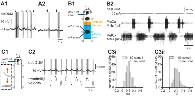
To monitor the activity of desDUM neurons during single-legged stepping, a low-friction treadmill (Gabriel et al., 2003) was positioned under the middle leg parallel to the longitudinal axis of the animal. Stance phase of the middle leg led to acceleration of the treadmill. Upward deflection of the tachometer trace represented the begin of stance phase during forward middle-leg stepping. Downward deflection indicated begin of a stance phase during backward middle-leg stepping. The maximum deflection in the tachometer trace refers to the end of a stance phase. When stepping sequences did not occur spontaneously, they were induced by brief tactile stimulation of the abdomen with a paint brush to activate the animal. Steps that occurred during, and just after tactile stimulation were not considered in the evaluation of their effect on desDUM neurons.
Passive movements of single middle legs that were standing on the treadmill were induced by movement of the treadmill via the motor or by hand. The initial position of the femur was anteriorly directed at about 70° inner angle in respect to the longitudinal axis of the animal. From this position, the leg was moved backwards by about 40°. Passive backward movement was followed by a pause of 1-2 s and subsequent passive forward movement of the leg to the initial position. During movements, the leg remained on the treadmill. To study the effects of six-legged walking on activity of desDUM neurons , animals were glued dorsal side up on a piece of balsa wood. All legs were left intact, and leg movement was not restricted. One treadmill on each side of the animal was placed
Materials and Methods
21
parallel to the animals’ longitudinal axis. Coordination of the legs during six-legged walking on two treadmills was mainly that of a tetrapod coordination pattern.
2.7 Pilocarpine-evoked rhythmic motor neuron activity
Influence of CPGs on activity of desDUM neurons
The following experiments were performed according to Borgmann et al. (2007). The thoracic cuticle was opened dorsally to gain access to the prothoracic and mesothoracic leg nerves, and the leg nerves were cut. A 3-mm segment of cuticle between the prothoracic and mesothoracic ganglia was removed. Tracheae and connectives were left intact. The resulting gap was filled with vaseline to create two separated wells, and both wells were filled with saline. The connectives were cut posterior to the mesothoracic ganglion. Activity in the mesothoracic nl2 and nl5 leg nerves was recorded with hook electrodes. The nl2 nerve contains axons of protractor (ProCx) MNs, and nl5 contains axons of the antagonistic retractor (RetCx) MNs. As soon as a stable intracellular recording from a desDUM neuron was obtained, the posterior well was filled with the muscarinic acetylcholine (ACh)-receptor agonist pilocarpine (5 mmol in saline). About 2- 5 min after pilocarpine application, rhythmic oscillatory activity could be observed in the antagonistic MN pools. This activity was used as an indicator for ongoing activity in leg- joint CPGs (Büschges et al., 1995). Responses in desDUM neurons were recorded intracellularly.
Output effects of desDUM neurons on CPG activity
The same preparation was used to study both inputs to desDUM neurons as well as the neurons effect on CPG neurons. After at least 5 min of pilocarpine-induced MN activity as control action potential (AP) generation in a single desDUM neuron was induced by depolarizing current injection to somata for 60 s. After that again followed at least 5 min of control, before the same desDUM neuron was activated again. For evaluation of nl2 and nl5 burst properties I used custom written spike2 scripts. Thereby, APs in the respective MN pools were identified by signal falling through a threshold that was adjusted manually fitting the signal to noise ratio as well as signal amplification in any given experiment. APs were thereupon transformed to uniform events for further analysis.
Bursts were defined as temporal aggregation of APs of excitatory MNs comprising at
Materials and Methods
22
least five APs with a maximum interspike-interval of 0.5 s. Period was the time from onset of one burst to onset of the next burst of a MN pool. Only experiments in with stable rhythmicity (alternating activity of antagonistic MN pools, maximum burst duration of 30 s) were considered for evaluation.
2.8 Stimulation of the femoral chordotonal organ (fCO)
Effect of fCO stimulation on desDUM neurons
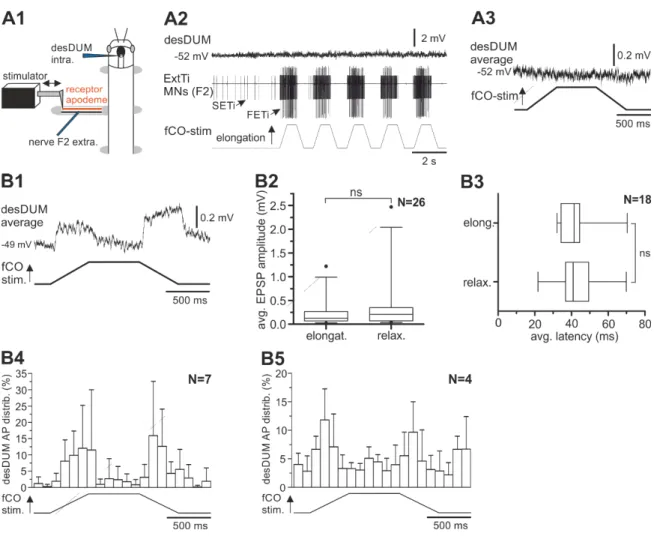
Stimulation of the femoral chordotonal organ (fCO) of the mesothoracic leg was performed according to Hellekes et al. (2012). The intact middle leg was amputated at the middle of the tibia. All other legs were removed at the level of mid-coxa. The remaining leg was fixed perpendicular to the longitudinal axis of the animal and glued to a platform resulting in an angle of about 120° between femur and tibia. A small window was cut in the dorsal cuticle of the femur to gain access to the nerve F2 containing extensor tibiae (ExtTi) MN axons, and the receptor apodeme of the fCO. A vaseline well around the leg was filled with saline (Weidler and Diecke, 1969), the apodeme of the fCO was cut, and the distal ending was attached to a clamp. Mechanical displacements of the apodeme parallel to the leg and towards or away from the body were produced by a piezoelectric actuator driven by a ramp generator (Electronics Workshop, Zoological Institute, University of Cologne). The fCO was activated with ramp-and-hold stimuli (2s duration) which produced displacements of the apodeme of 300 µm from the starting position.
These displacements corresponded to changes in FT joint angle (inner angle) from 120° to 60° (Weiland et al., 1986). ExtTi MN activity was monitored extracellularly from the leg nerve F2, which contains axons of the excitatory fast ExtTi MN (FETi), the slow ExtTi MN (SETi), and the inhibitory MN common inhibitor 1 (CI1) (Goldammer et al., 2012).
The single units of all three MNs could be readily distinguished in the extracellular recording trace by their characteristic AP amplitude. Successful stimulation of the fCO was monitored by the occurrence of resistance reflex responses characterized by activation of ExtTi MNs during elongation of the apodeme (Bässler, 1976; 1983).
Responses of desDUM neurons were recorded intracellularly. For evaluation of the effects of fCO stimulation by means of EPSP amplitude and latency of occurrence, averages of the time course of desDUM neuron membrane potential were created using spike2 software.
Materials and Methods
23
Output effects of desDUM neurons on fCO induced ExtTi MN activity
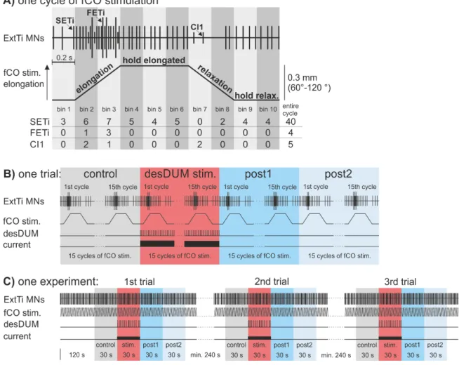
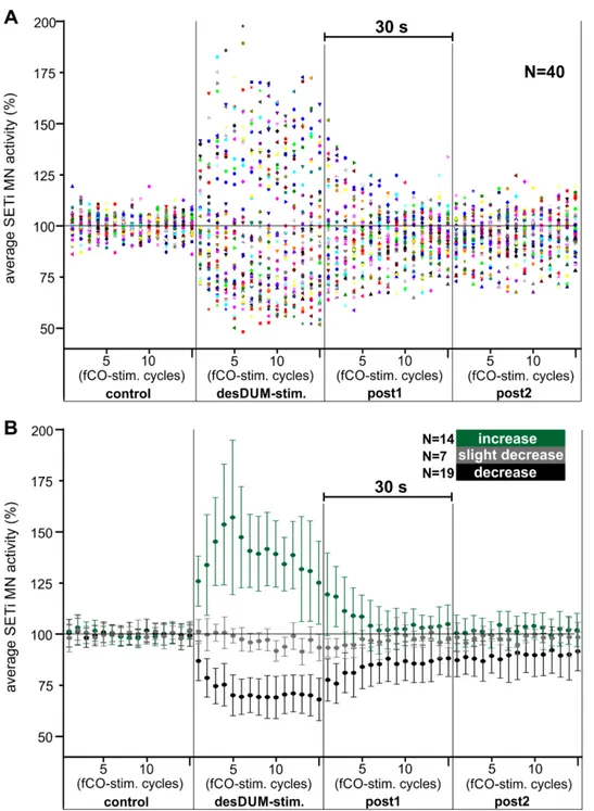
Reflexes in extensor MNs were induced by continuous fCO stimulation with afore mentioned properties. After at least 2 min of time for habituation of the reflex responses, a single desDUM neuron was activated by intracellular depolarizing current injection for 30 s (Fig. 3.1 B and C). Within 30 s there occur 15 cycles of fCO stimulation. In detail, desDUM neurons were stimulated with rectangular depolarizing current pulses with a frequency of 20 Hz and duty cycle of 20 ms. The amplitude of injected current was adjusted to yield AP generation in the stimulated desDUM neuron at a mean frequency of 25- 40 Hz. In some experiments the parameters of activation were altered. This is stated in the corresponding sections of the results part. In the experiments, 30 s of control prior to desDUM neuron activation (control), 30 s of desDUM neuron activation (desDUM stim.) and two consecutive 30 s sections (post1, post2) after desDUM neuron stimulation resembled one trial (Fig. 2.1B). Each experiment consisted of at least three trials with intervals of at least 4 min between single trials (Fig2.1C). For evaluation of the effects of desDUM neuron activation a custom written spike2 script was used. APs of FETi, SETi and CI1 MNs were identified by voltage signal falling through a threshold that was adjusted manually, fitting the signal to noise ratio, as well as signal amplification in a given experiment. APs of the respective MN units were then transformed to uniform events. As a basis for further evaluation, the APs of the three extensor MNs were each binned in 200-ms intervals, for every 2-s cycle of fCO stimulation (Fig. 2.1A). The effect of desDUM neurons on extensor MN activity 44 experiments was evaluated and illustrated in three different ways.
(I) Results section 3.3.1.1.3. In order to assess the effects on properties of fCO-induced reflex responses, histograms of activity of the single MN units over the 2-s time course of fCO stimulation were generated. APs of MNs were summed in 200 ms intervals for control section, desDUM neuron stimulation, post1, and post2 in the three trials of an experiment. To be able to compare in between animals and single experiments, the activity was normalized to the average activity of the 3rd control bin in each experiment (II) Results section 3.3.1.1.1. For investigation of the time course of effects of desDUM neuron stimulation, SETi APs were summed up over the 2-s of single cycles of fCO stimulation (Fig. 2.1A). Averages of the activity over trials were created for each of the 60 fCO stimulation cycles of one trial. To be able to compare in between experiments and
Materials and Methods
24
animals the activity during every fCO stimulation cycle was normalized to the average activity of the 15 control stim. cycles in each trial. Here the normalization was only possible for SETi MNs, because APs of FETi MNs and CI1 neurons were not generated during all control fCO stim. cycles.
(III) Results section 3.3.1.1.2. To better assess the net output of a given desDUM neuron subpopulation on all units of the ExtTi MN pool, the APs of the extensor MN units generated in the four 30-s sections of each trial were summed up and averaged over trials.
To be able to compare in between single experiments, the activity was normalized to control in each experiment.
Figure 2.1 Stimulation of the femoral chordotonal organ. (A) The fCO was stimulated by repetitive elongation and relaxation of the fCO receptor apodeme. The applied stimuli followed a 2 s ramp-and-hold function. Reflex responses in extensor MNs were monitored by recording from leg nerve F2. The single units of the extensor MN pool can be distinguished according to their distinct AP amplitudes in the extracellular recording (for details: see text). (B) and (C) I studied the effect of desDUM neuron activation on fCO stimulation-induced extensor MN activity. One experiment consisted of three trials (C). For evaluation of the effects, each trial was subdivided in 4 sections that each consisted of 15 fCO stim. cycles (for details: see text).


![Odor Modulation of Electrical and [Ca2+]i Activities in Neurons of the Olfactory Bulb](data:image/gif;base64,R0lGODlhAQABAIAAAP///wAAACH5BAEAAAAALAAAAAABAAEAAAICRAEAOw==)