Descending control of locomotion in the lamprey
Inaugural-Dissertation
zur
Erlangung des Doktorgrades
der Mathematisch-Naturwissenschaftlichen Fakultät der Universität zu Köln
vorgelegt von
Swantje Grätsch
aus Schleswig
Köln, Mai 2018
Berichterstatter/in:
Prof. Dr. Ansgar Büschges Prof. Dr. Silvia Daun Prof. Dr. Réjean Dubuc
Tag der mündlichen Prüfung :
16. Juli 2018
Contents
1. Zusammenfassung ...5
2. Summary...7
3. List of Abbreviations ...9
4. Introduction ...10
4.1. Locomotor control in vertebrates ...11
4.2. The control of locomotion in lampreys ...13
4.3. Aims and objectives ...17
5. Published studies ...20
5.1. Forebrain dopamine neurons project down to a brainstem region controlling locomotion ...20
5.2. Nigral glutamatergic neurons control the speed of locomotion ...29
5.3. A specific population of reticulospinal neurons controls the termination of locomotion...42
6. Study under review ...54
6.1. A brainstem neural substrate for stopping locomotion ...54
7. Discussion...85
7.1. Inputs to the MLR: Modulation of locomotion and control of locomotor speed...85
7.2. Outputs of the MLR: Termination of locomotion ...87
7.3. Significance for clinical research ...90
7.4. Concluding remarks and future perspectives ...91
8. Bibliography ...94
Acknowledgements ...106
Teilpublikationen ...108
Lebenslauf ...111
Erklärung ...113
1. Zusammenfassung
Lokomotion entsteht aus einem dynamischen Zusammenspiel dreierlei Komponenten: Den rhythmischen Bewegungsmustern, die von neuronalen Netzwerken im Rückenmark generiert werden, den absteigenden Einflüssen von supraspinalen Hirnstrukturen und den sensorischen Eingängen aus der Peripherie. Durch dieses Zusammenspiel können periodische Bewegungssequenzen generiert werden, die gestartet, aufrechterhalten und gestoppt werden müssen. Um die Bewegungskontrolle auf zellulärer Ebene untersuchen zu können, wurde in den letzten Jahrzehnten das Neunauge als Modellorganismus etabliert. In diesem basalen Wirbeltier wurden neuronale Netzwerke im Rückenmark identifiziert, zentrale Mustergeneratoren (ZMGs), die rhythmische Aktivität generieren und Muskelaktivität während der Fortbewegung steuern. Diese ZMGs werden von retikulospinalen (RS) Neuronen im Hirnstamm kontrolliert, welche wiederum von lokomotorischen Regionen, wie der mesenzephalen lokomotorischen Region (MLR), aktiviert werden. Die MLR kontrolliert die Initiierung und Aufrechterhaltung von Bewegung und spielt eine entscheidende Rolle bei der zielgerichteten Fortbewegung. Die Aktivität der MLR unterliegt dabei der Kontrolle von Hirnstrukturen im Vorderhirn, wie den Basalganglien. Diese Dissertation beschäftigt sich mit den absteigenden Eingängen, die die MLR aus dem Vorderhirn erreichen, sowie mit den absteigenden Projektionen der MLR zu unterschiedlichen RS Zellpopulationen im Hirnstamm. Hierfür wurden elektrophysiologische, neuroanatomische, bildgebende und Verhaltensversuche im Neunauge durchgeführt.
Klassischerweise werden Projektionen von dopaminergen Neuronen der substantia nigra pars compacta (SNc) so beschrieben, dass sie aufsteigend zum Striatum, der Eingangsstation der Basalganglien, führen. In der ersten Studie (Ryczko et al., 2013) konnten dopaminerge Neurone des posterior tumberculum (PT, homolog zur SNc in Säugetieren) identifiziert werden, die absteigend auf die MLR projizieren. Versuche in semi-intakten Präparationen ermöglichen eine Korrelation der RS Zellaktivität mit aktiven Schwimmbewegungen. Hierbei wurde beobachtet, dass eine elektrische Stimulation des PT zu Aktivität in RS Zellen und zu aktiven Schwimmbewegungen führt. Im selben experimentellen Aufbau wurde außerdem eine signifikante Erhöhung der Aktivität in RS Zellen und im Schwimmverhalten beobachtet, wenn Dopaminrezeptoren der MLR lokal aktiviert wurden. Auf der anderen Seite führte ein pharmakologisches Blockieren von D1 Rezeptoren in der MLR zu einer Reduzierung der RS Zellaktitivät und des Schwimmverhaltens. Somit konnte in diesem Teil der Arbeit gezeigt werden, dass absteigende dopaminerge Nervenbahnen des PT die MLR direkt innervieren und die Aktivität der MLR sowie des Schwimmverhaltens erhöhen.
Aufgrund von Vorstudien wurde bereits angenommen, dass neben den absteigenden dopaminergen
Projektionen auch glutamaterge Neurone des PT die MLR direkt innervieren. Diese glutamatergen
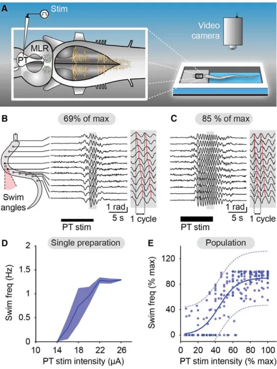
Projektionen wurde in der zweiten Studie untersucht (Ryczko et al., 2017). Eine wichtige Beobachtung dieser
Studie war, dass die Aktivität der MLR und der Bewegungsgeschwindigkeit durch eine elektrische PT
Stimulation graduell kontrolliert werden kann: je höher die Intensität der PT Stimulation, desto schneller
wurden Bewegungsabläufe ausgeführt. Die Blockierung von Glutamatrezeptoren in der MLR hatte eine
erhebliche Beeinträchtigung der Initiierung von Bewegungsabläufen zur Folge. Die Blockierung von D1 Dopaminrezeptoren in der MLR setzte die Schwimmgeschwindigkeit zwar signifikant herunter, eine graduelle Kontrolle der Schwimmgeschwindigkeit durch elektrische PT Stimulation war aber nach wie vor möglich.
Daraus ergibt sich, dass absteigende glutamaterge PT Neurone für die graduelle Kontrolle der Schwimmgeschwindigkeit verantwortlich sind.
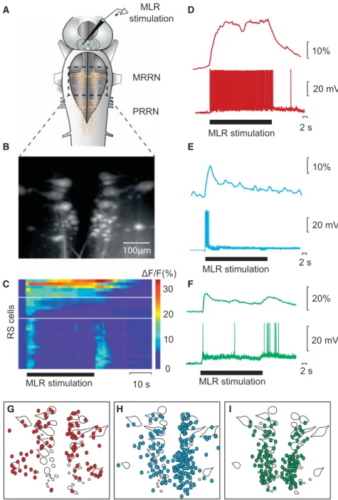
In der dritten Studie (Juvin*, Grätsch* et al., 2016) konnte gezeigt werden, dass RS Zellen nicht uniform auf eine elektrische MLR Stimulation antworten, sondern drei unterschiedliche Aktivitätsmuster aufweisen. Eine Population von RS Zellen wird kurz am Beginn einer MLR Stimulation aktiviert, während eine zweite Zellpopulation Aktionspotentiale während der gesamten MLR Stimulation generiert. Interessanterweise wurde eine dritte Gruppe von RS Zellen identifiziert, die eine Salve von Aktionspotenzialen am Anfang und eine weitere Salve nach dem Ende einer MLR Stimulation produziert. In semi-intakten Präparationen wurde gezeigt, dass diese letzte Salve von Aktionspotentialen stark mit dem Ende der Schwimmepisode korreliert.
Des Weitern wurde nachgewiesen, dass eine pharmakologische Aktivierung dieser RS Zellen Schwimmbewegungen beendet, während eine Inaktivierung dieser RS Zellen den Beendigungsprozess der Schwimmepisode stark beeinträchtigt. Da diese RS Zellen funktionell eng mit dem Ende von Bewegungsabläufen verknüpft ist, wurden sie Stopp Zellen genannt.
Es war bisher unklar, wie Stopp Zellen währen einer Bewegung aktiviert werden und sie wiesen keine Membraneigenschaften auf, die ihr charakteristisches Aktivitätsmuster erklären. Daher wurden sie in der vierten Studie (Grätsch et al., in Begutachtung) auf synaptische Eingänge untersucht, die die zweite Salve von Aktionspotenzialen auslösen könnten. In dieser Studie konnte gezeigt werden, dass durch eine Stimulation der MLR während einer Bewegungsepisode, Stopp Zellen rekrutiert werden und somit das Ende des Bewegungsablaufs kontrolliert wird. Elektrophysiologische und anatomische Versuche weisen außerdem darauf hin, dass eine monosynaptische Verbindung zwischen der MLR und Stopp Zellen besteht.
Teile dieser Arbeit wurden bereits in Fachzeitschriften publiziert (Ryczko et al., 2013; Ryczko et al., 2017;
Juvin*, Grätsch* et al., 2016) oder sind im Begutachtungsverfahren (Grätsch et al.).
1
2
* Ko-Erstautoren
2. Summary
Locomotion underlies a dynamic interplay of a basic motor pattern that is generated by spinal neural networks, descending control originating from supraspinal structures, and sensory feedback from the periphery. Locomotion usually occurs intermittently and thus, it must be initiated, maintained, and eventually stopped. Over the past decades, the lamprey has been used as an experimental model to define the cellular mechanisms controlling locomotion in vertebrates. In this model, spinal central pattern generators (CPGs) have been characterized and shown to generate rhythmic muscle contractions needed for body propulsion. The spinal CPGs are controlled by brainstem reticulospinal (RS) neurons, which are activated by upstream brain structures, such as the mesencephalic locomotor region (MLR). The MLR initiates and controls locomotion in a graded fashion and plays a role in goal-directed locomotion. Its activity is in turn controlled by forebrain structures, such as the basal ganglia. The focus of my thesis was to examine descending projections from forebrain structures to the MLR as well as MLR projections to different RS cell populations in the lamprey lower brainstem. For this, electrophysiological, neuroanatomical, Ca
2+- imaging, and behavioral experiments were performed.
In vertebrates, forebrain dopaminergic neurons of the substantia nigra pars compacta (SNc) are classically described to send ascending projections to the striatum, the input structure of the basal ganglia. In a first study (Ryczko et al., 2013), we identified in the lamprey a previously unknown descending dopaminergic pathway from the posterior tuberculum (PT; the homologue structure to the mammalian SNc) that directly innervates the MLR. Experiments were performed in semi-intact preparations, in which cellular activity can be correlated to active swimming movements of the intact body. It was demonstrated that electrical PT stimulation elicits RS cell activity as well as motor behavior. Both RS cell activity and locomotor output were significantly increased when dopamine was injected locally into the MLR. On the other hand, local injections of a D1 receptor antagonist in the MLR dramatically decreased RS cell activity and locomotor activity. It was concluded that this descending dopaminergic pathway provides extra excitation to the MLR and consequently increases the locomotor output.
It was thought that this newly identified dopaminergic pathway acts in parallel with a descending glutamatergic pathway from the PT to the MLR. In a second study (Ryczko et al., 2017), the glutamatergic projection was examined in detail. One important finding was that the PT controls MLR activity and consequently the locomotor speed in a graded fashion: increasing stimulation intensity of the PT leads to increasing MLR cell activity and locomotor speed. Local blockade of glutamate receptors in the MLR dramatically diminishes locomotor activity elicited by PT stimulation. Local injections of a D1 receptor antagonist in the MLR also decreases locomotor frequency but surprisingly, the graded control of locomotor speed was still present. It was concluded that the PT controls the locomotor speed in a graded fashion through direct descending glutamatergic projections to the MLR.
In a third study (Juvin*, Grätsch* et al., 2016), it was demonstrated that RS cells do not respond to MLR
stimulation uniformly, but with three distinct activity patterns. One RS cell population responds with a
transient burst of activity at the beginning of a MLR stimulation, a second group displays a sustained response throughout the MLR stimulation, and a third group of RS cells was shown to display two transient bursts of activity: a first burst of activity is generated at the beginning and a second burst occurs at the end of a MLR stimulation. These RS cells were recorded in semi-intact preparations, and it was demonstrated that the second burst of activity is strongly correlated to the end of a locomotor bout (‘termination burst’). Local application of glutamate on these RS cells was shown to stop ongoing swimming movements, whereas inactivation of glutamate receptors elicits a slower termination. As they contribute to the termination of locomotion, these RS cells are referred to as stop cells.
It was shown that the ‘termination burst’ does not underlie specific membrane properties of stop cells but rather synaptic inputs to those cells. The aim of a fourth study (Grätsch et al., under review) was to define the origin of these synaptic inputs. An important finding was that ongoing locomotion can be stopped by electrical and pharmacological MLR activation. When the animal is at rest, MLR stimulation elicits locomotion, but it produces very different effects if stimulated during locomotion. It stops swimming if it is stimulated at low intensity and prolongs swimming if stimulated at a higher intensity. Furthermore it was shown that MLR stimulation at low intensity also triggers the ‘termination burst’ in stop cells. Electrophysiological and anatomical experiments revealed that at least some connections between MLR and stop cells are monosynaptic.
Parts of this work are published in peer-reviewed journals (Ryczko et al., 2013; Ryczko et al., 2017; Juvin*, Grätsch* et al., 2016) or are under review (Grätsch et al.).
3* co-first authors
3. List of Abbreviations
AMPA: α-amino-3-hydroxy-5-methyl-4-isoxazolepropionic acid
AP5: (2R)-amino-5-phosphonovaleric acid; (2R)-amino-5-phosphonopentanoate; NMDA receptor antagonist ARRN: anterior rhombencephalic reticular nucleus
CNQX: 6-cyano-7-nitroquinoxaline-2,3-dione; AMPA/kianate receptor antagonist CNS: central nervous system
CPG: central pattern generator CuN: cuneiform nucleus
DLR: diencephalic locomotor region EPSP: excitatory postsynaptic potential GABA: gamma-aminobutyric acid GPe: globus pallidus externa GPi: globus pallidus interna
I
CAN: Calcium-activated nonselective cation current LDT: laterodorsal tegmental nucleus
MHR: mid-hindbrain neurons
MLR: mesencephalic locomotor region
MPTP: 1-methyl-4-phenyl-1,2,3,6-tetrahydropyridine MRN: mesencephalic reticular nucleus
MRRN: middle rhombencephalic reticular nucleus NMDA: N-methyl-D-aspartate
PD: Parkinson’s disease PPN: pedunclopontine nucleus
PRRN: posterior rhombencephalic reticular nucleus PT: posterior tuberculum
RS: reticulospinal
SCH29930: halobenzazepine; D1 receptor antagonist SNc: substantia nigra pars compacta
SNr: substantia nigra par reticulata
STN: subthalamic nucleus
4. Introduction
Locomotion is a complex motor behavior that plays a crucial role in our daily life. Animals have developed different locomotor strategies to survive in their environment and to explore it: a stick insect coordinates six legs while walking through shrub lands; a hummingbird uses its two wings to perform ultra-fast flight maneuvers; and a humpback whale can travel long distances by swimming through the ocean water. Even though the biomechanics of their movement are very different, the general scheme for locomotor control is very similar in invertebrate and
vertebrate species: Central pattern generators (CPGs) produce a basic motor pattern and control sequential muscle contractions, needed for body propulsion. These CPGs are in turn controlled by descending inputs from the central nervous system (CNS) and modulated by sensory feedback from the periphery (for review see Rossignol et al., 2006; Dubuc et al., 2008; Büschges et al, 2011;
Grillner and Robertson, 2017).
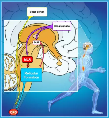
In vertebrates, the locomotor CPGs are located in the spinal cord and they receive synaptic inputs from reticulospinal (RS) neurons in the hindbrain (see Figure 1). These RS cells are command cells for locomotion and are controlled by upstream locomotor regions such as the mesencephalic and the
diencephalic locomotor region (MLR and DLR, respectively). The MLR controls the initiation of locomotion and is controlled by forebrain structures, including the basal ganglia. Additionally, the motor cortex is involved in fine adjustment of the locomotor output.
Figure 1. Neural control of locomotion in vertebrates.
Schematic representation of a human brain (sagittal view) with the
approximate locations and connections of selected supraspinal brain
structures that are relevant for locomotor control in vertebrates. (CPG,
central pattern generator; DLR, Diencephalic locomotor region, MLR,
Mesencephalic locomotor region; modified from Le Ray et al., 2011).
4.1. Locomotor control in vertebrates
Early motor control studies
The general concept of neural control of locomotion was developed based on discoveries made in cat studies that were performed in the 1960s and 1970s. Grillner and Zangger (1979) demonstrated after a complete low thoracic spinal transection in acute mesencephalic cats that neural networks in the spinal cord generate rhythmic activity and control sequential activation of muscle groups during body movements. Interestingly, it could be demonstrated that these neural networks have the capacity to generate an unchanged rhythmic activity in isolated conditions, namely after deafferentation of dorsal roots. For that reason, these spinal networks were referred to as ‘central pattern generators’ (Griller and Zangger, 1975). As described above, the spinal CPGs are controlled by supraspinal structures such as the MLR that activates spinal networks via RS cells in the hindbrain. The MLR itself was discovered in the late 1960 by the muscovite research group of Orlovskii. They discovered in decerebrated cats that locomotion can be elicited by electrically stimulating a brain region located at the junction of the mid- and hindbrain (Shik et al., 1966). In this study it was demonstrated that the locomotor output can be controlled in a graded fashion by stimulating this region electrically: stimulation at low intensities initiated walking in cats and increasing stimulation intensities systematically changed the locomotion pattern to trotting and then galloping gait. Since this brainstem region appeared to be dedicated to controlling locomotion, it was then named ‘mesencephalic locomotor region’. It was later confirmed that the MLR initiates locomotion not by directly projecting to the spinal cord, but by activating RS cells which in turn relay the locomotor command to spinal locomotor networks (Garcia-Rill and Skinner, 1987 a, b; Orlovskii, 1970; Steeves and Jordan, 1984). Garcia-Rill and Skinner (1987 b) demonstrated in cats that RS neurons in the medioventral medulla receive inputs from the MLR and project directly to the spinal cord. Additionally, they showed that electrical as well as pharmacological activation of this area initiates locomotor activity (Garcia-Rill and Skinner, 1987a). In conclusion, these important studies laid the foundation of today’s knowledge about the neural mechanisms involved in motor control.
Further development
Following studies were later performed in different model organisms and striking similarities were observed
in the neural organization of the locomotor networks throughout vertebrate species. For example, locomotor
CPGs were identified in the spinal cord of many different vertebrates and shown to generate a basic locomotor
pattern (e.g. Xenopus tadpole: Kahn and Roberts, 1982; rat: Kudo and Yamada, 1987; goldfish: Fetcho and
Svoboda, 1993; zebrafish: McDearmid and Drapeau, 2006; for review, see Grillner et al., 2003; Ryczko et al.,
2010).
RS cells were also shown to constitute the interface between the locomotor centers and spinal networks and provide mainly excitatory input to spinal interneurons and motor neurons (e.g. Peterson et al., 1979; Perrins et al., 2002; Bouvier et al., 2015; Capelli et al., 2017). Interestingly, multiple studies in different vertebrate models revealed that different groups of RS cells control various motor functions, such as locomotor initiation (Garcia-Rill and Skinner, 1987a; Kimura et al, 2013; Capelli et al., 2017), maintenance (Bretzner and Brownstone, 2013), termination (Bouvier et al., 2015; Perrins et al., 2002; Capelli et al, 2017), and steering (Thiele et al., 2014).
Importantly, the MLR has been shown to be highly conserved and has been identified in all vertebrate species tested (e.g. rats: Skinner and Garcia-Rill, 1984; mice: Lee et al., 2014; salamanders: Cabelguen et al., 2003;
ducks and geese: Sholomenko et al., 1991; lamprey: Sirota et al, 2000, for review, see Jordan, 1998; Dubuc et al., 2008). It is classically described to be located at the border between the midbrain and hindbrain and electrical, pharmacological, or optogenetic stimulation initiates stable locomotor bouts (Shik et al., 1966;
Garcia-Rill et al., 1985; Lee et al., 2014; Roseberry et al, 2016; Caggiano et al., 2018; Josset et al, 2018). The key characteristic of the MLR to control the locomotor speed in a graded fashion was also shown to be present in other species (Sirota et al., 2000; Cabelguen et al., 2003; Lee et al., 2014; for review, see Le Ray et al, 2011; Ryczko and Dubuc, 2013). The mammalian MLR comprises cholinergic, glutamatergic, and GABAergic neurons that are localized in different nuclei, the pedunculopontine nucleus (PPN) and the cuneiform nucleus (CuN) (Skinner and Garcia-Rill, 1984; Martinez-Gonzalez et al., 2011; Roseberry et al., 2016; for review, see Ryczko and Dubuc, 2013). It has not yet been resolved whether different sub-nuclei of the MLR control different motor functions, but this matter has been extensively studied. Sinnamon (1993) proposed that different MLR regions control different motor functions such as appetitive behavior that is used to approach a consummatory stimulus, escape behavior in response to threat, and exploratory behavior. Recent optogenetic studies support this hypothesis (Roseberry et al., 2016; Caggiano et al., 2018; Josset et al., 2018).
Examination of the functional role of different cell types in the MLR demonstrated that glutamatergic MLR cells drive locomotor activity, whereas cholinergic cells contribute to speed control. GABAergic cells inhibit glutamatergic MLR neurons, which leads to locomotor arrest (Roseberry et al., 2016). Caggiano and colleagues (2018) revealed that glutamatergic neurons in both PPN and CuN contribute to slow exploratory movements but only activation of glutamatergic CuN neurons can elicit high-speed escape-like behavior.
Similar observations were made by Josset and colleagues (2018), who demonstrated that optogenetic stimulations of glutamatergic CuN neurons trigger fast locomotion, as it is seen in escape behavior.
Furthermore, it was shown that both glutamatergic as well as cholinergic neurons in the PPN contribute and modulate slow walking movements, as observed in exploratory behavior (Josset et al., 2018).
The mammalian MLR in turn is controlled by forebrain structures, such as the basal ganglia, which are
involved in the selection of actions and motor programs (for review, see Kreitzer and Malenka, 2008; Grillner
and Robertson, 2016). At rest, GABAergic neurons from the substantia nigra pars reticulata (SNr) and the
globus pallidus interna (GPi), the output structures of the basal ganglia, keep the MLR under tonic inhibition
(Saitoh et al., 2003; Roseberry et al., 2016; for review, see Takakusaki et al., 2008). In order to generate and suppress goal-directed locomotion, the basal ganglia recruit their direct and indirect pathway respectively (Kravitz et al., 2010; Roseberry et al., 2016). The direct pathway is composed of GABAergic neurons that project from the striatum, the input structure of the basal ganglia, directly to the SNr and GPi. Activation of this direct pathway inhibits the GABAergic neurons of the SNr and GPi and thus disinhibits the MLR, which then leads to the initiation of locomotion. Striatal neurons of the indirect pathway, on the other hand, project to the globus pallidus externa (GPe), which in turn projects to the subthalamic nucleus (STN). The STN activates the GPi and the SNr, which then leads to suppression of motor activity (Kravitz et al., 2010; for review, see Grillner and Robertson, 2016; Roseberry and Kreitzer, 2017).
In the past decades, motor control studies have given a broad insight into the neural control of locomotor behavior. Cross-linking concepts that were found in invertebrates, basal vertebrates, and more recently- evolved vertebrates is one reason for this progress (for review, see Mullins et al., 2011). Furthermore, technological advances allowed the establishment of new techniques, such as Ca
2+imaging or optogenetic tools. The latter provide many advantages, since the functional role of neurons with specific genetic markers can be examined (e.g. Kimura et al., 2013; Lee, 2014; Thiele et al., 2014; Bouvier et al., 2015; Capelli et al., 2017; Caggiano et al., 2018; Josset et al., 2018). However, the mammalian nervous system is very complex and single cell recordings remain challenging, notably during ongoing locomotion. Therefore, studies in organisms with simpler nervous systems, like the lamprey, remain of great importance in order to reveal details about neural connectivity and properties involved in locomotor control.
4.2. The control of locomotion in lampreys
Studies performed in mammalian models could not yet bring detailed insights into the cellular organization and connectivity within the locomotor network. In the 1980s, Grillner and colleagues started to investigate the cellular organization of the spinal locomotor CPG in a basal vertebrate, the lamprey. The lamprey was chosen as an experimental model for several reasons. It is a basal vertebrate that diverged from the vertebrate phylum some 560 million years ago (Kumar and Hedges, 1998) and the anatomical organization of the lamprey and mammalian CNS is strikingly similar (for review, see Nieuwenhuys et al., 1998; Robertson et al., 2014 Grillner and Robertson, 2017). Another advantage was the simplicity of the undulary, limbless movements.
Lampreys swim in the horizontal plane and movements underlie reciprocal muscle contractions of the left and
right side of the body. For the most common forward propulsion, those contractions propagate along the body
axis like a mechanical wave that propagates from the rostral to the caudal body segments, with an
intersegmental phase lag of approximately 1% (Wallén and Williams, 1984). In very rare cases, the lamprey
performs backward swimming, characterized by undulary movements that start in caudal segments and
propagate rostrally with a phase lag of approximately -1% (Matsushima and Grillner, 1992; Islam et al.,
2006). Compared to the mammalian CNS, there are considerably fewer neurons present in the lamprey CNS and many are larger and thus more accessible for intracellular recording. Another advantage is that he lamprey brain can survive in vitro for a few days, which makes it very valuable for anatomical and physiological experiments. Moreover, a semi-intact preparation was developed in lampreys to study neural activity during active behavior (Sirota et al, 2000). Here, the brain is exposed and accessible to recording electrodes while the intact body is still attached and may perform active swimming movements, cellular activity in the intact brain can thus be correlated to the behavioral output. Over the past decades, these features allowed the development and the combination of multiple in vitro and in vivo techniques that are now used to examine neural networks from the single cell to the behavioral level (e.g. Derjean et al., 2010; Brocard et al., 2010; Ryczko et al., 2013;
Juvin et al., 2016).
The locomotor CPGs in the lamprey spinal cord
As mentioned above, Grillner and his group began to characterize the cellular organization of the locomotor CPGs in the lamprey spinal cord. Buchanan and Grillner (1987) discovered that excitatory glutamatergic premotor neurons build networks that intrinsically generate burst activity and excite ipsilateral motor neurons (Buchanan and Grillner, 1987). Rhythmic bursting activity can be generated by these networks even in isolated spinal cord preparations in which supraspinal and sensory inputs are removed. Pharmacological application of glutamate agonists to the spinal cord or electrical stimulation of supraspinal brain areas as well as RS cell axons induce stable bursting pattern in spinal ventral roots. This motor output is referred to as
‘fictive locomotion’ (Cohen and Wallén, 1980; Wallén and Williams, 1984). The rhythmogenetic networks were shown to be located in individual spinal cord segments and are interconnected through ipsilateral (intersegmental) and contralateral (intrasegmental) projections, originating from excitatory and inhibitory interneurons that allow rostro-caudal and left-right coordination of body segments (for review, see Grillner, 2003). For the propagation of the mechanical wave from rostral to caudal, intersegmental coordination of ipsilateral rhythmogenetic networks is needed. Excitatory interneurons of each segment project collateral axons to rostral and caudal segments (Dale, 1986). This interconnection allows the generation of rostro-caudal body undulations, but can also induce caudo-rostral body movements during backward swimming. Decisive for the direction of propagation of burst activity is the excitatory gradient of rostral and caudal spinal cord segments: if rostral segments are more excited than caudal segments, the wave of bursting activity propagates from rostral to caudal and vice versa (Matsushima and Griller, 1992). Glycinergic commissural interneurons coordinate alternating activity of rhythmogenetic networks located on the left and right side of one segment.
These neurons are activated by the excitatory interneurons of the rhythmogenetic networks and inhibit
contralateral CPG interneurons as well as contralateral motor neurons (Buchanan, 1982). Application of
strychnine, a glycinergic antagonist, on the isolated spinal cord does not prevent rhythmic bursting activity in
the spinal cord, but it results in a change from alternating bursting activity to simultaneous bilateral activity of
motor neurons within one segment (Cohen and Harris-Warrick, 1984). These results indicate that contralateral
inhibition is not essential to generate bursting activity in the hemi-segments but it is essential for coordinated left-right alternation of bursting activity in one segment.
The RS neurons in the lamprey
The majority of the discoveries described above were made in the isolated spinal cord preparation and pharmacological or electrical stimulations were used to induce ‘fictive locomotion’. In the intact animal however, the CPGs are activated by supraspinal inputs from the RS cells (Rovainen, 1974b; Buchanan et al., 1987a; Ohta and Grillner, 1989; Swain et al., 1993). RS cells are command cells for locomotion that receive and integrate sensory inputs from the olfactory, visual, vestibular, or mechanical systems (Derjean et al., 2010; Zompa and Dubuc, 1996; Deliagina et al., 1993; Deliagina et al., 1992a; b; Deliagina and Orlovskii, 2002; McClellan and Grillner, 1984; Dubuc et al., 1993a; b; for review, see Daghfous et al., 2016).
Furthermore, they receive central feedback from the spinal CPGs (Einum and Buchanan, 2005; Antri et al.,
2009; Buchanan, 2011). In the lamprey, there are approximately 2500 RS cells, which are located in the
brainstem and organized in four distinct nuclei: the mesencephalic reticular nucleus (MRN), the anterior
rhombencephalic reticular nucleus (ARRN), the middle rhombencephalic reticular nucleus (MRRN), and the
posterior rhombencephalic reticular nucleus (PRRN) (Bussières, 1994; Shaw et al., 2010). The reticular nuclei
of the lamprey were described to be homologous to those of other vertebrates: the ARRN and MRRN are
thought to be homologous to the mammalian nuclei pontis oralis and caudalis (Rovainen, 1967; Cruce and
Newman, 1984). The ARRN and MRRN contain identifiable pairs of RS cells, the Müller and the Mauthner
cells, which have been extensively studied in the past because of the large size of their cell bodies (Rovainen
1967a; Rovainen et al., 1973; for review, see Rovainen, 1978; Buchanan, 2001). The axons of Müller cells
project ipsilaterally in the middle axon tracts along the spinal cord, where they make en passage synapses with
spinal motor neurons and interneurons (Rovainen, 1976a; for review, see Buchanan, 2001). The axons of the
Mauthner cells project contralaterally. The PRRN is thought to be homologous to mammalian nucleus
gigantocellularis (Cruce and Newman, 1984). RS cells in the PRRN are not identifiable but their axons where
shown to project in the lateral tracts of the spinal cord (Shaw et al., 2010). The RS cells in the lamprey have
been shown to be functionally and neurochemically heterogenous. The majority of MRRN and PRRN cells
are glutamatergic (Buchanan et al., 1987b; Ohta and Grillner, 1989), but next to these excitatory cells, a few
glycinergic RS cells were identified that project to the spinal cord and synapse to spinal interneurons and
motor neurons (Wannier et al., 1995). Studies performed in different labs over the past years revealed that RS
cells control different locomotor functions, such as steering (Fagerstaedt et al., 2001; Kozlov et al., 2002),
locomotor speed (Brocard and Dubuc, 2003), postural control (Deliagina and Orlovsky, 2002; Zelenin et al.,
2007), and forward and backward swimming (Zelenin, 2011). It is noteworthy, that most of these studies
examined the activity of the Müller and Mauthner cells in the MRRN, because they were relatively easy to
target for intracellular recording. The use of Ca
2+-imaging allowed us to also examine activity of different
populations of RS cells, which will be presented in a following section (Juvin et al., 2016).
The mesencephalic locomotor region of the lamprey
Other than inputs from the sensory system and the spinal cord, RS cells receive descending inputs from upstream motor centers, such as the mesencephalic locomotor region and the diencephalic locomotor region (MLR and DLR respectively; El Manira et al., 1997; Sirota et al., 2000; Brocard et al., 2010). The MLR of lampreys comprises cholinergic and glutamatergic neurons that are localized in the PPN and laterodorsal tegmental nucleus (LDT). These neurons project bilaterally and symmetrically to RS cells in the brainstem via monosynaptic connections (Le Ray et al., 2003; Brocard et al., 2010). Whether the two neurotransmitter systems in the MLR contribute to different locomotor functions, as it has recently been shown in mice (Roseberry et al., 2016), has not been confirmed in lampreys. Yet, local applications of acetylcholine or nicotine elicit dose-dependent responses in RS cells and can initiate or accelerate locomotion in semi-intact preparations (Le Ray et al., 2003). Interestingly, blocking nicotinic receptors in the brainstem increased the threshold of MLR stimulation but did not prevent the initiation of locomotion as such, which indicates the cooperative nature of the two neurotransmitter systems that are present in the MLR (Le Ray et al., 2003).
Apart from the initiation of locomotion, the graded control of locomotor intensity is another characteristic of the MLR (Sirota et al., 2000). This mechanism works similarly to a rheostat: the stronger the MLR is activated, either electrically or pharmacologically, the higher is the activation of RS cells and consequently, the locomotor output (Sirota et al., 2000). Direct recruitment of different RS cell populations in the MRRN and PRRN (for slow and fast locomotor activity, respectively) underlies this fine control of the intensity of the locomotor output (Brocard and Dubuc, 2003). Additionally, a parallel pathway has been shown to boost locomotor activity (Smetana et al., 2010). Here, the MLR activates a group of muscarinoceptive hindbrain neurons, which in turn project to the RS cells in the MRRN (Smetana et al., 2010). These muscarinoceptive neurons provide extra excitation to RS cells in order to amplify the locomotor output. Interestingly, the MLR not only controls the locomotor output but also participates in other vital functions, by adjusting the activity in neural networks responsible for respiration or gating sensory inputs that reach RS cells (Gariépy et al., 2012;
Le Ray et al., 2010; for review, see Le Ray et al., 2011; Missaghi et al., 2016).
The basal ganglia of the lamprey
Like in mammalian species, the lamprey MLR is under the control of forebrain structures, such as the basal ganglia. In vertebrates, the basal ganglia are responsible for the selection of appropriate motor programs.
Interestingly, it was shown that the main structures that were identified in the mammalian basal ganglia are
also present in the lamprey nervous system (Stephenson- Jones et al., 2011; 2012; for review, see Grillner and
Robertson, 2017). GABAergic projections originating from the basal ganglia innervate the MLR and keep
them under tonic inhibition (Ménard et al., 2007; Stephenson-Jones et al., 2011; 2012; Pombal et al., 1997; for
review, see Robertson et al., 2014). Physiological experiments demonstrated that a blockade of GABAergic
receptors in the MLR induced well-coordinated swimming movements in a semi-intact preparation, whereas
activation of GABAergic receptors suppressed ongoing locomotion (Ménard et al., 2007). Meanwhile, the input structure of the basal ganglia, the striatum, receives input from the thalamus and from the pallium, the homologue structure of the mammalian cortex (Ericsson et al., 2013; Ocaña et al., 2015). Additionally, the striatum receives dopaminergic inputs from the posterior tuberculum (PT), the homologue of the mammalian substantia nigra pars compacta (SNc) and the ventral tegmental area (Pombal et al., 1997; Ryczko et al., 2013; Peréz-Frenández et al., 2014). As mentioned above for the mammalian basal ganglia, the afferent projections of the striatum form two distinct pathways: the direct pathway and the indirect pathway (Stephenson- Jones et al., 2011; 2012). GABAergic striatal neurons that express substance P constitute the direct pathway and project to the pallidal output structures of the basal ganglia (Stephenson-Jones et al., 2011). Like in the direct pathway in mammals, activation of this pathway in lampreys should suppress the GABAergic neurons of the basal ganglia output region and disinhibit the motor control regions which would in turn lead to locomotor activity. Striatal neurons that form the indirect pathway of the lamprey express enkephalin and project to the output region of the basal ganglia via the GPe and the STN (Ericsson et al., 2013; Stephenson- Jones et al., 2012). The circuitry is strikingly similar to the indirect pathway of mammals so it could be expected that activation of this pathway leads to disinhibition of the basal ganglia output region, which in turn would set the motor regions under tonic inhibition and suppress locomotor activity. Together these findings demonstrate that the building blocks and the connectivity within the basal ganglia were present in the first stages of vertebrate evolution (Stephenson-Jones et al., 2011; 2012; Ryczko et al., 2013) and it is tempting to suggest that this neural substrate for action selection has been used by all vertebrate species since then, with gradual modifications and complexification (for review, see Grillner and Robertson, 2017).
4.3. Aims and objectives
The locomotor network of the lamprey has been examined extensively in the past and many details are known about the descending control of locomotion. However, important details remain elusive such as detailed information regarding the descending input from the forebrain to the MLR and the output of the MLR to different RS cell populations. These topics are at the base of the specific aims of my thesis.
Classically, dopaminergic cells in the SNc were described to send ascending projections to the striatum, the
input regions of the basal ganglia (for review, see Ryczko and Dubuc, 2017). But in mammals, some studies
suggested the presence of a descending projection, possibly dopaminergic, from the SNc to the MLR (Rolland
et al., 2009; Beckstedt et al., 1979). Similarly in the lamprey, anatomical studies demonstrated that
dopaminergic neurons of the PT not only send ascending projections to the striatum but also descending
projections (Pombal et al., 1997). In a first study (Ryczko et al., 2013), we examined this dopaminergic
projection of the PT in more detail and identified a previously unknown descending dopaminergic pathway
from the PT to the MLR. Anatomical and physiological experiments were performed and confirmed the
functional relevance of this descending dopaminergic pathway in locomotor control. Dopaminergic inputs from the PT increase MLR activity and consequently the locomotor output via a D1 receptor-dependent mechanism.
Based on studies that were performed previously in the lamprey, it was thought that the PT not only sends dopaminergic projections to the MLR, but that an additional descending pathway provides a parallel excitatory input. For example, it has been shown that electrical stimulation of the PT provides a strong excitatory input to the MLR and that it can initiate locomotion (Derjean et al., 2010; Gariépy et al., 2012;
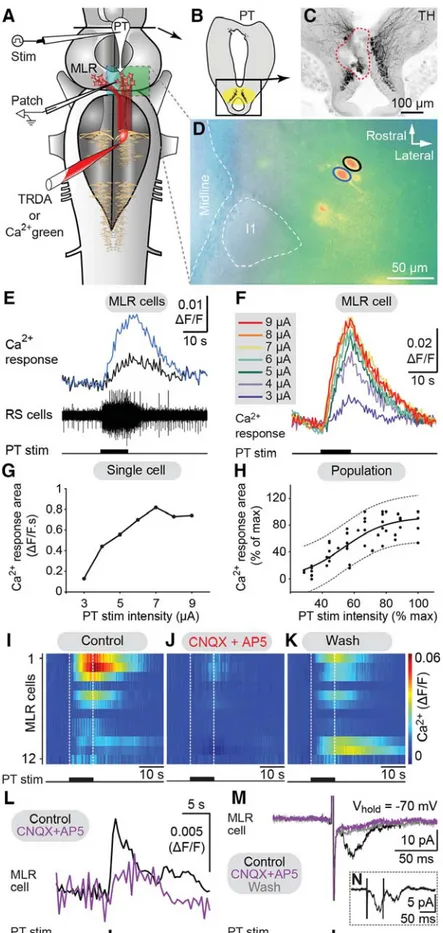
Ryczko et al., 2013). Intracellularly recorded MLR neurons respond to PT stimulation with fast excitatory postsynaptic potentials and a blockade of D1 receptors in the MLR reduces but does not prevent locomotion (Gariépy et al., 2012; Ryczko et al., 2013). Additionally, anatomical studies identified glutamatergic neurons in the PT and physiological studies revealed that activation of glutamatergic receptors in the MLR induces stable locomotor bouts (Sirota et al., 2000; Ménard et al., 2007; Villar-Cervino et al., 2011). Therefore, it seemed very likely that glutamate is present in this parallel pathway. The goal of a second study (Ryczko et al., 2017), was to examine this pathway from the PT to the MLR in more detail. Using anatomical, Ca
2+imaging, and electrophysiological techniques, the presence of a descending glutamatergic pathway from the PT to the MLR was confirmed. Moreover, it was shown to be responsible for the graded control of locomotor speed.
It had long been demonstrated that electrical stimulation of the MLR directly activates RS cells in the
hindbrain. In the lamprey, RS cells of larger size were preferably examined in the past and the aim of a third
study (Juvin*, Grätsch* et al., 2016) was to investigate RS cell responses to MLR stimulation in different
populations of RS cells. We found that MLR stimulation elicits three distinct patterns of activity in different
RS cell populations. One group of RS cells is transiently active at the beginning of the MLR stimulation and a
second group responds with a sustained activity throughout the whole MLR stimulation. A third group of RS
cells displayed two transient bursts of activity: one at the beginning and one at the end of the MLR
stimulation. Experiments in semi-intact preparations demonstrated that the second burst of activity is
correlated to the end of a swimming bout. We thus hypothesised that this RS cell population plays a role in
ending locomotor activity. These cells became of great interest because little was known at the time about the
neural mechanisms controlling the termination of locomotion (for review, see Klemm, 2001; Mullins et al.,
2011). Only recent studies brought insights into the brain regions and neurotransmitter systems that could be
involved in stopping locomotion (for review, see Roseberry and Kreitzer, 2017). In our study (Juvin*,
Grätsch* et al., 2016), we showed that the pharmacological activation of the RS cells displaying a second
burst of activity at the end of a swimming bout halted ongoing swimming. Their inactivation on the other hand
slowed the termination of locomotion down. We concluded that these cells played a crucial part in the
termination process of locomotion and named them ‘stop cells’.
It was not resolved how the stop cells are activated during ongoing locomotor movements, but synaptic inputs rather than membrane properties were suggested to play a significant role (Juvin*, Grätsch* et al., 2016). As the MLR provides major input to RS cells (Orlovskii, 1970; Steeves and Jordan, 1984; Le Ray et al., 2003;
Brocard and Dubuc, 2003; Brocard et al., 2010; Ryczko et al, 2016) it was considered to be a promising candidate for providing such a synaptic input to stop cells. In cats, it has been proposed that projections from the PPN to neurons in the nucleus reticularis points oralis that in turn activate RS neurons in the medullary reticular formation are responsible for suppression of muscle tone (for review, see Takakusaki, 2008). But details about this pathway have not yet been described. The aim of the fourth study was therefore to find the source that activates stop cells during ongoing locomotion. Using physiological and anatomical techniques, we could confirm that the MLR is able to activate RS stop cells during ongoing locomotion and can thus stop ongoing locomotion (Grätsch et al., under review).
4Altogether, my thesis investigated the descending control of locomotion in the lamprey and revealed details about the direct control of the MLR by forebrain structures as well as descending outputs of the MLR to different RS cell populations in the hindbrain. Furthermore, a neural substrate underlying the neural control of termination of locomotion was identified.
*
co-first authors
5. Published studies
5.1. Forebrain dopamine neurons project down to a brainstem region controlling locomotion
Ryczko, D., Grätsch, S., Auclair, F., Dubé, C., Bergeron, S., Alpert, M.H., Cone, J.J., Roitman, M.F., Alford, S., and Dubuc, R.
Published 2013 in Proceedings of the National Academy of Sciences, 110(34):E3235-42.
Author contributions
Designed research:
Dimitri Ryczko, François Auclair, and Réjean Dubuc.
Performed research:
Dimitri Ryczko, Swantje Grätsch, François Auclair, Catherine Dubé, Saskia Bergeron, Michael H. Alpert, Jackson J. Cone, Mitchell F. Roitman, Simon Alford, and Réjean Dubuc.
Analyzed data:
Dimitri Ryczko, Swantje Grätsch, François Auclair, Catherine Dubé, Saskia Bergeron, Michael H. Alpert, Jackson J. Cone, Mitchell F. Roitman, Simon Alford, and Réjean Dubuc.
Wrote the paper:
Dimitri Ryczko, François Auclair, and Réjean Dubuc.
This paper was reproduced with the permission from the publisher.
Forebrain dopamine neurons project down to a brainstem region controlling locomotion
Dimitri Ryczkoa, Swantje Grätscha, François Auclaira, Catherine Dubéa, Saskia Bergeronb, Michael H. Alpertc, Jackson J. Conec, Mitchell F. Roitmanc, Simon Alfordc, and Réjean Dubuca,b,1
aGroupe de Recherche sur le Système Nerveux Central, Département de Physiologie, Université de Montréal, Montréal, QC, Canada H3C 3J7;bGroupe de Recherche en Activité Physique Adaptée, Département de Kinanthropologie, Université du Québec à Montréal, Montréal, QC, Canada H3C 3P8; and
cLaboratory of Integrative Neuroscience, University of Illinois at Chicago, Chicago, IL 60607
Edited by Sten Grillner, Karolinska Institutet, Stockholm, Sweden, and approved June 28, 2013 (received for review January 18, 2013) The contribution of dopamine (DA) to locomotor control is tradi-
tionally attributed to ascending dopaminergic projections from the substantia nigra pars compacta and the ventral tegmental area to the basal ganglia, which in turn project down to the mesencephalic locomotor region (MLR), a brainstem region controlling locomotion in vertebrates. However, a dopaminergic innervation of the pedun- culopontine nucleus, considered part of the MLR, was recently identified in the monkey. The origin and role of this dopaminergic input are unknown. We addressed these questions in a basal vertebrate, the lamprey. Here we report a functional descending dopaminergic pathway from the posterior tuberculum (PT;
homologous to the substantia nigra pars compacta and/or ventral tegmental area of mammals) to the MLR. By using triple labeling, we found that dopaminergic cells from the PT not only project an ascending pathway to the striatum, but send a descending pro- jection to the MLR. In an isolated brain preparation, PT stimulation elicited excitatory synaptic inputs into patch-clamped MLR cells, accompanied by activity in reticulospinal cells. By using voltammetry coupled with electrophysiological recordings, we demonstrate that PT stimulation evoked DA release in the MLR, together with the activation of reticulospinal cells. In a semi-intact preparation, stimulation of the PT elicited reticulospinal activity together with locomotor movements. Microinjections of a D1 antagonist in the MLR decreased the locomotor output elicited by PT stimulation, whereas injection of DA had an opposite effect. It appears that this descending dopaminergic pathway has a modulatory role on MLR cells that are known to receive glutamatergic projections and promotes locomotor output.
motor system
|
Parkinson diseaseD
opamine (DA) neurons of the substantia nigra pars com- pacta (SNc) and ventral tegmental area (VTA) modulate motor behaviors, including locomotion, through ascending pro- jections to the basal ganglia, the output of which projects to the mesencephalic locomotor region (MLR) (1–3), a brainstem re- gion known to control locomotion in all vertebrate species tested to date (reviewed in ref. 4). DA is known to control the excit- ability of striatal cells, and a dysfunction of the ascending DA pathway to the striatum is considered to be the main cause for the motor deficits in Parkinson disease (1). However, there have been hints of descending DA projections that would be in position to directly modulate the MLR and hence locomotor activity. In monkeys, DA terminals of unknown origin were observed in the pedunculopontine nucleus (PPN) (5), considered part of the MLR (reviewed in ref. 4). In addition, there is an axonal projection from the SNc to the PPN in rats, but the transmitter system is un- known (6).We examined the DA system in a basal vertebrate, the lam- prey, and found a previously unknown descending DA pathway from the posterior tuberculum (PT) to the MLR, which com- prises the PPN and the laterodorsal tegmental nucleus (LDT) in lampreys (ref. 7; reviewed in ref. 4). In lampreys, the PT is considered homologous to the SNc and/or VTA of mammals
because of its DA projection to the striatum (3). Further, we de- termined a role for this DA pathway in the control of locomotion.
Results
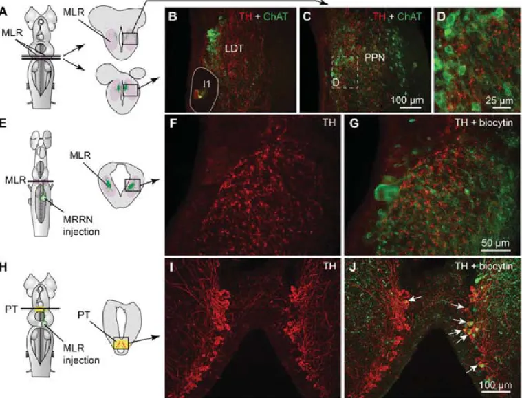
Descending DA Projections from the PT to the MLR.Immunofluo- rescence against tyrosine hydroxylase (TH) or against DA was used to visualize PT neurons containing DA. The distribution of TH and DA immunoreactive cell bodies and fibers were very similar in the PT and the MLR (Fig. S1A–F). Fibers and vari- cosities positive for TH (n= 8 animals) or DA (n= 3 animals) were present throughout the LDT and the PPN (Fig. 1BandC and Fig. S1 A–C), both considered parts of the MLR (ref. 7;
reviewed in ref. 4). TH-positive terminals were found in close proximity to cholinergic MLR cell bodies and dendrites (Fig. 1 A–D) and in the vicinity of MLR cells traced from the middle rhombencephalic reticular nucleus (MRRN;n= 5 animals; Fig.
1E–G). The location of tracer injection sites in the MRRN was verified by histologic examination (Fig. S2AandB). As the MLR cholinergic projection to the reticular formation can initiate loco- motion (7, 8), the juxtaposition of TH-positive terminals suggests that they are in position to directly modulate locomotor output.
We looked for the origin of this DA projection by using tracer injections in the MLR coupled with TH immunofluorescence.
The MLR was considered to overlap largely with the cholinergic neuronal population of the isthmic region, with the conspicuous Müller cell I1 lying at the caudal limit as a landmark (detailed description provided in ref. 9). The PT refers to a region of the caudal diencephalon located ventral to the pretectum. The PT contains a prominent population of dopaminergic neurons, some of them projecting to the striatum, that are intensely labeled by
Significance
We found in lampreys that dopaminergic cells from the pos- terior tuberculum (homologue of the mammalian substantia nigra pars compacta and/or ventral tegmental area) not only send ascending projections to the striatum, but also have a di- rect descending projection to a brainstem region controlling locomotion—the mesencephalic locomotor region—where it releases dopamine (DA). DA increased locomotor output through a D1 receptor-dependent mechanism. The presence of this descending dopaminergic projection may have considerable implication for our understanding of the role of DA in motor control under physiological and pathological (i.e. Parkinson disease) conditions.
Author contributions: D.R., F.A., and R.D. designed research; D.R., S.G., F.A., C.D., S.B., M.H.A., J.J.C., M.F.R., S.A., and R.D. performed research; D.R., S.G., F.A., C.D., S.B., M.H.A., J.J.C., M.F.R., S.A., and R.D. analyzed data; and D.R., F.A., and R.D. wrote the paper.
The authors declare no conflict of interest.
This article is a PNAS Direct Submission.
1To whom correspondence should be addressed. E-mail: rejean.dubuc@gmail.com.
This article contains supporting information online atwww.pnas.org/lookup/suppl/doi:10.
1073/pnas.1301125110/-/DCSupplemental.
www.pnas.org/cgi/doi/10.1073/pnas.1301125110 PNAS | Published online August 5, 2013 | E3235–E3242
NEUROSCIENCEPNASPLUS
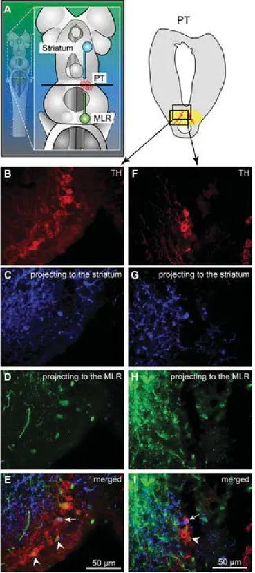
TH immunofluorescence (3). The rostrocaudal extent of the population in spawning phase animals is around 500 μm, spreading approximately 100 to 300μm from the midline (Fig. 1 H–J). The PT contained 189 ± 21 TH-positive cells (n = 12 animals). Many TH-positive cells of the PT were retrogradely labeled from MLR tracer injections (24 ± 3 cells, i.e., 15.4 ± 2.4%), largely on the ipsilateral side (2 ± 1 cells labeled con- tralaterally, i.e., 1.6±0.6%;n= 6 animals; Fig. 1H–J). Again, these results were confirmed by DA immunofluorescence (n= 3 preparations; Fig. S1 G–I). It is noteworthy that retrogradely labeled neurons were found only among the intensely labeled TH-positive population of the PT (outlined inFig. S1EandF), both in the dorsomedial portion that contains larger neurons and in the lateroventral portion that contains smaller neurons. No labeled cells were found in the periventricular TH (and DA) neuron population of the mammillary area, or in any other TH- positive cell population of the brain and spinal cord. By using triple labeling, we found that DA cells in the PT projecting to the striatum were intermingled with those projecting to the MLR (n= 4 preparations; Fig. 2). In all cases, occasional TH-positive neurons of the PT (n= 1–2 cells) were found to project to the
MLR and the striatum (Fig. 2E). Overall in the PT, TH-positive neurons with descending projections to the MLR (n = 16–52 cells) were, on average, 11±2 times more numerous than those with ascending projections to the striatum (n= 1–5 cells;n = 4 animals).
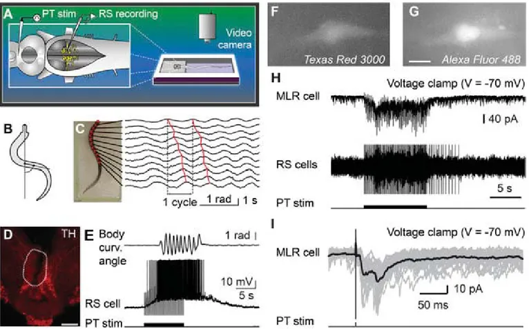
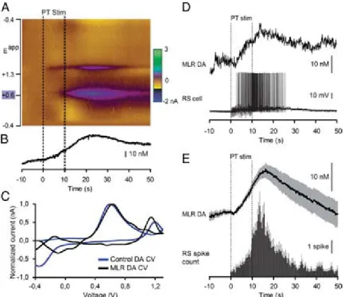
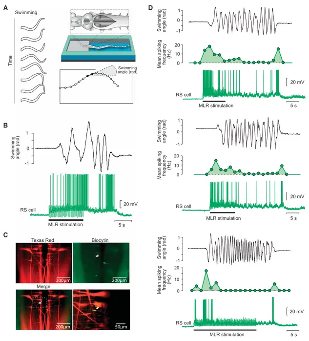
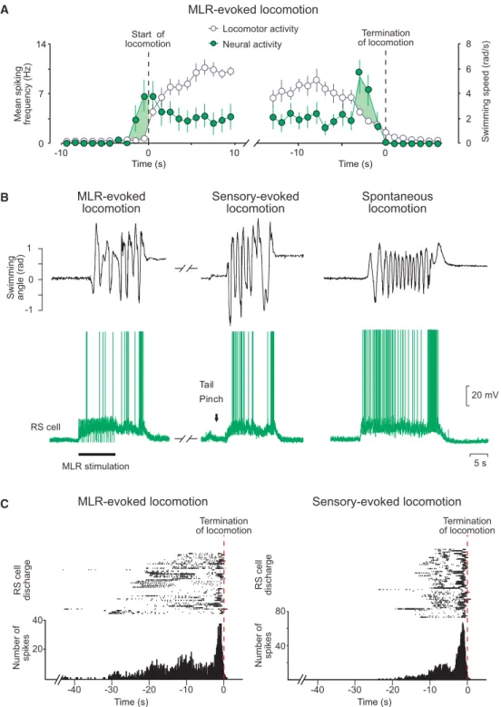
Activation of the Locomotor System by Stimulation of the PT.Phys- iological experiments were carried out to examine the role of this descending DA projection in motor control. For this purpose, a semi-intact preparation was used in which the activity of reticulospinal cells is recorded intracellularly while the body freely swims in the chamber. Trains of stimuli applied to the PT (10-s train, 4–5 Hz, 4–30μA, 2-ms pulses) elicited reticulospinal cell discharges and swimming (n= 13 preparations; Fig. 3A–E).
The location of the stimulation sites was confirmed by histologic examination (Fig. 3D). Chemical stimulation of the PT with local microinjections of 3.0 to 8.0 pmol of D-glutamate (5 mM, 17–43 pulses of 20 ms, 0.6–1.6 nL per microinjection) into the site used for electrical stimulation, was confirmed to elicit locomotion and reticulospinal discharges in one preparation. This is consistent with previous observations that electrical or chemical stimulation
Fig. 1. Dopaminergic (DA) neurons of the PT send descending projections to the MLR. (A–D) TH (red)-containingfibers and varicosities in proximity with MLR cells positive for ChAT (green) in the LDT and the PPN in adult lampreys. The giant I1 reticulospinal cell is outlined inB. (D) Magnification of the dashed rectangle inC. (E–G) Fibers and varicosities immunoreactive to TH (red) surrounded the laterally oriented dendrites of MLR cells retrogradely labeled from an injection of the tracer biocytin (green) in the reticulospinal population of the MRRN in larval lampreys. (H–J) A unilateral injection of the tracer biocytin in the MLR (H,Right, green) followed by immunofluorescence against TH (I, red) revealed double-labeled cells in the PT (white arrows,J). The innervation of the MLR by TH-positivefibers was very similar in larval and adult lampreys.
E3236 | www.pnas.org/cgi/doi/10.1073/pnas.1301125110 Ryczko et al.
with D-glutamate of this region initiates locomotion (10, 11). By using targeted whole-cell patch clamp recordings in an isolated brain preparation (Materials and Methods), we found that trains of stimuli (10-s train, 4–5 Hz, 10–35μA, 2-ms pulses) to the PT (Fig. S2 G and H) directly activate MLR cells projecting to reticulospinal neurons (n= 6 cells from six preparations; Fig. 3 F–I). When comparing simultaneous recordings from an MLR cell and reticulospinal cells (extracellular), we found a very similar activation following trains of stimuli to the PT (Fig. 3H).
Single stimuli to the PT evoked short-latency, large excitatory postsynaptic currents in whole-cell patch recorded MLR cells (Fig. 3I). Glutamatergic receptors are involved in these responses, as previously demonstrated (10), and the DA input from PT to the MLR could modulate this glutamatergic excitatory connection.
The rest of our study was aimed at examining this possibility.
Stimulation of the PT Evokes DA Release in the MLR.Fast-scanning cyclic voltammetry (12) was used to measure changes in DA concentration in the MLR while stimulating the PT (Fig. 4). The location of the recording site was confirmed to be within the MLR (Fig. S2J and K). A reticulospinal neuron was recorded intracellularly to monitor locomotor network activation. Trains of stimuli (10-s train, 5 Hz, 14–25μA, 2-ms pulses) in the PT (Fig. S2GandI) elicited a large increase in DA concentration in the MLR (n= 6 preparations; Fig. 4A–C). We found a strong positive correlation between the increase in DA concentration in the MLR elicited by PT stimulation and the increase in the number of spikes per unit time in reticulospinal cells recorded during the same trials (R= 0.91;P<0.001;n= 30 stimulations in six preparations; Fig. 4DandE). DA release in the MLR was also evoked together with reticulospinal spiking activity when chemically activating the PT with local microinjections of 60.5 pmol of D-glutamate (5 mM, 10 pulses of 100 ms, 12.1 nL per microinjection; n = 20 stimulations in four preparations; Fig.
S3). These data demonstrate that PT activation results in DA release in the MLR and suggest that DA release may contribute to locomotor output.
Blockade of the DA Inputs to the MLR Decreases Locomotor Output.
We then tested whether DA had an effect on the locomotor output elicited by PT stimulation (10-s train, 5 Hz, 12–30μA, 2-ms pulses). Bath-applying DA (10μM) onto the brain induced a 25% decrease in the PT stimulation intensity threshold re- quired to elicit locomotion in two of three semi-intact prepara- tions [reduction from 16 to 12μA in both cases (Fig. S4A–C); 20 μA in the remaining preparation in which no effect was ob- served]. For PT stimulation intensities above locomotor thresh- old (16–30 μA), bath-applied DA increased locomotor bout duration (+86.8±18.4%;P<0.001 vs. control), the number of locomotor cycles (+102.6±23.8%; P<0.001), and locomotor frequency (+25.8 ± 9.1%; P < 0.01; pooled data from three preparations, 18 bouts, and six intensities per preparation;Fig.
S4D–F). These effects were reduced after DA washout (P<0.05 or P < 0.001 vs. DA). Bath-applied DA also increased the number of reticulospinal spikes (+72.4±17.9%; P<0.001 vs.
control) and the duration of spiking activity (+58.4±14.7%;P<
0.001 vs. control). These increases were also reversed after ap- proximately 1 h of DA washout (P<0.01 vs. DA in both cases).
Next, we determined that DA has a direct excitatory effect on the MLR. Microinjections of 1.0 to 7.0 pmol of DA (5 mM,n= 5–38 pulses of 20 ms, 0.2–1.4 nL per microinjection) in the MLR
Fig. 2. TH-positive cells projecting to the striatum or to the MLR are intermingled in the PT as shown in triple-labeling experiments. (A) Dorsal view of a lamprey brain showing the injection sites of the tracers in the MLR (green) and in the striatum (blue). (B–EandF–I) Photomicrographs of two examples of transverse sections in the PT at the level indicated on the dia- gram inA. (Top Right) Diagram illustrates a cross-section at the level of the PT with the approximate location of the photomicrographic frames shown in B–I. DA cells of the PT were labeled with immunofluorescence against TH (red,A, B,andF). Some cells of the PT were retrogradely labeled by a uni- lateral injection of one tracer in the striatum (blue,A,C,andG). Some cells of the PT were retrogradely labeled by another tracer injection, this time in the MLR (green,A,D, andH). (E) The photomicrographs fromB–Dwere merged to show the three markers. White arrowheads indicate some
examples of TH-positive cells of the PT that project to the MLR, whereas the white arrow points to a TH-positive cell projecting to the MLR and the striatum. (I) The photomicrographs fromF–Hwere merged. The white ar- rowhead indicates a PT cell projecting to the MLR and containing TH. The white arrow points to a cell that projects to the striatum and contains TH.
Ryczko et al. PNAS | Published online August 5, 2013 | E3237
NEUROSCIENCEPNASPLUS
(as confirmed by histologic examination; Fig. S2J and L) in- creased the locomotor output elicited by trains of electrical stimulation (10-s train, 5 Hz, 4–7μA, 2-ms pulses) in the PT (Fig.
3D) in a semi-intact preparation (n = 25 injections in five preparations; Fig. 5 A–C). DA microinjections prolonged the locomotor bout duration (+56.7 ± 14.3%; P < 0.001) and in- creased the number of locomotor cycles (+50.8 ± 12.3%; P<
0.001). These effects were reversed after DA washout (P<0.05 vs. injection in both cases; Fig. 5C). DA injections in the MLR also increased the duration of reticulospinal cell spiking activity (+33.6± 11.8%;P < 0.01). This effect was reversed after ap- proximately 1 h of washout (P<0.01; Fig. 5C). The effects on locomotor threshold were not measured in these experiments.
The excitatory effect of DA microinjections in the MLR was lower than that of bath-applied DA, probably because of the single site of action of DA during local microinjections. For in- stance, local DA microinjections in the MLR did not significantly modify the locomotor frequency that remained at 95.2±3.0% of control (P > 0.05), or the number of reticulospinal spikes that remained at 119.5±11.1% of control (P>0.05).
D1 receptor activation is known to have excitatory effects on striatal cells in mammals (reviewed in ref. 13). Those receptors are also present in lampreys (14, 15). Targeted blocking of D1 receptors in the MLR (Fig. S2Jand L) dramatically decreased the locomotor output elicited by stimulation of the PT (10-s
train, 5 Hz, 7–11μA, 2-ms pulses; Fig. 3D). Local microinjection of 0.1 to 0.8 pmol of the D1 antagonist SCH 23390 (500μM, 6–43 pulses of 20 ms, 0.2–1.6 nL per microinjection) in the MLR (n= 25 injections infive preparations; Fig. 5D–F) decreased the duration of locomotor bouts (−48.4 ± 10.6%; P < 0.001 vs.
control), locomotor frequency (−32.2± 6.3%;P< 0.001), and the number of locomotor cycles (−60.0 ± 9.9%; P < 0.001).
These decreases in locomotor output were reversed after wash- out (for all parameters,P<0.001 vs. injection; Fig. 5F). Blocking D1 receptors in the MLR also decreased the duration of spiking activity in reticulospinal neurons (−43.8±9.4%;P <0.001 vs.
control), their discharge frequency (−30.8±9.9%;P<0.01), and their number of spikes (−41.6±11.9%;P<0.001). Recovery was obtained after approximately 1 h of washout (P <0.05 orP <
0.01 vs. injection; Fig. 5F).
Discussion
Newly Identified Descending DA Pathway.In this study, we provide evidence for a descending DA projection from the PT to the MLR that modulates motor output. This descending DA path- way supports DA release, which increases locomotor output, and D1 receptors are involved in this excitatory effect. It appears that this descending DA pathway amplifies the previously known excitatory glutamatergic inputs to MLR cells (10). Such a descending DA projection is also very likely to be present in
Fig. 3. Stimulation of the PT activates brainstem locomotor circuits. (A–E) In a semi-intact preparation, the PT was stimulated and the reticulospinal (RS) cells were recorded together with locomotor movements. (BandC) Body curvature oscillations during locomotion. (C) PT stimulation (10-s train, 5 Hz, 11μA, 2-ms pulses) elicited swimming as illustrated by the rostrocaudal mechanical wave. (D) The PT stimulation site was confirmed histologically by an electrolytic lesion (enclosed by white dashed line) that colocalized with TH-immunoreactive neurons (red). (Scale bar: 200μm.) (E) PT stimulation elicited reticulospinal activity together with swimming. Stimulation artifacts were clipped. (F–I) Whole-cell patch-clamp recordings of MLR neurons retrogradely labeled from an injection of dextran amines conjugated to Texas red (MW, 3,000 Da) in the MRRN. (FandG) MLR neuron labeled by the Texas red–dextran amine injection in the MRRN and by thefluorescent marker added to the patch pipette solution (Alexa Fluor 488 hydrazide). (Scale bar: 10μm.) (H) Trains of stimuli (10-s train, 5 Hz, 7μA, 2-ms pulses) activated the whole-cell patch-clamped MLR neuron concomitantly with reticulospinal neurons, which were used to monitor locomotor acti- vation. Action potentials were recorded extracellularly from reticulospinal neurons of the MRRN, and stimulation artifacts were clipped. (I) Single pulse PT stimulation (0.1 Hz, 7μA, 2-ms pulses) elicited short-latency excitatory postsynaptic currents in MLR neurons. (HandI) Data from two different animals.
E3238 | www.pnas.org/cgi/doi/10.1073/pnas.1301125110 Ryczko et al.