Human MicroRNA Pathway
DISSERTATION
ZUR ERLANGUNG DES
DOKTORGRADES DER NATURWISSENSCHAFTEN (DR. RER. NAT.) DER FAKULTÄT FÜR BIOLOGIE UND VORKLINISCHE MEDIZIN
DER UNIVERSITÄT REGENSBURG
Vorgelegt von Daniele Hasler
aus Rom (Italien)
Im Jahr 2018
Das Promotionsgesuch wurde eingereicht am: 01. März 2018
Die Arbeit wurde angeleitet von: Prof. Dr. Gunter Meister
Unterschrift:
“Mi manderete a scuola?” chiese Peter Pan.
“Sì”
“E poi in ufficio?”
“Credo di sì”
“E presto sarò un uomo?”
“Molto presto”
“Ma io non voglio andare a scuola e imparare cose serie…
non voglio diventare un uomo.
Oh,… pensa se un giorno dovessi svegliarmi e accorgermi di avere la barba!”
Peter Pan
I I
Contents
Contents I
Abstract V
Zusammenfassung VI
1. Introduction 1
1.1. TRNA Biology: Ancient But Still Attractive 1
1.1.1. Structure and Nucleotide Modifications Guarantee tRNA Functionality 2
1.1.2. Organisation of tRNA Genes 5
1.1.3. Transcription by Pol III 8
1.1.3.1. Pol III Transcription Is Driven by Different Promoter Types 8
1.1.3.2. Regulation of Pol III Transcript Levels 9
1.1.3.3. Transcription Termination by Pol III 11
1.1.4. Maturation of tRNA Ends 12
1.1.4.1. 5’ End Processing of Pre-tRNAs by RNase P 12
1.1.4.2. 3’ End Processing of Pre-tRNAs by RNase Z 13
1.1.4.3. Alternative Processing of Pre-tRNA Ends 16
1.1.5. CCA Addition by the tRNA Nucleotidyltransferase 18
1.1.6. Extravagant Splicing Mechanism for tRNA Introns 20
1.1.6.1. A Specialized Splicing Machinery Removes tRNA Introns 20
1.1.6.2. Diverse Strategies for Joining the tRNA-Splicing Intermediates 23
1.1.7. Mechanisms of tRNA Export from the Nucleus 24
1.1.8. Final Processing by Aminoacyl-tRNA Synthetases 27
1.1.8.1. Loading of the Cognate Amino Acid 27
1.1.8.2. ARSs Safeguard Correct tRNA Maturation at Several Steps 29
1.1.9. Quality Control and Turnover of tRNAs 30
1.1.10. Many Faces of tRNA Fragments 33
1.1.10.1. Pre-tRNAs as an Early Source of tRFs 34
1.1.10.2. Mature tRNA Halves 35
1.1.10.3. Puzzling Diversity of tRFs 38
1.2. The miRNA Pathway 42
1.2.1. The Nuclear Part of MiRNA Biogenesis 42
1.2.2. The Cytoplasmic Part of MiRNA Biogenesis 44
1.2.3. Ago Proteins, the Key Players of the MiRNA Pathway 46
1.2.4. An Interplay of Processes Leads to the Repression of Targeted Transcripts 47 1.2.5. Many Roads Lead to Ago: Generation of Non-canonical MiRNAs 48
2. Aims of the Study 53
I
II I
3. Results 55
3.1. Generation of a La-specific Antibody 55
3.2. La Affects the Cellular sRNA Population 56
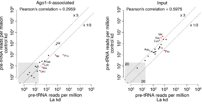
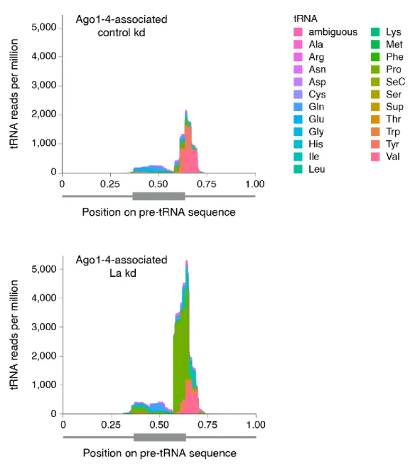
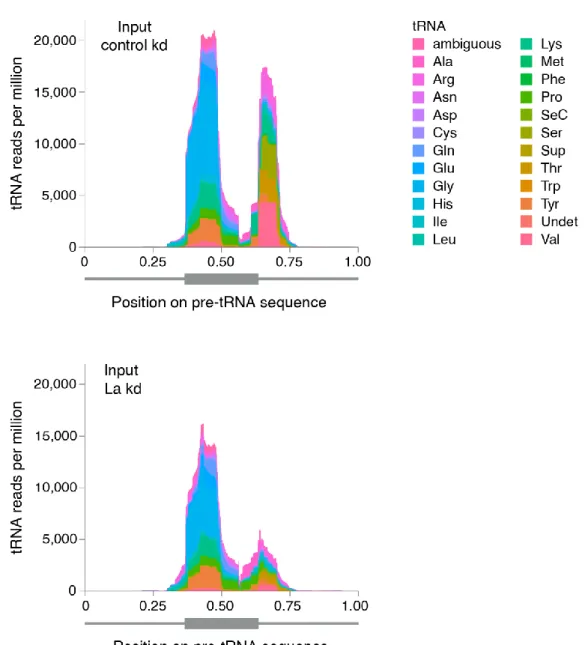
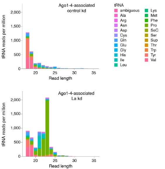
3.3. La Regulates the Abundance of Specific Ago-loaded tRNA Fragments 61
3.4. La-dependent Pre-tRNA Fragments Have MiRNA Characteristics 64
3.5. The Pre-tRNA Pro-CGG-2-1 Generates an Ago-loaded sRNA in the Absence of La 66
3.6. A Bona Fide MiRNA is Generated from Pre-tRNA-Ile-TAT-2-3 74
3.7. The sRNA-Ile Is Loaded into Functional Silencing Complexes 80
3.8. La is the Main Regulator for the Transition of Pre-tRNA-Ile-TAT-2-3 to miR-1983 85 3.9. Full-length La Protein is Required for the Interaction with Pre-tRNA-Ile-TAT-2-3 89 3.10. The Double-stranded Stem of Pre-tRNA-Ile-TAT-2-3 Reduces its Affinity to La 93 3.11. Viral Pol III Transcripts Sequester La and Influence the Cellular Landscape of sRNAs 96
4. Discussion 101
4.1. Crosstalk Between La and the MiRNA Machinery 101
4.2. Processing Products of tRNAs are Abundant and Affected by La Depletion 103 4.3. La is a Gatekeeper Preventing Mis-channeling of tRNA Fragments into the
Human MiRNA Pathway 105
4.4. Hidden Pitfalls in the Analysis of tRFs 108
4.5. Pre-tRNA-Ile-2-3 Escapes La Binding by an Alternative Folding 109 4.6. The Chimeric Pre-tRNA-Ile-TAT-2-3 Generates Either a Functional tRNA or MiR-1983 110
4.7. Making a Fool of La? 113
5. Material and Methods 116
5.1. Material 116
5.1.1. Consumables and Chemicals 116
5.1.2. Kits and Ready-made Solutions 116
5.1.3. Materials for Small-scale Purifications and Chromatography Columns 117
5.1.4. Laboratory Equipment 118
5.1.5. Buffers and Solutions 119
5.1.6. RNA and DNA Oligonucleotides 120
5.1.6.1. Oligonucleotides Used for Preparation of Deep-sequencing Libraries 120
5.1.6.2. RNA Oligonucleotides 120
5.1.6.3. DNA Oligonucleotides 122
5.1.7. Plasmids 126
5.1.8. Antibodies 128
5.1.9. Bacterial Strains and Cell Lines 129
5.1.9.1. Bacterial Strains 129
5.1.9.2. Mammalian Cell Lines and Growth Media 129
5.2. Methods 130
5.2.1. Molecular Biological Methods 130
5.2.1.1. Determination of Nucleic Acid Concentration 130
5.2.1.2. Polymerase Chain Reaction (PCR) 130
5.2.1.3. Agarose Gel Electrophoresis 131
5.2.2. Molecular Cloning 131
5.2.2.1. Restriction Digest of DNA and Dephosphorylation of Linearized Vectors 131
5.2.2.2. Annealing and Phosphorylation of DNA Fragments 131
5.2.2.3. DNA Insert Ligation into Vectors 132
5.2.2.4. Transformation and Cultivation of E. coli 133
5.2.2.5. Extraction of Plasmid DNA from E. coli and Test Digest 133
5.2.2.6. Mutagenesis PCR 134
5.2.2.7. Sequencing of Plasmid DNA 135
5.2.3. RNA-based Methods 135
5.2.3.1. Isolation of RNA 135
5.2.3.2. CDNA Synthesis and Quantitative Real-time PCR (qPCR) 135
5.2.3.3. Denaturing Polyacrylamide Gel Electrophoresis (PAGE) 136
5.2.3.4. In vitro Transcription 136
5.2.3.5. 32P-Labeling of Nucleic Acids 137
5.2.3.6. Northern Blot and Determination of Copy Number per Cell 137
5.2.3.7. Aminoacylation Assay 138
5.2.3.8. In silico RNA Methods 139
5.2.4. Biochemical Methods 139
5.2.4.1. Determination of Protein Concentration 139
5.2.4.2. SDS- PAGE 140
5.2.4.3. Coomassie staining 140
5.2.4.4. Western Blot 140
5.2.4.5. Immunoprecipitation, Ago-APP and Isolation of Co-precipitated RNAs 141
5.2.4.6. Small RNA Cloning and Data Analysis 142
5.2.4.7. PAR-CLIP Experiments and Data Analysis 143
5.2.4.8. Dicer Cleavage Assay 144
5.2.4.9. Expression and Purification of Recombinant Proteins 145
5.2.4.10. Generation and Purification of Polyclonal Antibodies 146
5.2.4.11. EMSA 147
5.2.5. Cell Culture Methods 147
5.2.5.1. Transfection of Mammalian Cells 147
5.2.5.2. Dual Luciferase Assay 148
6. Contributions 149
7. Data Publication 150
8. Abbreviations 151
9. List of Figures 158
10. List of Tables 160
11. References 161
12. Acknowledgments 200
V V
Abstract
The Lupus autoantigen La is an RNA-binding protein that stabilizes RNA polymerase III (Pol III) transcripts and supports correct RNA folding. In addition, La was shown to affect the biogenesis of mammalian microRNAs (miRNAs).
In this study, we have analyzed the consequences of La depletion on the Argonaute (Ago)-bound small RNA (sRNA) population in human cells. We find that in the absence of La, distinct tRNA fragments are loaded into Ago proteins. Thus, La functions as gatekeeper ensuring correct tRNA maturation and thereby protects the cell from tRNA fragments, which might be potentially harmful by acting as unintended miRNAs.
We further provide evidences, that viral non-coding RNAs (ncRNAs) perturb La’s gatekeeper function and induce the production of Ago-loaded tRNA fragments, mimicking the effects observed under La depletion.
Interestingly, one specific isoleucine precursor tRNA (pre-tRNA) produces both, a tRNA and a functional miRNA, even when La is present. We demonstrate that this specific pre- tRNA is able to partially escape from La binding due to its fully complementary 5’ leader and 3’ trailer sequences. The double-stranded RNA structure of this specific isoleucine pre-tRNA reduces the affinity to La and allows processing by components of the miRNA biogenesis machinery.
In sum, our study unraveled a novel aspect of La biology, showing that it supports correct
pathway selection and, by that, maturation of primary Pol III transcripts. Furthermore, we
characterized in molecular detail the biogenesis of a non-canonical, pre-tRNA-derived
miRNA.
Zusammenfassung
Das Protein Lupus Autoantigen La bindet an das 3’ Ende von primären RNA Polymerase III (Pol III) Transkripten und schützt sie somit vor exonukleolytischem Abbau. Zudem fördert La die korrekte Faltung der gebundenen Transkripte. Des Weiteren wurde La als ein Faktor beschrieben, der die Biogenese einiger mikroRNAs (miRNAs) reguliert.
In dieser Arbeit wurde der Einfluss von La auf die Population von kurzen RNAs untersucht, die mit Argonaut (Ago) Proteinen interagiert. Daraus ergab sich, dass La die Entstehung von einigen kurzen tRNA Fragmenten verhindert. In Abwesenheit von La akkumulieren diese tRNA Fragmente in Ago Protein-Komplexen und könnten daher fälschlicherweise als miRNAs wirken, was mögliche negativen Konsequenzen birgt.
Zusätzlich liefert unsere Studie Hinweise dafür, dass diese Kontroll-Funktion von La durch virale, nicht-kodierende RNAs beeinträchtigt wird. Die Expression solch viraler RNAs führte zur Produktion von Ago-interagierenden tRNA Fragmenten. Somit ähnelte dieser Effekt den Auswirkungen einer La Depletion.
Interessanterweise kann aus einer bestimmten Isoleucin Vorläufer-tRNA (pre-tRNA) sogar in Anwesenheit von La eine tRNA oder auch eine funktionelle miRNA entstehen.
Wir konnten zeigen, dass die strukturellen Besonderheiten dieser pre-tRNA eine effiziente Bindung durch La verhindern. Dies beruht darauf, dass die 5’ und 3’ Überhänge der Isoleucin pre-tRNA stark komplementär zueinander sind. Die dadurch entstehende doppelsträngige Struktur verursacht zwar eine verminderte Affinität zu La, ermöglicht jedoch gleichzeitig die Prozessierung durch Komponenten der miRNA Biogenese Maschinerie.
Zusammengefasst hat unsere Studie eine bis dahin unbekannte Funktion des Proteins La
aufgeklärt. La besitzt eine wegweisende Rolle in der Reifung von primären Pol III
Transkripten und verhindert dadurch, dass pre-tRNAs durch konkurrierende Prozesse
fehlgeleitet werden, z.B. in den miRNA-Biogeneseweg. Zudem wurde der molekulare
Mechanismus aufgeklärt, der dazu führt, dass eine nicht-kanonische miRNA aus einer
pre-tRNA entstehen kann.
1 1
1. Introduction
1.1. TRNA Biology: Ancient But Still Attractive
The fundamental role of tRNAs in protein translation was unravelled very early in the history of molecular biology (Hoagland et al., 1958). By translating the code of nucleic acids into the code of proteins, tRNAs act as the Rosetta Stone of the cell, allowing genetic information to be expressed. In order to fulfil this central role, tRNA functionality needs to be guaranteed. Although the final mode of action of tRNAs within translation is conserved among all domains of life, it is striking how frequently convergent evolution resulted in different mechanisms to ensure that the requirement of functional tRNAs is satisfied. This aspect of tRNA biology, in particular, still leaves many open questions.
Are some molecular mechanisms described in specific model organisms peculiar for them or are they shared as well by other species?
Not only do the details in tRNAs’ life cycle remain partially elusive but also the plethora of RNA base modifications and the responsible enzymes are detected and characterized in only a few organisms (El Yacoubi et al., 2012). Furthermore, the extent of tRNA modification dynamics and the resulting regulatory potential have been addressed by a handful of studies, but still require broader investigations (Emilsson et al., 1992; Noon et al., 2003; Chan et al., 2010; Yi and Pan, 2011).
In addition to their well-known function in translation, tRNAs fascinate also by their
many non-translational activities (reviewed in Schimmel, 2017), starting from the
chromatin insulator role of tRNA genes as shown for different eukaryotes (Kuhn et al.,
1991; Raab et al., 2012). Mature, aminoacylated tRNAs are involved as well in several
pathways, for example, tRNA-Arg and the arginyl-tRNA-protein transferase act in a
specific protein degradation pathway (Kaji, 1968; Balzi et al., 1990). Furthermore,
aminoacylated tRNA-Glu has been shown to serve as a substrate for the biosynthesis of
tetrapyrroles (i.e., chlorophyll and heme) in green plants, archaebacteria and many
eubacteria (Huang et al., 1984; Schön et al., 1986; Jahn et al., 1992). Recently it has been
reported that tRNA binding to cytochrome c leads to inhibition of caspase activation and
2
2 IN
INT TR RO OD DU UC CT TI IO ON N
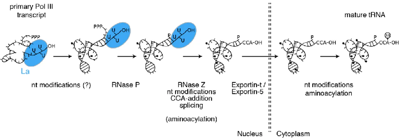
Figure 1.1: TRNA Maturation is a Multistep Process. Schematic representation of processing events and key enzymes required during the biogenesis of tRNAs. The La protein (blue) binds and protects the 3’ end of pre-tRNA transcripts supporting their correct folding. The order of the reactions, enzymes as well as cellular localization of the steps might vary under certain conditions and between different organisms.
therefore blocks apoptosis (Mei et al., 2010). Intriguingly, mature tRNAs can be hijacked by retroviruses and long terminal repeat (LTR) retrotransposons during their replication cycles, and are thereby used as primers for reverse transcription (Rosenthal and Zamecnik, 1973; Mak and Kleiman, 1997).
In the past decade a new aspect of tRNA biology has emerged, namely, the occurrence of tRNA-derived fragments. They seem to excerpt diverse biological roles and several modes of actions have been proposed. However, published data are not always satisfying and are often contradictory. In this regard, the elusive nature of tRNA-derived fragments is probably the most prominent challenge to tackle next within the tRNA field.
1.1.1. Structure and Nucleotide Modifications Guarantee tRNA Functionality
TRNAs are highly transcribed and represent one of the most abundant transcript class within a cell. It has been estimated that a single yeast cell contains three million tRNAs (Waldron and Lacroute, 1975). This tremendous amount of molecules is transcribed by RNA polymerase III (Pol III). The following sections aim to describe several processing steps tRNAs undergo before being loaded with the correct amino acid (Figure 1.1).
The outcome of tRNA biogenesis are structurally highly conserved molecules which have
co-evolved with the ribosome to match the requirements for protein synthesis (reviewed
in Tamura, 2015; Zhang and Ferré-D’Amaré, 2016). Importantly, the folding of tRNAs is
not only relevant for its fundamental role during translation, but its acquisition during the
maturation of tRNAs also governs several subsequent reactions (e.g., removal of the 5’
leader sequence from pre-tRNAs by RNase P) and structural determinants are continuously interrogated for quality control (e.g., by the tRNA nucleotidyltransferase).
The typical secondary structure of tRNAs resembles a cloverleaf and according to distinguishing attributes the different stem-loops have been named D-arm, T-arm and anticodon arm (Figure 1.2A). The loop of the D-arm contains several dihydrouridine modified nucleosides, while the loop of the T-arm almost universally starts with a ribothymidine, which is uncommon in RNAs. It is followed by a pseudouridine (Ψ) and a cytidine, whereby the T-arm is alternatively also referred to as TΨC-arm. The anticodon itself, consisting of three nucleotides pairing with the codon triplet on the mRNA, is located at the center of the anticodon loop. Between the anticodon arm and the T-arm a segment of variable length can occur (variable arm).
The first crystal structure of a tRNA was solved in the 70’s (Kim et al., 1973) and revealed the L-shape of this molecule. The acceptor stem and the T-arm form one segment, which is also called minihelix, while the D-arm together with the anticodon arm form the second part of the L-shape. These two branches are joined by the elbow which consists of the loops of the D- and T-arms (Figure 1.2B). Importantly, several Watson- Crick interactions as well as extensive unconventional base pairings occur within this region and are essential for stabilizing the overall tertiary structure of the tRNA (reviewed in Zhang and Ferré-D’Amaré, 2016).
Figure 1.2: Structural Characteristics of Mature tRNAs. (A) The secondary structure of tRNAs resembles a cloverleaf. D-arm, anticodon arm, variable arm, T-arm and the acceptor stem are depicted in different colors. The last four nucleotides at the 3’ end of the acceptor stem are indicated. They form a single-stranded overhang, whereby the terminal CCA is added post-transcriptionally. The preceding nucleotide (N) is termed discriminator base. (B) TRNAs adopt an L-shaped tertiary structure. The corresponding colors as in (A) are used. Figure adapted from Tamura (2015).
4
4 IN
INT TR RO OD DU UC CT TI IO ON N
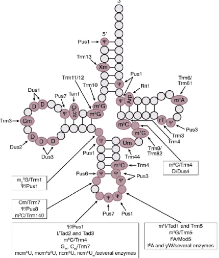
Figure 1.3: Frequent Modifications and Responsible Enzymes for Yeast Cytoplasmic tRNAs.
Schematic representation of a tRNA with circles indicating nucleotides and lines indicating base pairings.
Positions which are unmodified in all tRNAs are depicted with lighter circles, positions where modifications can occur are shown with darker circles. Figure adapted from El Yacoubi et al. (2012);
abbreviations used as listed therein.
A multitude of tRNA structures have been obtained meanwhile and some of them depict the tRNA bound by some processing and maturation enzymes or engaged at the ribosome during different translation steps (reviewed in Giegé et al., 2012). These data highlight the intrinsic flexibility of tRNAs, especially of the elbow, which accounts for the variability in the positioning of the two L-shaped segments relative to each other (Giegé, 2008;
Giegé et al., 2012).
Important for the correct folding (Helm et al., 1999) as well as for the structural
stabilization of tRNAs (Anderson et al., 1998, see also sections 1.1.4.3 and 1.1.9) is their
amazing repertoire of posttranscriptional modifications. Approximately 100 different
nucleoside variants have been identified. Their generation requires the activities of an
astonishing amount of highly specialized enzymes, which are very often responsible for
position-specific modification of certain bases (Figure 1.3).
Due to the complex chemical properties of some of these nucleotide variants it is frequent that they are obtained in multi-step reactions through the action of several proteins (reviewed in El Yacoubi et al., 2012; Hori, 2014). Despite all the efforts life has dedicated to the evolution of these elaborated modifications, only few of them are strictly essential or are related to severe phenotypes. These are mostly located within the anticodon loop (Björk et al., 2001; Wolf et al., 2002; El Yacoubi et al., 2009) and the lack of specific modification has been implicated in different human diseases as well (Kirino et al., 2004;
Wei et al., 2011; reviewed in Torres et al., 2014). TRNA modifications within the anticodon loop contribute in several ways to the accuracy of protein translation. First of all, modified nucleoside behave as identity determinants for the enzyme, specifically charging tRNAs with the correct amino (reviewed in El Yacoubi et al., 2012). Most importantly, tRNA modifications within the anticodon loop impact translation rates (Nedialkova and Leidel, 2015) and contribute to decoding accuracy by stabilizing codon- anticodon interactions. In this context they are required either for allowing wobble interactions or for ensuring stringent discrimination of closely related codons. This is particularly relevant for so-called split codon boxes whereby different amino acids (or a stop codon) are decoded depending only on the last nucleotide of the codon (Björk et al., 2007; Johansson et al., 2008).
1.1.2. Organisation of tRNA Genes
Currently, 610 tRNA genes are annotated at the genomic tRNA database (gtRNAdb) 2.0
for the human genome (Chan and Lowe, 2015). According to the amino acid they carry,
tRNA genes are grouped into different isotypes. Due to the degeneracy of the genetic
code, one specific amino acid can be encoded by several codons, which are recognized by
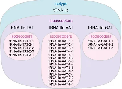
distinct tRNA isoacceptors. For the nomenclature of different tRNA isoacceptors, the
sequence of the anticodon is used (e.g., tRNA-Ile-TAT and tRNA-Ile-AAT). A specific
isoacceptor group consists of so-called isodecoders, i.e., tRNAs sharing a common
anticodon. Although originating from different genomic loci, some mature tRNA
isodecoders might have identical sequences. However, isodecoders can also slightly differ
in their tRNA body sequence. The hierarchical grouping of tRNA genes is exemplarily
shown in Figure 1.4 for the tRNA-Ile isotype.
6
6 IN
INT TR RO OD DU UC CT TI IO ON N
Of note, not all possible tRNA isoacceptors are encoded in the genome (Figure 1.5). This is possible because wobble interactions (i.e., non-standard Watson-Crick pairings) can occur at the third base within a codon with the first base of a tRNA’s anticodon. Thus, a smaller set of tRNA isoacceptors allows decoding of all 64 possible codon sequences.
Figure 1.5 also shows, that the number of certain tRNA isodecoder genes is over- represented for certain isoacceptors, e.g., for tRNA-Cys-GCA (38), compared to others, e.g, to tRNA-Ser-GGA (1). The reasons for this fact remain elusive, since tRNA copy numbers do not always correlate with the codon usage rates.
Regarding their distribution, tRNA genes are in general dispersed throughout the human genome, differing from the strictly clustered localization of rRNA repetitive units. Still, small clusters especially on chromosomes 1, 6 and 17 are present as well (Raab et al., 2012). In the fission yeast Schizosaccharomyces pombe clustering is more pronounced and tRNA genes are found at centromeric DNA sequences (Kuhn et al., 1991). It has been shown that binding of Pol III-specific transcription factors to these tRNA genes is sufficient to hinder spreading of repressive chromatin marks to distal regions.
Interestingly, the insulator function of tRNA genes is conserved in humans as well, although the small clusters are not present at centromeres (Raab et al., 2012).
Figure 1.4: TRNAs Are Hierarchically Grouped into Isotypes (blue), Isoacceptors (violet) and Isodecoders (pink). Illustration of the different terms used to categorize tRNAs. See main text for details.
Please note in the numbering of tRNA isodecoders that those sharing the same first number (e.g., tRNA-Ile- TAT-2-1, tRNA-Ile-TAT-2-2 and tRNA-Ile-TAT-2-3) have identical mature tRNA sequences.
Nevertheless, they can originate from distinct genomic loci which are unambiguously indicated by the second number.
Figure 1.5: Overview of Predicted Human tRNA Genes and Corresponding Codon Usage. The tRNA isotypes are grouped in different sets according to the number of anticodons used to decode the same amino acid. Potential suppressor tRNAs are shown in grey as no experimental evidence exists for their functionality in humans. Consider that the codons TGA, TAA and TAG are recognized as stop codons under normal conditions. Data obtained from the gtRNAdb 2.0 (Chan and Lowe, 2015).
8
8 IN
INT TR RO OD DU UC CT TI IO ON N
The proximity of tRNA loci to each other is not only visible in regard to their position on the genomic DNA but is also evident from their spatial localization within the nucleus.
The three-dimensional organization of tRNA genes has been studied in great detail in the budding yeast Saccharomyces cerevisiae and actively transcribed tRNA genes were detected in nucleoli (Thompson et al., 2003). TRNA genes were shown also in human cells to be enriched in nucleolus-associated chromatin domains (Németh et al., 2010), however, not many analyses have been performed in the human system.
1.1.3. Transcription by Pol III
1.1.3.1. Pol III Transcription Is Driven by Different Promoter Types
Pol III is the largest of the three RNA polymerases and, yet, it is structurally the least characterized. Only recently, structures at ~4 Å resolution have been achieved by cryo- electron microscopy (EM) (Hoffmann et al., 2015; Abascal-Palacios et al., 2018), although a first low resolution structure at 17 Å was published almost ten years before applying cryo-EM as well (Fernández-Tornero et al., 2007). The Pol III holoenzyme consists of 17 subunits, of which ten are unique to Pol III, while the other seven subunits are either shared by Pol I and Pol III or are present in all three RNA polymerases.
Transcription by Pol III is directed by three distinct promoter types (I, II and III) which differ in regard to cis regulatory elements; consequently, the composition of transcription factors involved varies among the different promoters (Figure 1.6). Common to all promoters is the requirement of the general transcription factor TFIIIB. This complex consists of the TATA box-binding protein (TBP), the Pol III-specific transcription factors TFIIIB component B'' homolog (BDP), and TFIIB-related factor 1 (BRF1) or TFIIB- related factor 2 (BRF2). While the binding of TBP to a TATA-box or TATA-like sequence is required for type III promoters, the presence of such sequence elements is not mandatory for the other two promoter types, but has been shown to increase transcription efficiency (Giuliodori et al., 2003). In addition, activity of type III promoters is facilitated by the binding of the small nuclear RNA-activating protein complex (SNAP
c) to a proximal sequence element (PSE) and by the binding of OCT1 and STAF to the distal sequence element (DSE) (reviewed in Schramm and Hernandez, 2002). Of note, the promoter type III is found only in multicellular organisms.
In contrast, the main characteristic of promoter type I and II is that the sequence elements
driving transcription are located within the transcribed sequence. These so called A-box,
B-box, C-box and intermediate elements (IE) are therefore very often embedded in the mature transcripts (e.g., in tRNAs). In the case of type II promoters, which drive the expression of all tRNA genes with a minor exception, the A-box and B-box form together a specific binding site for positioning the multisubunit complex TFIIIC (Figure 1.6).
1.1.3.2. Regulation of Pol III Transcript Levels
As mentioned before, TFIIIB consists of three different proteins (TBP, BDP and BRF1 or BRF2), where the exact composition depends on the promoter type. BRF1 is required for transcription from internal promoters (type I and II), while it is replaced by the vertebrate- specific BRF2 at type III gene-external promoters. This variance might account for the potential to regulate specifically gene expression of promoter type III genes
Figure 1.6: Type I, II and III of Pol III Promoter Differ in Sequence Elements and Required Transcription Factors. See main text for details. IE, intermediate element; DSE, distal sequence element;
PSE, proximal sequence element; TTTT transcription termination signal; TATA, TATA box or TATA-like sequence, parentheses indicate that not all type I and type II promoters contain a TATA element. Adapted from Dieci et al. (2007) and Oler et al. (2010).
1
10 0 IN
INT TR RO OD DU UC CT TI IO ON N
independently from the other promoter types. In fact, recent X-ray crystallographic structures of BRF2-TBP complexes bound to different natural promoter sequences shed light on a fascinating feature of BRF2: the presence of a redox-sensing regulatory module wherein a conserved cysteine residue (C361) can cycle through reduced and oxidized states. In case C361 is oxidized, assembly of the complex on DNA is severely compromised (Gouge et al., 2015). Consequently, BRF2-dependent transcription of promoter type III genes is reduced during oxidative stress. Of note, the only tRNAs possessing such promoters are tRNA-SeC gene loci, while all the other tRNAs are transcribed from type II promoters (Oler et al., 2010). Selenocystein is especially required for the synthesis of several proteins directly involved in the oxidative stress response.
However, the BRF2-dependent down regulation of tRNA-SeC levels during prolonged oxidative stress conditions and the resulting decrease of selenoproteins expression, lead to the induction of apoptosis (Gouge et al., 2015). This protective mechanism seems to be overcome in several types of cancers, where oncogenic BRF2 overexpression is observed (Cabarcas and Schramm, 2011).
The regulation of tRNA-SeC expression by the BRF2 redox-sensor is probably the only known mechanism explaining how tRNA levels can be modulated at the transcriptional level. Indeed, several pieces of evidence emerged during the past years suggest that the protein synthesis machinery is not fuelled by a constant tRNA pool (reviewed in Kirchner and Ignatova, 2014; Orioli, 2017). First indications came from microarray-based analyses of different tissues, demonstrating remarked levels of tRNA heterogeneity among the tested samples (Dittmar et al., 2006). Furthermore, chromatin immunoprecipitation (ChIP) experiments revealed considerable variance in the tRNA loci occupied by Pol III.
Also, the total amount of in silico predicted tRNA genes bound by Pol III varied between
four different cell lines, ranging from ~30% to ~60% (Oler et al., 2010). Finally, a broad
microarray-based study extensively demonstrated changes in the tRNA pool among
proliferative, differentiating and senescent cells (Gingold et al., 2014). The tRNA content
of several cancer types and cancer stages was also characterized recently, providing first
evidences for the relevance of differing tRNA levels. These changes correlated with a
shift in the codon-usage signature of genes involved in the driving processes of the
analyzed conditions (Gingold et al., 2014; Goodarzi et al., 2016). A similar adaptive
dynamic in tRNA supply and codon-usage demand has been proposed as well for cell
cycle‐regulated genes (Frenkel-Morgenstern et al., 2012). All these findings depict the
coordinated adaptation of the expressed tRNA subset and the codon-usage requested
under specific situations. According to such a system, the translation of the requested proteins will be favoured in contrast to unintended gene products (Gingold et al., 2014;
Quax et al., 2015). Still, the chicken or the egg question remains, regarding the driving force for this co-evolution process: did the tRNA pool adapt to a varying demand or did the bias in codon-usage of important genes react to the altered tRNA composition? In both cases, the molecular mechanisms resulting in the differential tRNA expression remain largely elusive. The core elements of tRNA genes are very similar to each other and motif analyses of Pol III bound versus non-bound tRNA genes did not reveal any differences in A-box and B-box sequence composition (Oler et al., 2010) or only modest variances (Canella et al., 2010). However, when similar analyses were performed with tRNAs enriched during differentiation or proliferation, the sequence motif of the B-box differed between the two subsets and therefore might be a first indication of the underlying regulatory mechanism (Gingold et al., 2014).
1.1.3.3. Transcription Termination by Pol III
Similarly to the relatively unsophisticated organisation of Pol III promoters, transcription termination by Pol III is driven by a direct mechanism, simply requiring a short oligo(dT) tract on the non-template strand (Arimbasseri and Maraia, 2015). Neither post- translational modification of the polymerase nor the activity of trans-acting factors is required for Pol III termination unlike for Pol II (Arimbasseri et al., 2013). Although the short oligo(dT) tract is the universal terminator for Pol III, the minimum length for efficient termination varies among different species. The yeast S. cerevisiae requires six or more Ts for efficient termination (Allison and Hall, 1985), while in vertebrates four Ts are generally sufficient (Bogenhagen and Brown, 1981; Cozzarelli et al., 1983). At the same time, sequence analyses of human tRNA loci revealed the presence of strong terminator sequences (i.e., having more than four Ts) more downstream than observed in S. cerevisiae (Orioli et al., 2011). The more proximal terminator sequences instead appear to be weaker in humans. This, together with other findings, suggests that transcription of tRNA genes in vertebrates is more prone to read-through events resulting in 3’ extended transcripts. The strong secondary terminators ensure thereby stringent termination (Arimbasseri et al., 2013).
The context surrounding the stretch of four Ts influences the ability of Pol III to terminate
transcription, at least in some situations (Gunnery et al., 1999). The ability of Pol III to
1
12 2 IN
INT TR RO OD DU UC CT TI IO ON N
discriminate permissive oligo(dT) tracts is fundamental for the production of Lys-TTT tRNAs. Beside the required three Us in the anticodon, an invariant U immediately upstream of them is present, resulting in four consecutive Ts on the non-template strand (Mazabraud et al., 1987). On the other side, termination at non-canonical oligo(dT)- stretches has been observed as well (Orioli et al., 2011).
The underlying principle of transcription termination is based on two distinct processes, namely pausing of the polymerase and transcript release. The intrinsic nature of the oligo(dT) tract already accounts for slowing down Pol III, as the base pairing of the nascent oligo(rU) on the RNA with the oligo(dA) tract on the template DNA strand is the weakest possible interaction compared to other RNA:DNA hybrids (Arimbasseri et al., 2013). This characteristic leads to the destabilization of the elongating polymerase complex. However, it is not sufficient to release the transcript and the requirement of the Pol III endonucleolytic cleavage activity (Huang et al., 2005; Arimbasseri and Maraia, 2013) and the role of proximal RNA secondary structures (Nielsen et al., 2013;
Arimbasseri et al., 2014; Nielsen and Zenkin, 2014; Gogakos et al., 2017) are still a matter of debate.
As more and more details are being unravelled regarding the potent properties of Pol III transcript termination, it should still be considered that the 3’ ends of the transcripts are heterogeneous and differ in the number of Us incorporated before being released (Arimbasseri et al., 2013).
1.1.4. Maturation of tRNA Ends
1.1.4.1. 5’ End Processing of Pre-tRNAs by RNase P
As described above, the primary Pol III transcripts all terminate on a stretch of Us, which
is immediately bound by the Lupus autoantigen La protein at the transcription site
(Fairley et al., 2005). This interaction is not only fundamental for protecting the 3’ end
from exonucleolytic degradation but is also required for the efficient processing of Pol III
transcript, in particular of tRNAs. RNA chaperone activities have been attributed to the
La protein and it therefore facilitates correct folding of the bound pre-tRNAs (Pannone et
al., 1998; Chakshusmathi et al., 2003; Belisova et al., 2005; Kucera et al., 2011; Naeeni et
al., 2012). In addition, the interaction with La governs the hierarchy of the different tRNA
processing events. Upon phosphorylation of La the 5’ end of the pre-tRNA becomes
accessible for the RNase P complex which removes the 5’ leader sequence by endonucleolytic cleavage (Fan et al., 1998; Intine et al., 2000).
The human RNase P is a ribonucleoprotein (RNP) complex composed of ten protein subunits and the catalytic (Kikovska et al., 2007) RNA H1 (or RPPH1), which is transcribed as well by Pol III and localizes at least in part in the nucleoli and in the nucleoplasm (Jarrous et al., 1999). First studies performed with the RNA moiety of bacterial RNase P clearly demonstrated that the RNA is endonucleolytically active on its own, and was therefore the first true ribozyme being characterized (Guerrier-Takada et al., 1983). Of note, bacterial RNase P consists of the catalytic RNA and one single protein, while the number of protein components increased during evolution (Lai et al., 2010). Studies performed in yeast demonstrated that the protein subunits of RNase P are required for cell viability, and that RNase P activity was decreased in vivo upon conditional depletion of single subunits (Lygerou et al., 1994; Chu et al., 1997; Dichtl and Tollervey, 1997; Stolc and Altman, 1997; Chamberlain et al., 1998). Still, the exact role of the protein components and the reason for the increased complexity of the eukaryotic complexes remain an open question, although the protein subunits might be involved in stabilization and folding of the RNA H1 (Esakova et al., 2008), and might increase the binding affinity of the RNase P for its substrates (Esakova and Krasilnikov, 2010). In addition, eukaryotic RNase P has acquired novel regulatory functions which might depend at least in part on the protein subunits (Reiner et al., 2006). Interestingly, the 5’
ends of mitochondrial tRNAs are cleaved by a protein only heterotrimeric complex which thus represents the only known exception to the ubiquitous RNA-catalysed RNase P enzymes (Holzmann et al., 2008).
1.1.4.2. 3’ End Processing of Pre-tRNAs by RNase Z
The canonical processing of the 3’ end of pre-tRNAs requires the endonucleolytic
cleavage activity of a single protein termed RNase Z. However, the characterization of
this endoribonuclease lags behind the deeper knowledge about the multisubunit RNase P
complex. In particular, most studies regarding RNase Z activity and substrate specificity
have been conducted in vitro and only in spare cases the in vivo requirement of RNase Z
for the removal of the 3’ trailer from pre-tRNAs has been demonstrated (Pellegrini et al.,
2003; Dubrovsky et al., 2004; Brzezniak et al., 2011; Skowronek et al., 2014; Xie and
Dubrovsky, 2015; Reinhard et al., 2017).
1
14 4 IN
INT TR RO OD DU UC CT TI IO ON N
Two forms of RNAse Z exist, a short version composed of 280-360 amino acids (referred to as RNase Z
S) and a long version composed of 750-930 amino acids (referred to as RNase Z
L) (Späth et al., 2007). RNase Z
Sis found in representatives from all three domains of life, while RNase Z
Lis present only in eukaryotes. The latter very likely originated from gene duplication of RNase Z
S, since sequence similarities to RNase Z
Sare found in the N- as well as in the C-terminal part of RNase Z
L(Takaku et al., 2004).
Remarkably, the genomes of eukaryotic organisms in general encode at least one RNase Z
Lprotein (e.g., in S. cerevisiae and D. melanogaster), while distribution of the short RNase Z
Sform varies among eukaryotes. In humans, for example, both forms of RNase Z are present, whereby RNase Z
Sis encoded by the ELAC1 gene and RNase Z
Lby ELAC2.
Localization studies showed that RNase Z
Sis found in the cytosol, while RNase Z
Lin the nucleus and in mitochondria. Interestingly, the dual localization of the latter protein is achieved by the alternative use of two translation initiation sites. This results in an RNase Z
Lvariant containing a mitochondrial targeting sequence and a slightly shortened RNase Z
Lvariant lacking 15 N-terminal amino acids and, therefore, most of the mitochondrial targeting sequence (Rossmanith, 2011). Since RNase Z
Lis the only RNase Z form found in some eukaryotes, it has been speculated that RNase Z
Lis sufficient for the biogenesis of nuclear- and mitochondrial-encoded tRNAs. However, it has been not clarified whether both RNase Z forms might have redundant functions at least under certain conditions, or if the RNase Z
Sencoded in some eukaryotic genomes has acquired a completely different specialization. Although these two proteins still remain largely uncharacterized in vivo, recent biochemical evidences confirmed the role of RNase Z
Lin the processing of mitochondrial tRNAs (Brzezniak et al., 2011; Reinhard et al., 2017). Furthermore, mutations in ELAC2 have been identified to occur in prostate cancer (Tavtigian et al., 2001) and in other diseases (Haack et al., 2013; Akawi et al., 2016).
An important aspect of 3’ processing of pre-tRNAs is the fact that eukaryotic tRNA genes
do not encode the 3’ terminal CCA which is mandatory for aminoacylation of mature
tRNAs. Hence, eukaryotic RNase Z enzymes are required to cleave pre-tRNAs
immediately after the discriminator base, which is the unpaired nucleotide 3’ to the last
base pair (bp) of the acceptor stem. The tRNA nucleotidyltransferase successively
proceeds with the non-templated addition of the 3’ CCA end. The situation is different in
archea and bacteria: some organisms encode only CCA-containing tRNA genes, others
encode CCA-containing and CCA-less genes. Those organisms which do not encode any
RNase Z only possess tRNAs with the CCA end already encoded in the genome. The 3’
processing of such tRNAs relies on the action of 3’→5’ exoribonucleases. Although some other bacteria like, for example, Escherichia coli also encode the CCA end within all tRNAs genes, they still possess RNase Z proteins. They have been shown to be catalytically active in vitro and can cleave off the trailer of CCA-less pre-tRNA substrates. However, some evidence suggests that their physiological substrates are other RNA transcripts, unrelated to tRNAs (Perwez and Kushner, 2006). In those organisms encoding CCA-containing and CCA-less pre-tRNAs, the action of RNase Z is restricted, with some minor exceptions, to the CCA-less pre-tRNA subpopulation alone. In this way it has been demonstrated that the presence of the CCA motif downstream of the discriminator base inhibits RNase Z activity (Pellegrini et al., 2003; Dutta et al., 2013). A similar anti-determinant property of the terminal CCA sequence has been observed as well for several eukaryotic RNase Z enzymes and might therefore prevent mature tRNAs from being pinched at the acceptor stem (Nashimoto, 1997; Mohan et al., 1999).
From the structural point of view, RNase Z enzymes are quite exotic endoribonucleases, as they are characterized by a metallo-hydrolase domain of the β-lactamase family. Such a domain is found in only one other RNA processing enzyme, namely, in the cleavage and polyadenylation specificity factor 73 (CPSF-73) protein which is the endoribonuclease cleaving the mRNA 3’ ends and allowing subsequent polyadenylation (Mandel et al., 2006). These proteins exhibit zinc-dependent phosphodiesterase activity.
A peculiarity of RNase Z is the so-called exosite, which is a flexible arm that protrudes from the main protein body as elucidated by the crystal structures of the RNase Z
Sfrom different organisms (de la Sierra-Gallay et al., 2005; Ishii et al., 2005; Kostelecky et al., 2006). Although this element does not participate in the catalytic reaction, it was later shown to clamp the pre-tRNA by several hydrogen bond contacts (de la Sierra-Gallay et al., 2006).
RNase Z
Sforms homodimers, whereby the one subunit might account for substrate
recognition and the other for cleavage (de la Sierra-Gallay et al., 2005, 2006). Instead, the
longer RNase Z
Lacts as a monomer but has sequence similarities to RNase Z
Sin its N-
and C-terminal parts. This fact suggests that RNase Z
Lhas lost the necessity to form
homodimers by linking the two components within the same protein. It further seems that
the two halves of RNase Z
Lhave acquired specialization, as the N-terminal part has lost
catalytic activity but still possesses the exosite, and, on the contrary, the C-terminal part is
functionally active but does not contain the exosite (Takaku et al., 2003, 2004; Ma et al.,
2017).
1
16 6 IN
INT TR RO OD DU UC CT TI IO ON N
1.1.4.3. Alternative Processing of Pre-tRNA Ends
Most of the experimental evidence corroborates a model whereby pre-tRNA 5’ end processing precedes 3’ end processing (O’Connor and Peebles, 1991). In particular, the interaction of La with the newly synthesized pre-tRNAs buries their 3’ ends hindering removal of the trailer (Teplova et al., 2006), while it is conceivable with the recruitment of RNase P (Fan et al., 1998; Intine et al., 2003). In addition, a decrease of RNase Z processivity has been reported for substrates with 5’ extensions, suggesting that the removal of the leader sequence is preferred before cleavage of the trailer (Pellegrini et al., 2003).
Still, some exceptions to this 5’-before-3’ processing rule have been reported. In particular, studies conducted in S. cerevisiae wild type and mutant strains revealed that pre-tRNA-Trp transcripts undergo first maturation of the 3’ end, but still interact with the La homologous protein 1 (Lhp1), the budding yeast ortholog of the human La protein (Kufel and Tollervey, 2003).
Not surprising instead is the more substantial effect manifested by yeast Lhp1 depletion strains concerning the general tRNA maturation process. Due to the loss of the Lhp1’s protective function, the 3’ ends of pre-tRNAs are readily trimmed by exonucleases, while the 5’ leader is removed only subsequently (Yoo and Wolin, 1997). Thus, reversal of the 5’-before-3’ order in favor of the 3’-before-5’ processing can be observed in the absence of Lhp1 and acts as a backup mechanism to keep tRNA biogenesis running.
However, 3’-before-5’ processing might not only intervene for rescuing the perturbed function of Lhp1. Some evidence indicates that a fraction of pre-tRNAs is processed on a Lhp1-independent manner even under normal growth conditions, but this alternative processing pathway is overshadowed by the canonical 5’-before-3’ order (reviewed in Maraia and Lamichhane, 2011). Such a situation could originate from the intrinsic property of Pol III transcription, namely the length heterogeneity of the terminal oligo(U) tail of pre-tRNAs. Analyses of pre-tRNAs from wild type S. pombe cells indicated in fact that these transcripts terminate on shorter U-stretches than noticed in yeast strain encoding a mutated Pol III subunit. The mutant strain exhibited reduced Pol III 3’
cleavage activity resulting in abnormal transcription termination characterized by longer
terminal oligo(U) tracts. These particular pre-tRNAs showed strong association with the
yeast La ortholog and their maturation obeyed the 5’-before-3’ hierarchy (Huang et al.,
2005). Contrarily, in the wild type condition a larger fraction of pre-tRNAs with few 3’
terminal Us might exist and might escape binding by La proteins, whose availability appears to be a limiting factor (Huang et al., 2005; Maraia and Lamichhane, 2011). The maturation of such pre-tRNAs would consequently also be independent of RNase Z and rely on the action of diverse exoribonucleases as shown for pre-tRNAs with particularly short 3’ trailers (Skowronek et al., 2014). As suggested also by others, pre-tRNAs would thus be subject to a permanent competition between binding by La and exonucleolytic processing (Copela et al., 2008).
The knowledge about the exoribunucleases involved in the 3’ trimming of pre-tRNAs is still relatively spare, and their identification required elaborated experimental strategies.
All studies performed so far have been carried out in yeast due to the versatility of genetic tools available for monitoring tRNA 3’ end formation (Rijal et al., 2015). A major issue consists in the occurrence of several closely related exoribonucleases and which might therefore be able to adopt redundant functions. In this context, Rex1 was the first 3’→5’
exoribonuclease experimentally linked to the processing of selected pre-tRNAs (van Hoof et al., 2000; Copela et al., 2008; Ozanick et al., 2009; Skowronek et al., 2014), while the contribution of its homolog Rex2 seems to play a minor role (Skowronek et al., 2014).
Of note, in Rex1 deletion strains the accumulation of polyadenylated pre-tRNA intermediates was observed (Copela et al., 2008; Ozanick et al., 2009; Skowronek et al., 2014), similar to previous findings indicating that hypomodified initiator pre-tRNA-Met gets polyadenylated by Trf4, a subunit of the Trf4/Air2/Mtr4 polyadenylation (TRAMP) complex (Kadaba et al., 2004). In the latter case, it has been shown that the structural instability caused by the lacking modification activates the nuclear surveillance machinery, and that the polyadenylated initiator pre-tRNA-Met is degraded by the nuclear exosome (Kadaba et al., 2004). It has been therefore speculated that in Rex1 deletion strains, some pre-tRNA transcripts would accumulate unprocessed 3’ ends, and that this feature is recognized as well by the nuclear surveillance machinery (Ozanick et al., 2009).
Alternatively, Trf4-mediated polyadenylation of some pre-tRNAs and subsequent trimming by Rex1 might be not related to degradation processes but might be the initial steps during the maturation of the 3’ end (Ozanick et al., 2009). In such a scenario polyadenylation might be a prerequisite for Rex1 exonucleolytic activity, but this aspect has not been well addressed so far.
It is evident that a caveat for studying the role of exonucleases in tRNA maturation
processes is to discern these activities from the effects directed to degrade tRNAs within
quality control pathways (Copela et al., 2008; Ozanick et al., 2009; Hopper and Huang,
1
18 8 IN
INT TR RO OD DU UC CT TI IO ON N
2015). In this regard, it is challenging to interpret the observation that yeast strains depleted for the nuclear exosome specific Rrp6 subunit accumulate different pre-tRNA precursors (Copela et al., 2008; Skowronek et al., 2014) as well as a structurally aberrant initiator pre-tRNA-Met (Kadaba et al., 2004). For more detail regarding the quality control pathway of tRNAs see section 1.1.9.
1.1.5. CCA Addition by the tRNA Nucleotidyltransferase
The eukaryotic end-processed tRNAs acquire their 3’ CCA end post-transcriptionally by the action of the tRNA nucleotidyltransferase (Hecht et al., 1958; Deutscher, 1972). This process has been shown to take place in the nucleus (Solari and Deutscher, 1982; Lund and Dahlberg, 1998). However, the enzyme resides as well in the cytoplasm where it is able to heal damaged tRNAs, similar to its bacterial counterpart (Mukerji and Deutscher, 1972). Indeed, those prokaryotes which encode the terminal CCA within the tRNA genes also possess tRNA nucleotidyltransferase for repairing the tRNA 3’ ends (Zhu and Deutscher, 1987). Furthermore, a third isoform of the tRNA nucleotidyltransferase is imported into mitochondria accounting for CCA addition to the tRNAs encoded in the genome of these organelles (Mukerji and Deutscher, 1972; Chen et al., 1992).
An intriguing characteristic of tRNA nucleotidyltransferases is their ability to specifically and exactly add the CCA overhang in a stepwise reaction without the requirement of any nucleic acid template. Interestingly, this property is shared by two distinct classes of tRNA nucleotidyltransferases which seem to have evolved independently from each other (Aravind and Koonin, 1999). Class I tRNA nucleotidyltransferases are found in archea and they share similarities to the eukaryotic poly(A) polymerases. Class II enzymes, instead, are found in bacteria and eukaryotes. The crystal structures of representatives from both classes have been solved (reviewed in Xiong and Steitz, 2006 and Betat and Mörl, 2015), unraveling extensive structural and mechanistic differences. Nevertheless, both enzyme types generate an environment which mimics Watson-Crick base pairing for selecting the correct nucleotide to incorporate; however, they exploit different strategies.
The active site of class II enzymes forms a selectivity filter only via the side chains of its
own amino acids. This mechanism is therefore referred to as “protein templating” (Li et
al., 2002; Tomita et al., 2004). Contrarily, in class I enzymes the side chain of a specific
amino acid as well as interactions with the phosphate backbone of the bound tRNA
substrate contribute to the correct nucleotide selection (Xiong and Steitz, 2004).
During polymerization, tRNA nucleotidyltransferases undergo a specificity switch after the incorporation of the second C nucleotide and are prompted for the addition of the terminal A nucleotide. This exceptional feature distinguishes tRNA nucleotidyltransferases from the other template-independent polymerases, namely poly(A) polymerases, terminal RNA uridylyltransferases (TUTase) and the terminal deoxynucleotidyl transferase, as these enzymes simply add homopolymeric sequences to their targets. Mechanistically, this process has been unraveled thanks to the structural information acquired from class I tRNA nucleotidyltransferases. It has been described that the nucleotide binding pocket of the enzymes is subjected to changes in size and shape consequent to the progressive extension of the tRNA 3’ end in such a manner that only an ATP molecule can be accommodated in the active site for the final reaction. This conformational resetting is supported by the intrinsic flexibility of the so-called head domain which bears the catalytic center. Thus, during CCA addition the tRNA substrate is maintained in the same position (Shi et al., 1998a), while the head domain is repositioned progressively on the extending tRNA end (Xiong and Steitz, 2004).
Of note, although the overall structure of class I and class II tRNA nucleotidyltransferases exhibits great differences, both enzymes are able to recognize their substrates on a similar way. TRNA nucleotidyltransferases are obligated to recognize their substrates independently of their sequences in order to attach the invariant CCA overhang to the 3’
ends of all tRNAs. Thus, it is not surprising that both enzyme classes almost exclusively interact with the sugar-phosphate backbone of the tRNAs and that their peculiar shape is the main determinant for substrate binding. TRNAs have an L shaped tertiary structure, whereby the acceptor stem together with the T-arm form the so-called minihelix, while the other edge is formed by the anticodon stem-loop and the D-arm. In this regard, tRNA nucleotidyltransferases have developed an extended cleft which is perfectly suited to accommodate the minihelix structure in such a manner, that the 3’ end of the tRNA is positioned within the active site of the head domain (Tomita et al., 2004; Xiong and Steitz, 2004). However, an interesting finding was done when in vitro CCA-addition assays were performed with an excess of the enzyme in respect to the RNA substrates.
This condition ensured single-turnover kinetics and revealed that constructs consisting
only of the minihelix portion were not suitable substrates for tRNA
nucleotidyltransferases (Dupasquier et al., 2008), despite previous observations achieved
under different experimental setups (Shi et al., 1998b). Even more intriguing is the fact
that a full-length tRNA with a nick in the anticodon loop turned out as well to be an
2
20 0 IN
INT TR RO OD DU UC CT TI IO ON N
unsuited substrate, although the anticodon loop is not contacted at all by the tRNA nucleotidyltransferases. Thus, it has been postulated that these enzymes possess the innate ability to sense structural stability of the tRNAs and are therefore involved in quality control mechanisms (Dupasquier et al., 2008).
This hypothesis has been confirmed by the discovery of a tRNA nucleotidyltransferase- dependent tRNA degradation pathway, whereby structurally unstable tRNAs and unfunctional tRNA-like sRNAs are marked by a second CCA-addition cycle. The resulting CCACCA 3’ end behaves like a degradation tag which might recruit several exonucleases (Wilusz et al., 2011). Recent and comprehensive crystallographic studies have revealed how tRNA nucleotidyltransferases are able to prevent damaged and therefore potentially harmful tRNAs from becoming available for protein synthesis. Once the first CCA has been added, the head domain of the tRNA nucleotidyltransferase adopts a conformation which is competent for a second CCA-addition cycle and a new nucleotide for the polymerization reaction is bound. By doing so, the head domain generates a clockwise screw motion leading to the overwinding and compression of the bound substrate. A stable tRNA is resistant to this tension and is released without further consequences. Instead, the torque force applied on defective tRNAs causes the on-enzyme refolding of the bound RNA, whereby a small bulge is ejected in proximity of the 3’ end.
If the substrate adopts such a conformation the tRNA nucleotidyltransferase is consequently able to catalyze a second CCA-addition cycle. The enzyme therefore employs a strategy resembling the action of a vise for interrogating the structural property of the bound tRNA (Kuhn et al., 2015).
1.1.6. Extravagant Splicing Mechanism for tRNA Introns 1.1.6.1. A Specialized Splicing Machinery Removes tRNA Introns
Of the 606 annotated human tRNAs, 34 contain an intron sequence (Chan and Lowe,
2015). The proportion of intron-containing to intron-less tRNA genes varies substantially
across different organisms. In S. cerevisiae for example, 21.5% of the tRNAs harbor
introns, compared to 5.6% of the human genome (Chan and Lowe, 2015). Also the length
of the embedded introns differs and usually ranges between 6 and 133 nucleotides,
whereby extraordinary long introns of about 200 nucleotides have been predicted in some
insects (Chan and Lowe, 2015; Lu et al., 2015). The position within the body of
eukaryotic tRNAs instead is extremely conserved and introns mostly start at the second
nucleotide 3’ to the anticodon sequence. Some particular exceptions have been found instead in archea, whereby the most extreme case is represented by the so-called “split tRNA genes”. Here, mature tRNAs are produced by the ligation of two halves originating from completely independent transcripts (Randau et al., 2005a, 2005b).
Apart from such exotic tRNA precursors, it has been shown by chemical and enzymatic probing of several yeast pre-tRNAs that the presence of the introns does not significantly affect their secondary and tertiary structure compared to their mature species. The intron sequence generates a double-stranded segment by base-pairing with elements of the anticodon loop and thereby forms a helix just extending the anticodon stem (Swerdlow and Guthrie, 1984; Lee and Knapp, 1985). While archeal pre-tRNA introns are characterized by a bulge-helix-bulge motifs present at both splice sites (Daniels et al., 1985; Thompson and Daniels, 1990; Marck and Grosjean, 2003), the structural features of eukaryotic intron-containing pre-tRNAs are less sophisticated. In this regard, the presence of a so-called A-I (for anticodon-intron) interaction upstream of the 5’ splice sites is required, as well as the typical tRNA folding (Mattoccia et al., 1988; Reyes and Abelson, 1988; Baldi et al., 1992). It is therefore not surprising that splicing of pre-tRNA introns in archea and in eukaryotes exhibits some mechanistic differences, for example in the positioning of the splicing enzymes on their substrates (reviewed in Belfort and Weiner, 1997). The rest of this section will focus on the molecular aspects of tRNA splicing in eukaryotes.
In these organisms, removal of pre-tRNA introns relies on the function of a dedicated tRNA splicing endonuclease (TSEN) complex, which is not related to the spliceosome required for pre-mRNA processing. First described in yeast (Rauhut et al., 1990; Trotta et al., 1997) and later in humans (Paushkin et al., 2004), the eukaryotic TSEN complex is a heterotetramer composed of Sen2, Sen34, Sen54 and Sen15 subunits, whereof Sen2 and Sen34 are catalytically active. Each of these two subunits catalyzes a phosphoester transfer reaction, resulting in the cleaved intron and in the 5’ and 3’ tRNA halfes. These bear respectively at the resected ends a 2’,3’-cyclic phosphate and a 5’-OH (Peebles et al., 1983). Studies conducted in yeast mutant strains argued that Sen2 is responsible for the cleavage reaction at the 5’ site (Ho et al., 1990), while Sen34 is designated to cut at the 3’
site (Trotta et al., 1997). In addition, it has been proposed that Sen34 is involved in positioning the RNA within the active site of Sen2, therefore supporting cleavage at the 5’
site as well (Trotta et al., 2006).
2