PHYSICOCHEMICAL METHODS OF INVESTIGATING NATURAL PRODUCTS
H.
w.
THOMPSONSt. John' s College, Oxford University, U.K.
INTRODUCTION
When one reflects on the achievements of organic chemists who studied natural products more than twenty-five years ago, one cannot fail to be increasingly impressed. Then, structural determination involved a lengthy programme of extraction and purification, chemical degradation and synthesis. In the determination of purity, or proof of identity, a few simple properties were available, such as melting point, boiling point, mixed melting point; refractive index, or specific optical rotatory power. Since that time, the introduction of new physical methods has revolutionized the subject and opened up a new era, not only by speeding up the work, but also by making it possible to deal with srnaller quantities of material and by providing detailed information of a kind not previously obtainable, such as, for example, certain aspects of stereochemistry.
The variety and scope of these physical methods are now vast, and each has its own particular sphere of application. Although in some respects the information provided by one method simply confirms that obtained by another, it is usually desirable to have all available, since each has certain specific advan tages.
EXTRACTION AND SEPARATION
The main applications of physical methods are: ( 1) for extraction, separation and purification; (2) for structural determination; and (3) for quantitative assay. The older, standard procedure of solvent extraction has been developed into the more powerful method of counter-current separation1, in which differences in the distribution of solutes between two or more solvents are applied, greater volumes can be used, and repeated partition can be achieved by automatic and continuous mechanical opera- tions. In this way, it has been possible to concentrate and extract such substances as hormones, vitamins, antibiotics and peptides2•
Some compounds, expecially those of high molecular weight, are weil suited to separation by electrophoresis3• This method is based upon the differences in mobility of charged colloidal particles under an electric field gradient, _and commercial instruments are now available for the separation and estimation of very complex compounds.
Other methods of separation include molecular distillation, dialysis, sublimation and freeze-drying, ditfusion, and ultracentrifugation. Most important of all, however, is chromatography of one kind or another4•
Simple absorption chromatography has now been extended by the use of a large variety of column materials, such as metallic oxides, silica or silicic
2-(3-4}-7
H. W. THOMPSON
acid, or cellulose. The use of more than one liquid phase in partition chromatography5, and graded elution6, has been much developed. A recent advance in this connection is the use of capillary columns7, which make it possible to use much smaller quantities and to obtain sharper separation. The choice of column material and liquid phases is determined semi-empirically, although some general principles can be laid down with regard to the type of substances being separated. Paper chromatography has been improved in several ways, by using ascending ftow, centrifugal methods8, or electrophoresis, and by the use-ofautomatic inspection devices.
In many respects, however, and especially for volatile substances and when small quantities are involved, gas-liquid (vapour phase) chromatography has proved superior to other methods9 • Here again, the choice of column material and liquid phase is somewhat empirical and is decided by the particular case. Recent developments have been mainly concerned with the outflow detector equipment. _ In a few cases, automatic titration can be used. Hot wire or thermistor detectors rely on the difference in thermal conductivity of different vapours, and the gas density balance has been developed to a high degree of sensitivity10• The electrical conductivity of a flame of burning hydrogen through which an organic vapour is passing has also been ap}Jlied in the thermal ionization detector to give remarkable sensitivity11• In another very sensitive method 12, argon, used as the carrier gas, is excited to a metastable state by a radioactive source such as strontium-90 or krypton-85, and then ionizes organic vapours by collision, the amount of ionization being recorded by the current under a constant applied voltage.
When the separation of radioactive molecules is involved, such as arises with compounds containing carbon-14, the effiuent vapours · can be made to excite a phosphor or scintillation counter, and the radiation thus. emitted can then be detected continuously with a photoelectric multiplier13• All these devices detect and estimate the amount of the ernerging vapours.
An obvious advance for some purposes would be the simultaneaus qualitative identification of the compounds. Recently, it has been suggested that differences in electron affinity of molecules might be used for this purpose14, or at any rate to separate them into structural types.
X-RAY ANALYSIS
Of all the methods of structural determination, undoubtedly the most far- reaching in scope is X-ray diffraction by crystalline solids. In principle, it is capable of revealing the whole molecular structure, both the spatial atomic arrangement and the interatomic distances. X-rays are scattered by the electron clouds of atoms, and the intensity distribution in the diffrac- tion pattern should, therefore, Iead to an electron density contour map.
Measurements in differentdimensionswill then serve to build up the entire structure, although, as a rule, the hydrogen atoms cannot be seen and must be inferred. A remarkable analysis by Friedrichson and Mathieson 15 established the formula of the alkaloid cryptopleurine without prior chemical information about the substance.
Very considerable computations have usually been necessary to obtain a fit between the observed pattern and an assumed molecular model. Yet this computation is not the only difficulty. Calculation of the intensity
pattern involves both the atomic structural amplitude factors and the corresponding phase angles. The latter cannot be determined directly, and this gives rise to what has been called the phase problem. If the main features of the structure are already known from chemical work, and the crystals are centrosymmetric, the problern can sometimes be solved without too much difficulty, but this is not usually the case. The difficulties may . be avoided if one or more heavy atoms are present in, or can conveniently be introduced into, the molecule: according to the particularly heavy atom and type of crystal structure, the molecular structure can be solved more or less exactly. Rough rules have been suggested for the most desirable relative mass of the heavy atom. Another possible way of getting round the phase problern is by use of two or more isomorphaus compounds.
Of course, in spite of these difficulties, the structure of many important molecules has been settled in recent years16• These include steroids, terpenes, antibiotics, alkaloids such as strychnine17, colchicine18 and calycanthine19 , natural pigments, proteins and nucleosides, and some progress has been made with viruses. Some of these determinations have been important in a wider sense. For example; in 1932 Bernal20 first suggested that the structure assumed earlier for the main steroid skeleton in ergosterol was inconsistent with the X-ray pattern, and his results led to the formula which is now accepted. Subsequently, complete X-ray deter- minations have been carried out on cholesterol2\ calciferoJ2 2 and lumis- terol23, in each case by use of a derivative containing a heavy atom. X-ray powder patterns are now available for a very large number of steroids as a guide to identity24, supplementing other common properties if required.
An exhaustive and refined X-ray analysis ofpenicillin25, using several heavy atom salts, gave the conclusive evidence for the ß-lactam structure; and the recent detailed solution 26 of the complex vitamin B12 ( containing nearly 200 atoms) and other molecules studied by Hodgkin and her co-workers has shown the enormous power of the method. Very recently, remarkable results have been reported for myoglobin 27.
It seems likely that significant improvements in the technique will be made during the coming years, not only in diagnostic precision but also in the speed of corriputation. If the intensities are to be measured more accurately, Geiger or scintillation counters should replace photographic methods, although considerable labour will still be necessary. However, with more accurate intensity data and refinement of the Fourier analysis, hydrogen atoms may be more definitely located, and the new automatic computing instruments should speed up the whole operation.
It is certain, however, that, for reasons of convenience, other physical methods will be used for structural work for some time to come.
ULTRA-VIOLETABSORPTION
Of the methods based upon optical transitions between molecular energy levels, visible or ultra-violet absorption in the region 7000-2000A is perhaps the oldest. Absorption of electronic excitation energy is primarily involved, and, although the wavelength and intensity of the absorptionband is fairly characteristic of the group of atoms forming the chromophore28 , it often happens that different structures absorb at about the same wavelength.
H. W. THOMPSON
At present, the method is empirical, for it is impossible, in general, to calculate the values of molecular electronic energy states and the transition proba- bilities. The absorption is primarily a function of the electron space cloud, and the spectra are, therefore, far less characteristic of a nuclear skeleton than, for example, the infra-red absorption spectra. On the other hand, the distinctive properties of the ultra-violet absorption band systems sometimes provide a more direct indication of a dass of compound than the vibrational spectra. An example of this is the differentiation of aliphatic and aromatic structures, or of saturated and unsaturated systems.
Saturated compounds, as a rule, do not absorb at wavelengths above 2000A. Singlechromophores such as 0=0, 0=0, S=O, N02 absorb in the ultra-violet, and the extinction coefficients vary over a wide range.
They are sometimes so high that only very small (f.Lg) quantities of material are needed to measure the absorption spectrum. Structural investigations rely not only on the fact that the rough position and intensity of the absorp- tion bands may indicate the main chromophore, but also on the fact that the specific small variations in these properties may indicate the relationship of the key group to its particular environment. Factors which affect the position and intensity include the nature and position of neighbouring substituents, cumulation or conjugation, the size and type of the ring in which the chromophore occurs, steric factors and even other structural features such as stereochemical conformation. Solvents, too, have distinct effects, and more work on these would be valuable, not only to provide more reference data, but also as a possible additional means of diagnosis.
Sometimes a particular chromophore absorbs weakly, but conversion into a convenient derivative Ieads to greatly intensified absorption. This occurs, for example, when a ketone is converted into its semicarbazone.
The introduction of photoelectric recording has greatly simplified the measurement of ultra-violet absorption spectra, and routine instruments using prisms or gratings, with single or double beam are now available.
I t is doubtful whether an extension of the conventional range into the vacuum ultra-violet below 2000A will be profitable for general purposes, since, apart from other experimental difficulties, some of the usual solvents will cease to be usable. Also, since it is known that ultra-violet absorption spectra are sharpened up if measurements are made on substances in the form of solids at very low temperatures, it might be thought desirable to apply this method in structural work. Here again, however, it is unlikely that much will be gained in most cases, for the use of low temperatures tends to bring out detailed vibrational structure of an electronic band system, and for our present purpose this may be less important than the location of the main electronic absorption Ievel.
Much work has concerned double or triple bond systems, especially when they are conjugated, since conjugation shifts the absorption to Ionger wavelengths. In polyenes, such as
C6H5 • (CH=CH)n • C6H5 or CH3 • (CH=CH)n • COOH,
the position and intensity of absorption is determined by the length of the conjugated chain. This has been useful in the investigation of carotenoids, and in a similar way with polyacetylenes29• Thus, the very characteristic
absorption of the triacetylene systern in conjugation with a diene was used to establish the structures of isornycomycin (I) 30 and of rnycomycin, and these are in accord with the equally characteristic infra-red absorption bands of these compounds. In ß-ionone (III), the greater extent of conjugation cornpared with that in cx-ionone. (II) leads to absorption at Ionger wave- lengths31. Sirnilarly, it was possible to decide 32 in favour of the structure (IV) rather than (V) for patulin. The cx,ß-unsaturated ketonic side-chain
CH3-C:;:::: C-C =C-C =:C-CH =CH-CH==CH-CH2-COOH (I)
"o
o: o cb
CH{V)
in helvolic acid can be identified 33, but sorne infra-red and nuclear rnagnetic resonance studies 34 have shown that the ultra-violet absorption spectrum does not provide the whole story.
In steroids containing two C=C double bonds, the location of the absorption band depends on whether these bonds are in the sarne or in different rings 35. For exarnple, ergosterot (VI) and cholesta-3,5-diene (VII) have absorption rnaxirna at 2820A and 2340A respectively. Sirnilarly, cx, ß-unsaturated ketones among the steroids can be distinguished.
HO
kN ~
(VI)
~ ~
(VII)
With polynuclear arornatic ring syste1ns 36, or with tropolones and azulenes, characteristic absorption occurs: as a general principle, the shift is to Ionger wavelengths as the cornplexity of the fused ring systern increases. Similar results are found with heterocyclic cornpounds. The spectrurn has been used to exarnine the position of attachrnent of a sugar residue to a purine skeleton in sorne nucleosides 37, and spectral comparison with N-acyl indoles was irnportant in fixing the structure ofstrychnine 38. The pyrrole pigrnents have also been rnuch studied, with special reference to chlorophyll and haernin 39. The detection40 of the 5,6-dirnethyl-benzirninazole skeleton by
H. W. THOMPSON
its ultra-violet absorption was important in the early work on vitamin B12 ; and the presence of the 1 ,4-naphthoquinone structure was detected41 in the vitamins K by this means. In establishing the structure of terramycin, spectral differences between 5-hydroxy- and 7-hydroxy-indanones were used to fix the position of the hydroxyl group42 •
Some correlations between ultra-violet spectral features and stereo- chemical factors have been found. For example, in the polyenes, and in acyclic or monocyclic dienes, cis or trans structures have different spectral characteristics43, although the details are rather complex. In simple a-substituted cyclohexanones, the absorption maximum of the carbonyl group is shifted slightly to shorter or Ionger wavelength, depending on whether the substituent is equatorial or axial44• It is possible that other methods described below provide more satisfactory criteria in these cases.
Quantitative determination of many natural products by ultra-violet absorption is now a standard procedure45• It has been invaluable with some of the vitamins, sterals and chlorophylls, and recently for the deter- mination of sugars and amino-acids after interaction or co-ordination· with reagents so as tobring the absorptionband into a convenient spectral region.
INFRA-RED ABSORPTION
The infra-red absorption spectrum of a complex molecule can be used:
(1) to discover the presence of particular groups, in the earlier stages of enquiry; (2) to discriminate between the alternative overall structures which have been suggested by detailed chemical work, even between different stereochemical forms; (3) to establish the identity of two specimens; and (4) for quantitative determination. Of these uses, the first two depend upon the principle that some of the nuclear skeletal yibrations can be localized within a bond or small group of atoms, the oscillation taking place almost independently of the remainder of the nuclear skeleton. This can only be an approximation, but it is sufficiently satisfied in the case of X-H vibrations, or those of multiple bonds such as C=O, C-N, P=Ü, or in certain other deformations of X-H bonds. There must be no " mass "
effects or coupling between vibrations of different parts of the skeleton, nor complications from Fermi resonance. Characteristic absorption bands may then be found, usually in the region 200-3500 cm-1 (2·8-50 p,).
Proof of identity is based on the fact that any complex molecule gives rise to a vibrational array and spectral pattern which is characteristic for the whole nuclear skeleton. This pattern involves both the positions and the relative intensities of absorption bands, and forms a " fingerprint " of the molecule. While a pair of optical enantio.morphs show identical spectra, all other structures should give different spectra; this often applies to Stereoisomers which are closely similar, even with the same configuration but different conformations46 • In solution, if interactions between dextro- rotatary and laevo-rotatary forms do not occur, a racemate should have the same spectrum as either enantiomorph, and this may avoid the need for optical resolution when only proof of identity is required.
Frequency correlation charts are, of course, well-known47• They should, however, be used cautiously, for unexpected shifts of band frequency or changes of band intensity often arise. To a spectroscopist, the dogmatic
444
assurance shown by some organic chemists in the interpretation of the spectra is sometimes surprising. Care is also needed in making com- parisons of spectra measured in different states of aggregation, or in different solvents, where one or another form of interaction may occur. Indeed, solvent effects may even prove useful in some specific cases for confirmatory diagnosis. Unfortunately, all this work must at present remain empirical, for we cannot yet calculate and predict exactly either the molecular vibration frequencies or the band intensities. This difficulty arises from insufficient knowledge of the internal molecular free field rather than from the com- plexity of the mathematical problern itself.
In recent years there have been a number of advances on the experimental side. Commercial recording spectrometers have been developed with better resolving power and speed, using prisms or gratings, not only for the conventional region 2·5-15 f--t, but also tor Ionger wavelengths to 40 11- and for the region 1-3 1-'-in which overtone bands are sometimes useful48• Where appropriate, the new fast detectors such as photoconductive cells have been introduced, and for much routine organic chemistry eheaper instruments of high quality have proved invaluable. The pressed disc technique (in which solid samples are embedded in an alkali halide matrix) has been developed, but great care must be exercised in using it owing to the effects of absorbed water vapour, of grinding and other factors which are not yet fully understood. It has proved useful, combined with the freeze-drying technique, in the study of lipids49• Cavity-type absorption cells (in which the sample is introduced into a hole drilled within a rock-salt crystal) have made it possible to use very small quantities of material, and such cells are being adapted to take off successive fractions from a chromatographic column. The reftecting microscope50 has also been more widely used, with samples of 10 11-g. Some of the difficulties of measuring infra-red spectra in aqueous solutions have also been overcome, and interesting results have been obtained with nucleoproteins, nucleic acids, polypeptides, amino-acids and carbohydrates, studied in both water and deuterium oxide51• The intense absorption bands of water lie at 3500, 1650 and 800 cm-1 but by the use of very thin layers between plates of such materials as calcium ftuoride and silver chloride, and double beam compensation methods, the spectral features of the solute can be pieced together. In some cases, variations with pH or temperature have been found. One difficulty of using deuterium oxide as a solvent is the possibility of exchange with hydrogen in the solute. In other cases, such deuterium exchange has been used deliberately to help in sorting out complex spectral features and vibrationa1 band assignments. Differential spectroscopy, by which parts of a comp1ex spectrum ean be cancelled out, leaving the features of less dominant components clearer for examination, has been applied successfully in the study of some natural oils.
In structura1 studies of natural products by means of infra-red absorption spectra the groups most commonly sought are OH, NH, CH3 , CH2, CH, C=O and C=C, present in saturated or unsaturated systems, in open con- jugated chains, or in rings of varying size and type. While a strong band
at 3300-700 cm-1 usually provides confirmatory evidence for an OH or NH group, ambiguity can arise as a result either of hydrogen bonding or of
445
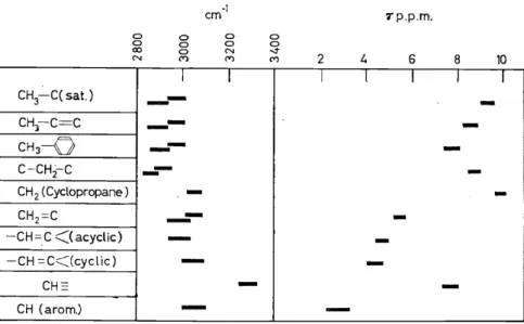
differences in the residue to which the OH or NH group is attached. The Stretchingvibration bands of C-H bonds between 2800-3300 cm-1 can to some extent be used to distinguish between saturated, olefinic and acetylenic types, and the higher frequency in a strained cyclopropane ring has been particularly useful (Figure 1). In all these cases, however, it seems that
CH3-C(sat.) CHl-C=C CH3
-Q
C-CH;2-C
CH 2 ( Cyclopropane ) CH2=C
-CH=.c <<acyclic) -CH =C<Jcyclic)
CH::=
CH (arom.)
0 0 CX) N
0 0 0
<")
--
I-- -
- -
- - -
- --
cm -1 0 0 N
<")
I
- -
0 0 ....:t
<") 2
I
-
7'p.p.m.
4 6
l I
- -
-
8 10
I I
- -
- - - -
Figure 1. Characteristic C-H vibration frequencies and corresponding proton resonance shifts greater discrimination can be achieved by using the chemical shift effect in the nuclear magnetic resonance spectra which will be discussed below.
This chemical shift effect not only serves to distinguish types of OH, NH and CH groups in most cases, but is also able to reveal details of, for example, the skeleton to which a CH3 group is attached.
It is sometimes possible to resolve ambiguities in interpreting the C-H stretching vibration bands by taking into account also the bands due to deformation vibrations at Ionger wavelengths. These bands are sometimes significantly displaced by the contiguity of electronegative atoms or other inductive influences, but here again the more recent nuclear resonance spectra may prove more convincing.
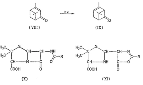
The Stretchingvibration ofthe C=O group has been exhaustively studied, and its absorption band shifts considerably in different classes of com- pound52. This has been much used to decide between possible alternatives suggested by chemical work. For example, ultra-violet irradiation of verbonone (VIII) Ieads to the isomeric ketone chrysanthenone (IX); the very high C=O frequency of this (1785 cm-1) indicates a strained cyclo- butanone ring, and other spectral features (frequencies of 3030 and 1660 cm-1 are associated with =CH and R1R 2C=CHR3 groupings respectively) support the structure shown53• In the early studies on penicillin, much work centred on the C=O absorption bands 1600-1800 cm-1 which might have been attributable to an amide, fused ß-lactam (X) or oxazolone (XI).
Detailed studies on many amides and oxazolones were rather indecisive, but
examination of some model fused ß-lactams led Shell laboratories, Emery- ville, to be the first to · support the lactam-amide formula (X) which was subsequently confirmed by X-ray analysis54•
~0 rao
(VIII) (IX)
HJC
/s,
' c /
..._CH-CH-NHJC/1
I I j:-R
CH--NH C - 0
I II
COOH 0
(X) (XI)
Among many other recent uses of C==Ü bands are the differentiation of y-and 8-aldonolactones55, determination of the structure of ketoflavones56,
studies on ketolactone oxidation products of camphor57, and the analysis of tissue and serum lipids58• Sphingolipids show bands of an amide group at 1655 and 1550 cm-1 which are absent with all other lipids. Cephalin and lecithin have an ester group band at 1740 cm-1, and can themselves be distinguished by other bands in the region of 1000 cm-1• All these bands can be used for quantitative analysis.
The spectral characteristics of C=C and C==C bonds, alone, conjugated or cumulated, arealso useful. For example, they provide convincing support for the structure of mycomycin (XII)59 (Table 1). The characteristic
H-C==C-C_C-CH=C=CH-CH=CH-CH=CH-CH2--COOR
(XII)
vibration of the C-N near 2250 cm-1 was used60 to detect this group in vitamin B12 •
Table I. Spectral properties of mycomycin
Grouping Spectral characteristics
-C=CH R'-C==C-R"
-CH=C=CH- -C :=C-C:=C- -COOR
3280, 2040 cm-1 2200 cm-1 1930 cm-1
u.v.
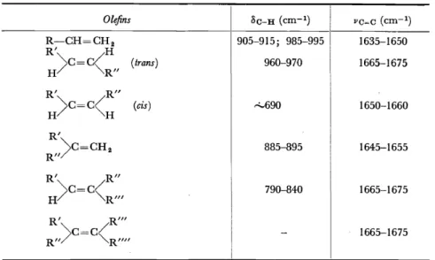
2560, 2670, 2810 A 1733 cm-1Another important set of bands is provided by the substituted oldins61.
These occur in the region of 10 p. and are associated with bending motions of olefinic C-H bonds ( Table 2). Either alone, or taken tagether with the
Table 2. Frequencies of characteristic bands in the infra-red absorption spectrum of olefins, associated with C-H deformation ( 8c-H) and C= C stretching (vc=c)
Olefins Sc-H (cm-1) VC=C (cm-1)
R-CH=CH 2 905-915; 985-995 1635-1650
R' H
) c - c ( (trans) 960-970 1665-1675
H R"
R' R"
H)c=c(H (cis) "-690 1650-1660
R'
)c=CH2 885-895 1645-1655
R"
R' R"
) c - c / 790-840 1665-1675
H - "'-R"' R' R"'
) c - c / 1665-1675
R" - "'-R""
C=C stretching vibration bands, these bands have been widely used in studying complex molecules, for they appear to apply reliably whether the unsaturated group is in a side-chain or in a ring. In this way, the iso- propenyl (XIII) or isopropylidene (XIV) end groups of simple terpenes
H3C""
hC-CH 2- H2C/
(XIII)
(Band frequencies: 890 and 1645 cm-1)
(XIV)
(Band frequencies: 810 and 1670 cm-1)
were investigated62• The cis or trans structure of the -CH=CH- group has been examined in many cases. Thus the double bond in the side-chain of ~22-ergostene (XV) 63 is shown to be trans by the band at 970 cm- 1.
The corresponding bonds in calciferol (XVI), in tachysterol, and in the precursor of calciferol have also been examined in the same way64 • The absence ofthe band at 965 cm-1 indicates a cis structure for jasmone (XVII) and cinerolone65, and a band at 899 cm-1 has been assigned to a terminal methylene group in nyctanthic acid66 (XVIII) a seed extract related to the tetracyclic terpenes. The same characteristics have been used in attempts to determine the cis-trans relationships in ~,w-diphenyl polyenes (XIX) and in ß-carotene (XX)67 • With simple cis or trans fatty acids and Iipids this differentiation is straightforward.
448
t;:H3 CH3
I
I
/CHJCH--CH==CH--CH--CH
c~ 'c~
(XV) (XVI)
(XVII)
o-(CH=CH)n
-o
(XVIII)(XIX)
(XX)
Many of these simple correlations have been used, together with ultra- violet and nuclear resonance data, in establishing the structures of the interesting new plant growth promoting factor, giberellic acid, and the associated giberellins68, and, together with other characteristic vibration frequencies of linkages involving nitrogen atoms, in fixing the structure of highly complex alkaloids such as lycoctanine and its derivatives69 •
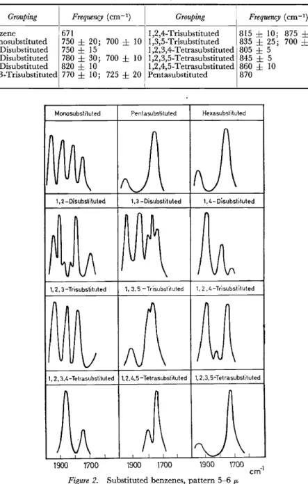
Some intense infra-red bands in the region 11-15 IL occur with sub- stituted aromatic rings 70• These are associated with out-of-plane bending motions of the residual C-H bonds, and sometimes provide immediate evidence about the positions of the substituents. Moreover, the rules often appear to apply for fused aromatic ring systems71 • The approximate frequencies of these bands are given in Table 3. However, these bands are somewhat unsatisfactory criteria for the presence of substituted aromatic rings, not only because of the rather wide frequency variation in some cases, but also since their intensity varies in an unpredictable way as the nature of the substituents is changed. A more reliable indication is the general pattern of bands between 1600-2000 cm-1 arising from combinations of the out-of-plane skeletal vibrations72 (see Figure 2).
449
Table 3. Characteristic bands in the infra-red absorption spectrum of aromatic rings, associated with C-H deformations
Grouping Frequency (cm- 1)
I Grouping Frequency (cm-1)
Benzene 67I I ,2,4-Trisubstituted 8I5 ± 10; 875
±
5Monosubstituted 750
±
20; 700±
10 1 ,3,5-Trisubstituted 835±
25; 700±
25 I,2-Disubstituted 750±
I5 I ,2,3,4-Tetrasubstituted 805±
51 ,3-Disubstituted 780
±
30; 700±
IO 1 ,2,3,5-Tetrasubstituted 845±
5 I,4-Disubstituted 820±
10 I ,2,4,5-Tetrasubstituted 860±
10 1 ,2,3-Trisubstituted 770±
10; 725±
20 Pentasubstituted 870Monosubsliluted Pentasubsti tuted Hexasubstituted
~
1, 2 -DisubstitutedJ\ J\
1,3 -Disubstituted 1, 4- Disubstituted~
1, 2, 3-Tr·,substituted Invn~
1, 3. 5 -Trisubst"Jtuted 1,~
2, 4-Tr"1subsl ituted~ ~ n ~
M
I
V u V N
1, 2,3.4-Tetrasubsti\u\ed 1. 2.4.5-Te\rasubst'ttuted 1, 2,3, 5-Tetrasubs\ituted
~ ~ ~)
n1900 1700 1900 1700 1900 1700 cm-1 Figure 2. Substituted benzenes, pattern 5-6 11-
The dass of natural products in which infra-red absorption has been most studied is the steroids73• Different side-chains attached at the C-17
· position in this perhydrocyclopentenophenanthrene system give rise to the pregnanes, bile acids, cholestanes and ergostanes. With all these
PHYSICOCHEMICAL METHODS OF INVESTIGATION
compounds, information can be obtained about the structure and conforma- tion of the nuclear skeleton, and about the side-chains. In saturated systems, C=O groups in rings A,B,C or at C-20 in the side-chain have about the same vibration frequency, but in the five-membered D ring the value is noticeably higher. Conjugated unsaturation in all cases lowers the carbonyl group frequency significantly. In side-chain ester groups, this frequency is higher than that in the ketones, and may coincide with that in the D ring, but the latter also has a band in the region 1100-1200 cm-1•
Some bands of the ring CH 2 groups are affected in definite ways by adjacent carbonyl groups. Many other correlations, including some for the steroid lactones, have been worked out74, and more complex polycyclic terpenes are now being examined in the same way.
The relation between the infra-red absorption spectrum and stereo- chemical facto:r;s is particularly interesting. First, closely related diastereo- isomers have different spectra, although these are c~mplex. Thus chol- estane and coprostane, differing only in the trans or cis arrangement at the fusion of rings A and B, have different spectra46• Also, spectral differences have been established empirically between compounds containing a sub- stituent group in the axial or equatorial position.
Thus the acetate band at 1240 cm-1 in 3-acetoxy- steroids is single if the group is equatorial, but split into several peaks if it is axial75• Axial OH groups in the 3-position usually give a band near 1000 cm-1 (probably determined by the C-0 band vibration), butthislies higher at 1040 cm-1 ifthe group is equatorial. This variation offrequency can be interpreted in terms of the likely effect of motions involving a greater or less amount of bond stretching or bending. The rules are not without exceptions, but have been applied to establish the axial orientation of the OH group in 1 uminestan-3 ß-ol76.
Similar correlations between conformation and spectrum have been found with the decalols77, and in deuterium-substituted steroids the C-D bond frequency varies for axial and equatorial positions78 • Also, when ahalogen atom is introduced in an equatorial position on the carbon atom next to a carbonyl group, the carbonyl group band shifts to higher frequency, whereas no such effect occurs when the group is axiaF9• This result is paralleled by data on the ultra-violet absorption spectra referred to above.
Similar conformation effects have been examined for the OH groups in carbohydra tes so.
Reference has been made already to infra-red work on lipids, and there are many applications in biochemistry81 • Amino-acids, polypeptides and proteins have been much studied, and important spectral differences have been found between the open and closed chain forms of polypeptides82•
Measurements with polarized radiation on crystalline materials or oriented fibres can provide information about the relative orientation of N-H and C=Ü bonds and about the type of hydrogen bonding involved. There is a serious difficulty of principle here, since the directions of dipole vector change during a vibration may not coincide exactly with a preconceived bond direction, but even semi-quantitative measurements from the band intensities with polarized radiation are valuable. Deoxyribonucleic acid can be distinguished from ribonucleic acid by its band at 1020 cm-1, and
its spectrum is consistent with the structure derived from X-ray work83•
Preliminary studies of bacteria and viruses show differences in composition which are worth further examination84.
Many ambiguities arise in the application· of the vibration frequency correlation rules. Attempts have been made to remove some of them by using the intrinsic intensities of the absorption bands85. In some cases this may be possible. For example, the intensity of the Stretching vibration band of the N-H group in different classes of compounds varies con- siderably86. In aliphatic secondary amines it is low, but in heterocyclic bases, such as carbazole, it is high. In fact, the very low intensity of this group in certain alkaloids has caused anxiety. If the first overtone bands are also studied, an even better differentiation of the NH types is obtained.
Similarly, there appears to be a significant difference in the 0-H band intensity in phenols on the one hand and in alcohols on the other87 , and regularities occur in the intensities of different kinds of C-H bands88• With steroids, intensities can also be used to discriminate between carbonyl groups in different ring positions and in the side-chain89• An interesting example has been found with cisoid and transoid a:,ß-unsaturated ketones90•
With cisoid types, the intensities of C=C and C=O bands are about equal, but with transoid types the C=O band is relatively much stronger. Although the attached groups give rise to minor variations, the rule seems tobe reliable enough to apply in complex compounds. For instance, cholestan-5-en-4- one (cisoid) can be distinguished from cholestan-4-en-3-one (transoid) in this way.
However, accumulating evidence suggests that, in general, the band intensities (which are, of course, determined by different factors from those which control the vibration frequencies) are very susceptible to electronic effects of neighbouring atoms and groups, so much indeed as to Iimit their present value. A more detailed examination of these electronic influences is being carried out in the hope that the variations can be predicted or at any rate reconciled with structural details91•
NUCLEAR MAGNETIC RESONANCE
Nuclear magnetic resonance spectroscopy is, perhaps, the most important new method for investigating details of molecular structure. It makes use ofthe magnetic effect set up by a spinning charged nucleus to get information about the chemical environment in which the particular nucleus occurs.
Nuclei of even atomic mass and even atomic number, such as 12C, 160 and
32S, do not spin, and this is indeed fortunate as far as applications to organic chemistry are concerned, since otherwise the nuclear resonance spectrum arising from a carbon chain might be too complex for general use. Nuclei such as IH, 19F, 13Q, 15N and s1p have a spin moment I = 1/2, and we are primarily concerned with these. With other spinning nuclei, such as 2H, HN, 170 and 35Cl, complications arise, partly as a result of the effects of their quadrupole moments.
A nucleus of spin angular momentum I gives rise to an equivalent magnetic moment fL which can be oriented in (2I
+
1) directions if placed in a homo- geneous magnetic field H. The energy difference between two such adjacent Ievels is given by the relationPHYSICOCHEMICAL METHODS OF INVESTIGATION
hv=l
JLHIn a proton (lH) with a spin I = I /2, only two orientations are possible, and the small difference in energy Ievel associated with each of them Ieads to a slight difference in population of the Ievels in accordance with the Boltzmann factor. In a given field strength H, therefore, there is a particular resonant frequency corresponding to the quantum of energy which is required to tip the nucleus from one orientation to the other. Alternatively, for a given supplied frequency, there will be a specific field strength corresponding to the resonant condition. It is usual, for experimental convenience, to maintain a constant frequency and vary the applied field strength.
Several factors can Iead to broadening of the absorption line, especially with solids, but in liquids and gases these are minimized as a result of molecular motions, and sharp resonance lines arise.
The use of this phenomenon in molecular structural determination is based upon two additional effects. First, the electronic space clouds surrounding a spinning nucleus exert a screening effect which makes the effective magnetic field strength different from that applied.
H = Happi. (1 - a)
A slightly different field strength from that relating to a " free " nucleus must, therefore, be applied to obtain resonance at a fixed absorbed fre- quency. The extent of the so-called chemical shift varies with the par- ticular skeletal environment, and can be correlated with the ionic character of particular bonds, with the inductive and mesomeric effect of substituents, and with the magnetic anisotropy of the molecule concerned.
In practice, it is not possible to measure absolute values of field strength with the same accuracy as it is to determine differences of field strength.
Chemical shifts are, therefore, measured with reference to standard reference lines, such as are provided by water,. cyclohexane, benzene or tetramethyl- silane. Also, since the chemical shift is proportional to the field strength, it is desirable to represent it as the dimensionless unit
Hsubst. - Href.
S
=
H=
asubst. - Uref.ref.
I t can also be expressed as
S - ö. X 106
- oscillator frequency
in which Ö. is the frequency shift between sample and reference corresponding to the change in field strength, S then being expressed in parts per million.
In different nuclei, 8 may vary between a few parts per million (as with protons) and a few per cent (as with cobalt). Recently, another quantity
H. W. THOMPSON
-r ( = 10 - ö) has been suggested for use with the proton taking tetra ..
methylsilane as the reference substance, so that a set of simple numbers are obtained for the shifts of all except the most acidic protons.
A difficul ty arises here, however, which has led to some confusion in the correlation of many results so far obtained. If the reference substance is used internally, mixed with the sample, there must be no interactions. If it is used externally, in a separate probe, corrections must be applied to allow for differences in the bulk susceptibili ties of the two samples. U nfor- tunately, both external and internal standards have been used in the past, as well as different reference substances, and it is not always possible to correct different recorded data so as to use them tagether in making com- parisons of chemical shifts. In the illustrative examples, which follow, various scales of reference will be used.
The second effect which is important with organic molecules arises from a spin-spin interaction between neighbouring nuclei. As a result of this, the resonant line for a proton may be split by the magnetic disturbance of the other neighbouring protons into a pattern which is highly characteristic, and which makes it possible to designate the nature of the adjacent atomic groupings. This effect, which is independent of the applied magnetic field, is transmitted through the electron space cloud of the molecule and diminishes rapidly as the distance between the interacting nuclei is increased.
A point of particular importance is that the integrated intensity of a nuclear resonance absorption line is a direct measure of the number of nuclei of the type concerned. If the relative intensities of lines at different resonant frequencies are compared, such as those which arise with protons subject to different chemical shifts, it is at once possible to count the number of groups of different kinds, and this is strikingly useful in solving certain structures. Certainly here the method has considerable advantages over the use of the infra-red absorption spectrum.
The design of equipment for these measurements is partly determined by whether low or high resolution is required92• It is essential, however, to have a high degree of magnetic stability and homogeneity. Permanent magnets, thermostatically controlled, have stability, but electromagnets can have higher field strength, and may be required for less sensitive heavier nuclei. Greater field homogeneity over the required area can be obtained with correcting coils on the pole pieces, and it is usual to spin the sample to obtain an even greater effective field homogeneity. The sweep rate used in searching for the signal must be carefully controlled in relation to the relaxation time in the sample.
Usually 0·5 cm3 of liquid sample is adequate, but much smaller quan- tities have sometimes been used. For proton resonance, suitable solvents are carbon tetrachloride, tetrachloroethylene, carbon disulphide, deutero- chloroform, trifluoroacetic acid, dimethyl sulphoxide, trichloroacetonitrile, dimethyl formamide, water, cyclohexane, benezene, dioxane and acetone.
Many of these have been used to give an internal reference signal, although cyclohexane and tetramethylsilane are now regarded as the most suitable.
A change of solvent sometimes causes shifts which are useful in splitting up overlapping lines. The limited solubility of some compounds introduces a difficulty, since, in order to obtain a satisfactory signal strength, larger
samples or a higher field ~trength will be required, and both are undesirable in the interest of a uniform and stable field.
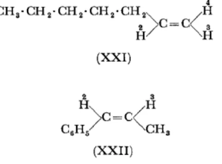
As already explained, the proton resonance signals in C-H bonds of saturated, unsaturated or aromatic hydrocarbons give very characteristic chemical shifts. In hept-1-ene (XXI), or in cis-propenylbenzene (XXII), for example, each type of proton gives a distinct signal93 (see Table 4).
4
CH3·CH2·CH2·CH2·CH2 2
>
C=C"-/H aH "-H (XXI)
Table 4. Characteristic chemical shifts ofproton magnetic resonance signals in C--H bonds
2
Hept-1-ene (XXI): H
3
H
4
H CH2-C=C
CH2 (sat.) CH3
2
cis-Propenylbenzene: H
3
H
CH (arom.) CH3
-1·1 -0-4 -0·2 +2·8 +3·5 +4·0 -1·2 -0·5 -2·0 +3·5
Similarly in ethylphenylacetate, distinct signals are obtained for the CH3 , CH2, CH2CO and C6H5 protons, split in some cases by spin interactions94•
In substituted benzene rings, the aromatic proton shifts differ according to the relative position and nature of substituents, and in polynuclear fused aromatic hydrocarbons or in azulenes the protons in different locations can be distinguished. The same applies to protons in heterocyclic rings such as furans, pyridines and thiophenes, and the chemical shifts are in some cases made more distinctive by spin interactions. The differences in pattern between glyoxalines and pyrazoles was used recently to confirm the presence of a pyrazole ring in an amino-acid extracted from water-melon seed95• The presence of a 2,3-disubstituted pyridine ring in a new alkaloid has also been established in this way96, and of a 1,4-dihydropyridine skeleton in a phosphopyridine nucleotide97•
With protons attached to elements other than carbon, or with CH3 or CH 2 groups attached to different kinds of group, the chemical shifts vary over a wide range98• Many ofthe earlier data require revision or correction, but it is certain that striking differences occur between groups which are often difficult to distinguish or established by other methods. For example, unless hydrogen bonding complicates the situation too much, it is possible to distinguish between the 0 H groups in alcohols and phenols, the SH groups in mercaptans or thiophenols, or the NH groups in different types of amine. Infra-red methods are not always conclusive in these instances.
The relative intensities of the signals often provide additional evidence.
For example, a cresol derivative99 was shown to possess structure (XXIII) since, apart from the phenolic and ring protons, it gave three CH3 resonances of relative intensities 3 :3 :6. The alternative structure (XXIV) would have given five such resonances of intensities 1 :2:3:3:3.
B~:Q::H
H3C-~-0-CH3
CH3 (XXIII)
B~~:H
H3C- C -O-CH2-CH3 H I
(XXIV)
Thujic acid methy1 ester provides a similar example100• This compound gave three resonances of relative intensities 5 :3 :6 ( due to
=
CH, COOCH3 and C(CH3) 2 respectively), thus confirming structure (XXV) rather than (XXVI), since the latter would have given four resonances of relative intensities 3:2:3:6 (due to =CH, CH, COOCH3 and C(CH3) 2 respectively).The spin-spin patterns are often complex, and can be much affected by the relative values of the chemical shifts of the groups between which spin interactions are occurring. The principles, however, can be illustrated by some simple cases. For example, in the molecule HD, with the spins of proton and deuteron 1/2 and 1 unit respectively, the proton can, so to speak, " see " three orientations of the deuteron (
+
1, 0, -1) and the proton signal is a trip1et. The deuteron " sees " two orientations of the proton( +
1/2,- 1/2) and its signal is a doublet. In 31P 19F3 , the fluorine nucleusPHYSICOCHEMICAL METHODS OF INVESTIGATION
" sees" two orientations of the phosphorus nucleus (
+
1/2, - 1 /2) giving a doublet, whereas the phosphorus nudeus "sees" four combinations of the fluorirre species (+
3/2,+
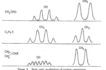
1/2, - 1/2, - 3/2), ofwhich two have three times the statistical weight of the other pair, the P resonance therefore being a quartet, 1 :3:3:1.CH
CH
Figure 3. Spin-spin multiplets of proton resonance
In ethyl alcohol or diethyl ether, the CH3 and CH2 groups of the ethyl radical split into three and four components, with relative intensities corresponding to the statistical weights ofthe Ievels (see Figure 3). n-Propyl and isopropyl groups attached to a residue X can be distinguished. In the simplest case, the iso-propyl group will give two lines for the CH3 groups which " see" two orientations of the adjacent proton, and the CH group will have seven lines due to the sets of orientations of the six protons in the methyl groups (
+
3,+
2,+
1, 0, - 1, - 2, - 3), as shown in Figure 3.In the n-propyl group ofCH3·CH2·CH2·X, the methyl groupwill give a triplet, the cx-methylene group a triplet, and the ß-methylene group a set of twelve lines. Chemical shifts determined by X may complicate the general appearance, however, and complete resolution of the multiplets is not always obtained. In CH3 · CHO, a quartet and cloubiet are found associated respectively with the CHO and CH3 groups (see Figure 3). The -CH 2-CH 2- or -CH 2-CH3 groups in ring ethers of the type (XXVII) or (XXVIII) can be distinguished; the ~CH-CH3 group gives a quartet (CH, 1 :3:3:1) and doublet (CH3, 1 :1), but the -CH2CH2- group only
- C - 0
'
I >H-CH,
- C - O
I
(XXVII) (XXVIII)
a single line which may be split to a greater or less extent as the general symmetry of the attached residue is disturbed.
These principles have been applied in structural determinations of many natural products. Evidence for the group ~C-CH-CH=CH- has
0 -
Ibeen important in fixing the structure of giberellic acid101 (XXIX). The characteristic chemical shifts of protons in the groups CH2=0:::: and
~CH found with Feist's acid, and the absence of CH3 resonances at high field, show102 that this substance contains a saturated cyclopropane ring with an exocyclic methylene group, and possesses structure (XXX) rather
H
H~c
I o
HO-C%
oc/1
CH3 (XXIX)
H-.. ... c....r /COOH
HzC=<I
/c ....
H....r ''COOH
(XXX) (XXXI)
than (XXXI). This is confirmed by the intensity distribution, formula (XXX) giv_ing three lines of equal intensity, and (XXXI) three lines of relative intensity 2: 1 :3.
Among alkaloids which have been studied, myosmine has a set of four nuclear resonance lines of equal intensity due to the four different pyridine protons, with three other lines at higher field, each of which has twice the intensity of the pyridine protons103• This fixes the structure as (XXXII) rather than (XXXIII). The side-chain group -CH(CH3) 2 and the position of the methoxy-group have been determined in lunacrine (XXXIV) 104, and the N-CH3 group has been detected in aspidospermine105•
Much useful information has been obtained from the proton resonance spectra of essential oils and glycerides, by using the characteristics of groups such as CH (aromatic), OH, CH3CO, -CH=, CH2 and CH3 groups in different environments106 • Sterculic acid was recently shown to contain a cyclopropene ring in the middle of a long fatty-acid chain 10 7•
(XXXII) (XXXIII)
~'tH
(CH3),y~ÄoAH
H3CO bH3 (XXXIV)
The nature of the end-groups in carotenoid-type molecules such as a spirilloxanthin and the paprika pigments have been determined108• The tentative structure originally proposed for spirilloxanthin was (XXXV), where R
=
R'=
(a). However, the spectrum is only explained if the end groups are (b). In the paprika ketones109, characteristics of CH3 groups on saturated quaternary carbon atoms have led to a similar revision of ideas about the end groups. The stereochemistry of these conjugated chain systems has also been examined in relation to the nuclear resonance spectra by reference to simpler related molecules such as the dimethyl muconates.With methyl photosantonate (XXXVI) 110, the resonance signals due to
(CH3) 2C= and =CH-CH2-COOCH3 proved invaluable in fixing the
structure; and in "'-santonin (XXXVII), a sesquiterpene Iactone, one olefinic proton has been recognized among a large number of saturated group protons111•
The chemical shifts of CH3 groups in helvolic acid (XXXVIII) have led to the determination of the end-group in the side-chain, one such group being assigned to the C-24 position 3 4 ,
With steroids in general, characteristic resonances occur for CH3 groups on quaternary carbon atoms, and for acetoxy-, olefinic CH=, and other groups in different locations in the skeleton 112• These appear to offer great
R
~
R'(XXXV)
~-~aco-& c:;•H
(a) (b)
(XXXVI)
(XXXVII)possibilities for structural determination. The chemical shifts are also affected by stereochemical conformation, so that axial and equatorial H atoms can be distinguished. Such conformational effects in the proton resonance spectra have also been found with acetylated pyranoses and inosito1s 113, and are confirmed by resu1ts on similar conformations in such compounds as trioxane114, the cis- and trans,...decalins 115, the dimethylcyclo- hexanes116, and 2-acetoxy-1 ,3-dimethoxycyclohexanones 117.
COOH
0
(XXXVIII)
The use of spinning nuclei other than protons is still in its early stages of development, but this offers great possibi1ities, · especially perhaps with 13C, 19F and 31P which have a spin I= 1/2, and no quadrupole moment.
The signal strengths are intrinsically weaker, so that !arger samples are needed, as weil as better detecting equipment. Also, owing to the greater relaxation times, the sweep rate has to be controlled carefully. However, the chemical shifts are much greater than those for protons, and they are very sensitive to structural changes. In 13C, the chemical shifts referred to a benzene carbon nucleus as zero vary from about
+
120 in hydrocarbons to - 80 in ketones 118. The location of these 13C resonances is far removed from that of protons, and the spin coupling with bonded hydrogen atoms Ieads to characteristic multiplets. With CH3 · COOH, using the natural abundance of 13C, the carbon nucleus of the COOH group appears as a single resonance, and that of the CH3 group as a quartet at higher fields.In dimethyl acetylene di-carboxylate there are resonances characteristic of the -C0-0-, -C==C-, and -OCH3 groups. In mesity1ene, the chemical shifts of the 2, 4 and 6 carbon atoms are convincingly different from those of the non-equivalent 1, 3 and 5 carbon atoms.
Resonances of 31P nuclei in different environments show even !arger
·chemical shifts119• The experimental difficulties are at present considerable, but it may be possible to discover by this means features of phosphoryl compounds. Indeed, all these results encourage the hope that, by a study ofresonances ofseveral nuclei such as 1H, 13C and 14N in the same molecule, it may be possible to determine a sufficiently large number of structural units in that molecule to fix its whole structure without recourse to much chemical work on degradation or synthesis.
Other refinements.in technique seem likely to remove some of the present difficulties. One is the double irradiation method 120• When spin inter- actions Iead to a highly complex spectrum, it may be possible to decouple these spins by irradiation with a field close to the resonant frequency of one of the nuclei, thus simplifying the spectrum and its use for diagnosis.
Another way of enhancing chemical shifts in certain cases is by adding para1nagnetic salts which co-ordinate with hydroxyl or other groups 121.
A still more interesting development is electronic-nuclear double resonance, in which the substance is irradiated with intense microwave radiation at a frequency which excites the electron resonance of any paramagnetic material present. Strong interactions may come into play which Iead to a large increase in the intensity of the nuclear resonance absorption, and the effect may be useful in studying some of the heavier nuclei.
Electron paramagnetic resonance itself has been applied to the study of certain natural productsl 22. These include, for example, the porphyrin types in which a paramagnetic ion is surrounded by an organic skeleton, as in haemoglobin, myoglobin, chlorophyll or the phthalocyanines. In this way, important information can be obtained about the symmetry of the central part of the structure and about the type of bonding to the central metallic atom.
OPTICAL ROTA TORY DISPERSION
Optical rotation has long been used as a criterion for purity, proof of identity, for the determination of enantiomorphic type, and, to some extent, as an indication of the position of a functional group or of the relative configuration of different asymmetric centres in a molecule. Empirical rules have been proposed for computing the optical rotation of compounds within a dass of compounds such as the steroids by addition of definite amounts for different structural units present in the molecular skeleton 123•
In this way, using rough measures for C=C, OH, CH3CO and C=O groups at different positions, useful corroborative evidence about the structure of certain steroids was obtained.
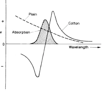
Optical rotation is equivalent to circular birefringence, and occurs when a substance transmits the left- and right-hand components of a beam of circularly polarized light with unequal velocity. If these components are absorbed unequally, the optical rotation will vary with wavelength, and classical equations have been proposed to express this. Indeed, some workers have used the optical rotation at two wavelengths as a refinement for structural work and for a criterion of purity. The variation of specific rotation with wavelength becomes greater as the absorption band is approached, and, as it is traversed, a Cotton effect may be observed, the