Brenna et al.: Automatic liquid Chromatograph for haemoglobins 437 J. Clin. Chem. Clin. Biochem.
Vol. 25, 1987, pp. 437-440
© 1987 Walter de Gruyter & Co.
Berlin · New York
Assessment of an Automatic Liquid Chromatograph for Foetal and Abnormal Haemoglobins
By S. Brenna, L. Prencipe, S. Granata and N. Montalbetti
Laboratory of Clinical Biochemistry and Hematology Ca' Granda — Niguarda Hospital, Milan, Italy
(Received December 4, 1986/March 12, 1987)
Summary: The DIAMAT TM (Bio-Rad) analyser is a microprocessor-operated HPLC System using a silica- based weak cation exchange column with three phosphate buffers of increasing ionic strength äs the Step gradient mobile phase. A dual wavelength detector measures absorbance at 415 and 690 nm; each sample is completely processed in 8 minutes. The Instrument effectively separates and quantifies HbF in a discrete peak. We have verified that the HbF assay is linear up to about 65% values.
We calculated within-run imprecision for n = 20 in 3 different haemolysed blood samples; the results are shown below äs percent of total haemoglobin:
A) Mean = 1.06 SD = 0.048 CV% = 4.5 B) Mean = 1.90 SD = 0.041 CV% = 2.1 C) Mean = 8.93 SD = 0.047 CV% = 0.5 Between-run imprecision for similar samples (n = 18) was:
D) Mean = 3.74 SD = 0.20 CV% = 5.5 E) Mean = 9.94 SD = 0.15 CV% = 1.5
Accuracy was assessed in different series by correlating Hb values (y) with those obtained by the alkali denatürätion test (x). The regression line equation was y = 1.03 — 0.33 (r = 0.999, n = 62).
The DIAMAT TM Instrument also reveals the presence of any HbS and cälculates its peak area correctly, äs we found by electrpphoretic reassay in heterozygoüs subjects. We also noted the presence of other abnormal haemoglobins characterized by specific retention times.
Introduction . ,. _ / ~ , 1_
concentrations exceeding 70% of total Hb occur m In normal adult blood, foetal haemoglobin (HbF) homozygous ß-thalassaemia, -ß-thalassaemia, and accounts for not more thän 2% of total haemoglobin. hereditary persistence of foetal haemoglobin.
HbF up to 20% of total can be found in heterozygpus
cases of ß-thalassaemia, hereditary persistence of foe- One widely used method for assaying HbF is based on tal haemoglobin (HPFH), and in double hetero- its resistance to alkali denatürätion (1); this method, zygosis with ß-thalassaemia plus one haemoglobin however, is poorly sensitive to high HbF concentra- variant such äs HbC, HbS or HbE. Finally, HbF tipns.
J. Clin. Chem. Clin. Biochem. / Vol. 25,1987 / No. 7
438 Brenna et al.: Automatic liquid Chromatograph for haemoglobins Two methods used routinely are haemoglobin electro-
phoresis on cellulose acetate at an alkaline pH (2) and on agar citrate at an acidic pH (3), each being suitable for separating different haemoglobins.
Isoelectric focusing affords excellent resolution (4);
but the method is remarkably cpmplex and its use in the clinical chemistry laboratory is restricted for the time being to research work.
Other methods for assaying HbF are radial immuno- diffusion (5), cation-exchange micro-chromatögra^
phy for eluting "fast" haemoglobins (6), and some variants of ion-exchange high-performance liquid chromatography (HPLC) (7, 8).
The DIAMAT TM apparatus is a high performance liquid chromatography analyser originally intended for the automatic assay of glycated haemoglobins.
The same apparatus, however, also quantifies HbF in a separate peak (fig. 1), äs well äs HbS, if any^
and other abnormal peaks.
\ \
The possibility of complete automation of these as- says, offered by the DIAMAT TM Instrument, prompted us to test it for accuracy and reliability in the screening of abnormal haemoglobins, primarily HbF. The more laborious methods of electrophoresis and alkali resistance were used for confirmation.
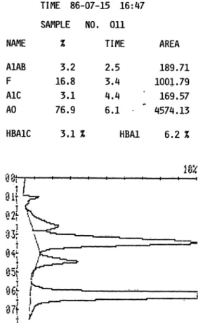
TIME 86-07-15 16:47 SAMPLE NO, 011
NAME % TIME AREA 189.71 1001.79 169.57 A1AB
F A1C AO
3.2 16.8 3.1 76.9
2.5 3.4 4.4 6.1
HBA1C 3.1 % HBA1 6.2
Materials and Methods
High performance liquid chromatography analyser The -Rad DIAMAT TM Instrument is an automatic ana- lyser utilizing the principles of ion-exchange high-performance liquid chromatography for assaying glycated haemoglobin. The apparatus features a single piston pump with a step-gradient valve system that allows three phosphate buffers of increasing ionic strength and different pH to riin through the analytical column in a tiined sequence.
First solution:
Second solution:
Third solution,
phosphate buffer, phosphate büffer, phosphate büffer,
90 mmol/1 pH = 5.9 140 mmol/1 pH = 5.8 270 mmol/1 pH = 5.7
Fig. 1. Printout of sample with high HbF.
The column (4 mm i.d. 15 cm) is packed with a weak cation-exchange resin in beäd form; the support is silica with a carboxymethyl functional group. Buffers are eluted in the following order:
buffer 1: 1.7 minutes, buffer 2: 2.0 minutes, buffer 3: 2.1 minutes and buffer l: 2.2 minutes.
Thus, each sample is completely processed in 8 minutes. Hae- molysed blood samples are stored in the refrigeräted autosam- pler chamber until a 5 portion is äutomatically injected intq the column thennostated at 23 °C.
Readings are made at 415 and 690 mn, to eliminate problems of turbidity or light source instability.
All operätions are controlled by a microprocessor. The printout is a summary of HbAjC and HbAj (a -f b + c) values äs well äs of the relative (area) percentages of all resolved haemoglobins.
Alkali denaturation
We used the method of Singer et al. (11).
Alkaline-pH haemoglobin electrophoresis
For this we used the eompletely automatic OLYMPUS AES 2000 a:pparatus. Electrophoresis was conducted on SARTO- RIUS cellulose acetate Strips at 4 mA for 35 minutes in TRIS^
EDTA-glycine buffer at pH 9.2 with semi-micfo seeding, and Strips were stained with Ponceau Red.
Sample treatment
For comparison and reproducibility tests in the DIAMAT TM series we used fresh venous blood samples with EDTA added äs änticoagulant. Before analysis, the blood was haemplysed and diluted l: 200 with a solution of polyoxyethylene ether in borate buffer, volume fraction 0.001.
In such haemolysates, even if stored at —20 °C, HbF is not very statole from day to day; thus fof assessing between-run precisiön we used two blood samples pf high and low HbF content stored in the refrigerator,
For haemoglobin electrophoresis, suitable portions of whole blood were washed repeatedly with isotonic saline solution. We then treated l volume of red blood cells with l volume of water and 1/2 volume of tolüene.
J. Clin, Chem. Clin. Biochem. / Vol. 25,1987 / No. 7
Brenna et al.: Automatic liquid Chromatograph for haemoglobins 439 Results
Linearity
To verify the linearity of HbF assays we mixed a sample of neonatal blood (HbF = 85%) in 20 dif- ferent portions with adult blood of equal haemoglo- bin content and no appreciable HbF content, tihus making 20 serial dilutions of HbF from 0 to 85%.
From the first 4 points (0 to 12% HbF) we calculated the linear regression for the HbF values expected from dilution and those actually produced by the DIAMAT TM Instrument.
The remaining points were never more than 5% away from the theoretical line, except for HbF contents exceeding 65%.
Precision
Within-run precision was assessed in 20 replicates of 3 blood samples with different HbF contents.
HbF readings are expressed äs percentage of total Hb:
Sample A: mean = 1.06, SD = 0.048, CV% = 4.5 Sample B: rnean = 1.90, SD = 0.041, CV% = 2.1 Sample C: mean = 8.93, SD = 0.047, CV% = 0.5 The between-run precision test (18 replicates of 2 pathological samples) gave:
Sample D: mean = 3.74, SD = 0.20, CV% = 5.5 Sample E: mean = 9.94, SD = 0.15, CV% = 1.5 Method comparison
Accuracy was assessed in different series by correlat- ing HbF values (y) with those obtained with the alkali-denäturation method (x) in the ränge of 0 to 70% HbF. The linear regression equation was:
y * 1.03 - 0.33; r = 0.999; N = 62 Carry-over
We investigated sample related carry-over in the DI- AMAT TM Instrument by performing 5 determina^
tions of a high-HbF blood sample (70%) followed by 5 determinations of a Iow-HbF sample (0.5%).
We repeated the experiment 10 times, and then calcu- lated the mean value of the f atip:
Y _ (l* - 1s) (hs - 1s)
The resulting "carry-over constant" of 1.4 l O"3
indicates the absence of sample-to-sample carry-over or incomplete cotumn elution.
Table l shows the results of HbS assays in some subjects with the HbS trait.
Tab. 1. HbS percentage in 4 subjects with HbS-trait Patient
AB CD
DIAMAT TM 46 % 45.9%
41.8%
73 %
Electrophoresis 47 %44.3%
42 %72 %
Discussion
Among the various programs offered by the DI- AMAT TM Instrument, the basic routine program affords the elution and assay of glycated haemoglobin fractions and HbF in only 8 minutes.
Chromatography proved efficient and precise, es- pecially for pathologic HbF values.
The HbF assay is linear up to 65% values; compari- son with the alkali-resistance method revealed good correlation, even at high HbF values.
Thus the DIAMAT TM apparatus affords ready de- tection of most% cases of ß-thalassaemia and HbF persistence without recourse to the more laborious and time-consuming method based on alkali resist- ance.
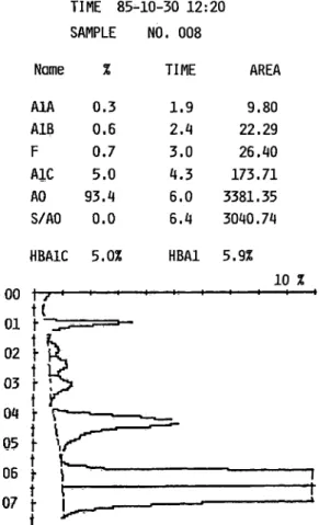
The same DIAMAT TM program also reveals HbS, which is eluted next to HbA (fig. 2). In such cases the Instrument does not indicate the percentage of
TIME 85-10-30 12:20 SAMPLE NO. 008
Name
A1A A1B F A1C AO S/AO
0.3 0.6 0.7 5.0 93. t*
0.0
TIME 1.9 2.4 3.0 6.0 6.4
AREA 9.80 22.29 26.40 173.71 3381.35 3040.74 HBA1C 5.0% HBA1 5.9%
UU 7~" ' ' ' 10 %
uj. r .^ ' "^t t
02 } Ji
4 1>
03 f «S
O/i l-t f T 1 t l .^ '
05 \ \fS~^
.. t ?^_
Üb f \
' \ l*~ — ·
'1J — ·-
1
Fig. 2. Printout of sample with HbS.
J. Clin. Chem. Clin. Biochein. / Vol. 25,1987,/ No. 7
440 Brenna et al.: Automatic liquid Chromatograph for haemoglobins HbS directly but only its peak area, from which the
percentage relative to total haemoglobin is readily calculated. The HbS area calculated by the Integrator is in good aggreement with the percentage found by electrophoresis.
As for the possible detection and assay of other haemoglobin subfractions, we are currently testing some Software modifications and different elution buffers; preliminary results appear promising for HbC and HbA?.
References
1. Betke, K., Märt, H. P. & Schlicht, I. (1959) Nature 184, 1877-1878.
2. Briere, R. O., Golias, T. & Batsakis, J. (1965) Am. J. Clin.
Pathpl. 44, 695-701.
3. Robinson, A. R., Rohson, M., Harrison, A. P. & Zvelzer, W. W. (1957) J. Lab. Clin. Med. 50, 745-752.
4. Basset, P., Braconnier, R & Rosa, J. (1982) J. Chromat.
227, 267-340.
5. Kohn, J. & Pagne, B. V. (1972) J. Clin. Pathol. 25, 830-831.
6. Abraham, E. C., Carver, J., Döbler, J., Milner, P. F. &
Huisman, T. H. J. (1979) Hemöglobin i, 341-351.
7. Gardiner, M. B., Carver, J., Abraham, B. L., Wilson, J.
B. & Huisman, t. H. J. (1982) Hemöglobin 6, 1^13.
8. Wüson, J. B., Headlee, M. E. & Huisman, T. H. J. (1983) J. Lab. Clin. Med. 702,174-186.
9. Kfuiswijk, K. T., Diaz, D. P. & Hpltkamp, A. C. (1981) Clin. Chem. 27, 641-642.
10. Moore, N. A., Hickey, T. M. & Kouns, D. M. (1985) Clin.
Chem. 6, 413.
Dr. Sergio Brenna via Campo dei Fiori, 32 1-20026 Novate Milanese
J. Clin. Chem. Clin. Biochem. / Vol. 25,1987 / No. 7