AUS DEM LEHRSTUHL FÜR HUMANGENETIK
PROF. DR. RER. NAT. BERNHARD WEBER DER FAKULTÄT FÜR MEDIZIN DER UNIVERSITÄT REGENSBURG
LIS1 -assoziierte klassische Lissenzephalie: Eine retrospektive, multizentrische Studie zum epileptischen Phänotyp und Ansprechen auf Antikonvulsiva
Inaugural – Dissertation zur Erlangung des Doktorgrades
der Medizin
der
Fakultät für Medizin der Universität Regensburg
vorgelegt von Christiane Regina Pröpper
2017
Dekan: Prof. Dr. Dr. Torsten E. Reichert 1. Berichterstatter: Prof. Dr. Ute Hehr
2. Berichterstatter: Prof. Dr. Gerhard Schuierer
Tag der mündlichen Prüfung: 12.04.2018
3
Publikation 4
Deutsche Zusammenfassung 12
1. Einleitung 12
2. Patienten und Methoden 14
2.1. Patientenkollektiv 14
2.2. Klinische Evaluation 14
2.3. Genetische Untersuchung 15
2.4. Statistische Auswertung 15
3. Ergebnisse 16
3.1. Patientenkollektiv 16
3.2. Klinische und neuroradiologische Diagnostik 16 3.3. Genotyp-Phänotyp-Korrelation und klinische Präsentation 19 3.4. Epilepsie und Elektroenzephalografie (EEG) 20
3.5. Evaluation der antikonvulsiven Therapie 21
4. Diskussion 25
5. Zusammenfassung 29
6. Abkürzungsverzeichnis 30
7. Literaturverzeichnis 32
8. Danksagung 35
Publikation
4
Publikation:
Brain & Development 38 (2016) 399–406
www.elsevier.com/locate/braindev
Original article
LIS1-associated classic lissencephaly: A retrospective, multicenter survey of the epileptogenic phenotype and response
to antiepileptic drugs
Saskia M. Herbst
a,*,1, Christiane R. Proepper
a,1, Tobias Geis
b, Ingo Borggraefe
c, Andreas Hahn
d, Otfried Debus
e, Martin Haeussler
f, Gero von Gersdorff
g,
Gerhard Kurlemann
h, Matthias Ensslen
c, Nathalie Beaud
i, Joerg Budde
j, Michael Gilbert
k, Ralf Heiming
l, Rita Morgner
m, Heike Philippi
n, Sophia Ross
o,
Gertrud Strobl-Wildemann
p, Kerstin Muelleder
q, Paul Vosschulte
r, Deborah J. Morris-Rosendahl
s, Gerhard Schuierer
t, Ute Hehr
aaCenter for and Institute of Human Genetics, University of Regensburg, Regensburg, Germany
bDepartment of Pediatric Neurology, Klinik St. Hedwig, University Children’s Hospital Regensburg (KUNO), Regensburg, Germany
c Department of Pediatric Neurology, Developmental Medicine and Social Pediatrics, Children’s Hospital, University of Munich, Munich, Germany
d Department of Child Neurology, Gießen, Germany
eClemenshospital, Children’s Hospital, Münster, Germany
fFrühdiagnosezentrum Würzburg, University Children’s Hospital, Würzburg, Germany
g University Hospital Cologne, Köln, Germany
h University Children’s Hospital Muenster, Department of General Pediatrics, Neuropediatrics, Münster, Germany
i Department of Neuropediatrics, Westküstenklinikum Heide, Heide, Germany
j Department of Pediatrics St. Hedwig, St. Josefskrankenhaus Freiburg, Freiburg, Germany
k Pediatric Practice, Werne, Germany
l Pediatric Practice, Barsinghausen, Germany
mPediatric Practice, Kirchberg, Germany
n Center of Developmental Neurology Frankfurt, Frankfurt, Germany
o Pediatric Neurology, University Children’s Hospital Erlangen, Erlangen, Germany
p MVZ Human Genetics, Ulm, Germany
q Landes- Frauen- und Kinderklinik Linz, Linz, Austria
r Pediatric Practice, Münster, Germany
s Genomic Medicine, National Heart and Lung Institute, Imperial College London, Royal Brompton Hospital, London, United Kingdom
t Center for Neuroradiology, Bezirksklinikum Regensburg, University Medical Center, Regensburg, Germany Received 6 August 2015; received in revised form 30 September 2015; accepted 1 October 2015
Abstract
Background: Patients with LIS1-associated classic lissencephaly typically present with severe psychomotor retardation and drug- resistant epilepsy within the first year.
Aim: To analyze the epileptogenic phenotype and response to antiepileptic therapy in LIS1-associated classic lissencephaly.
* Corresponding author at: Center for and Institute of Human Genetics, University of Regensburg, Franz-Josef-Strauss-Allee 11, 93053
Regensburg, Germany. Tel.: +49 941 944 5408; fax: +49 941 944 5402.
E-mail address: saskia.herbst@ukr.de (S.M. Herbst).
1 These authors contributed equally to the article.
http://dx.doi.org/10.1016/j.braindev.2015.10.001
0387-7604/©2015 The Japanese Society of Child Neurology. Published by Elsevier B.V. All rights reserved.
5
S.M. Herbst et al./Brain & Development 38 (2016) 399–406
Method: Retrospective evaluation of 22 patients (8 months–24 years) with genetically and radiologically confirmed LIS1- associated classic lissencephaly in 16 study centers.
Results: All patients in our cohort developed drug-resistant epilepsy. In 82% onset of seizures was noted within the first six months of life, most frequently with infantile spasms. Later in infancy the epileptogentic phenotype became more variable and included different forms of focal seizures as well generalized as tonic–clonic seizures, with generalized tonic–clonic seizures being the predominant type. Lamotrigine and valproate were rated most successful with good or partial response rates in 88–100% of the patients. Both were evaluated significantly better than levetiracetam (p < 0.05) and sulthiame (p < 0.01) in the neuropediatric assessment and better than levetiracetam, sulthiame (p < 0.05) and topiramate (p < 0.01) in the family survey. Phenobarbital and vigabatrin achieved good or partial response in 62–83% of the patients.
Conclusion: Our findings suggest that patients with LIS1-associated lissencephaly might benefit most from lamotrigine, val- proate, vigabatrin or phenobarbital.
© 2015 The Japanese Society of Child Neurology. Published by Elsevier B.V. All rights reserved.
Keywords: Lissencephaly; LIS1; Epilepsy; Treatment; Brain malformation; Genotype-phenotype relationship
1. Introduction
Classic lissencephaly is a rare brain malformation caused by defective neuronal migration during embryonic development. Birth prevalence is estimated to be approximately 1–4:100,000 newborns [1]. Affected children typically present with severe psychomotor retardation and drug-resistant epilepsy. The seizure disorder commonly manifests in the first months of life with infantile spasms and later includes various seizure types, sometimes progressing to Lennox–Gastaut syndrome [2–4]. Treatment of frequently daily seizures is a major concern for the families and their attending child neurologist. Feeding problems, respiratory infec- tions and status epilepticus are thought to contribute to the high mortality rate of approximately 50% up to the age of 10 years [2,3].
The etiology of classic lissencephaly is heterogeneous and includes Miller–Dieker syndrome as well as isolated monogenic forms. Currently mutations in seven core genes are known to cause classic lissencephaly. In approximately 70% of patients mutations in the LIS1 (PAFAH1B1) gene are detected [5]. While heterozygous deletions or intragenic mutations in LIS1 lead to the iso- lated lissencephaly sequence (ILS), variable microdele- tions of 17p.13.3 including the LIS1 gene and additional critical genes (e.g. YWHAE) cause Miller– Dieker syndrome [6].
Lissencephaly is radiologically characterized by a smooth and thickened cerebral cortex with reduced or absence of gyration (pachygyria/agyria) instead of the characteristic gyri and sulci of the normal human and primate brain [7].
There is a remarkable genotype phenotype correla- tion with mutations in the different genes resulting in gene-specific distinct cerebral malformations as seen in magnetic resonance imaging (MRI) as well as gene- specific changes in the unique layering of the cerebral cortex [8,9].
We therefore postulated that the genetic alterations underlying classic lissencephaly not only determine the genotype-specific cortical architecture but may also contribute to the individual epileptogenic phenotype and response to antiepileptic therapy.
To our knowledge, the efficacy of antiepileptic treat- ment in classic lissencephaly so far was described only in the context of case reports [10–12], in cohorts of patients with intractable epilepsy in general [13] or in animal models for lissencephaly [14,15].
Given the difficulties of performing a prospective randomized study in infants with this rare brain malformation, it was the aim of this retrospective study to systematically describe the epileptogenic phenotype and the response to antiepileptic therapy in a homoge- nous patient cohort with genetically and radiologically confirmed LIS1-associated lissencephaly.
2. Patients and methods 2.1. Patients
Patients were recruited from the genetically assessed lissencephaly cohort in our diagnostic laboratory (n = 11) and through the homepage of the patient sup- port group LISS e.V. (n = 11). Inclusion criteria for the patients of the present study were: (1) classic lissen- cephaly (type I lissencephaly) diagnosed by cerebral magnetic resonance imaging (cMRI) and (2) confirmed pathogenic LIS1 mutation by genetic testing. Available cMRIs of ten patients were re-evaluated by a neuroradi- ologist (G.S.) and were graded according to Dobyns’ lis- sencephaly patterning scale [16].
The study was conducted in accordance with The Code of Ethics of the World Medical Association (Declaration of Helsinki) and was approved by the ethic committee of the University hospital Regensburg, Germany. A written informed consent was given by all parents as legal guardians.
400
Publikation
6
S.M. Herbst et al./Brain & Development 38 (2016) 399–406
2.2. Clinical evaluation
Standardizedneuropediatric (n = 18) and family ques- tionnaires (n = 22) with or without additional telephone interviews (n = 8) were conducted to evaluate the psychomotor development and the disease progression with a main focus on epilepsy and antiepileptic therapy.
In addition, standardized neuropediatric assessments (n = 18) were performed in 16 German and Austrian study centers. In order to study the genotype-specific response to various antiepileptic drugs (AEDs) standard- ized neuropediatric (n = 18) and family surveys (n = 22) were analyzed. Since classic lissencephaly is considered to be invariable associated with intractable epilepsy, we chose to evaluate the efficacy of antiepileptic drugs in this special patient cohort on an ordinal ranking scale instead of determining the responder rate commonly defined as seizure reduction >50%. Families and attending neurope- diatricians were therefore asked to independently evaluate the efficacy of current and previous prescribed antiepileptic drugs on an ordinal ranking scale (categories: ‘‘good response”, ‘‘partial response”, ‘‘no response” and ‘‘clinical aggravation”). AEDs prescribed to less than six patients were not taken into account (Supplementary material, Table 2).
2.3. Genetic testing
Prior to our study the genetic analysis of the patients’
DNA samples had already been performed in a diagnos- tic setting either in our own laboratory (n = 11) or in other cooperating laboratories in Germany and Austria (n = 11). The underlying LIS1 mutations were detected either by fluorescence in situ hybridization (FISH), mul- tiplex ligation-dependent amplification (MLPA; MRC Holland Kit (P061-C1 vs.10)), qPCR (as described by Borozdin [17]) or Sanger sequencing of the LIS1 gene (PAFAH1B1).
2.4. Statistical analysis
All data were analyzed using SPSS Software (IBM SPSS Statistics for Windows, Version 22.0, Armonk, NY (USA)). Data were tested to be normally distributed with the Kolmogorov–Smirnov test. Normally dis- tributed variables were referred to in terms of mean and standard deviation, and non-normally distributed data by median and interquartile range (IQR). Analysis of the efficacy of the antiepileptic drugs was performed using the Kruskal–Wallis test combined with the Dunn–Bonferroni post hoc test (two-tailed testing).
Our study was created as an exploratory study, hence we considered an unadjusted P value of p < 0.05 to be significant (without multiplicity adjustment) [18].
3. Results 3.1. Patient cohort
Our cohort consisted of 22 unrelated patients (14 males, 8 females) with LIS1-related classic lissence- phaly recruited in 16 German and Austrian study centers. Age at the time of data collection ranged between 8 months and 24 years (mean: 9.4 ± 6.9 years).
One patient died at the age of 9.1 years from infection with fever-triggered exacerbation of the seizure disorder.
3.2. Clinical diagnosis and neuroimaging
In our cohort classic lissencephaly was diagnosed at a median age of 4.3 months (IQR: 3.2). One patient was diagnosed prenatally and three patients (14%) within the first six weeks of life, while the majority of patients (82%) were diagnosed postnatally beyond the age of six weeks. First abnormalities in behavior, psychomotor development or onset of seizures were noticed at a mean age of 3.2 ± 2.8 months. For all 22 patients cerebral MR imaging was performed at a median age of 5.0 months (range: 20 days–14.7 years), confirming either general- ized (n = 4), occipitally pronounced (n = 14) or classic lissencephaly (not further specified) (n = 4).
In all ten re-evaluated cMR images the lissencephaly was more severe occipitally and MR grading according to Dobyns’ lissencephaly patterning scale [16]
ranged between grade 2 and 4: grade 2 (n = 2), grade 3 (n = 6) and grade 4 (n = 2) (Supplementary material, Fig. 1 and Table 1). The most common additional brain anomaly identified was an abnormal corpus callosum (flattening of the body and mild hypoplasia of the splenium) in seven patients (Supplementary material, Fig. 1). Furthermore, hippocampal malrotation was diagnosed in two patients, dilated ventricles in two and an enlarged extracerebral space in one individual.
Moreover, cavum Vergae was a common variant encountered in our cohort (50%). The cerebellum appeared normal in all ten patients. Interestingly, gyra- tion of the orbitofrontal region appeared to be relatively unaffected in all re-evaluated cMRIs, indicating more severely compromised neuronal migration in the occipi- tal regions compared to the frontobasal cortex (Supple- mentary material, Fig. 1 A3–C3). For the remaining 12 patients written neuroradiological cMRI reports were available (11 comprehensive reports and one short report) and described dilated ventricles in three, signal alteration of the white matter in three and hypoplasia of the corpus callosum in two patients. Moreover, an enlarged extracerebral space and cerebellar hypoplasia were noted in one patient each.
401
7
S.M. Herbst et al./Brain & Development 38 (2016) 399–406
402 3.3. Genotype–phenotype relationship and clinical
performance
In our cohort 45% (n = 10) of patients had exon or whole gene deletions of the LIS1 gene, including two patients with Miller–Dieker syndrome due to a typical microdeletion in 17p13.3, detected by FISH (Supplementary material, Table 1). In addition, two patients were found to carry exonic partial duplications of the LIS1 critical region (9%). In 18% of the patients a nonsense mutation was identified (n = 4); one of these patients showed a postzygotic mosaicism for a nonsense mutation with approximately 20% mutant alleles in the analyzed genomic DNA from peripheral blood. In 9%
each of the overall cohort a missense mutation (n = 2), a splice site mutation (n = 2) and a frameshift mutation (n = 2) was identified (full mutation list in Supplemen- tary material, Table 1).
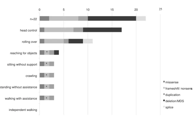
Psychomotor development at the time of the study (median age: 9.4 years, range 8 months-24 years) revealed severe impairment in all patients with LIS1- associated lissencephaly. While early developmental steps such as head control were mastered by more than half of the patients, later milestones of the first year were rarely obtained (Fig. 1).
77.3% of the patients showed good head control, while 22.7% rarely or never achieved this ability. Simi- larly, the ability to roll over was always observed in eight patients (36.4%) and sometimes in three patients (13.6%). Only a small percentage of the LIS1-cohort was able to reach for objects (18.2%). Developmental steps of the second half of the first year, including sit- ting, crawling and standing without assistance were obtained by less than 15% of the patients. Only three patients were at least sometimes able to walk with assis- tance, while no patient could walk independently. Inter- estingly, these three patients had an underlying 5´ missense mutation (p.Arg20Ser, patient LIS1-01), a small 18 kb duplication (including intron 1, exon 2 and intron 2, patient LIS1-09) and low grade mosaicism for a nonsense mutation (p.W74*, patient LIS1-22;
mutation in 20% of peripheral blood cells). Further- more, cMR imaging for two of them (LIS1-01 and LIS1-09) confirmed a milder cerebral phenotype accord- ing to Dobyns grade 4. In contrast, all other 19 patients in our cohort at a median age of 7.6 years (range:
8 months–24 years) did not reach milestones beyond the first half of the first year (Fig. 1).
3.4. Epilepsy and electroencephalography (EEG)
All patients in our cohort developed drug-resistant epilepsy. In 82% the onset of seizures was within the first six months of life (n = 18). Age at onset of epilepsy ranged from 2 months to 24 months, with a median age of 5.0 months (IQR: 2.1).
In the majority of patients the first reported seizure type was infantile spasms (n = 10/14; 71%), in two patients accompanied by myoclonic seizures. Three patients (21%) presented with generalized tonic–clonic and one with generalized tonic seizures at the time of manifestation.
At the time of data collection the majority of patients (n = 11/17; 65%) suffered from a variety of seizure forms with generalized tonic–clonic seizures (n = 12; 71%) being the most predominant type. In addition, focal sei- zures (n = 6; 35%), infantile spasms (n = 4; 24%), absence (n = 3; 18%), myoclonic (n = 3; 18%), tonic (n = 3; 18%), clonic (n = 1; 6%) and atonic seizures (n = 1; 6%) were reported.
Electroencephalography (EEG) showed abnormal interictal findings in all patients including hypsarrhyth- mia (n = 9/20; 45%), regional abnormalities or regional epilepsy-typical potentials (ETPs) (n = 11/20; 55%) as well as generalized abnormalities or generalized ETPs (n = 10/20; 50%).
Despite anticonvulsive polytherapy all patients still suffered from seizures. However, the median daily num- ber of seizures could be reduced from 4.0 (IQR: 10.5) at onset of epilepsy to 1.5 (IQR: 4.9) at the time of data col- lection. At this time 65% of the patients had daily seizures (n = 13), 30% had more than one seizure per week (n = 6) and only one patient had at least one seizure per month.
For two children a loss of earlier obtained skills during periods of increased epileptic activity was reported.
3.5. Evaluation of anticonvulsive therapy
Statistical analysis was performed for all antiepileptic drugs prescribed to P6 patients to reveal a rank order for both the neuropediatric (Fig. 2A) and the family evaluation (Fig. 2B).
In both evaluations lamotrigine (LTG) was rated the most successful anticonvulsive drug with (partial) response rates of 87.5–100%. Efficacy of LTG was eval- uated to be significantly better than levetiracetam (LEV) and sulthiame (STM) (p < 0.05 and p < 0.01 respec- tively) in the neuropediatric assessment and significantly better than oxcarbazepine (OXC), LEV, STM (p < 0.05) and topiramate (TPM) (p < 0.01) in the family survey.
The second ranked drug, valproate (VPA), was eval- uated with (partial) response rates of 88.2–91.7%. Effi- cacy of VPA was rated significantly better than LEV and STM (p < 0.05) in both surveys and showed signif- icant differences from TPM (p < 0.01) in the family evaluation.
At the time of data collection five severely affected patients were taking the combination therapy LTG and VPA. In four patients this combination reduced the median number of daily seizures by 91.2% from 17/d (IQR: 26.4) to 1.5/d (IQR: 7.0) (no frequency data was available from the fifth patient). In addition to VPA
402
Publikation
8
20
5 10 15
0 n=22
head control
rolling over
reaching for objects
sitting without support
crawling
standing without assistance
walking with assistance
independent walking
missense frameshift/ nonsens s duplication deletion/MDS splice
*
*
*
*
*
and LTG four of these five patients received additional anticonvulsive treatment: stiripentol + clobazam (n = 1), vigabatrin (VGB) (n = 1) and rufinamide (n = 1). Interestingly, a 17-year-old patient was treated with vagus nerve stimulation (VNS) in addition to his
continuing and unchanged combination of VPA + LTG for two years. VNS was reported to result in
decreased seizure frequency and improved EEG results without serious side effects in this patient.
Phenobarbital (PB) and VGB were ranked third or fourth in both the family and the neuropediatric assessments. PB revealed a good treatment response in 33.3–41.7% and a partial treatment response in 41.7%–44.4%. Similar efficacy was reported for VGB with partial or good response rates in 61.6–68.8% in both surveys. Both PB and VGB were rated significantly better than the worst ranked drug in both surveys (STM in the neuropediatric assessment (p < 0.01) and TPM in the family evaluation (p < 0.05)).
In contrast, no response or even clinical aggravation was noted by the attending neuropediatricians in 71.4%
for OXC (n = 7) and 60.0% for LEV (n = 10). More- over, STM had virtually no treatment response in any patient in this neuropediatric evaluation: no response was noted in 83.3% and even clinical aggravation in 16.7% (n = 6). Similar poor efficacy of these three drugs were reported in the family survey with no response or
clinical aggravation noted in 46.2% for LEV (n = 13), in 50.0% for OXC (n = 8) and 54.6% for STM (n = 11).
Overall the rank order revealed a similar efficacy ranking for the anticonvulsant drugs in the family and in the neuropediatric survey. Only TPM therapy was rated with divergent results. Treatment response to TPM was rated worst in the family survey with no response in 52.9% and even clinical aggravation in 5.9%. Partial response was described in 29.4% and good response in 11.8% (n = 17). In the neuropediatric assess- ment, however, TPM ranked 5th with good response in 18.2%, partial response in 45.5% and no response in 36.4% (n = 11).
4. Discussion
Psychomotor development of patients with LIS1- related lissencephaly is affected early and most severely and milestones of the second half of the first year of life are rarely mastered. Sitting, crawling and walking with assistance were only reported in three patients with milder mutations. This may indicate that patients with 5´ missense mutations and small 5´ duplications before the coiled coil domain may have a better develop- mental potential compared to deletions and other muta- tions that predict a premature protein termination.
Interestingly, a similar genotype–phenotype relationship
S.M. Herbst et al./Brain & Development 38 (2016) 399–406
Fig. 1. Genotype–phenotype correlation in LIS1-associated lissencephaly. Only 3/22 patients were able to obtain psychomotor milestones of the second half of the first year of life, these included one patient with a 18 kb duplication (cMRI: grade 4a), one patient with missense mutation (cMRI:
grade 4a) and one patient (*) with a mosaic nonsense mutation (20% mutant alleles in peripheral blood cells).
25
403
9 B
Family evaluationwas previously reported for the lissencephaly grading in cMR imaging; as lissencephaly grade 4 was documented for two patients with 5´ missense mutations (before the coiled coil domain [19]) and more severe cMRI pheno- types (grade 2–3) in patients with large deletions [20].
The main challenge in treating patients with LIS1-associated lissencephaly is their severe epilepsy, which is frequently resistant to medical polytherapy.
In approximately 90% of our cohort onset of epilepsy was noted within the first year of life, which is similar to previous published data [2,3]. Likewise, drug-resistant epilepsy as defined by the ILAE [21] was present in all of our 22 patients. Effective antiepileptic treatment is therefore crucial in order to provide optimal conditions for the development of basic psychomotor and mental skills. However, data about the efficacy of antiepileptic medication in patients with LIS1- associated lissencephaly is sparse [10–12]. Leventer et al. described five patients with LIS1-related lissence- phaly and epileptic seizures. Three of them were reported to be resistant to multiple antiepileptic therapies [11]. In this study we propose, that due to a genotype specific altered layering of the lissencephalic cortex a genotype-specific approach to antiepileptic
therapy may reduce the number of seizures and thus improve the overall quality of life for these patients.
Infantile spasms were the most frequent seizure form at the time of manifestation in our study population as well as in other previously published patient cohorts with classic lissencephaly [3,4]. According to our data, standard treatment protocols for infantile spasms such as VGB, ACTH and steroids may also be effective in the early disease course for patients with LIS1- associated lissencephaly. This is in line with Mokánszki et al., who reported good response to ACTH in a child with infantile spasms and lissencephaly due to a LIS1 frameshift mutation [12]. Furthermore, good response to VGB in the treatment of infantile spasms was reported in children with tuberous sclerosis [22].
Later in infancy seizure symptomology became more variable and included different forms of focal seizures as well generalized tonic–clonic seizures. In these infants early prescription of LTG and VPA appears promising.
Both drugs were rated as the best two drugs by both families and attending neuropediatricians with (partial) response rates of more than 85%. LTG is considered to be an effective drug for generalized tonic–clonic seizures as well as a broad spectrum of partial seizures
S.M. Herbst et al./Brain & Development 38 (2016) 399–406
A
Neuropediatric evaluationFig. 2. (A) Efficacy of antiepileptic medication (neuropediatric evaluation). (B) Efficacy of antiepileptic medication (family evaluation). Therapeutic efficacy of antiepileptic drugs prescribed to P6 patients in descending order of the Kruskal–Wallis rank sum: good response = green, partial response = yellow, no response = red, clinical aggravation = dark red. Statistical differences are indicated with ‘*’ (p < 0.05) and ‘**’ (p < 0.01).
404
Publikation
10
S.M. Herbst et al./Brain & Development 38 (2016) 399–406
[23]. In addition, it is one of a small number of FDA-approved therapies for seizures associated with severe Lennox–Gastaut syndrome [24].
Moreover, PB, which is more commonly prescribed in the neonatal period [25], may be quite useful even in older patients with LIS1 mutation since it achieved response rates of more than 75%.
Collectively our data indicate that combining VPA + LTG may constitute a very promising regime for patients with LIS1-associated lissencephaly. This combi- nation is reported to have synergistic effects [26]. In our cohort five severely affected patients were taking VPA + LTG at the time of the survey and this combination therapy was reduced the median number of daily seizures from 17to1.5 (91.2%). Inaddition toVPA + LTG, four patients received additional anticonvulsive treatment including one case of vagus nerve stimulation. Further studies are needed to explore whether complementary therapeutic concepts such as vagus nerve stimulation, which has been reported to be effective in patients with other drug-resistant lesional epilepsies [27], are also effec- tive in patients with LIS1-related lissencephaly.
The neuropediatric and family evaluations revealed a similar rank order for most drugs analyzed. TPM, how- ever, was rated quite differently among families and neu- ropediatricians. In the family survey TPM was ranked last with no response or clinical aggravation in 58.8%, whereas among neuropediatricians TPM was rated to beatleast partially effectivein 63.7%.These controversial results may be in part explained by the severe side effects that were reported. Families may have rated the anticonvulsive efficacy of TPM worse due to the common dose-dependent severe drowsiness and sedation [28]. One family noted specifically in the telephone interview that under TPM treatment their affected child slept for more than 20 h per day and was barely responsive at all.
Further studies are needed to analyze whether TPM treatment may have more severe side effects such as severe sedation in the specific context of LIS1-associated lissencephaly.
Despite anticonvulsive polytherapy about two thirds of our LIS1-cohort still suffered from daily seizures.
Interestingly, six patients with less than daily seizures at the time of data collection were taking at least one of the top four ranked medications (VPA, LTG, VGB, PB), including one patient with the combination of VPA + LTG.
We are well aware of the major limitations of this first retrospective study with 22 patients regarding possible spontaneous and age-related fluctuation of seizure fre- quency. Moreover, no systematic comparison of the dosages, the serum levels of active drug metabolites, or the overlapping time frame in which several drugs were taken simultaneously, was possible. However, due to the rarity of this orphan disease, achieving the gold stan- dard of good clinical practice is very difficult.
In this initial study the evaluation of the AEDs is lim- ited to the subjective estimation of the attending neuro- pediatricians and the affected families. Nevertheless, it provides exciting insights into the effects of various antiepileptic treatment schemes commonly applied to patients with LIS1-associated lissencephaly on both sei- zure frequency and their impact on the quality of life as important clinical endpoints for this patient cohort. It must now be complemented by prospective studies, which will include the assessment of the seizure reduc- tion rate for larger patient cohorts with this rare genetic disorder in order to ultimately improve seizure control and overall quality of life for these patients.
5. Conclusion
LIS1-associated lissencephaly uniformly results in severe psychomotor impairment and early drug- resistant epilepsy commonly starting within the first year of life. Affected patients might benefit most from antiepileptic therapy with LTG, VPA, VGB or PB.
The results of our study are limited to patients with LIS1-associated lissencephaly and need to be confirmed by larger prospective studies. Moreover, additional studies are needed to determine the epileptogenic pheno- type for patients with even rarer forms of classic lissen- cephaly e.g. resulting from mutations in the DCX and TUBA1A gene.
6. Conflict of interest
All authors declare no conflict of interest.
Acknowledgments
We would like to give a special and warm thank you to all patients, their families and attending physicians taking part in our study and to the patient support group LISS e.V. for their tremendous help in recruiting families for our study.
Appendix A. Supplementary data
Supplementary data associated with this article can be found, in the online version, at http://dx.doi.org/10.
1016/j.braindev.2015.10.001.
References
[1] Dobyns WB, Das S. LIS1-associated lissencephaly subcortical band heterotopia. GeneReviews®. Seattle: University of Washington; 2009 [updated 2014].
[2] Dobyns WB. The clinical patterns and molecular genetics of lissencephaly and subcortical band heterotopia. Epilepsia 2010;
51:5–9.
405
11
S.M. Herbst et al./Brain & Development 38 (2016) 399–406
[3] de Wit M-CY, de Rijk-van Andel J, Halley DJ, Poddighe PJ, Arts WFM, de Coo IFM. Long-term follow-up of type 1 lissencephaly:
survival is related to neuroimaging abnormalities. Dev Med Child Neurol 2011;53:417–21.
[4] Saillour Y, Carion N, Quelin C, Leger P-L, Boddaert N, Elie C, et al. LIS1-related isolated lissencephaly. Arch Neurol 2009;66:
1007–15.
[5] Hehr U, Schuierer G. Genetic assessment of cortical malforma- tions. Neuropediatrics 2011;42:43–50.
[6] Cardoso C, Leventer RJ, Ward HL, Toyo-oka K, Chung J, Gross A, et al. Refinement of a 400-kb critical region allows genotypic differentiation between isolated lissencephaly, Miller–Dieker syn- drome, and other phenotypes secondary to deletions of 17p13.3.
Am J Hum Genet 2003;72:918–30.
[7] Dobyns WB, Reiner O, Carrozzo R, Ledbetter H. Lissencephaly – a human brain malformation associated with deletion of the LIS1 gene located at chromosome 17p13. JAMA 1993;270:
2838–42.
[8] Forman MS, Squier W, Dobyns WB, Golden JA. Genotypically defined lissencephalies show distinct pathologies. J Neuropathol Exp Neurol 2005;64:847–57.
[9] Kato M, Dobyns WB. Lissencephaly and the molecular basis of neuronal migration. Hum Mol Genet 2003;12:R89–96.
[10] Inui T, Kobayashi T, Kobayashi S, Sato R, Endo W, Kikuchi A, et al. Efficacy of long term weekly ACTH therapy for intractable epilepsy. Brain Dev 2015;37:449–54.
[11] Leventer RJ, Cardoso C, Ledbetter DH, Dobyns WB. LIS1 missense mutations cause milder lissencephaly phenotypes includ- ing a child with normal IQ. Neurology 2001;57:416–22.
[12] Mokáanszki A, Ko¨rhegyi I, Szabó N, Bereg E, Gergev G, Balogh E, et al. Lissencephaly and band heterotopia: LIS1, TUBA1A, and DCX mutations in Hungary. J Child Neurol 2012;27:
1534–40.
[13] Klinkenberg S, Aalbers MW, Vles JSH, Cornips EMJ, Rijkers K, Leenen L, et al. Vagus nerve stimulation in children with intractable epilepsy: a randomized controlled trial. Dev Med Child Neurol 2012;54:855–61.
[14] Yamada M, Yoshida Y, Mori D, Takitoh T, Kengaku M, Umeshima H, et al. Inhibition of calpain increases LIS1 expres- sion and partially rescues in vivo phenotypes in a mouse model of lissencephaly. Nat Med 2009;15:1202–7.
[15] Majkowski J. Drug Effects on afterdischarge and seizure thresh- old in lissencephalic ferrets: an epilepsy model for drug evalua- tion. Epilepsia 1983;24:678–85.
[16] Dobyns WB, Truwit CL, Ross ME, Matsumoto N, Pilz DT, Ledbetter DH, et al. Differences in the gyral pattern distinguish chromosome 17-linked and X-linked lissencephaly. Neurology 1999;53:270–7.
[17] Borozdin W, Boehm D, Leipoldt M, Wilhelm C, Reardon W, Clayton-Smith J, et al. SALL4 deletions are a common cause of Okihiro and acro-renal-ocular syndromes and confirm haploin- sufficiency as the pathogenic mechanism. J Med Genet 2004;41:
e113.
[18] Bender R, Lange S. Adjusting for multiple testing–when and how?
J Clin Epidemiol 2001;54:343–9.
[19] Uyanik G, Morris-Rosendahl DJ, Stiegler J, Klapecki J, Gross C, Berman Y, et al. Location and type of mutation in the LIS1 gene do not predict phenotypic severity. Neurology 2007;69:442–7.
[20] Cardoso C, Leventer RJ, Dowling JJ, Ward HL, Chung J, Petras KS, et al. Clinical and molecular basis of classical lissencephaly:
mutations in the LIS1 gene (PAFAH1B1). Hum Mutat 2002;19:
4–15.
[21] Kwan P, Arzimanoglou A, Berg AT, Brodie MJ, Allen Hauser W, Mathern G, et al. Definition of drug resistant epilepsy: consensus proposal by the ad hoc Task Force of the ILAE Commission on Therapeutic Strategies. Epilepsia 2010;51:1069–77.
[22] Vigevano F, Cilio MR. Vigabatrin versus ACTH as first-line treatment for infantile spasms: a randomized, prospective study.
Epilepsia 1997;38:1270–4.
[23] Beran RG, Berkovic SF, Dunagan FM, Vajda FJE, Danta G, Black AB, et al. Double-blind, placebo-controlled, crossover study of lamotrigine in treatment-resistant generalised epilepsy.
Epilepsia 1998;39:1329–33.
[24] Montouris GD, Wheless JW, Glauser TA. The efficacy and tolerability of pharmacologic treatment options for Lennox– Gastaut syndrome. Epilepsia 2014;55:10–20.
[25] Hellström-Westas L, Boylan G, Agren J. Systematic review of neonatal seizure management strategies provides guidance on anti-epileptic treatment. Acta Paediatr 2015;104:123–9.
[26] Brodie MJ, Yuen AWC. Lamotrigine substitution study: evidence for synergism with sodium valproate? Epilepsy Res 1997;26:423–32.
[27] Hajnsek S, Petelin Z, Poljakovic Z, Mrak G, Paladino J, Desnica A. Vagus nerve stimulation in the treatment of patients with pharmacoresistant epilepsy: our experiences. Coll Antropol 2011;35:755–60.
[28] Uldall P, Buchholt JM. Clinical experiences with topiramate in children with intractable epilepsy. Eur J Paediatr Neurol 1999;3:
105–11.
406
Einleitung
12
Deutsche Zusammenfassung:
LIS1 -assoziierte klassische Lissenzephalie: Eine retrospektive, multizentrische Studie zum epileptischen Phänotyp und Ansprechen auf Antikonvulsiva
1. Einleitung
Die klassische Lissenzephalie zählt mit einer Prävalenz von etwa 1 – 4:100.000 Neugeborenen zu den seltenen Hirnfehlbildungen.
1Ursächlich für die Erkrankung ist eine Migrationsstörung kortikaler Neurone während der Embryonalentwicklung. Die Entwicklung des menschlichen Cortex durch radiäre Wanderung neuronaler Vorläuferzellen von ihrem Entstehungsort im Bereich der Ventrikelwand in Richtung Hirnoberfläche ist bei Kindern mit klassischer Lissenzephalie gestört, sodass anstelle der gesunden, gefalteten Großhirnrinde ein abnorm geschichteter und verdickter Cortex mit glatter Oberfläche entsteht (lissós = griech. glatt, enképhalos = griech. Gehirn).
2,3Betroffene Kinder weisen in der Regel eine erhebliche psychomotorische Retardierung auf und leiden an einer schweren, nicht selten therapieresistenten Epilepsie. Diese manifestiert sich für gewöhnlich in den ersten Lebensmonaten in Form von Blitz-Nick-Salaam (BNS)- Anfällen und umfasst mit zunehmendem Alter verschiedene Anfallsformen bis hin zu komplexen Epilepsieformen wie dem Lennox-Gastaut-Syndrom.
4,5,6Die Therapie der meist täglichen Anfälle stellt eine große Herausforderung für betroffene Familien und behandelnde Neuropädiater dar. Fütterungsprobleme, Atemwegsinfektionen und Status epilepticus werden als Hauptursachen für die hohe Todesrate von circa 50 % bis zu einem Alter von zehn Jahren angenommen.
4,5Die Ätiologie der klassischen Lissenzephalie ist heterogen und umfasst syndromale Formen
wie das Miller-Dieker-Syndrom und isolierte monogene Formen. Mutationen in mehreren
Genen konnten bereits als ursächlich für die klassische Lissenzephalie identifiziert werden,
wobei in ungefähr 70 % das LIS1-Gen (auch PAFAH1B1 genannt) betroffen ist.
7Während
heterozygote Deletionen und Mutationen im LIS1-Gen zur sogenannten isolierten
Lissenzephalie Sequenz (ILS) führen, verursachen variable Mikrodeletionen des
Genabschnitts 17p.13.3, welche das LIS1-Gen und andere kritische Gene (u. a. das YWHAE-
Gen) einschließen, das Miller-Dieker-Syndrom.
8Einleitung
13 Radiologisch stellt sich die klassische Lissenzephalie in Form eines glatten und verdickten Cortex mit deutlich reduzierter (Pachygyrie) oder abwesender Gyrierung (Agyrie) anstelle der charakteristischen Windungen und Furchungen des gesunden menschlichen Gehirns dar.
9,10Wissenschaftliche Arbeiten der vergangenen Jahre konnten eine ausgeprägte Genotyp- Phänotyp-Korrelation feststellen: Mutationen in einem bestimmten Gen resultieren sowohl in einem genspezifischen charakteristischen makroskopischen Bild, das sich mithilfe der Magnetresonanztomografie darstellen lässt, als auch in genspezifischen Veränderungen des mikroskopischen Kortexaufbaus.
9,11,12Daher postulieren wir, dass die für die Lissenzephalie verantwortlichen genetischen Veränderungen nicht nur die Genotyp-spezifische kortikale Architektur bedingen, sondern auch den individuellen epileptischen Phänotyp sowie das Ansprechen auf die antikonvulsive Therapie beeinflussen.
Bislang wurde die Effektivität der antikonvulsiven Therapie bei Patienten mit klassischer
Lissenzephalie nur im Rahmen von einzelnen Fallberichten,
13,14,15für Patientenkohorten mit
komplexen, therapieresistenten Epilepsieformen im Allgemeinen
16,17,18sowie in Tiermodellen
für Epilepsie
19,20untersucht. Aufgrund der Seltenheit dieser Hirnfehlbildung ist es jedoch
schwierig, eine prospektive, randomisierte Studie mit betroffenen Kindern durchzuführen,
sodass wir uns für ein retrospektives Studiendesign entschieden. Ziel der vorliegenden Arbeit
war es, den epileptischen Phänotyp sowie das Therapieansprechen in unserer homogenen
Kohorte von Patienten mit genetisch und radiologisch gesicherter klassischer LIS1-
assoziierter Lissenzephalie erstmals systematisch zu charakterisieren.
Patienten und Methoden
14 2. Patienten und Methoden
2.1. Patientenkollektiv
Die Patientenrekrutierung erfolgte aus der Datenbank genetisch diagnostizierter Patienten unseres humangenetischen Zentrums (n = 11) sowie über die Homepage der Selbsthilfegruppe Liss e.V. (n = 11). Einschlusskriterium für die Patienten dieser Studie war die Diagnose einer klassischen Lissenzephalie (Typ 1-Lissenzephalie), gesichert durch 1) eine kraniale Magnetresonanztomografie (cranial magnet resonance tomography, cMRT) und 2) eine pathogene Mutation im LIS1-Gen in der genetischen Untersuchung. Verfügbare cMRT-Bilder von zehn Patienten wurden in Kooperation mit dem Zentrum für Neuroradiologie Regensburg reevaluiert und nach Dobyns´ lissencephaly patterning scale eingestuft.
21Unsere Studie wurde in Übereinstimmung mit der Ethikerklärung des Weltärztebundes (Deklaration von Helsinki) durchgeführt, zudem lag ein positives Votum von der Ethikkommission des Universitätsklinikums Regensburg (Ethikantrag Nummer: 13-101- 0090) vor. Schriftliche Einverständniserklärungen aller Eltern als Erziehungsberechtigte ihrer Kinder wurden eingeholt.
2.2. Klinische Evaluation
Die Datenerhebung zur Evaluation der psychomotorischen Entwicklung der Patienten sowie ihrem Krankheitsverlauf mit besonderem Fokus auf die Epilepsie und deren antikonvulsive Therapie erfolgte mittels standardisierter neuropädiatrischer (n = 18) und Familien- Fragebögen (n = 22). Ergänzend wurde bei Bedarf ein standardisiertes Telefoninterview durchgeführt (n = 8). Des Weiteren wurden standardisierte neuropädiatrische Untersuchungsbefunde (n = 18) der Patienten in 16 Studienzentren in Deutschland und Österreich erhoben.
Zur Evaluation des genspezifischen Ansprechens auf verschiedene antikonvulsive
Therapeutika wurden die standardisierten neuropädiatrischen und Familien-Fragebögen
ausgewertet. Die Familien sowie die behandelnden Neuropädiater wurden gebeten, die aktuell
sowie in der Vergangenheit angewandten antikonvulsiven Medikamente für das betroffene
Kind auf einer Skala mit den Kategorien „gutes Ansprechen“, „partielles Ansprechen “, „kein
Ansprechen“ und „klinische Verschlechterung“ zu bewerten. In Anbetracht der
bekanntermaßen häufig therapieresistenten Epilepsieformen bei Patienten mit klassischer
Lissenzephalie, entschieden wir uns, die Bewertung der antikonvulsiven Therapie mithilfe
einer Ordinalskala durchzuführen. Die alternativ häufig für die Evaluation von
Patienten und Methoden
15 Antikonvulsiva verwendete Definition der Ansprechrate als Anfallsreduktion um 50 % erschien uns in diesem Kollektiv nicht sinnvoll. Medikamente, die von weniger als sechs Patienten eingenommen wurden, berücksichtigten wir nicht in der statistischen Auswertung (siehe Tab. 2)
2.3. Genetische Untersuchung
Die genetische Untersuchung und Diagnosesicherung der in unsere Studie eingeschlossenen Patienten wurde bereits vor Studienbeginn in unserem humangenetischen Zentrum (n = 11) bzw. in anderen kooperierenden humangenetischen Laboren in Deutschland und Österreich (n = 11) durchgeführt. Die für das Krankheitsbild ursächlichen Mutationen im LIS1-Gen (PAFAH1B1) wurden entweder mit Fluoreszenz-In-Situ-Hybridisierung (FISH), Multiplex Ligation-dependent Probe Amplification (MLPA; MRC Holland Kit (P061-C1 vs.10)), quantitativer Polymerase-Kettenreaktion (qPCR; wie beschrieben durch Borozdin
22) oder Sanger-Sequenzierung des LIS1-Gens detektiert.
2.4. Statistische Auswertung
Die Datenauswertung erfolgte mithilfe des Statistikprogramms SPSS Software (IBM SPSS Statistics for Windows, Version 22.0, Armonk, New York, USA). Die Daten wurden unter Verwendung des Kolmogorov-Smirnov-Testes auf Normalverteilung untersucht.
Normalverteilte Daten wurden in der vorliegenden Arbeit mithilfe von Mittelwert (MW) und
Standardabweichung (Standard Deviation, SD) beschrieben, während nicht normalverteilte
Daten mit Median (M) und Interquartilsabstand (Interquartile Range, IQR) angegeben
wurden. Zur weiteren Beschreibung der Ergebnisse wurde bei Bedarf die Spannweite (Range,
R) genannt. Die Evaluation der Effektivität der antikonvulsiven Therapie wurde mithilfe des
Kruskal-Wallis Testes in Kombination mit einem post-hoc-Test (Dunn-Bonferroni-Test,
zweiseitige Testung) durchgeführt. Unsere Studie wurde als exploratorische Studie angelegt,
sodass ein nicht-adjustierter p-Wert von p < 0,05 als signifikant angenommen wurde (ohne
Adjustierung bezüglich Multiplizität).
23Ergebnisse
16 3. Ergebnisse
3.1. Patientenkollektiv
Unser Kollektiv bestand aus 22 nicht-verwandten Patienten (14 männliche, 8 weibliche) mit LIS1-assoziierter klassischer Lissenzephalie, die wir aus 16 deutschen und österreichischen Studienzentren rekrutierten. Das Alter der Patienten zum Zeitpunkt der Datenerhebung lag zwischen 8 Monaten und 24 Jahren (MW: 9,4 ± 6,9 Jahre). Ein Patient starb im Alter von 9,1 Jahren an einer Infektion mit Fieber-getriggerter Exazerbation seiner Epilepsie.
3.2. Klinische und neuroradiologische Diagnostik
Die Diagnose der Lissenzephalie wurde in unserer Patientenkohorte im medianen Alter von 4,3 Monaten (IQR: 3,2) gestellt. Bei einem Patienten erfolgte die Diagnosestellung bereits pränatal, bei drei Patienten (14 %) innerhalb der ersten sechs Lebenswochen und bei der Mehrheit der Patienten (82 %) nach der sechsten Lebenswoche. Erste klinische Auffälligkeiten (v.a. im Verhalten und in der psychomotorischen Entwicklung) oder die Manifestation erster Krampfanfälle wurden im durchschnittlichen Alter von 3,2 ± 2,8 Monaten beschrieben.
Die Diagnose Lissenzephalie wurde bei allen 22 Patienten durch eine Magnetresonanztomografie bestätigt, welche im medianen Alter von 5,0 Monaten (R: 20 Tage – 14,7 Jahre) durchgeführt wurde. Diese zeigte eine generalisierte Lissenzephalie (n = 4), okzipital betonte (n = 14) oder nicht näher differenzierte, klassische Lissenzephalie (n = 4). In allen zehn reevaluierten cMRT-Bildern war die Lissenzephalie stärker okzipital betont und das MRT-Grading entsprechend der Dobyns´ lissenzephaly patterning scale
21ergab eine Lissenzephalie zwischen Grad 2 und 4, darunter Grad 2 (n = 2), Grad 3 (n = 6) und Grad 4 (n = 2) (siehe Abb. 1 und Tab. 1). Als häufigste, zusätzlich vorhandene Hirnfehlbildung lag ein abnormes Corpus callosum (Abflachung des Corpus und milde Hypoplasie des Splenium) vor, dass bei sieben Patienten diagnostiziert wurde (siehe Abb. 1). Des Weiteren zeigten sich eine Malrotation des Hippokampus bei zwei Patienten, erweiterte Ventrikel bei zwei und ein erweiterter extrazerebraler Raum bei einem Patienten. Ein Cavum Vergae als nicht- pathologische Variante konnten wir bei 50 % der Patienten unserer Kohorte identifizieren. In allen zehn reevaluierten MRT-Aufnahmen erschien das Kleinhirn unauffällig.
Interessanterweise stellte sich die Gyrierung der orbitofrontalen Region in allen zehn cMRT-
Bildern relativ wenig beeinträchtigt dar, sodass von einer stärkeren Beeinträchtigung der
neuronalen Migration im okzipitalen Bereich im Vergleich zum frontobasalen Cortex
Ergebnisse
17 auszugehen ist (siehe Abb. 1, A(3) – C(3)). Für die weiteren zwölf Patienten lagen uns schriftliche neuroradiologische MRT-Befunde vor (elf ausführliche und ein kurzer Bericht).
Darin wurden erweiterte Ventrikel bei drei Patienten beschrieben, eine Signalanhebung der weißen Substanz bei drei und eine Hypoplasie des Corpus callosum bei zwei Patienten.
Weiterhin wurde ein vergrößerter extrazerebraler Raum bei einem Patienten sowie eine Kleinhirnhypoplasie bei einem anderen Patienten diagnostiziert.
Abbildung 1: cMRT-Befunde bei klassischer LIS1-assoziierter Lissenzephalie.
A
Patient mit Lissenzephalie Grad 2a: Diffuse Agyrie mit einigen flachen Sulci über den frontalen und temporalen Polen, Cavum Vergae. (1) axiales cMRT, (2) koronares cMRT, (3) axiales cMRT auf Höhe der Orbitae.
B Patient mit Lissenzephalie Grad 3a: Gemischte frontale Pachygyrie und okzipitale Agyrie, Cavum Vergae.
(1) axiales cMRT, (2) koronares cMRT, (3) axiales cMRT auf Höhe der Orbitae.
C
Patient mit Lissenzephalie Grad 4a: Diffuse okzipital ausgeprägte Pachygyrie, Cavum Vergae. (1) axiales cMRT, (2) koronares cMRT, (3) axiales cMRT auf Höhe der Orbitae.
A(3), B(3), C(3) Relativ geringe Beeinträchtigung der Gyrierung im Bereich der orbitofrontalen Region bei allen
drei Patienten und Graden.
Ergebnisse
18
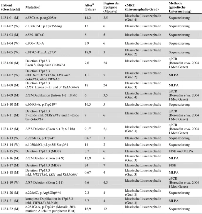
Tabelle 1: Klinische und genetische Daten der 22 Patienten mit LIS1-assoziierter klassischer Lissenzephalie.
Patient
(Geschlecht) Mutationi Alterii
(Jahre)
Beginn der Epilepsie (Monate)
cMRT
(Lissenzephalie-Grad)
Methode (genetische Untersuchung) LIS1-01 (M) c.58C>A, p.Arg20Ser 14,2 3,5 klassische Lissenzephalie
(Grad 4) Sequenzierung
LIS1-02 (W) c.1066T>C, p.Cys356Arg 13 6 klassische Lissenzephalie Sequenzierung
LIS1-03 (M) c.569-10T>C 8 5 klassische Lissenzephalie Sequenzierung
LIS1-04 (W) c.900+1G>A 2,9 6 klassische Lissenzephalie Sequenzierung
LIS1-05 (W) c.817C>T, p.Arg273* 18,9 3 klassische Lissenzephalie
(Grad 2) Sequenzierung
LIS1-06 (M) Deletion 17p13.3
Exon 8, Stop nach GARNL4 7,6 24 klassische Lissenzephalie qPCR
(Borozdin et al. 2004 J Med Genet) LIS1-07 (W)
Deletion 17p13.3
inkl. HIC, METTL16, LIS1 und
GARNL4, ohne YWHAE 1,1 5 klassische Lissenzephalie
(Grad 2) MLPA
LIS1-08 (M) Deletion 17p13.3
(LIS1: Exons 3–11 und 3´ KIAA0664) 18 24 klassische Lissenzephalie
(Grad 3) MLPA
LIS1-09 (M) LIS1-Duplikation (Intron 1-2; 18 kb) 6 3,5 klassische Lissenzephalie (Grad 4)
qPCR
(Borozdin et al. 2004 J Med Genet) LIS1-10 (M) c.656G>A, p.Trp219* 16,5 5 klassische Lissenzephalie Sequenzierung LIS1-11 (M)
Deletion 17p13.3
5´-Ende inkl. SERPINF1 und 3´-Ende
bis GARNL4 7 6 klassische Lissenzephalie qPCR
(Borozdin et al. 2004 J Med Genet) LIS1-12 (M) LIS1-Deletion (Exon 6 + 7; 6.2 kb) 9,1iii 2,1 klassische Lissenzephalie
(Grad 3)
qPCR
(Borozdin et al. 2004 J Med Genet) LIS1-13 (W) c.282delG, p.Trp94* 0,67 3 klassische Lissenzephalie Sequenzierung LIS1-14 (W) c.1050delG, p.Lys351Ser fs*4 14 2 klassische Lissenzephalie Sequenzierung LIS1-15 (W) Deletion 17p13.3 (MDS) 3,7 6 klassische Lissenzephalie FISH und MLPA LIS1-16 (M) LIS1-Deletion (Exon 8 + 9) 12,9 6 klassische Lissenzephalie
(Grad 3) MLPA
LIS1-17 (M) Deletion 17p13.3 (MDS) 24 7 klassische Lissenzephalie FISH
LIS1-18 (M) Deletion 17p13.3
inkl. METTL16, LIS1 und KIAA0664 0,67 4 klassische Lissenzephalie
(Grad 3) MLPA
LIS1-19 (W) LIS1-Deletion (Exon 2-11) 6,6 4,5 klassische Lissenzephalie qPCR
(Borozdin et al. 2004 J Med Genet) LIS1-20 (M) c.22delC, p.Arg8Glufs*4 2,2 4 klassische Lissenzephalie
(Grad 3) Sequenzierung
LIS1-21 (M) komplexe Duplikation in 17p13.3
inkl. YWHAE (38.9 kb) 3,7 4 klassische Lissenzephalie
(Grad 3) MLPA
LIS1-22 (M) c.281G>A, p.Trp94* (Mosaik, 20%
mutierte Allele im peripheren Blut) 16,9 12 klassische Lissenzephalie Sequenzierung
igemäß der Referenz: NM_000430.3 # MLPA= MRC Holland Kit (P061-C1 vs.10)
ii Alter zum Zeitpunkt der Datenerhebung
iii Patient starb im Alter von 9,1 Jahren
Ergebnisse
19 3.3. Genotyp-Phänotyp-Korrelation und klinische Präsentation
Zehn Patienten in unserem Kollektiv (45 %) wiesen Exon-Deletionen oder komplette Deletionen des LIS1-Gens auf, darunter auch zwei Patienten mit Miller-Dieker-Syndrom, welche die typische, durch FISH bestätigte Mikrodeletion im Genabschnitt 17p13.3 besaßen (siehe Tab. 1). Des Weiteren wurde bei zwei Patienten (9 %) eine partielle Exon-Duplikation der LIS1-kritischen Region identifiziert. Eine Stopp-Mutation wurde bei vier Patienten (18 %) detektiert, darunter ein Patient mit Mosaik für eine Stopp-Mutation in circa 20 % der analysierten genomischen DNA im peripheren Blut. Bei jeweils zwei Patienten der Kohorte (9 %) wurden eine Missense-Mutation, eine Spleiß-Mutation und eine Frameshift-Mutation identifiziert (siehe Tab. 1).
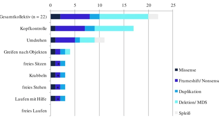
Die psychomotorische Entwicklung aller Patienten unserer LIS1-Kohorte war zum Zeitpunkt der Datenerhebung bei einem medianen Patientenalter von 9,4 Jahren (R: 8 Monate – 24 Jahre) schwer beeinträchtigt. Während erste Entwicklungsschritte wie das Erlangen der Kopfkontrolle noch von der Mehrheit der Patienten erreicht wurden, erlernten nur wenige Patienten spätere Entwicklungsschritte des ersten Lebensjahres (siehe Abb. 2). 77,3 % der Patienten verfügte über eine gute Kopfkontrolle, aber 22,7 % besaß diese Fähigkeit nur selten oder nie. Selbstständiges Umdrehen beherrschten acht Patienten (36,4 %) immer und drei Patienten (13,6 %) gelegentlich. Nur ein kleiner Anteil von 18,2 % unserer LIS1-Kohorte erlernte, nach Objekten zu greifen. Meilensteine der zweiten Hälfte des ersten Lebensjahres wie Sitzen, Krabbeln und freies Stehen wurden von weniger als 15 % der Patienten erreicht.
Nur drei Patienten gelang es, gelegentlich mit Hilfe zu laufen, aber kein Patient war dazu
ohne Unterstützung fähig. Interessanterweise besaßen diese drei Patienten eine
zugrundeliegende 5´-Missense-Mutation (p.Arg20Ser; Patient LIS1-01), eine kleine 18-kb-
Duplikation (einschließlich Intron 1, Exon 2 und Intron 2; Patient LIS1-09) und ein
geringgradiges Mosaik für eine Stopp-Mutation (p.W74*, Mutation in 20 % der peripheren
Blutzellen; Patient LIS1-22). Außerdem wiesen die cMRT-Bilder für zwei dieser Patienten
(LIS1-01, LIS1-09) einen milden zerebralen Phänotyp auf und wurden als Dobyns´ Grad 4
eingestuft. Im Gegensatz dazu hatten alle anderen 19 Patienten unseres Kollektivs mit einem
medianen Alter von 7,6 Jahren (R: 8 Monate – 24 Jahre) bislang keine Meilensteine der
zweiten Hälfte des ersten Lebensjahres erlernt (siehe Abb. 2).
Ergebnisse
20
Abbildung 2: Genotyp-Phänotyp-Korrelation bei LIS1-assoziierter klassischer Lissenzephalie.
Nur 3 von 22 Patienten waren fähig, psychomotorische Meilensteine der zweiten Hälfte des ersten Lebensjahres zu erlernen, darunter ein Patient mit einer 18-kb-Duplikation (cMRT: Grad 4a), ein Patient mit einer Misssense- Mutation (cMRT: Grad 4a) sowie ein Patient (*) mit einem Mosaik einer Stopp-Mutation (mit ca. 20 % mutierten Allelen im peripheren Blut).
3.4. Epilepsie und Elektroenzephalografie (EEG)
Alle Patienten unserer LIS1-Kohorte entwickelten eine pharmakoresistente Epilepsie. In 82 % (n = 18) manifestierte sich die Epilepsie innerhalb der ersten sechs Lebensmonate. Das Alter der Kinder zu Beginn der Krampfanfälle lag zwischen 2 und 24 Monaten, mit einem medianen Alter von 5 Monaten (IQR: 2,1). Bei der Mehrheit der Patienten manifestierte sich die Epilepsie in Form von Blitz-Nick-Salaam (BNS)-Anfällen (n = 10/14, 71 %), bei zwei Patienten wurden zusätzlich myoklonische Anfälle beschrieben. Drei Patienten (21 %) litten zu Beginn an generalisierten tonisch-klonischen Anfällen und ein weiterer Patient (7 %) an generalisierten tonischen Anfällen.
Zum Zeitpunkt der Datenerhebung litt die Mehrzahl der Patienten (n = 11/17, 65 %) an einer
0 5 10 15 20 25
Gesamtkollektiv (n = 22) Kopfkontrolle Umdrehen Greifen nach Objekten freies Sitzen Krabbeln freies Stehen Laufen mit Hilfe freies Laufen
Missense
Frameshift/ Nonsense Duplikation
Deletion/ MDS Spleiß